Evaluation of phycocyanin production by marine microalgae Arthrospira
platensis, grown in fish wastewater
Avaliação da produção de ficocianina pela microalga marinha Arthrospira
platensis, cultivada em efluente piscícola
DOI:10.34117/bjdv6n4-270
Recebimento dos originais: 10/03/2020 Aceitação para publicação: 22/04/2020
Igor Gabriel Rodrigues Ferreira Gomes
Mestre pela Universidade Federal do Ceará
Departamento de Engenharia de Pesca, Campus do Pici, Universidade Federal do Ceará, 60.455-760, Fortaleza, Ceará, Brasil
E-mail: gabrielrfgomes@gmail.com
Francisco Regivânio do Nascimento Andrade
Mestre pela Universidade Federal do Ceará
Programa de Pós-Graduação em Biotecnologia de Recursos Naturais, Campus do Pici, Universidade Federal do Ceará, 60.455-760, Fortaleza, Ceará, Brasil
E-mail: regi.andrade.biotec@gmail.com
Gabriel de Mesquita Facundo
Mestre pela Universidade Federal do Ceará
Programa de Pós-Graduação em Engenharia de Pesca, Campus do Pici, Universidade Federal do Ceará, 60.455-760, Fortaleza, Ceará, Brasil
E-mail: gabriel_biel@hotmail.com
Carlos Henrique Profírio Marques
Doutor pela Universidade Federal do Ceará
Instituto Federal de Educação, Ciência e Tecnologia do Acre, 69.980-000, Cruzeiro do Sul, Acre, Brasil
E-mail: chnanet@yahoo.com.br
Leonardo Freitas Galvão de Albuquerque
Doutor pela Universidade Federal do Ceará
Instituto Federal de Educação, Ciência e Tecnologia do Ceará, 60.115-282, Morada Nova, Ceará, Brasil
E-mail: leonardo.freitas@ifce.edu.br
José William Alves da Silva
Doutor pela Universidade Federal do Ceará
Instituto Federal de Educação, Ciência e Tecnologia do Ceará, 62.930-000, Aracati, Ceará, Brasil E-mail: jose.william@ifce.edu.br
Rafael Lustosa Maciel
Doutor pela Universidade Federal do Ceará
Instituto Federal de Educação, Ciência e Tecnologia do Amazonas, 69.800-000, Humaitá, Amazonas, Brasil
E-mail: maciel.rlm@hotmail.com
João Felipe Nogueira Matias
Doutor pela Universidade Federal do Ceará
Departamento de Engenharia de Pesca, Campus do Pici, Universidade Federal do Ceará, 60.455-760, Fortaleza, Ceará, Brasil
E-mail: jfn.matias@gmail.com
Elenise Gonçalves de Oliveira
Doutora pela Universidade Estadual de São Paulo
Departamento de Engenharia de Pesca, Campus do Pici, Universidade Federal do Ceará, 60.455-760, Fortaleza, Ceará, Brasil
E-mail: elenisego@yahoo.com.br
Francisco Hiran Farias Costa
Doutor pela Universidade Federal do Ceará
Departamento de Engenharia de Pesca, Campus do Pici, Universidade Federal do Ceará, 60.455-760, Fortaleza, Ceará, Brasil
E-mail: hiranfcosta@gmail.com
ABSTRACT
Phycocyanins are water-soluble proteins that work as accessory pigments and have several properties, such as immunostimulant action, cholesterol reducing effect, anti-inflammatory, antiviral, anticancer, antioxidant effects, among other uses. This study aimed to evaluate the accumulation of phycocyanin and biomass production of the microalgae Arthrospira (Spirulina) platensis grownin fish effluent. The experiment was carried out in two phases. The first developed in an indoor environment with controlled conditions, while the second, in a recirculation system, in an outdoor environment, using for both phases the Venkataraman media (control) and Nile tilapia effluent. There was no significant difference (p>0.05) between the performance of algae grown under the same conditions. Phycocyanin concentrations were higher for the treatments using modified Venkataraman medium. The purification process by ion exchange chromatography resulted in higher pigment concentrations for the eluted fractions with 0.2 M NaCl for all crude phycocyanin extracts.
Keywords: Phycobiliprotein, Extraction, Cyanophyceae.
RESUMO
As ficocianinas são proteínas solúveis em água que funcionam como pigmentos acessórios e apresentam diversas propriedades, como ação imunoestimulante, efeito redutor do colesterol, efeitos anti-inflamatório, antiviral, anticâncer, antioxidante, dentre outros usos. O presente estudo teve como objetivo avaliar a produção de biomassa da microalga Arthrospira (Spirulina) platensis e acúmulo de ficocianina, a partir de cultivo em efluente piscícola. O experimento foi carreado em duas fases, sendo a primeira desenvolvida em ambiente indoor com condições controladas, enquanto a segunda, em sistema de recirculação, em ambiente outdoor, utilizando para ambas as fases, os meios Venkataraman (controle) e efluente de tilápias do Nilo. Não houve diferença significativa (p>0,05) entre o desempenho das algas cultivadas nas mesmas condições. As concentrações de ficocianina foram superiores para os tratamentos que utilizaram o meio Venkataraman modificado. O processo
de purificação por cromatografia de troca iônica resultou em maiores concentrações do pigmento para as frações eluídas com NaCl 0,2 M para todos os extratos brutos de ficocianina.
Palavras-chave: Ficobiliproteína, Extração, Cianofícea.
1 INTRODUCTION
The fast increase in population has put in risk the capacity to maintain world food security, what means regular and permanent access to quality food for the world population. Currently, the majority of protein supply, at a global level, comes from vegetable and animal sources, in a scenario where agriculture and livestock are responsible for 20 to 30% of all environmental impact and almost 30% of greenhouse gas production generated worldwide (VERMEULEN; CAMPBELL; INGRAM, 2012; FAO, 2018).
Thus, the need for a generalized change in eating habits becomes evident, in order to induce a change in the present paradigm in food generation and to mitigate the pressure that the current production systems cause on the environment. To this end, new technologies and products must be taken into account, with greater use of techniques and productive processes that are more efficient and have less environmental impact (SMETANA et al., 2015; CHAUDHARY; GUSTAFSON; MATHYS, 2018).
Microalgae have been increasingly considered as an option with great potential to meet the demand for quality protein. In addition to being used as a food supply for human nutrition, these organisms can be used in animal feed and the preparation of diet. In addition to direct consumption, they have been attracting the attention of different industries, being sources of important bioactive compounds of great added value (HARISKOS; POSTEN, 2014; RUIZ et al., 2016; WELLS et al., 2016).
Arthrospira platensis, still known commercially as Spirulina¸ has been in great evidence,
mainly due to its great potential as a natural food supplement, reaching protein concentrations of 70% of its live weight, besides being, admittedly, an important source of essential amino acids, minerals, vitamins, essential fatty acids, and antioxidant pigments, being exploited for human as well as animal feed, and also exploited for biochemical applications (TEIMOURI; AMIRKOLAIE; YEGANEH, 2013; CAPORGNO; MATHYS, 2018).
Despite the high production, microalgae-based proteins are not economically competitive compared to conventional sources. Thus, as a strategy for maximizing economic potential, a cascade extraction process can be adopted, which prioritizes compounds with high-added value, but with a low relative volume of biomass, followed by compounds with low-added value, but high volume (BECKER, 2007; BÖCKER et al., 2020).
An example of a compound of high economic value is phycocyanin, a protein in the group of phycobiliproteins, which can be extracted from Arthrospira platensis. Phycobiliproteins are a group of water-soluble proteins endowed with bright and highly fluorescent coloring, which are part of the photosynthetic complex commonly presented by cyanobacteria. These compounds are considered natural compounds of high commercial value because they have a wide spectrum of applications, being used in nutraceutical, pharmaceutical, food, and cosmetic applications (PANDEY; PANDEY; SHARMA, 2013; MANIRAFASHA et al., 2018; BÖCKER et al., 2019).
To achieve a high degree of quality in the production of this pigment, a combination of multiple steps, involving different factors and criteria, is necessary. Aspects such as microorganism, culture medium and cultivation conditions are of great importance and significantly affect the result. Thus, studies are needed to evaluate the optimization of culture media and culture conditions and their effects on biomass production and subsequent pigment accumulation (BOROWITZKA, 2013; MARTELLI et al., 2014; SMETANA et al., 2017).
Thus, the objective of this study was to evaluate the production of marine microalgae
Arthrospira platensis in effluent from the cultivation of Oreochromis niloticus under two different
cultivation conditions and the consequent production and purity of the phycocyanin pigment.
2 MATERIALSAND METHODS
The Arthrospira platensis microalgae was obtained in the Planktology Laboratory of the Center of Biotechnology Applied to Aquaculture - CEBIAQUA of the Department of Fisheries Engineering of the Federal University of Ceará, where it was kept in modified Venkataraman medium. The experiment was divided in to two phases. The first stage consisted of the cultivation of A. platensis in a small-scale indoor system with controlled a biotic conditions. The second was characterized by the cultivation of the microalgae in a larger scale outdoor system, which consisted of an open aquaculture recirculation system (RAS) and exposed to natural conditions. For both phases two treatments were performed, being tested the modified Venkataraman culture medium and effluent from the cultivation of Oreochromis niloticus. In the end, the production and productivity of the microalgae biomass were evaluated, as well as the production of the phycocyanin pigment.
The effluent used was obtained from a cultivation of Nile tilapia (Oreochromis niloticus), which were grown in concrete ponds protected from direct sunlight. Prior to the experiment, the concentrations of phosphate (5.6 mg.L-1), ammonia (2.4 mg.L-1), nitrite (0.9 mg.L-1) and nitrate (9.7 mg.L-1) were measured. For the composition of modified Venkataraman medium, NaCl (5 g.L-1), NaHCO3 (10 g.L-1), urea (1 g.L-1) and triple superphosphate (0.5 g.L-1) were used.
For the indoor phase, the culture was performed in a stationary way (OHSE et al., 2009), in triplicate, using translucent containers with a usable volume of 18L. For the preparation of the modified Venkataraman medium, the reagents were slowly mixed, dissolved and then stored in polyethylene containers for 24 h. The effluent was filtered into a synthetic mesh with 60 µm opening and subsequently added NaCl (5 g.L-1). Both culture media were then autoclaved for 15 minutes at 120 ºC. The culture conditions were kept constant, with a temperature of 25 ± 1 ºC, illuminance of 78.1 µE.cm-2.s-1 and air flow of 3.0 ± 1.0 L ar.min-1.
For the outdoor phase, cultivation was also performed in a stationary way (OHSE et al., 2009). This stage was sized in order to design large-scale production conditions. For that, two open water recirculation systems were built, identical and individual, both consisting of five circular polyethylene ponds (0.5 m³), which were interconnected by a gravity drainage system that transported the effluent to a main tank (1 m³) located at a lower level than the others, from where it was recirculated to the other pumping culture tanks (3.6 m³.h-1). Thus, both systems had a total volume of
3.5 m³. Due to the high volume of systems, autoclaving of the culture media was not feasible. For the modified Venkataraman medium, the reagents were added to the system, which had previously been filled with fresh water and slowly mixed and dissolved. The effluent was drained and left to rest for 72 hours to decant the organic matter, filtered into a 60 µm mesh and then transferred to the culture unit, where it was added NaCl (5g.L-1). The abiotic parameters presented mean values for temperature
of 30.4 ± 1.9 ºC, illuminance of 1,450 µE.cm-2.s-1, and pH of 9.5 ± 0.1 and 8.7 ± 0.2 for the
Venkataraman and effluent treatments, respectively.
A digital bench meter was used to evaluate the hydrogen potential (pH). The temperature was measured by using a sensor attached to a portable digital oximeter, expressed in degrees Celsius (°C). For the characterization of the effluent, an evaluation of nitrogen and phosphate compounds was performed by spectrophotometry in a Hach DR 2700™ device. The analyses were made according to the procedures recommended by the manufacturer.
Daily monitoring of the culture was performed by spectrophotometry in a Hach DR 2700™ device, through readings analyzed at 680 nm to assess absorbance. The number of trichomes was also counted by scanning on a glass slide under a common optical microscope with 100x magnification, depending on the length of the filaments. The mean values of absorbance and respective numbers of trichomes were submitted to correlation and regression analysis, considering a level of significance (α) equal to 5%, using the BioEstat 5.3 software.
To obtain the production and productivity values in dry biomass, aliquots at the beginning and end of the cultivation were obtained. Equation 1 was used for the calculation of productivity.
Where P = productivity (g·L-1·dia-1); X0 = initial biomass (g·L-1); Xi = biomass at time i (g·L -1); ti = time interval (days) between X
0 e Xi.
After the complete development of the microalgae in the systems, the biomass was collected by filtering the culture in a 60 µm mesh, and then submitted to washing with deionized water to remove the salt. To obtain the dry biomass, the wet algae was dried in a laboratory oven with air circulation at 60 ºC for 24 hours. The dry biomass was weighed for yield (g.L-1) and productivity (g.L-1.day-1).
The extraction process of phycocyanin was conducted in different steps. For the biomass produced in the indoor systems, first the washing was performed with distilled water, in order to eliminate the salts in excess. Then, the washed biomass was frozen at -18 °C for a period of 24 hours. After this period, the biomass was thawed at a temperature of 4 °C. Then, 3 g of the thawed wet material of each treatment was weighed and hydrated with 100 mL of 0.1 M sodium acetate buffer solution, pH of 5.0. For extraction, the mixture was left under constant agitation for 48 hours at room temperature. After extraction, the mixture was then centrifuged 4,700 rpm at 4 ºC for 30 minutes. After centrifugation, the supernatant was separated and then dialyzed exhaustively against distilled water to be submitted to ion-exchange chromatography.
For the biomass produced in the outdoor systems, drying at 60 ºC was performed in a laboratory oven with air circulation for 24 hours. After obtaining the dry material, 3 g were hydrated in 100 mL of 0.1 M sodium acetate buffer solution, pH of 5. For extraction, the mixture was left under constant agitation for 48 hours at room temperature. After extraction, the mixture was then centrifuged 4,700 rpm at 4 ºC for 30 minutes and the supernatant was separated and then dialyzed exhaustively against distilled water to be submitted to ion exchange chromatography.
The phycocyanin concentration in the extracts was determined according to Bennett and Bogorad (1973) using equation 2, which follows below.
𝑃𝐶 = [𝐴𝐵𝑆615 – 0,474(𝐴𝐵𝑆652)]
5,34 (2)
Where PC = phycocyanin concentration (mg.mL-1); ABS615 = measurement of sample
absorbance at 615 nm; ABS652 = measurement of sample absorbance at 652 nm.
The purity degree of phycocyanin extract was determined according to Abalde et al. (1998) using equation 3.
𝑃𝐸 = 𝐴𝐵𝑆615 𝐴𝐵𝑆280
(3)
Where PE = purity degree of the extract; ABS615 = measurement of sample absorbance at 615
nm; ABS280 = measurement of sample absorbance at 280 nm.
𝑌(%) = PC 𝑥 V
DB (4)
Where Y = extraction yield in %; PC = phycocyanin concentration (mg.mL-1); V = buffer
volume (mL); DB = dry biomass.
For purification of phycocyanin, an ion exchange chromatography by DEAE-cellulose column was performed. For this, the column was previously balanced with the extraction buffer, 0.1 M sodium acetate, pH 5. Subsequently, 3 mL of crude phycobiliprotein extract were applied on top of the column (SOARES, 2005). The column flow was previously adjusted to 1 mL.min-1, and fractions of 1 mL were collected. Column elution was performed step by step, using the NaCl buffer in concentrations 0.2, 0.5 and 1.5 M. The presence of phycocyanin, allophycocyanin and proteins were monitored by spectrophotometry at 615, 652 and 280 nm, respectively (SILVEIRA et al., 2007). The data were submitted to simple variance analysis (ANOVA). In cases where there was a significant difference, the means of treatment were compared by Tukey's test (p≤0, 05) for a 5% significance level.
3 RESULTS
The linear regression equations and coefficients of determination (R²) between the absorbance values (ABS680nm) and the trichome count obtained from the cultivation of A. platensis in
Venkataraman medium and fish effluent in indoor system are shown in Table 1. For the outdoor systems, it was not possible to establish a correlation, therefore, equation 5 below was used, which was described by Coelho (2012) when evaluating the growth of the same microalgae strain in an integrated system with the cultivation of Nile tilapia. Thus, the absorbance values were substituted in the equations, and it was possible to calculate the cell concentrations and plot the growth curves for each culture (Figures 1 and 2).
OD (trichomes.mL-1) = [(OD680nm + 0,127) / 0,179] x 105 (5)
Where: OD = optical density; DO680 = optical density at 680 nm.
In the first phase of the experiment, for the Venkataraman media and fish effluent, respectively, results of final dry biomass production were 0.077 ± 0.0283 g.L-1 and 0.074 ± 0.005 g.L-1, for productivity, 0.008 ± 0.001 g.L-1. day-1 and 0.005 ± 0.001 g.L-1.day-1, for phycocyanin production (Table 2), 0.320 ± 0.001 mg.mL-1 and 0.160 ± 0.007 mg.mL-1 and 64.40 ± 0.04 mg. g-1 and 31.70 ± 1.37 mg.g-1, for yield, 10.51 ± 0.01% and 5.18 ± 0.22%, and for extract purity, 2.88 ± 0.01 and 1.89 ± 0.06 (Table 3). There was no significant difference (p>0.05) between treatments for microalgae production and productivity, however, the values for phycocyanin production, purity degree of the extract and yield were higher (p<0.05) for Venkataraman treatment.
In the second phase of the experiment, for the Venkataraman media and fish effluent, respectively, results of final dry biomass production were 0.148 ± 0.019 g.L-1 and 0.140 ± 0.022 g.L
-1, for productivity, 0.011 ± 0.001 g.L-1. day-1 and 0.008 ± 0.001 g.L-1.day-1 (Table 2), for phycocyanin
production, of 1.210 ± 0.008 mg.mL-1 and 0.470 ± 0.002 mg.mL-1 and 39.60 ± 0.26 mg. g-1 and 15.30 ± 0.08 mg.g-1, for yield, 40.40 ± 0.12% and 15.61 ± 0.22%, and for extract purity, 0.74 ± 0.01 and 0.36 ± 0.01 (Table 3). There was no significant difference (p>0.05) between treatments for microalgae production and productivity. However, the values for phycocyanin production, purity degree of the extract and yield were higher (p<0.05) for Venkataraman treatment.
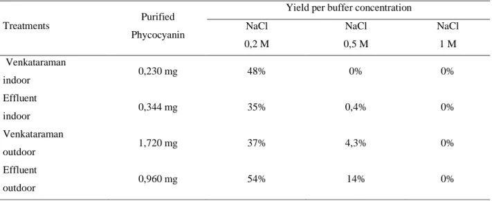
The purification of raw phycocyanin extract by ion exchange chromatography in a DEAE cellulose column resulted in a pigment recovery of 0.23 and 0.34 mg for indoor Venkataraman and effluent, respectively, and 1.72 and 0.96 mg for outdoor Venkataraman and effluent, respectively. The fractions eluted with 0.2M NaCl buffer showed higher performance for all treatments. Table 4 shows the amount of purified pigment per treatment, yields per treatment and concentration of the buffer used.
4 DISCUSSION
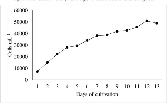
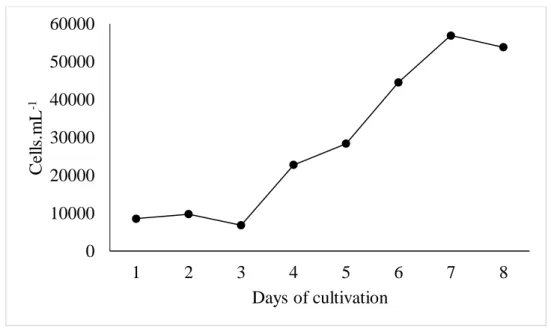
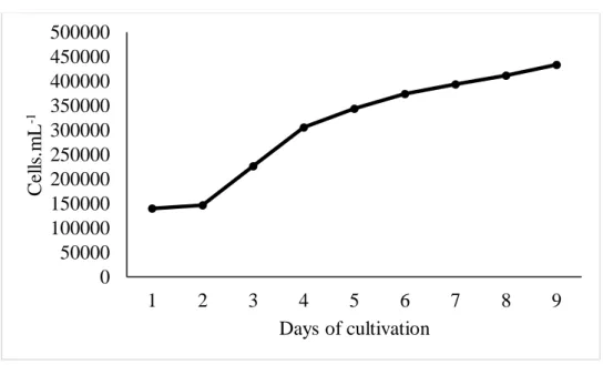
The treatments of the indoor phase exhibited development phases with distinct duration. The effluent treatment showed immediate on set of the exponential growth phase (Figure 1), while the Venkataraman treatment showed induction phase with 2 days duration (Figure 2). In spite of this, this last treatment reached the peak of cellular density in 7 days (Figure 2), while the first (Figure 1), in 12, although there is no significant difference (p>0.05) between productivity and final production results (Table 2). Both treatments of the outdoor phase presented induction phase lasting 1 day. However, the effluent treatment (Figure 3) presented an exponential growth phase lasting only 1 day, while the Venkataraman treatment (Figure 4) lasted 2 days. Thus, this last treatment reached the peak of cellular density within 9 days, while the first, in 13, although no significant difference (p>0.05) is observed for productivity and final production.
Studies indicate that the photoperiod, intensity and light source directly affect the growth of
A. platenis. Saboya (2010), when evaluating the effect of different photoperiods on the carotenoid
profile in the microalgae A. platensis cultivated in Venkataraman medium, identified that the use of constant lighting can increase the duration of the lag phase period, indicating the possible occurrence of photoinhibition. Lee, Erickson and Yang (1987), using urea as a source of nitrogen, observed that the greatest bioenergetic gains occurred in conditions of lower light intensity, while the opposite phenomenon occurred in conditions of higher light intensity. Other authors point to better results with the use of constant lighting in addition to the use of LED lamps, or in a set of fluorescent lamps (HO
et al., 2018; PRATES et al., 2018). Despite providing a greater gain in growth, the continuous use of
artificial lighting is expensive and may affect the economic viability of the activity (GOVINDJEE et
al., 1979).
The results obtained in the final production, which were measured on the day of the microalgae harvest, were 0.077 ± 0.02 g.L-1 and 0.074 ± 0.00 g.L-1 for the indoor Venkataraman and effluent treatments, respectively, with no significant difference (p>0.05) between them, and 0.148 ± 0.019 g. L-1 and 0.140 ± 0.02 g.L-1 for outdoor Venkataraman and effluent treatments, respectively, with no significant difference (p>0.05) between them. The productivity values found were 0.008 ± 0.001 g.L-1.day-1 and 0.005 ± 0.001 g.L-1.day-1 for indoor Venkataraman and effluent treatments, respectively, with no significant difference (p>0.05) and 0.011 ± 0.001 g. L-1.day-1 and 0.008 ± 0.001 g.L-1.day-1 for outdoor Venkataraman and effluent treatments, respectively, with no significant difference between them (p>0.05).
The results obtained in this study are in accordance with those obtained by Coelho (2014), that, when evaluating the growth of A. platensis in effluent produced by tilapia culture, found dry biomass yields ranging from 62.50 ± 29.52 mg.L-1 to 90.28 ± 24.51 mg.L-1, and Nogueira (2012),
that obtained a final yield of 0.22 g.L-1 when cultivating the same cyanophyce an using an internal
system of tilapia effluent. Olguín (2001) achieved a biomass production of A. platensis between 0.35 ± 0.03 g.L-1 to 0.77 ± 0.02 g.L-1 when evaluating the effect of low light flux (66 and 144 μmol.m-2.s -1) and medium with nitrogen deficiency on the development of this microalgae. The pH of the culture
medium also plays a key role on the behavioral dynamics during cultivation (SCHERHOLZ; CURTIS, 2013). In the present research, although there is no significant difference (p>0.05) between the results for final production and productivity between cultures in Venkataraman medium and effluent in both phases, it is worth noting that cultures with Venkataraman medium reached maximum values in a shorter time. This result was probably due to the use of sodium bicarbonate (NaHCO3) in
Venkataraman medium, which is easily dissociated in HCO3- and contributes to better nitrogen availability. The same author points to a higher level of productivity and rate of carbon dioxide (CO2)
fixation for this microalgae when the pH of the culture is close to 8.5, having reached a maximum yield of 72 mg.L-1.d-1, which was 14% higher than when pH of 9.5 was used. This indicates that, although the pH of the effluent medium was close to the ideal, when the values for this parameter were 9.20 ± 0.11 and 8.37 ± 0.17 for indoor Venkataraman and effluent, respectively, and 9.50 ± 0.10 and 8.70 ± 0.20 for outdoor Venkataraman and effluent, respectively, the general composition of the medium had a greater influence on the development of the microalgae.
The extraction process resulted in phycocyanin concentrations in the extracts with values of 0.32 ± 0.01 mg.mL-1 and 0.16 ± 0.01 mg.mL-1 for indoor Venkataraman and effluent treatments,
respectively, with a significant difference (p<0.05) between them; and 1.21 ± 0.01 mg. mL-1 and 0.47 ± 0.01 mg.mL-1 for outdoor Venkataraman and effluent treatments, respectively, with significant difference (p<0.05) between them. When evaluating the pigment concentration in the dry matter, results of 64.4 ± 0.0 mg.g-1 and 31,7 ± 1.4 mg.g-1 for indoor Venkataraman and effluent treatments, respectively, were observed with significant difference (p<0.05) between them, and 45.1 ± 0.3 mg.g
-1 and 9.8 ± 0.1 mg.g-1 for outdoor Venkataraman and effluent treatments, respectively, with
significant difference (p<0.05) between them. The yields obtained in the extractions were 10.51 ± 0.20% and 5.18 ± 1.40% for the indoor Venkataraman and effluent treatments, respectively, with a significant difference (p<0.05) between them, and 40.40 ± 0.12% and 15.61 ± 0.22% for the outdoor Venkataraman and effluent treatments, respectively, with a significant difference (p<0.05) between them. The degree of purity of the extracts was 2.88 ± 0.00 and 1.89 ± 0.06 for indoor Venkataraman and effluent treatments, respectively, with a significant difference (p<0.05) between them; and 0.74 ± 0.01 and 0.36 ± 0.01 for outdoor Venkataraman and effluent treatments, respectively, with a significant difference (p<0.05) between them.
Different studies point to different levels of c-phycocyanin production by the microalgae A.
platensis. Wan et al. (2016) achieved values ranging from 10 to 15% yield, while Mehar et al. (2019)
obtained results ranging from 10 to 14% yield, with the maximum value obtained at a pH of 8.5 and the minimum value at 7.5. Chen et al. (2016), reported 17% of yield when using CO2 supplementation. Manirafasha et al. (2018) found values of 0.234 mg.mL-1 when testing different
metabolic stress conditions for increased biomass production and accumulation of this phycobiliproteins. Chen et al. (2010) found phycocyanin concentration of 149 mg.g-1 when testing the use of green spectrum Light Emitting Diodes (LED). Prates et al. (2017) found phycocyanin concentration values ranging from 46.36 to 126.39 mg.g-1 when testing LED's of different colors. Sloth et al. (2017) achieved pigment production ranging from 11.4 ± 0.8 to 20.8 ± 1.0 mg.g-1 using
Galdieria sulfuraria biomass. Whereas Lee et al. (2017) achieved maximum productivity of 57.4
mg.L-1.day-1using the biomass of Nostoc sp.
The results obtained with the use of Venkataraman medium were superior (p<0.05) in both phases. The microalgae optimize the energy storage according to the metabolic strategy that allows them to better adapt to the culture conditions, consequently modifying their content of carbohydrates, lipids and proteins. The nitrogen source and its availability in the medium severely affect the development of microalgae. A low nitrogen supply inhibits the production of compounds such as proteins, causing a drop in the production of important biochemical compounds, which can influence the photosynthetic activity of these organisms such as the chlorophyll and phycocyanin pigments. Urea in an alkaline medium under goes fast dissociation releasing ammonia and CO2. Ammonia is
better absorbed by microalgae, since its metabolism demands less energy consumption to the detriment of nitrogen sources that use nitrate salts, for example. The final concentration of phycocyanin is strongly influenced by the extraction method. In the literature there are reports of several extraction methods, which combine different steps. Besides quantity, quality is strongly influenced by the selection of the appropriate extraction method (SASSANO et al., 2004; MATSUDO, 2009; MARKOU, 2012; LU et al., 2018; SHANTHI; PREMALATHA; ANANTHARAMAN, 2018; MAO; GUO, 2018; İLTER et al., 2018).
The number of studies evaluating the feasibility of cultivating microalgae in low-cost systems has been growing. Given that culture media represent high costs in the production process, their replacement by alternative media may be feasible. Several studies have been evaluating the cultivation of this cyanophycean in different types of wastewater, whether from industries, agriculture, or even aquaculture (MEZZOMO et al., 2010; OLGUÍN, 2012; EL-KASSAS; HENEASH; HUSSEIN, 2015; RUIZ-MARTINEZ et al., 2012; SLOTH et al., 2017).
The purification of crude phycocyanin extract using ion exchange chromatography in a DEAE-cellulase column resulted in a higher amount of pigments purified in the fractions eluted with 0.2 M NaCl (Table 4). Soares (2005), when performing phycocyanin purification procedure by ion exchange chromatography, using NaCl at concentrations 0.5, 1.6 and 2.0 M, found the most significant results in the eluted fractions with the lowest concentration. Molina (2011) observed the highest pigment concentrations when performing the elution with NaCl 0.1 and 0.2 M, corroborating with the results found in this study. The advantage in using salts with lower concentrations for purification lies in the reduction of costs and greater facility in removing traces of salt by dialysis processes, for example. The appropriate choice of purification methods is essential, since it is known that the costs of phycocyanin purification processes are around 50 to 90% of total production costs (PATIL; RAGHAVARAO, 2007).
5 CONCLUSIONS
The use of fish effluent for the cultivation of Arthrospira platensis was effective, providing a final production similar to the Venkataraman medium, however, it increases the cultivation time necessary to reach the maximum values. The concentrations of phycocyanin extracted from the biomass produced in the effluent were significant, however, lower than those found in the biomass produced in Venkataraman medium. The purification of crudephycocyanin extract from ion exchange chromatography in DEAE-cellulase column proved to be effective, with the largest quantities of purified pigment obtained in the eluted fractions with 0.2 M NaCl.
Despite the less substantial pigment production, the use of the effluent may become a more viable alternative from the commercial point of view, as it is a low-cost waste from another activity, and may help in the mitigation of potential environmental impacts caused by tilapia culture. Therefore, it is necessary to carry out economic feasibility studies to establish the cost benefit of the biomass production of Arthrospira platensis and the phycocyanin pigment coming from the fish effluent.
ACKNOWLEDGEMENTS
We are indebted to the Coordination for the Improvement of Higher Education Personnel (CAPES) Improvement of Higher Education Personnel (CAPES) who provided I.G.R.F. Gomes with the scholarship for her M.Sc. degree.
Table 1 Linear regression equations and coefficients of determination (R²) between the absorbance values at 680 nm and the trichome count of A. platensis grown in indoor systems
Figure 1Growth curve of A. platensis grown in fish effluent in indoor system
0 10000 20000 30000 40000 50000 60000 1 2 3 4 5 6 7 8 9 10 11 12 13 C el ls .m L -1 Days of cultivation
Treatments Linear regression equations R²
Ventakaraman indoor Y = 836.8 x OD680 – 70,748 0,8917
Figure 2 Growth curve of A. platensis grown in Venkataraman in indoor system
Table 2 Abiotic and kinetic parameters of A. platensis cultures exposed to different systems
Parameters Treatments Venkataraman indoor Effluent indoor Venkataraman outdoor Effluent outdoor Final production (g.L-1) 0,077 ± 0,020 a 0,074 ± 0,005a 0,148 ±0,019b 0,140 ± 0,022b Productivity (g.L-1.dia-1) 0,008 ± 0,001 ab 0,005 ± 0,001a 0,011 ± 0,001b 0,008 ± 0,001 ab
Different lower case letters in the same line indicate a significant difference between the means by Tukey's test (p<0.05)
Table 3 Results of phycocyanin concentrations in the extract, phycocyanin concentration in dry biomass, the purity of the extract and the yields obtained
Parameters Treatments Venkataraman indoor Effluent indoor Venkataraman outdoor Effluent outdoor Phycocyanin (mg.mL-1) 0,32 ± 0.01 a 0,16 ± 0,01b 1,21 ± 0,01c 0,47 ± 0,01d Phycocyanin (mg.g-1) 64,40 ± 0,040 a 31,70 ± 1,37b 39,60 ± 0,26c 15,30 ± 0,08d Yeild (%) 10,51 ± 0,01a 5,18 ± 0,22b 40,40 ± 0,12c 15,61 ± 0,22d Purity of the extract 2,88 ± 0,01a 1,89 ± 0,06b 0,74 ± 0,01c 0,36 ± 0,01d
Different lower case letters in the same line indicate a significant difference between the means by Tukey's test (p<0.05)
0 10000 20000 30000 40000 50000 60000 1 2 3 4 5 6 7 8 C el ls .m L -1 Days of cultivation
Table 4 Results of the DEAE cellulose ion exchange chromatography purification process
Treatments Purified
Phycocyanin
Yield per buffer concentration NaCl 0,2 M NaCl 0,5 M NaCl 1 M Venkataraman indoor 0,230 mg 48% 0% 0% Effluent indoor 0,344 mg 35% 0,4% 0% Venkataraman outdoor 1,720 mg 37% 4,3% 0% Effluent outdoor 0,960 mg 54% 14% 0%
Figure 3 Growth curve of A. platensis grown in fish effluent in outdoor system
0 50000 100000 150000 200000 250000 300000 350000 400000 450000 500000 0 1 2 3 4 5 6 7 8 9 10 11 12 13 14 C el ls .m L -1 Days of cultivation
Figure 4 Growth curve of A. platensis grown in Venkataraman in outdoor system
REFERENCES
ABALDE, J.; BETANCOURT, L., TORRES, E.; CID, A.; BARWELL, C. Purification and characterization of phycocyanin from marine cyanobacterium Synechococcus sp. IO9201. Plant Science, v.136, n.1, p.109-120, 1998.
BECKER, E.W. Microalgae as a source of protein. Biotechnology Advances, v. 25, n. 2, p. 207-210, 2007.
BENNETT, A.; BOGORAD, L. Complementary chromatic adaptation in a filamentous bluegreen alga. Journal of Cell Biology, v. 58, n. 2, p.419-435, 1973.
BÖCKER, L.; HOSTETTLERA, T.; DIENERB, M.; EDERC, S.; DEMUTHC, T.; ADAMCIKB, J.; REINEKED, K.; LEEBD, E.; NYSTRÖMC, L.; MATHYS,A. Time-temperature-resolved functional and structural changes of phycocyanin extracted from Arthrospira platensis/Spirulina. Food Chemistry, v. 316, 2020.
BÖCKER, L.; ORTMANN, S.; SURBER, J.; LEEB, E.; REINEKE, K.; MATHYS, A. Biphasic short time heat degradation of the blue microalgae protein phycocyanin from Arthrospira platensis. Innovative Food Science & Emerging Technologies, v. 52, p. 116-121, 2019.
BOROWITZKA, M. A. High-value products from microalgae-their development and commercialization. Journal of Applied Phycology, v. 25, n.3, p. 743-756, 2013.
0 50000 100000 150000 200000 250000 300000 350000 400000 450000 500000 1 2 3 4 5 6 7 8 9 C el ls .m L -1 Days of cultivation
CAPORGNO, M. P.; MATHYS, A. Trends in Microalgae Incorporation Into Innovative Food Products With Potential Health Benefits. Frontiers in Nutrition, v. 5, p. 1-10, 2018.
CHAUDHARY, A.; GUSTAFSON, D.; MATHYS, A. Multi-indicator sustainability assessment of global food systems. Nature Communications, v. 9, p. 1-13, 2018.
CHEN, C. Y.; KAO, P. C.; TAN, C. H.; SHOW, P. L.; CHEAH, W. Y.; LEE, W. L.; CHANG, J. S. Using an innovative pH-stat CO2 feeding strategy to enhance cell growth and C-phycocyanin
production from Spirulina platensis. Biochemistry Engineer Journal, v. 112, p. 78-85, 2016.
CHEN, H.B.; WU, J. Y.; WANG, C. F.; FU, C. C.; SHIEH, C. J.; CHEN, C. I.; WANG, C. Y.; LIU, Y.C. Modeling on chlorophyll a and phycocyanin production by Spirulina platensis under various light-emitting diodes. Biochemistry Engineer Journal, v. 53, p. 52-56, 2010.
EL-KASSAS, H. Y.; HENEASH, A. M. M.; HUSSEIN, N. R. Cultivation of Arthrospira (Spirulina)
platensis using confectionary wastes for aquaculture feeding. Journal of Genetic Engineering and
Biotechnology, v. 13, n. 2, p.145-155, 2015.
FAO. The State of World Fisheries and Aquaculture 2018 - Meeting the sustainable development goals. Roma. 2018. 227 p.
GOVINDJEE, S.; WONG, D.; PRÉZELIN, B. B.; SWEENEY, B. M. Chlorophyll-a fluorescence of
Gonyaulax-Polyedra grown on a light-dark cycle and after transfer to constant light. Photochemistry
and Photobiology, v. 30, n. 3, p. 405-411, 1979.
HARISKOS, I.; POSTEN, C. Biorefinery of microalgae - opportunities and constraints for different production scenarios. Biotechnology Journal, v. 9, n. 6, p. 739-752, 2014.
HO, S.; LIAO, J.; CHEN, C.; CHANG, J. Combining light strategies with recycled medium to enhance the economic feasibility of phycocyanin production with Spirulina platensis. Bioresource Technology, v. 247, p. 669-675, 2018.
JUTUR, P.P.; NESAMMA, A. A.; SHAIKH, K. M. Algae-derived marine oligosaccharides and their biological applications. Frontiers in Marine Science, v. 3, p. 1-5, 2016.
LEE N.; OH H.; KIM, HE.; AHN, C. Higher production of C-phycocyanin by nitrogen-free (diazotrophic) cultivation of Nostoc sp. NK and simplified extraction by dark-cold shock. Bioresource Technology, v. 227, p. 164-170, 2017.
LEE, I.; ERICKSON, L. E.; YANG, S. S. Kinetics and bionergetics of light-limited photoautotrophic growth of Spirulina platensis. Biotechnology and Bioengineering, v. 29, p. 832–843, 1987.
LI, T.; XU, J.; GAO, B.;XIANG, B.; LI, A.;ZHANG, Z. Morphology, growth, biochemical composition and photosynthetic performance of Chlorella vulgaris (Trebouxiophyceae) under low and high nitrogen supplies. Algal Research, v. 16, p. 481-491, 2016.
LU, W.; ALAM, M. A.; LUO, W.; ASMATULU, E. Integrating Spirulina platensis cultivation and aerobic composting exhaust for carbon mitigation and biomass production. Bioresource Technology, v. 271, p. 59-65, 2018.
MANIRAFASHA, E.; MURWANASHYAKAA, T.; NDIKUBWIMANAC, T.; NURRASHID AHMEDA, N. R.; LI, J.; LU, Y.; ZENG, X.; LING, X.; JING, K. Enhancement of cell growth and phycocyanin production in Arthrospira (Spirulina) platensis by metabolic stress and nitrate fed-batch. Bioresource Technology, v. 255, p. 293-301, 2018.
MAO, R.; GUO, S. Performance of the mixed LED light quality on the growth and energy efficiency of Arthrospira platensis. Applied Microbiology and Biotechnology, v. 102, p. 5245–5254, 2018. MARKOU, G. Alteration of the biomass composition of Arthrospira (Spirulina) platensis under various amounts of limited phosphorus. Bioresource Technology, v. 116, p. 533-53, 2012.
MARTELLI, G.; FOLLI, C.; VISAI, L.; DAGLIA, M.; FERRARI, D. Thermal stability improvement of blue colorant C-Phycocyanin from Spirulina platensis for food industry applications. Process Biochemistry, v. 49, n. 1, p. 154-159, 2014.
MATSUDO, M. C.; BEZERRA, T. P.; SATO, S.; PEREGO, P; CONVERTI, A.; CARVALHO, J. C. M. Repeated fed-batch cultivation of Arthrospira (Spirulina) platensis using urea as nitrogen source. Biochemical Engineering Journal. v. 43, p. 52-57, 2009.
MEHAR, J.; SHEKH, A.; NETHRAVATHY, M. U.; SARADA, R.; CHAUHAN, V. S. Automation of pilot-scale open raceway pond: A case study of CO2-fed pH control on Spirulina biomass, protein and phycocyanin production. Journal of CO2 Utilization, v. 33, p. 384-393, 2019.
MEZZOMO, N.; SAGGIORATO, A. G.; SIEBERT, R.; TATSCH, P.; OLIVEIRA, L.; HEMKEMEIER, M. C.; COSTA, M.; BERTOLIN, L. A. V.; ELITA, T. COLLA, L. M. Cultivation of microalgae Spirulina platensis (Arthrospira platensis) from biological treatment of swine wastewater. Food Science and Technology, v. 30, n. 1, p. 173-178, 2010.
MORENO, J. F.; CORZO, N.; MONTILLA, A.; VILLAMIEL, M.; OLANO, A. Current state and latest advances in the concept, production and functionality of prebiotic oligosaccharides. Current
Opinion in Food Science, v. 13, p. 50–5, 2017.
NOGUEIRA, S. M. S. Tratamento de efluentes de cultivos de tilápia do nilo (Oreochromis niloticus) com a microalga Spirulina platensis. 2012. 59 f. Dissertação (Mestrado em Engenharia de Pesca) - Universidade Federal do Ceará, Fortaleza, 2012.
OLGUÍN, E. J.; GALICIA, S.; ANGULO-GUERRERO, O.; HERNANDEZ, E. The effect of low light flux and nitrogen deficiency on the chemical composition of Spirulina sp. (Arthrospira) grown on digested pig waste. Bioresource Technology, v. 77, p. 19-24, 2001.
PANDEY, V.; PANDEY, A.; SHARMA V. Biotechnological applications of cyanobacterial phycobiliproteins. International Journal of Current Microbiology and Applied Sciences, v. 2, p. 89-97, 2013.
PATIL, G.; RAGHAVARAO, K. S. M. S. Aqueous two phase extraction for purification of Cphycocyanin. Biochemical Engineering Journal, v. 34, p.156-164, 2007.
PRATES, D. F.; RADMANN, E. M.; DUARTE, J. H.; MORAIS, M. G.; COSTA, J. A. V. Spirulina cultivated under different light emitting diodes: Enhanced cell growth and phycocyanin production. Bioresource Technology, v. 256, p. 38-43, 2018.
RAPOSO, M. F.; MORAIS, A. M. M. B.; MORAIS, R. M. F. Emergent Sources of Prebiotics: Seaweeds and Microalgae. Marine Drugs, v. 48, n. 2, p. 1-27, 2016.
RUIZ-MARTINEZ A.; MARTIN GARCIA N.; ROMERO I.; SECO A.; FERRER J. Microalgae cultivation in wastewater: Nutrient removal from anaerobic membrane bioreactor effluent. Bioresource Technology, v. 126, p. 247-253, 2012.
SABOYA, J. P. S. Teores de β-Caroteno e α-tocoferol presentes na microalga Spirulina (Arthrospira)
platensis cultivada com diferentes fotoperíodos. 2010. Dissertação (Mestrado em Engenharia de
Pesca) – Centro de Ciências Agrárias, Universidade Federal do Ceará, Fortaleza. 2010.
SASSANO, C. E. N. Cultivo de Spirulina platensis por processo contínuo utilizando cloreto de amônio como fonte de nitrogênio. 2004. 116 f. Tese de Doutorado – Universidade de São Paulo, Faculdade de Ciências Farmacêuticas, São Paulo.
SCHERHOLZ, M.L.; CURTIS, W.R. Achieving pH control in microalgal cultures through fed-batch addition of stoichiometrically-balanced growth media. BMC Biotechnology, v. 13, p. 1-15, 2013. SHANTHI, G.; PREMALATHA, M.; ANANTHARAMAN, N. Effects of L-amino acids as organic nitrogen source on the growth rate, biochemical composition and polyphenol content of Spirulina
platensis. Algal Resarch, v. 35, p. 471-478, 2018.
SHARMA N.; SHARMA P. Industrial and biotechnological applications of algae: a review. Journal
of Advances in Plant Biology, v. 1, n. 1, p. 1–26, 2017.
SILVEIRA, S. T.; BURKERT, J. F. M.; COSTA, J. A. V.; BURKERT, S. J.; KELIL, S. J. Optimization of phycocyanin extraction from Spirulina platensis using factorial design. Bioresource Technology, v. 98, p. 1629-1634, 2007.
SLOTH, J.K.; JENSEN, H.C.; PLEISSNER, D;. N.; ERIKSEN.T. Growth and phycocyanin synthesis in the heterotrophic microalga Galdieria sulphuraria on substrates made of food waste from restaurants and bakeries. Bioresource Technology, v. 238, p. 296-305, 2017.
SMETANA, S.; MATHYS, A.; KNOCH, A.; HEINZ, V. Meat alternatives: life cycle assessment of most known meat substitutes. The International Journal of Life Cycle Assessment, v. 20, p. 1254– 67, 2015.
SMETANA, S.; SANDMANN, M.; ROHN, S.; PLEISSNER, D.; HEINZ, V. Autotrophic and heterotrophic microalgae and cyanobacteria cultivation for food and feed: life cycle assessment. Bioresource Technology, v. 245, p. 162-170, 2017.
SOARES, N. N. Cultivo e extração de pigmentos das microalgas Spirulina platensis e
Haematococcus pluvialis. 2005. 56 f. Dissertação (Mestrado em Engenharia de Pesca) – Centro de
Ciências Agrárias, Engenharia de Pesca, Universidade Federal do Ceará, Fortaleza, 2005.
TEIMOURI, M; AMIRKOLAIE, A. K; YEGANEH, S. The effects of Spirulina platensis meal as a feed supplement on growth performance and pigmentation of rainbow trout (Oncorhynchus mykiss). Aquaculture, v.396-399, n.1, p.14-19, 2013.
VERMEULEN, S. J.; CAMPBELL, B. M.; INGRAM, J. S. I. Climate change and food systems.
WAN, M. X.; WANG, Z. Y.; ZHANG, Z.; WANG, J.; LI, S. L.; YU, A. Q.; LI Y. G. A novel paradigm for the high-efficient production of phycocyanin from Galdieria sulphuraria. Bioresource. Technology, v. 218, p. 272-27, 2016.
WELLS, M. L.; POTIN, P.; CRAGIE, J. S.; RAVEN, J. A.; MERCHANT, S. S.; HELLIWELL, K. E.; SMITH, A. G.; CAMIRE, M. E.; BRAWLEY, S. H. Algae as nutritional and functional food sources: revisiting our understanding. Journal of Applied Phycology, v. 29, p.949–982, 2017. WELLS, M. L.; POTIN, P.; CRAIGIE, J. S.; RAVEN, J. A.; MERCHANT, S. S.; HELLIWELL, K. E.; SMITH, A. G.; CAMIRE, M. E.; BRAWLEY, S. H.Algae as nutritional and functional food sources: revisiting our understanding. Journal of Applied Phycology, v. 29, n. 2, p. 949-982, 2016. WU, G.; FANZO, J.; MILLER, D. D.; PINGALI, P.; POST, M.; STEINER, J. L.; THALACKER-MERCER, A. E. Production and supply of high-quality food protein for human consumption: sustainability, challenges, and innovations. Annals of the New York Academy of Sciences, v. 1321, p. 1-19, 2014.