• Nenhum resultado encontrado

Biocompatibility assessment of calcium-silicate based endodontic sealers: in vitro studies

N/A
N/A
Protected

Academic year: 2021

Share "Biocompatibility assessment of calcium-silicate based endodontic sealers: in vitro studies"

Copied!
116
0
0

Texto

(1)

BIOCOMPATIBILITY ASSESSMENT OF

CALCIUM-SILICATE BASED ENDODONTIC SEALERS:

IN VITRO STUDIES

Fábio Miguel dos Santos Costa

Master’s Degree in Biomedical Engineering

Faculty of Engineering, University of Porto

Porto, July 2014

(2)
(3)

BIOCOMPATIBILITY ASSESSMENT OF

CALCIUM-SILICATE BASED ENDODONTIC SEALERS:

IN VITRO STUDIES

Fábio Miguel dos Santos Costa

Supervisor:

Prof. Maria Helena Raposo Fernandes

Faculty of Dental Medicine, University of Porto

Co-supervisor:

Prof. Pedro Sousa Gomes

Faculty of Dental Medicine, University of Porto

(4)
(5)

i

To My Parents and Brother

“The most beautiful experience we can have is the mysterious – the fundamental emotion which stands at the cradle of true art and true science.” ― Albert Einstein, Albert Einstein

(6)

ii The work presented in this dissertation would not have been possible without the collaboration and close association of many people and entities to which I would like to gratefully and sincerely thank:

to my supervisor Prof. Maria Helena Fernandes for the mentorship, guidance, availability, understanding and patience;

to my co-supervisor Prof. Pedro Sousa Gomes for the guidance and vast knowledge and skill about the materials used;

to the Laboratory for Bone Metabolism and Regeneration of FMDUP for all the support in the development and concretization of this project;

to Dr. Mónica Garcia for the knowledge transmitted about the laboratory techniques; to my laboratory colleagues José Carlos Silva, Stella Terena, and specially to Elisabete Gonçalves, for the stimulating discussions and their friendship, support and encouragement;

to my friends who were always there when I needed them the most and for providing moral support and motivation, which drives me to give my best;

to my family for the support they provided me through my entire life and their absolute confidence in me. The knowledge that they will always be there to pick up the pieces is what allows me to repeatedly risk getting shattered.

(7)

iii

Abstract

Calcium silicate-based materials are used in various endodontic procedures and became popularized with the appearance of MTA in 1993 as a root-end filling material to use in pulp capping, pulpotomy, root perforation repair, apexification, internal resorption or apical barrier formation for teeth with necrotic pulps and open apexes. These numerous clinical applications of MTA are due to its inherent advantages including biocompatibility and regenerative capabilities. Several modifications of MTA have been created along the years to improve some aspects of the physicochemical and biological profile of the original formulation.

Root canal sealers are intended to be contained within the root canal space, but they may extrude through the apical constriction, or eluents from the sealers may come into contact with periradicular tissues. This might cause irritation and delay in wound healing. Moreover, the contact of root canal filling materials with periapical tissues can also affect the normal bone metabolism and regeneration. Regarding this, it is known that angiogenesis is critical in the bone environment for the normal bone remodelling and regenerative events. Accordingly, biological evaluation of endodontic sealers benefit from the biocompatibility profile concerning bone formation and angiogenesis.

In this context, the aim of this study was to compare four calcium silicate-based sealers, namely ProRoot® MTA, MTA PlusTM, MTA Fillapex® and BiodentineTM regarding the effects of the sealers’ extracts on (i) osteoblastic cell behaviour, by using human bone-marrow derived mesenchymal stem cell cultures and an ex vivo osteogenic assay (regeneration of defects created on the parietal bone of neonatal mice), and (ii) endothelial cell behaviour, by using human endothelial cell cultures and an in vivo angiogenesis assay (chorioallantoic membrane – CAM - assay). The effects of the sealers’ extracts were dependent on the concentration and the time of exposure. Regarding the in vitro studies, the toxicity profile was similar for endothelial and mesenchymal stem cells regarding proliferation and functional parameters, however, endothelial cells were more sensitive to the toxic effects observed at the higher extract concentrations. ProRoot MTA and MTA Plus presented the best citocompatibility profile, followed by Biodentine and MTA Fillapex. This pattern of toxicity was also observed in the ex vivo regeneration of the rat parietal bone defects. However, on the CAM assay, no significant differences were observed on the angiogenic response elicited by the extracts of the four sealers. Overall, this study corroborates the low toxicity of calcium silicate-based sealers, but ProRoot MTA and MTA Plus presented the best biocompatibility profile.

(8)

iv Os materiais à base de silicato de cálcio são utilizados em vários procedimentos endodônticos e tornaram-se conhecidos com o aparecimento do MTA em 1993, como um material de preenchimento radicular para uso em capeamento pulpar, pulpotomia, reparação de perfuração de raiz, apicificação, reabsorção interna ou formação de barreira apical de dentes com necrose pulpar e ápices abertos. Estas inúmeras aplicações clínicas do MTA devem-se às suas vantagens inerentes, incluindo biocompatibilidade e capacidades regenerativas. Várias modificações do MTA foram criadas ao longo dos anos para melhorar alguns aspectos de perfil físico-químico e biológico da formulação original.

Os cimentos endodônticos devem ficar contidos dentro do espaço do canal radicular, no entanto pode haver extrusão através da constrição apical, ou os eluentes dos cimentos podem entrar em contato com os tecidos perirradiculares. Isto pode provocar irritação e atraso na cicatrização. Além disso, o contato dos materiais de preenchimento do canal radicular com os tecidos periapicais também pode afetar o metabolismo e regeneração óssea. Relativamente a este aspecto, sabe-se que a angiogénese é crítica para o ambiente ósseo durante a remodelação óssea normal e os eventos regenerativos.

Deste modo, a avaliação biológica de cimentos endodônticos beneficia do perfil de biocompatibilidade relativo à formação óssea e angiogénese. Neste contexto, o objetivo deste estudo foi comparar quatro cimentos à base de silicato de cálcio, ou seja, ProRoot® MTA, MTA PlusTM, MTA Fillapex® e BiodentineTM relativamente aos efeitos dos extratos dos cimentos sobre (i) o comportamento de células osteoblásticas, usando células estaminais mesenquimais derivadas de medula óssea humana e um ensaio ex vivo osteogénico (regeneração de defeitos criados no osso parietal de ratos neonatais), e (ii) o comportamento de células endoteliais, através da utilização de culturas de células endoteliais humanas e um ensaio in vivo de angiogénese (ensaio da membrana corioalantóide, CAM). Os efeitos dos extractos foram dependentes da concentração e do tempo de exposição. Considerando os estudos in vitro, o perfil de toxicidade foi semelhante para células endoteliais e células estaminais mesenquimais em termos de proliferação e parâmetros funcionais, no entanto, observou-se maior sensibilidade das células endoteliais aos efeitos tóxicos das concentrações mais elevadas dos extractos. O ProRoot MTA e o MTA Plus apresentaram o melhor perfil citocompatibilidade, seguido pela Biodentine e pelo MTA Fillapex. Este padrão de toxicidade também foi observado na regeneração ex vivo dos defeitos de osso parietal de rato. No entanto, no ensaio CAM, não foram observadas diferenças significativas na resposta angiogénica provocada pelos extratos dos quatro cimentos. Em conclusão, este estudo corrobora a baixa toxicidade dos cimentos à base de silicato de cálcio, mas o ProRoot MTA e o MTA Plus apresentarem o melhor perfil de biocompatibilidade.

(9)

v

Index

Acknowledgments ... ii Abstract ... iii Resumo ... iv Index ... v

Figure Index ... viii

Graphic Index ... x

Table Index ... xi

Abbreviation List ... xii

I: LITERATUTE OVERVIEW ... 1

1.1- Structure of the teeth ... 3

1.2- Structure of the supporting structures of the teeth ... 5

1.2.1- Alveolar process ... 5

1.2.2- Bone cells ... 6

1.2.3- Bone metabolism ... 8

1.2.4- Osteogenesis and angiogenesis ... 9

1.2.5- Interaction of endothelium and bone ... 9

1.2.6- Bone healing and angiogenesis ... 10

1.3- Endodontics ... 12

1.3.1- Endodontic treatment procedures ... 12

1.3.2- Repair in the pulp ... 14

1.4- Root canal filling materials ... 15

1.4.1- Root canal filling materials requirements ... 15

1.4.2- Gutta-percha cones ... 16

1.4.3- Sealers ... 18

1.4.3.1- Zinc-oxide eugenol (ZnOE) sealers ... 18

1.4.3.2- Polyketone sealers ... 19

1.4.3.3- Epoxy resin sealers ... 19

1.4.3.4- Glass ionomer cement (GIC) sealers ... 21

1.4.3.5- Calcium hydroxide (CH) sealers ... 22

1.4.3.6- Calcium silicate (CS)-based sealers ... 24

1.4.3.6.1- Chemical properties ... 24

1.4.3.6.2- Physical properties ... 25

1.4.3.6.3- Antibacterial and antifungal properties ... 26

1.4.3.6.4- Portland cement ... 26

1.5- Types of calcium silicate-based sealers ... 29

1.5.1- ProRoot MTA ... 30

1.5.2- MTA Plus ... 31

1.5.3- MTA Fillapex ... 31

1.5.4- Biodentine ... 32

II: RESEARCH HYPOTHESIS AND OBJECTIVES ... 33

III: MATERIALS AND METHODS ... 37

(10)

vi

3.1- Cell culture ... 41

3.2- Cell proliferation assay (DNA content) ... 41

4- Effect of the sealers’ extracts on Osteoblastic cell behaviour ... 42

4.1- Human mesenchymal stem cells ... 42

4.1.1- Cell culture ... 42

4.1.2- Characterization of the cell response ... 42

4.1.2.1- Cell proliferation assay (DNA content)... 42

4.1.2.2- F-Actin cytoskeleton and cell morphology ... 43

4.1.2.3- Total Protein Content... 43

4.1.2.4- Alkaline phosphatase activity ... 43

4.1.2.5- Alkaline phosphatase staining ... 44

4.1.2.6- Collagen staining and quantification ... 44

4.2- Ex vivo Osteogenic assay ... 44

4.2.1- Alkaline phosphatase staining ... 45

4.2.2- Collagen staining ... 45

5- Effect of the sealers’ extracts on Endothelial cells and Angiogenesis ... 46

5.1- Human endothelial cells ... 46

5.1.1- Cell culture ... 46

5.1.2- Characterization of the cell response ... 46

5.1.2.1- Cell proliferation assay (DNA content)... 46

5.1.2.2- Matrigel coating assay ... 46

5.2- In vivo Angiogenesis Assay ... 47

6- Statistical analysis ... 48

IV: RESULTS ... 49

1- Cytotoxicity evaluation of the sealers’ extracts ... 51

1.1- Cell proliferation ... 51

2- Effect of the sealer’s extracts on osteoblastic cell behaviour ... 52

2.1- Human mesenchymal stem cells ... 52

2.1.1- Cell proliferation ... 52

2.1.2- F-actin cytoskeleton and cell morphology ... 53

2.1.3- Alkaline phosphatase activity ... 54

2.1.4- Alkaline phosphatase staining ... 55

2.1.5- Collagen staining and quantification... 59

2.2- Ex vivo osteogenic assay ... 64

2.2.1- Alkaline phosphatase staining ... 66

2.2.2- Collagen staining ... 67

3- Effect of the sealer’s extracts on endothelial cells and angiogenesis ... 68

3.1- Human endothelial cells ... 68

3.1.1- Cell proliferation ... 68

3.1.2- Matrigel coating assay ... 69

3.2- In vivo angiogenesis assay ... 70

(11)

vii

VI: CONCLUSION ... 85 VII: REFERENCES ... 89

(12)

viii

Figure 1- Teeth anatomy. ... 3

Figure 2- (A) Scheme of alveolar bone; (B) Histological section of a tooth with the alveolar bone structures highlighted. ... 5

Figure 3- Bone cells origin. ... 6

Figure 4- Bone healing steps. ... 11

Figure 5- Root canal therapy stages... 13

Figure 6- (A) Root canal filling with gutta-percha; (B) Commercial gutta-percha. ... 17

Figure 7- Examples of ZnOE sealers: (A) Tubli-Seal; (B) Pulp Canal Sealer. ... 19

Figure 8- Examples of epoxy resin sealers: (A) EZ Fill; (B) AH26; (C) AH Plus. ... 20

Figure 9- Examples of GIC: (A) Glass Ionomer Base Cement; (B) Ketac Cem; (C) Ketac Fil Plus .. 21

Figure 10- Examples of CH-based sealers: (A) CRCS; (B) Apexit; (C) Sealapex; (D) Dycal; (E) Calcicur; (F) Calcimol LC. ... 23

Figure 11- Clinical applications of MTA ... 24

Figure 12- ProRoot® MTA (Dentsply; Tulsa Dental Products, Tulsa, OK, USA). ... 30

Figure 13- MTA PlusTM (Prevest Denpro, Jammu, India). ... 31

Figure 14- MTA Fillapex® (Angelus, Londrina, PR, Brazil). ... 31

Figure 15- BiodentineTM (Septodont, Saint Maur de Fossés, France). ... 32

Figure 16- Sealers’ preparation method: (A) ProRoot MTA; (B) MTA Plus; (C) MTA Fillapex; (D) Biodentine. ... 40

Figure 17- Placement of the sealers over the wells surface with the addition of α-MEM. ... 40

Figure 18- Procedure to obtain the parietal bone of neonatal mice. ... 45

Figure 19- CAM assay procedure on day 4 ... 47

Figure 20- CAM assay procedure on days 8 (A and B) and 11 (C through F) ... 48

Figure 21- CLSM imaging of hMSCs cultures at day 7 in the absence (control) and presence of extracts of the endodontic sealers (extract dilutions 1:20 to 1:2)... 53

Figure 22- ALP staining of human mesenchymal stem cell cultures exposed to the extract of ProRoot MTA (1:20 to 1:2 dilutions) for 7, 14 and 21 days. Magnification: 100x. ... 55

Figure 23- ALP staining of human mesenchymal stem cell cultures exposed to the extract of MTA Plus (1:20 to 1:2 dilutions) for 7, 14 and 21 days. Magnification: 100x. ... 56

Figure 24- ALP staining of human mesenchymal stem cell cultures exposed to the extract of MTA Fillapex (1:20 to 1:2 dilutions) for 7, 14 and 21 days. Magnification: 100x. ... 57

Figure 25- ALP staining of human mesenchymal stem cell cultures exposed to the extract of Biodentine (1:20 to 1:2 dilutions) for 7, 14 and 21 days. Magnification: 100x. ... 58

Figure 26- Collagen staining of human mesenchymal stem cell cultures exposed to the extract of ProRoot MTA (1:20 to 1:2 dilutions) for 7, 14 and 21 days. Magnification: 100x. ... 59

Figure 27- Collagen staining of human mesenchymal stem cell cultures exposed to the extract of MTA Plus (1:20 to 1:2 dilutions) for 7, 14 and 21 days. Magnification: 100x. ... 60

Figure 28- Collagen staining of human mesenchymal stem cell cultures exposed to the extract of MTA Fillapex (1:20 to 1:2 dilutions) for 7, 14 and 21 days. Magnification: 100x. ... 61

Figure 29- Collagen staining of human mesenchymal stem cell cultures exposed to the extract of Biodentine (1:20 to 1:2 dilutions) for 7, 14 and 21 days. Magnification: 100x. ... 62

Figure 30- Ex vivo regeneration of neonatal rat parietal bone defects in the presence of the sealers’ extracts (1:20 and 1:5 dilutions) over a period of 14 days. Magnification: 1x. Scale bar: 0.5mm. ... 65

(13)

ix

Figure 31- ALP staining of the osteoblastic cell cultures outgrown from the rat parietal bone cultured in the presence of the sealers’ extracts (1:20 and 1:5 dilutions), at days 7 and 14. Magnification: 40x. ... 66 Figure 32- Collagen staining of the osteoblastic cell cultures outgrown from the rat parietal bone cultured in the presence of the sealers’ extracts (1:20 and 1:5 dilutions), at days 7 and 14. Magnification: 40x. ... 67 Figure 33- Matrigel assay performed on human umbilical vein endothelial cell cultures exposed to the sealers’ extracts (1:20 and 1:5 dilutions). Magnification: 100x. ... 69 Figure 34- Representative images of CAMs containing the undiluted extract of the four

(14)

x

Graphic 1- Proliferation of L929 fibroblasts cultured for 7 days, in the absence (control) and in the presence of the extracts from the endodontic sealers. *- significantly different from control (p≤0.05) ... 51 Graphic 2- Proliferation of human mesenchymal stem cells cultured for 21 days, in the absence (control) and in the presence of the extracts from the endodontic sealers. *- significantly different from control (p≤0.05) ... 52 Graphic 3- ALP activity of human mesenchymal stem cells cultured for 21 days, in the absence (control) and in the presence of the extracts from the endodontic sealers. *- significantly different from control (p≤0.05) ... 54 Graphic 4- Quantification of the collagen staining in the human mesenchymal stem cell cultures in the absence (control) and in the presence of the sealers’ extracts for 7, 14 and 21 days. *- significantly different from control (p≤0.05) ... 63 Graphic 5- Proliferation of human umbilical vein endothelial cells cultured for 7 days, in the absence (control) and in the presence of the extracts of the endodontic sealers. *- significantly different from control (p≤0.05) ... 68

(15)

xi

Table Index

Table 1- Electron probe microanalysis results of GMTA and WMTA [63] ... 24 Table 2- Manufacturers and composition of calcium silicate-based root canal sealers. ... 39 Table 3- Sealers’ preparation characteristics. ... 39

(16)

xii AA – Ascorbic acid

ADT – Agar diffusion test ALP – Alkaline phosphatase

AMTA – Angelus mineral trioxide aggregate BADGE – Bisphenol-A-diglycidylether CAM – Chorioallantoic membrane CH – Calcium hydroxide

CLSM – Confocal laser scanning microscopy CS – Calcium silicate

FBS – Fetal bovine serum

FDA – Food and Drug Administration Fungi - Fungizone

GIC – Glass ionomer cement

GMTA – Grey mineral trioxide aggregate hBMSC – human bone marrow stromal cells hDPSCs – Human dental pulp stem cells HIF – hypoxia-inducible factor

hMSCs – Human mesenchymal stem cells HUVECs – Human umbilical endothelial cells M199 – Medium 199

MMP-9 – metalloproteinase-9 MTA – Mineral trioxide aggregate NaOH – Sodium hydroxide OPC – Ordinary Portland cement PBS – Phosphate buffer saline PDL – Periodontal ligament PEST – Penicillin-streptomycin PC – Portland cement

RCS – Root canal sealer RCT – Root canal treatment

SEM – Scanning electron microscopy VEGF – Vascular endothelial growth factor WPC – White Portland cement

WMTA – White mineral trioxide aggregate ZnOE – Zinc oxide eugenol

(17)
(18)
(19)

3

1.1- Structure of the teeth

A tooth can be promptly divided in two parts: the crown that is the part that is visible in the oral cavity and the root that extends into the bone of the jaw, anchoring the tooth in position. These are composed of the following structures: enamel, dentin, cementum and dental pulp (Figure 1).

Figure 1- Teeth anatomy.

Enamel is the visible dental tissue of a tooth and the hardest and most highly mineralized substance in the human body. It is composed of 96% of inorganic material (primarily of the mineral hydroxyapatite, which is a crystalline calcium phosphate) and accounts not only for its strength but also for its brittleness [1]; and 4% of water and organic material.

Dentin is the softer material that supports the enamel, decays more rapidly and is subject to severe cavities if not properly treated, but it still acts as a protective layer and supports the crown of the tooth. It forms most of the tooth and is composed of approximately 70% inorganic material in the form of hydroxyapatite crystals, 15-20% of organic matrix consisting of collagen, 1-2% of noncollagenous proteins and the remaining 10-12% of water [2].

When the dentin is fully formed, it’s called primary dentin and constitutes the bulk of the tooth, being characterized by the presence of dentinal tubules which normally extend from the dentin-enamel and dentin-cementum junction to the pulp. These tubules may serve entry regions for external irritants into the pulp. These contain dentin liquor, which is a fluid from the pulp tissue filling out the hollows of the dentin, odontoblastic processes, unmyelinated nerve endings and unmineralized and mineralized collagen fibers. Around the tubules there is a dense, highly mineralized tissue with a noncollagenous matrix called peritubular dentin, and between the tubules there is a mineralized collagen tissue called intertubular dentin. Dentinogenesis continues, but at a slower rate, even after the teeth are fully form.

Cementum is a specialized bone like substance covering the root of a tooth with a yellowish coloration. It is softer than enamel and slightly softer than dentin, being thickest at the root apex. It is approximately composed of 45% inorganic material (mainly hydroxyapatite), 33% organic material (mainly collagen) and 22% water. Cementum is excreted by cementoblasts within the root, which develop from undifferentiated mesenchymal cells in the connective tissue of the dental follicle. Its principal role is to serve as a medium by which the periodontal ligaments can attach to the tooth for stability. Cementum is acellular at the cementoenamel junction (⅔ of the root) and cellular (also more permeable) at the root apex (⅓ of the root apex) [2].

(20)

4

The dental pulp is the soft central part of the tooth which consists of a richly vascularized and highly innervated connective tissue. It is surrounded by dentin, has a form that mimics the outer contour of the various teeth and is in communication with periodontium and the rest of the body through the apical foramen and accessory canals, from where the blood vessels and nerves enter. Other characteristic element is the presence of dentin-forming cells called odontoblasts, which form a continuous and tightly packed sheet (odontoblastic layer) of regularly aligned and polarized cells, located at the periphery of the pulp with cytoplasmic processes extending into the tubules of the predentin and dentin. These are post-mitotic cells apparently incapable of further cell division and their rate of repopulation is extremely slow under physiological conditions [2].

The subodontoblastic region is characterized by a cell-free zone and a cell-rich zone. The last differ from odontoblasts since they have a bipolar, sometimes multipolar arrangement. They resemble fibroblasts of the central pulp and are speculated to have specific functions, being considered preodontoblasts for being capable of proliferation and differentiation into new odontoblasts. In the bulk of the pulp tissue, three main types of cells are present:

 inactive mesenchymal cells (when stimulated undergo cell division with the progeny developing into odontoblasts);

 fibroblasts (most numerous and responsible for extracellular matrix and collagen production and turnover);

 fibrocytes (that probably play a role in the maintenance of collagen fibres);

 other cellular elements belong to the vascular and neural system and occasionally appear inflammatory cells such as lymphocytes, plasma cells and macrophages.

The extracellular matrix of the pulp has a mucoid consistency organized as a heterogeneous colloid with soluble and insoluble components which serves as a matrix in which cells, fibres, and blood vessels are embedded. There are two main types of fibres in the pulp: collagen fibres, broadly present in the intercellular matrix; and elastic fibres, confined to the walls of larger blood vessels.

Blood vessels the size of arterioles branch off the dental artery and enter the pulp through the apical foramen and possibly through accessory canals, spread in the tissue, diminish in size, and finally become capillaries which provide the odontoblasts and other cells of the pulp with and adequate supply of nutrients.

The innervation of the pulp comprises afferent nerves which conduct sensory impulses (myelinated A-fibres surrounded by Schwann cells that enter the pulp with blood vessels) and autonomic nerves (unmyelinated C-fibres that enclose singly or in groups by Schwann cells, that enter in the pulp with sensory fibres) that are mainly involved in neurogenic modulation of the blood flow, but also in the transmission of pain. Nonetheless, the physiological need for the vast number of nerves in the coronal pulp is not immediately understood [2].

The dentin and the pulp of the tooth form the endodontium, as both develop from the dental papilla. Although the dentin mineralizes and the pulp remains a soft tissue, they maintain an intimate structural and functional relationship throughout the life of the tooth. The pulp contains all the cells of the endodontium and only cellular extensions, odontoblast processes, and nerve endings are found in the dentin. Thus, tissue reactions in the dentin are dependent to a great extent on the activity of cells in the pulp and pulpal reactions may be significantly modified by tissue changes in the dentin [2].

(21)

5

1.2- Structure of the supporting structures of the teeth

1.2.1- Alveolar process

The alveolar process is that thickened ridge of bone of the jaws containing the tooth sockets (dental alveoli). The alveolar bone or process is divided into the alveolar bone proper (bundle bone) which provides tooth support and the supporting alveolar bone (cortical bone and trabecular bone) (Figure 2). Microscopically, both the alveolar bone proper and the supporting alveolar bone have the same components: fibres, cells, intercellular substances, nerves, blood vessels, and lymphatics.

Figure 2- (A) Scheme of alveolar bone; (B) Histological section of a tooth with the alveolar bone structures highlighted.

The cortical plate and alveolar bone meet at the alveolar crest (usually 1.5 to 2 mm below the level of the cementoenamel junction on the tooth it surrounds). Alveolar bone comprises inner and outer components and it is perforated by many foramina, which transmit nerves and vessels, thus it sometimes is referred to as cribriform plate. Radiographically, alveolar bone also is referred to as the lamina dura because of an increased radiopacity which is due to the presence of thick bone without trabeculations that x-rays must penetrate and not to increased mineral content [3].

Embedded within the bundle bone are the extrinsic collagen fibre bundles of the periodontal ligament (PDL), which, as in cellular cementum are mineralized only at their periphery. Bundle bone thus provides attachment for the PDL fibre bundles that insert into it. Histologically, bundle bone generally is described as containing less intrinsic collagen fibrils than lamellar bone, but in some cases the alveolar bone can be made up almost completely of bundle bone. This is a simplistic description, however, because the tooth constantly is making minor movements, and therefore the bone of the socket wall constantly must adapt to many forms of stress. Thus practically all histologic forms of bone can be observed lining the alveolus, even in the same field in the same section. This considerable variation reflects the functional plasticity of alveolar bone [3].

The cortical bone or cortical plate consists of surface layers of lamellar bone supported by compact haversian system bone of variable thickness. The cortical plate is generally thinner in the maxilla and thickest on the buccal aspect of mandibular premolar and molars. The

(22)

6

trabecular bone or spongy bone occupying the central part of the alveolar bone also consists of bone disposed in lamellae. Yellow marrow, rich in adipose cells, generally fills the intratrabecular spaces, although sometimes there also can be some red or hematopoietic marrow. Trabecular bone is absent in the region of the anterior teeth, and in this case, the cortical plate and alveolar bone are fused together [3].

The mineral content of alveolar bone is mostly calcium hydroxyapatite, which is similar to that found in higher percentages in both enamel and dentin, but is most similar to the levels in cementum (50%). Like all bone, mature alveolar bone is by weight 60% mineralized or inorganic material, 25% organic material, and 15% water. The minerals of potassium, manganese, magnesium, silica, iron, zinc, selenium, boron, phosphorus, sulfur, chromium, and others are also present but in smaller amounts. It is important to note that alveolar bone is more easily remodeled than cementum, thus allowing orthodontic tooth movement. When viewing a stained histological section, the remodeled alveolar bone shows arrest lines and reversal lines, as does all bone tissue.

1.2.2- Bone cells

There are mainly four different cell types in the bone tissue that can be classified by:

 Embryologic origin: cells from osteoblastic lineage (osteoblasts, osteocytes and bone lining cells) derived from mesenchymal stem cells (MSC) and osteoclasts derived from bone marrow hematopoietic cells (Figure 3);

 Activity: bone forming cells and bone resorbing cells

 Location: osteoblasts, osteoclasts and bone lining cells are found along the bone surface while osteocytes can only be found in the interior, entrapped by the mineralized extracellular matrix. The development and differentiation of osteoblasts and osteoclasts are controlled by growth factors and cytokines produced in the bone marrow microenvironment and by adhesion molecules which mediate cell-cell and cell-matrix interactions [4].

(23)

7 The osteoblast lineage cells are responsible for the bone tissue formation during embryonic development, growth, remodelling, fracture repair or any bone defect. The differentiation process starts with an osteoprogenitor and then there’s a progressive differentiation – preosteoblast, osteoblast and osteocyte.

Osteoblasts can migrate from neighbouring tissues or through the vascular system into the target area and are responsible for the production of the extracellular bone matrix (osteoid) and its subsequent mineralization through hydroxyapatite (HA) deposition that occur when the content of these vesicles is released by exocytosis. These cells are responsible for the production of bone tissue specific proteins, cell adhesion proteins, bone growth factors, proteoglycans and for storing enzymes, calcium and phosphate ions in vesicles. Osteoblasts can remain active cells; become embedded by the extracellular matrix that is subsequently mineralized (turning to osteocytes); or become relatively inactive and, eventually, develop into bone lining cells [5].

Osteocytes are the most abundant cell type in bone and are considered the most mature osteoblastic cells, resulting from the process whereby the osteoblasts become entrapped by mineralized bone matrix and stop its production. As osteocytes mature, they become smaller by cytoplasmic loss and by becoming entrapped deeper within the tissue. The lacunae where these cells are located have collagen fibrils which support cytoplasmic process that are responsible for intercellular crosstalk, via the established canaliculi. Besides allowing cell-mediated exchanges of minerals, this network is also believed to sense mechanical deformation within bone that leads to the activation of bone formation or resorption. Osteocytes are non-mitotic cells and their turnover is only achieved during the remodelling process (a combining process of bone resorption and formation). There is a direct relationship between the amount of osteocytes and the bone formation process where the higher bone formation, the greater the amount of trapped osteoblasts, so the number of formed osteocytes will increase.

Bone lining cells are inactive osteoblasts with dense nuclei and a more flattened shape, which cover most of areas in the bone tissue surface not being remodelled. These cells compactly associated with each other and with osteocytes by thigh junctions or cytoplasmic extensions and have less cytoplasmic organelles than osteoblasts, as they are metabolically less active. Its function is not clear but they seem to be responsible for the removal of the osteoid thin layer that coats the surface bone, thus exposing the bone to bone resorption performed by osteoclasts. Further, they also seem to take part in typical osteoblasts functions, under appropriate activation.

Osteoclasts are giant multinucleated bone-resorbing cells resulting from the fusion of several mononuclear haematopoietic precursor cells. Generally these cells are located on the surface of bone undergoing resorption and present a high mobility which allows them to move along the bone surface, from one site to another [6].

Osteoclasts are responsible for the bone resorption process, which occurs through the release of acidic substances that form an acid microenvironment and dissolve the bone tissue [7]. The products originating from bone resorption are endocyted by osteoclasts and then transported and released by the cell antiresorptive surface.

(24)

8

The bone matrix is composed by an organic component and an inorganic component. The organic part makes 40% of the dry weight of bone and is composed by [8]:

 collagenous proteins – which are responsible for tensile strength and are synthesized, secreted and assembled by osteoblasts, while in the extracellular environment. Collagen type I, the most abundant, forms a triple helix of polypeptide chains that are organized into highly ordered fibrils establishing several crosslinks that contribute to the formation of a porous structure from which the bone derives its yield strength.

 proteoglycans - composed of glycosaminoglycan-protein complexes are responsible for compressive strength and inhibiting mineralization.

 matrix proteins (e.g. osteocalcin, osteonectin and osteopontin) - include non-collagenous proteins that are responsible for promoting mineralization and bone formation.

 cytokines and growth factors - present in small amounts are responsible for helping the bone cells differentiation, activation, growth, and turnover.

The inorganic part makes 60% of the dry weight of bone and is composed, essentially of calcium hydroxyapatite (HA). Calcium (Ca2+) and phosphate (PO43-) ions form crystals called HA crystals - Ca10(PO4)6(OH)2, arranged along the collagen fibrils and surrounded by proteoglycans and glycoproteins. Magnesium, sodium, potassium, bicarbonate and citrate ions also exist in the bone matrix but in lowest amount. The combination of HA with collagen fibrils gives the hardness and strength to the bone tissue that characterize it [9].

1.2.3- Bone metabolism

Bone tissue is being continuously remodeled, a lifelong process that maintains skeleton integrity and where old bone is removed from the skeleton by osteoclasts – bone resorption – and new bone is formed by osteoblasts to replace it – ossification [5].

Cortical bone remodels by osteoclastic tunneling through the bone, forming a cutting cone. The osteoclastic resorption is followed by layering capillaries, osteoblasts, lamellae and the cement line. The osteoblasts lay down the osteoid to fill the cutting cone until they become entrapped and differentiate into osteocytes, which will lead to the formation of a new Haversian system. Thus, cortical bone continues to change over time and its area decreases as age increases, while the medullary canal volume increases with age.

Cancellous bone has a lack of mature osteons and a large surface are of trabeculae, so it remodels by recruitment of osteoclasts to the bone surface, followed by their activation. Following, osteoblasts start to lay down extracellular matrix that is subsequently mineralized.

The bone metabolism is the complex sequence of bone turnover (osteoclastogenesis) and bone formation (osteoblastogenesis). The metabolic functions of bone largely involve the homeostasis of calcium and phosphate. Release or absorption of calcium by bone is largely regulated by calcitonin, parathyroid hormone, estrogen and androgens, vitamin D, and other cytokines, growth factors, prostaglandins and other molecules (REFs).

(25)

9

1.2.4- Osteogenesis and angiogenesis

Skeletal development and damage repair includes the coordination of multiple events such as migration, differentiation, and activation of multiple cell types and tissues [10]. The development of a microvasculature and microcirculation is critical for the homeostasis and regeneration of living bone, without which, the tissue would simply degenerate and die [11].

The vasculature transports oxygen, nutrients, soluble factors and numerous cell types to all tissues in the body. In mammalian embryonic development, the nascent vascular networks develop by aggregation of de novo angioblasts into a primitive vascular plexus –

vasculogenesis – that undergoes a complex remodelling process in which sprouting, bridging

and growth from existing vessels – angiogenesis – lead to the onset of a functional circulatory system [12].

The factors and events that lead to the normal development of the vasculature are recapitulated during situations of neoangiogenesis in the adult. The main pro-angiogenic agents are vascular endothelial growth factor (VEGF), basic fibroblast growth factor (bFGF), various members of the transforming growth factor beta (TGF-β) family and hypoxia (through hypoxia-inducible transcription factor, HIF). Other factors with angiogenic properties include the angiopoietins (Ang-1), hepatocyte growth factor (HGF), platelet-derived growth factor (PDGF-BB), insulin-like growth factor family (IGF-1, IGF-2) and the neurotrophins (NGF) [13].

In the mature established vasculature, the endothelium plays a pivotal role in the maintenance of homeostasis of the surrounding tissue providing the communicative network to neighbouring tissues to respond to requirements as needed. Furthermore, the vasculature provides the necessary factors such as growth factors, hormones, cytokines, chemokines and metabolites needed by the surrounding tissue and acts, when needed, as a barrier to limit the movement of molecules and cells. Signals and attractant factors expressed on the bone endothelium help recruit circulating cells, particularly haematopoietic and mesenchymal stem cells [14]. Thus, any alteration in the vascular supply to the bone tissue can lead to skeletal pathologies such as osteonecrosis, osteomylitis and osteoporosis.

1.2.5- Interaction of endothelium and bone

In the cartilaginous region during embryogenesis, endothelial cells develop into immature vascular networks that enter the bone structure via cartilage canals already established in the expanding cortical bone. During postnatal bone growth, canal and vessel formation for blood vessels develops simultaneously at the growth plate region [15].

The bone vasculature comprises an arteriole, which divides into a complex anastomosing network of capillaries and sinusoidal vessels characterized by a highly permeable thin wall consisting of a single layer of endothelial cells, which are supported in part by a vascular membrane, pericytes, and reticular cells. These capillaries then double back and connect to a single venule, which follows the arteriole route returning to the perichondral plexus [16]. These vascular networks supply nutrients required by the chondrocytes, and mesenchymal cells for chondrogenesis and osteogenesis in order to maintain a functional secondary ossification centre [17]. Therefore, the development of these vessel networks is integral to the homeostasis of bone.

(26)

10

 the homeostasis of bone integrity. For instance, human umbilical vein endothelial cells (HUVECs) conditioned medium was found to enhance the proliferation of human bone marrow stromal cells (hBMSCs) and when hBMSCs and HUVECs are co-cultured in vitro, the expression and activity of the early osteoblastic marker alkaline phosphatase (ALP) is elevated, but only when cell populations are grown in direct contact [18]. HUVEC can communicate with hBMSC via a gap junctional channel and affect dexamethasone-induced hBMSC differentiation into osteoblasts, by initiating the recruitment of osteoprogenitor cells at sites of bone remodelling and maintaining them in a pre-osteoblastic stage, hence avoiding mineral deposition within the vessel. Once these osteoblast precursors have migrated from the vessels towards a remodelling/bone forming site, the onset of rapid differentiation should occur into mature functional osteoblasts intent on laying down new osteoid.

 the functions of the osteoclastic lineage where osteoclast precursors need to adhere and migrate through the endothelium to areas of bone resorption. Alterations in the micro-vascular supply network will ultimately affect the tightly regulated resorption sequence resulting in decreases in bone formation, regeneration and repair, as well as altered osteointegration of orthopaedic and dental implants [19].

Bone endothelial cells display their own distinctive characteristics, with a capacity to respond to bone regulators such as cytokines, estrogen, PTH and others.

Within this context, VEGF plays a fundamental role. VEGF has multifarious activities in bone development, regulating the survival and activity of endothelial, chondrogenic and osteogenic cells, and promoting vascularisation during endochondral bone formation [20]. Elevated levels of VEGF and HIF-α overexpressing osteoblasts have been demonstrated to be critical in coupling angiogenesis and osteogenesis during long bone formation [21]. Although VEGFs’ main effects are on endothelial cells, they also bind to VEGF receptors expressed on monocytes, neurons, chondrocytes and osteoblasts [22]. VEGF binds to the extracellular matrix and is made available by matrix metalloproteinase-9 (MMP-9) which is critical for early bone development [23]. It has been hypothesized that MMP-9 makes VEGF bioavailable from hypertrophic cartilage, functionalizing as a chemoattractant to promote vascular invasion.

Hypoxia and VEGF lead to an upregulation of bone morphogenic protein 2 (BMP-2) in microvascular endothelial cells demonstrating the intricate signalling pathways affecting the interactive relationship of endothelial cells and cells of the osteoblastic lineage [24]. These stress/strain and oxygen sensitive pathways may play a role in the response of endothelial cells at a site of healing and/or remodelling and hence be at the forefront of the repairing mechanistic cascade producing numerous factors such as BMPs that can act reciprocally on the osteoblast lineage and affect the rate of bone tissue repair.

1.2.6- Bone healing and angiogenesis

Bone has the unique capacity to regenerate without the development of a fibrous scar, which assists on the maintenance of the mechanical properties of the skeleton.

Following damage and/or trauma with damage to the musculoskeletal system, the following events take place in an ordered fashion (Figure 4):

(27)

11

 disruption of the vasculature leads to acute necrosis and hypoxia of the surrounding tissue, which leads to the activation of thrombotic factors in the coagulation cascade, resulting in haematoma formation [19];

 the inflammatory response and tissue breakdown activate factors that recruit osteoprogenitor and mesenchymal cells to the damaged site;

 the stimulation of the endosteal circulation in the damaged bone allows mesenchymal cells associated with growing capillaries to invade the wound region from the endosteum and bone marrow [25];

 the transiently formed granulation tissue at the edge is replaced by fibrocartilage;

 the periosteum directly stimulated intramembranous bone formation, leading the establishment of an external callus [26]; while internally, the tissue is being mineralised to form woven bone;

 the cell mediated remodelling cascade, involving BMP-2 and 4, VEGF, bFGF, TGF-β and PDGF is activated. Osteoclastic removal of necrotic bone tissue is accompanied by angiogenesis, which in turn is followed by the replacement of the large fracture callus by lamellar bone;

 the callus size is reduced and the normal vascular supply is restored.

Figure 4- Bone healing steps.

VEGF expression is detected on chondroblasts, chondrocytes, osteoprogenitor cells and osteoblasts in the callus, where it is highly expressed in angioblasts, osteoprogenitor and osteoblast cells during the first seven days of healing but decreases after eleven days [25]. Additionally, osteoclasts release heparinase that induces the release of the active form of VEGF from heparin, activating not only angiogenesis, but also osteoclast recruitment, differentiation and activity leading to the remodelling of the callus during endochondral ossification [27].

The critical sequential timing of osteoclast differentiation and activation, angiogenesis, recruitment of osteoprogenitor cells and the release of growth factors in osteogenesis and damage repair maybe enhanced by the synchronized endogenous production of angiogenic and osteogenic mediators [19].

Disruption of the normal bone vasculature can result in skeletal problems such as osteopetrosis, metastatic bone disease and inflammatory bone loss in rheumatoid arthritis and apical periodontitis. Thus, we are now beginning to understand the intimate relationship

(28)

12

between the vasculature and bone, and the important role played in maintaining bone homeostasis, offering the possibility of novel new approaches to solving the problems of fracture healing, particularly in delayed and non-union pathologies.

1.3- Endodontics

The word endodontic is derived from the Greek language endo "inside" and odons "tooth" meaning it concerns processes that take place primarily within the pulpal chamber. Endodontics comprises not only theoretical thinking but also the skills and the practical thinking needed for clinical and moral judgment [28].

Historically, the main task of treatment has been to cure toothache as the consequence of inflammatory reactions in the pulp and periapical tissue. A commonly used method to remedy pain was to cauterize the pulp by a red-hot wire or by chemicals, such as arsenic, to devitalize the pulp. Removal of pulp procedures were introduced with the creation of hooked instruments and the advent of local anesthesia made treatment a painless procedure.

Pain relief is still a primary goal of endodontic treatment, but patients may also want to exclude the compromised tooth which means that intra- and extraradicular infections should be eradicated and that materials implanted in the root canal should not cause adverse tissue reactions. Using modern endodontic treatment procedures, these goals can be accomplished to a great extent [2].

1.3.1- Endodontic treatment procedures

The vital pulp is protected from injurious elements in the oral cavity by enamel, dentin and an intact periodontium. When these barriers are breached for any reason, microorganisms and the substances they produce may enter in direct contact with the pulp and adversely affect its stable condition [2].

One of the most common diseases of the world dental caries which result from bacterial infections caused by the mutans streptococci, most prominently Streptococcus mutans and

Streptococcus sobrinus, and lactobacilli, that cause demineralization and destruction of the

hard tissue encasement of the tooth, usually by production of acid by bacterial fermentation of the food debris accumulated on the tooth surface. If demineralization exceeds the buffering effect of saliva and other remineralization factors, these hard tissues progressively break down, producing cavities and holes in the teeth. Like any connective tissue, the pulp responds with inflammation (pulpitis) to neutralize and eliminate the agents and organize subsequent repair of the damaged tissue. The pulp may react in a manner that allows it to sustain the irritation and remain in a functional state. However, when caries has extended to the vicinity of the pulp, the response may take a destructive course and result in severe pain and death of the tissue (pulp necrosis that can be partial or total) [2].

If the pulp is in a state where an inflammatory breakdown is deemed imminent, there are measures that need to be taken in order to prevent an infectious development in the root canal system. Pulpectomy is the recommended procedure and involves the surgically removal and replacement of the pulp with a root filling material to prevent microbial growth and multiplication, within the pulpal chamber and pulp canals. It is done under local anesthesia and with the use of specially designed root canal instruments that remove the diseased pulp and

(29)

13 prepare the canal system so that it can be filled properly [29]. It may also be carried out when a pulp is directly exposed to the oral environment which may occur after:

 infectious pulpitis due to caries or exposure of the dentin or pulp;

 traumatic pulpitis due to traumatic injuries of the teeth;

 iatrogenic pulpitis due to improper and sometimes proper dental treatment. If the exposure is recent and the pulp judged not to be seriously inflamed it may not have to be removed. If the open wound is treated with a proper dressing and protected from the oral environment by pulp capping, healing and repair of the wound are possible [29].

The necrotic pulp is defenceless against microbial invasion and due to an open direct exposure (either by uncovered dentinal tubules, cracks in the enamel and dentin or lateral canals exposed as a result of progressive marginal periodontitis), microorganisms in the oral cavity will reach the pulp chamber. The specific environment in the root canal, characterized by the degrading pulp tissue and lack of oxygen, will favour a microbiota dominated by proteolytic, anaerobic bacteria that organize in clusters and in communities, attached to the root canal walls and inside the dentinal tubules of the root. This environment confers protection from host defence mechanisms and favours a fast bacteria proliferation. Microorganisms attempt to invade the periodontal tissues and may do so before the host defence has been effectively organized via the apical foramen or any other exit from the root canal. Then, bacteria will usually be held back by the immune-inflammatory response, but not eliminated from the root canal space, which lead to the formation of a chronic inflammatory lesion. This evolution may or may not be associated with pain, tooth tenderness and various degrees of swelling, and remain for as long as no treatment is initiated [2].

To minimize the development of this clinical condition root canal treatment (RCT) (Figure 5) is performed to treat the necrotic pulp and is aimed to eliminate the intracanal infection, by cleaning the canal with files to remove the microbiota and its growth substrate. The instruments used cannot reach all parts of the canal system due to the complex anatomy of the root, therefore, antimicrobial substances are added to perform canal disinfection. The canal system is finally sealed with a root filling to avoid reinfection and to prevent surviving microorganisms from growing.

(30)

14

Pulpectomy and RCT do not always lead to a successful clinical outcome. Treatment failures may occur, as a tooth may continue to be tender or periapical inflammation may persist which will allow original organisms to survive in the root canal or new bacteria to enter via leakage along the margins of the coronal restoration. The root canal may be following retreated [2]:

 non-surgically, where the root filling is removed, the canal is reinstrumented, antimicrobial substances are applied to eliminate the microbiota and the space is refilled. It may also be carried out to improve the technical quality of previous root fillings.

 surgically when the presence of crowns, bridges and posts may mean it is not feasible to reach the root canal in a conventional way. A mucoperiosteal flap is raised and access to the apical part of the root made through the bone. It often involves cutting of the root tip, instrumentation of the apical root portion and placement of a filling at the apical end.

1.3.2- Repair in the pulp

When reparative dentistry is possible, it is because pulpal inflammation heals when external irritants from a carious lesion are removed, and the resultant cavity is treated with a well-sealed restoration. The tissue reaction characterizing pulpal repair is usually similar to those in other connective tissues with the difference that the odontoblasts form secondary dentin in response to external irritants offering limited protection of the pulp. It is called reparative dentin, however, since it forms during the period of external irritation and tissue destruction, and not during the period of repair, having little bearing on the process [2].

When external irritants have been removed, pulpal repair may occur and could be complete if the inflammatory reaction is mild. However, if the inflammation is severe there may be extensive and irreversible tissue changes in the pulp where the inflamed tissue may be replaced by a fibrous tissue with only a few cells and the odontoblast layer may be completely destroyed in the area of reaction [2].

Abscesses are often present in the pulp of carious teeth and they are said to be a sign of irreversible pulpal inflammation, which may not be true. If the exogenous irritants are removed and the abscess is small (which often occurs within the pulp), it may become filled with a granulation tissue, and then by a fibrous tissue, or even become calcified.

A predictable reaction in immature teeth with ischemic necrosis is revascularization of the pulp with new vessels starting to grow within the necrotic tissue 4 days after, in the apical half after 10 days and in the coronal pulp and the pulp horns, after 30 days. The root canal and pulp chamber contain again vital, cell-rich connective tissue that it is not pulp tissue but periodontal tissue grown into the pulp space from the periodontal ligament. After a period of a few months to a year, bone- and cementum-like tissue will form. However, since the new pulp tissue does not contain odontoblasts, dentin formation will not occur. Nonetheless, if the root sheath has remained vital at the apex, induction of new odontoblasts may be verified , which in turn leads to dentin formation and to the organization of a complete root tip [2].

The hard tissue formation in revascularized teeth will ultimately lead to a clinically and radiographically complete obliteration of the pulp chamber and root canal space. However, microscopic inclusions of soft tissue will always be present in the hard tissue throughout the original pulp space.

(31)

15

1.4- Root canal filling materials

Bacteria play a significant role in the etiology and pathogenesis of apical periodontitis. The root canal has to be provided with a tight and long-lasting obturation to prevent bacteria and toxic products from spreading from or through the canal system, to the periapical area. Root canal obturation involves the three-dimensional filling of the entire root canal system with a root canal filling material and has two main purposes:

 elimination of all possible leakage pathways from the oral cavity or the periradicular tissues into the root canal system;

 sealing the root canal system of any irritants that remain after appropriate shaping and cleaning of the canals, thereby isolating these irritants.

Root canal filling materials may be divided into three main types [2]:

 gutta-percha cones that are prefabricated filling materials of a given shape and size;

 sealers that are pastes and cements mixed and hardened by a chemical setting reaction after a given amount of time which varies between preparations;

 a combination of cones and sealers that is currently recommended.

Less commonly used are thermoplastic materials (in combination with a sealer) heated or melted for better adaptation to the root canal wall and injected in a liquid state, and then hardened by cooling.

1.4.1- Root canal filling materials requirements

Root canal filing materials may be considered as implants; therefore, they should fulfil the requirements of such a device concerning technical, biological, handling and biocompatibility properties [2].

Technical properties are related with sealing aspects, taking into account that the success

of a material depends on the prevention of infection or reinfection of the apical and lateral periodontal ligament and the adjacent bone. The material should be easily introduced into the canal and seal the canal laterally as well as apically; it should not shrink after being inserted and it should not be soluble in tissue fluids being impervious to moisture; it should not have pores and minimize water absorption, and should not stain tooth structure.

These materials should have a good sealing ability because apical and coronal leakage facilitates bacterial growth. In vitro test methods used to analyze leakage include dye penetration, in association with pressure, centrifugation or vacuum, however results are contradictory for the same material using different methods [30]. In vivo usage tests provide more relevant results but are more difficult to perform with the inclusion of more uncontrollable variables. This evaluation require a set of different test methods and this information should be regarded with caution as it is necessary other properties to characterize the clinical usefulness of the new material.

Biological properties are related with preventing local and systemic tissue irritation for

both the patient and dental personnel and with stimulating regeneration of the apical region. The material should not cause or adverse reactions or allergies both for patients and dental personnel; it should not cause irritation of local tissues; it should be sterile or quickly and easily

(32)

16

sterilized before application, should possess antimicrobial properties; and it should stimulate the periapical healing process.

The material should have an acceptable level of biocompatibility and according to regulations, successfully pass a clinical risk assessment before being allowed to be marketed.

Different in vitro test methods have been designed to test biocompatibility [2]:

 cytotoxicity tests are a measure of the basic material properties regarding their ability to cause cell damage in terms of cell viability, proliferation and functional activity;

 genotoxicity/mutation assays analyses the possible influence of a material on the genetic information of cells;

 antimicrobial assays test the antimicrobial properties by assessing the efficacy of the materials on the inhibition of the proliferation of different bacterial strains. The clinical relevance of these in vitro test methods is limited, because they do not take into account the complexity of a living organism, as well as the particular clinical situation of the apical region. So, this evaluation allows for the exclusion of potential toxic products, despite that further evaluation, within in vivo setting is mandatory to determine the biocompatibility of a material.

In vivo biocompatibility test methods are mainly performed on laboratory animals with

implantation of a material into the subcutaneous/muscle tissues of rats, mice or rabbits to test the local toxic potential. Other tests include endodontic usage tests in dogs and monkeys where the material is applied as used later within the clinical settings, i.e., for filling root canals and where the apical repair and the formation of hard tissue after treatment of teeth with open apices is analysed. Although these tests are closer to the clinical situation than in vitro tests, they also have disadvantages because results depend strongly on the treatment method and there are indications that they do not provide a sensitive discrimination among endodontic materials with different chemical composition [2].

None of the test models described so far for assessing the biological properties of root canal filling materials can be said to be identical to the clinical situation under which the material is used. Clinical trials and x-rays with the human tooth are essential but unsatisfactory as they do not allow for histological evaluation. This demonstrates that the biocompatibility of a new material cannot be evaluated by one test alone, but a set of tests if necessary to cover the different aspects of biocompatibility, and that further the assessment of the material behaviour throughout routine clinical practice is fundamental [2].

Handling properties shall facilitate the actual use of the material and the control of the

technique or treatment result. The material should have radiopacity needed for x-ray control; setting conditions must be adjusted to the special situation (regular fillings require slow setting allowing for condensation and eventual correction after x-ray control and retrograde fillings require fast setting for better moisture control during the operation); it should be easy to apply and remove if necessary using solvents, heat or mechanical instrumentation.

1.4.2- Gutta-percha cones

In the past, silver cones were broadly used but their application was limited by the bad sealing qualities and high corrosion leading to tooth discoloration and local reactions. Titanium cones have good biocompatibility however show low radiopacity and poor adaptation to the

(33)

17 root canal wall. Gutta-percha cones are the material of choice for filling the major part of the canal volume but do not fit the intricacies of the shape of the canal and therefore must be compacted and used together with sealers to improve the adaptation and sealing capabilities (Figure 6A) [2].

Figure 6- (A) Root canal filling with gutta-percha; (B) Commercial gutta-percha.

Gutta-percha is a natural product consisting of purified coagulated exudate of mazer wood trees (Isonandra percha) from the Malay archipelago or from South America. It is a high-molecular-weight polymer based on the isoprene monomer (Figure 6B). There are two forms relevant for dental products: β-form used in most cones and the α-form used for injectable products because of its improved flow characteristics. Their composition varies considerably between manufacturers [31]: formerly, cadmium-based dyes were added to provide a yellow color, which should facilitate removal; now, preparations use other colorants free of cadmium compounds. Some contain calcium hydroxide or chlorhexidine to enhance their antibacterial activity and stimulate apical healing. They are supplied in different sizes (length, diameter, taper) and may be used cold in combination with a sealer, in a heated state, allowing closer adaptation to the canal walls, or liquefied at temperatures of 70°C or 160/200°C and injected directly into the root canal [2].

Gutta-percha cones are elastic/flexible at room temperature, and becomes plastic at about 60°C, which insures the constancy of its volume under the oral cavity. Heating leads to expansion, a fact that reduces the sealing quality of warm or liquid gutta-percha application (when used without a sealer). They do not display systemic toxicity and allergic reactions are extremely rare. In cell culture studies, they proved to be non-cytotoxic or only a little cytotoxic depending on the product [32]. Generally, it is well tolerated by animal tissues, inducing the formation of a collagenous capsule with almost no inflammation, and revealed some antimicrobial properties [2].

These cones are usually supplied in a non-sterile way and their radiopacity is considered to be sufficient. Storage in disinfectants may have a negative influence on the mechanical properties of the cones and should be avoided. They should be stored cool and dark in order to prevent enhanced hardening and brittleness due to further crystallization and/or oxidation. The cones can be removed mechanically owing to its comparatively soft consistency [2].

(34)

18

1.4.3- Sealers

Root canal sealers (RCS) are used to fill voids and minor discrepancies of fit between the gutta-percha cones and the root canal wall. The use of sealers without gutta-percha cones leads to increased leakage due to the shrinking of the sealers during setting, which develops into pore formation and enhances the solubility of the sealer. Therefore, the use of sealers without any cone, as recommended in the past, is today obsolete [2].

Many endodontic sealers are now used in clinical practice, however, none of them meets all the appropriate requirements. To overcome the shortcomings of these materials, several materials have been proposed for clinical use in endodontic practice [33]. Sealers commonly used in clinical practice nowadays include zinc-oxide eugenol (ZnOE), polyketone, epoxy resin, glass ionomer cements (GIC), calcium hydroxide (CH) and calcium silicate (CH) sealers.

1.4.3.1- Zinc-oxide eugenol (ZnOE) sealers

ZnOE sealers have been used for many years with an ample clinical experience. However, sealing ability and biological properties are, in general, inferior compared with other RCS having a tendency for disintegration. Formaldehyde-releasing ZnOE RCS should not be used because of their inherent toxicity potential [28].

ZnOE sealers comprise a large group of different preparations with, sometimes, addition of thymol or thymol iodide to increase the antimicrobial effects, hydroxyapatite or calcium hydroxide to improve apical healing and colophony to give body structure, to impart adhesiveness to the sealer and to reduce the solubility/disintegration. Modified ZnOE preparations are tipically composed of: around 60% zinc oxide, 35% alumina and 5% natural resin (powder); 62.5% ortho-erhoxy benzoic acid and 37.5% eugenol (liquid). These preparations harden in a humid environment by forming a ZnOE chelate compound: the mix sets within 24h and the setting reaction is reversible, releasing eugenol and zinc ions under hydrolytic conditions [2].

Several studies showed apical leakage that increased with storage time in thick layers more than in thin layers [34]. Sealing properties were inferior in comparison to other sealers as coronal leakage was greater for a ZnOE sealer than for a CH sealer, probably due to the relatively high solubility of the ZnOE sealer [35]. Modified ZnOE cements appear to have better sealing abilities.

As for the biological properties, systemic toxicity was evaluated to be low despite known allergens released include eugenol and formaldehyde [36]. Eugenol is cytotoxic which has been shown frequently for ZnOE with different cell culture systems, especially after mixing but also following setting. Formaldehyde-containing ZnOE sealers were classified as highly/extremely cytotoxic even after several elutions of the hardened species and caused permanent damage of the nerve in vivo. In addition to allergic reactions, they may influence the immune system evoking and accentuating an inflammatory reaction in the apical region [37]. A ZnOE sealer has shown an initial inflammatory effect on the periapical tissues after teeth obturation, in ferret root canals, whereas only three out of ten showed inflammation after root filling with a calcium hydroxide formulation [38]. Antimicrobial properties can be demonstrated even 7 days after mixing on a variety of microorganisms, including Enterococcus faecalis suspensions and anaerobic bacteria [39].

Imagem

Figure 2- (A) Scheme of alveolar bone; (B) Histological section of a tooth with the alveolar bone  structures highlighted
Figure 3- Bone cells origin.
Figure 4- Bone healing steps.
Figure 6- (A) Root canal filling with gutta-percha; (B) Commercial gutta-percha.
+7

Referências

Documentos relacionados

 O tratamento com ayahuasca na maior dose (fêmeas) aumentou significativamente os níveis de BDNF na formação hipocampal em relação ao peso total do encéfalo. No grupo

Table 1 presents the annual number of students enrolled on Portuguese higher education by subsystem and type of education from the academic year of 2006/07 (first year

Isso sugere que um processo de descentralização dos poderes dentro de um sistema federal não implica automaticamente em desconcentração do poder econômico, indicando que

Mas uma coisa sabe, tem de ficar em casa de quarentena para se proteger a si, à família, aos amigos, a todos os por- tugueses e a todas as pessoas do mundo inteirinho, porque o

Este trabalho teve como objetivos mostrar através de revisão bibliográfica a agregação de Indicadores ambientais, a realidade das praias urbanas de Fortaleza, utilizando o

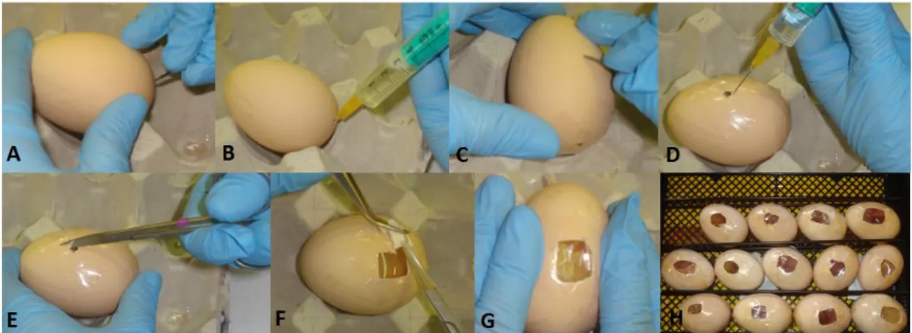
The aim of this study was to evaluate the physicochemical properties and the apical dentin bond strength of the tricalcium silicate-based Biodentine in comparison to white MTA

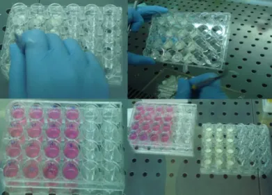
In our study, it was used materials (experimental calcium- based root canal sealers based on MTA and salicylate resins) with different chemical composition, structure, and physical

In this study, two calcium silicate MTA based cements, each containing a different radiopacifying agent alternative to bismuth oxide, were compared with conventional MTA