The aim of this study was to compare the in vitro cytotoxicity of white mineral trioxide aggregate (MTA), MTA Fillapex® and Portland cement (PC) on human cultured periodontal
ligament fibroblasts. Periodontal ligament fibroblast culture was established and the cells were used for cytotoxic tests after the fourth passage. Cell density was set at 1.25 x10⁴
cells/well in 96-well plates. Endodontic material extracts were prepared by placing sealer/ cement specimens (5x3mm) in 1mL of culture medium for 72 h. The extracts were then serially two-fold diluted and inserted into the cell-seeded wells for 24, 48 and 72 h. MTT assay was employed for analysis of cell viability. Cell supernatants were tested for nitric oxide using the Griess reagent system. MTA presented cytotoxic effect in undiluted extracts at 24 and 72 h. MTA Fillapex® presented the highest cytotoxic levels with important cell
viability reduction for pure extracts and at ½ and ¼ dilutions. In this study, PC did not induce alterations in fibroblast viability. Nitric oxide was detected in extract-treated cell supernatants and also in the extracts only, suggesting presence of nitrite in the soluble content of the tested materials. In the present study, MTA Fillapex displayed the highest cytotoxic effect on periodontal ligament fibroblasts followed by white MTA and PC.
In Vitro
Cytotoxicity of White MTA,
MTA Fillapex
®
and Portland Cement on
Human Periodontal Ligament Fibroblasts
Patrícia Yoshino1, Celso Kenji Nishiyama1, Karin Cristina da Silva Modena2, Carlos Ferreira Santos3, Carla Renata Sipert3
1Endodontics Division, Hospital
for Rehabilitation of Craniofacial Anomalies, University of São Paulo (HRAC-USP), Bauru, SP, Brazil
2Department of Operative Dentistry,
Endodontics and Dental Materials, Bauru Dental School, USP - University of São Paulo, Bauru, SP, Brazil
3Department of Biological Sciences,
Bauru Dental School, USP - University of São Paulo, Bauru, SP, Brazil
Correspondence: Profa. Dra. Carla Renata Sipert, Alameda Octávio Pinheiro Brisolla, 9-75, 17012-901 Bauru, SP, Brasil. Tel: +55-14-3235-8276. e-mail: [email protected]
Key Words: fibroblasts, nitric oxide, periapical tissue, toxicity tests.
Introduction
The study of dental materials has received great attention in Dentistry because their toxic compounds may damage the surrounding tissues, interfere on the healing process or cause allergic reactions. Concerning the materials employed for endodontic procedures, special care must be taken when choosing irrigants, intracanal dressings and filling materials, as they will be placed in close contact with live tissues such as dental pulp remnants, periodontal ligament and alveolar bone (1).
During an endodontic treatment, the periapical tissues are usually exposed not only to regular root canal filling materials, but also to materials used during surgical procedures like sealing of perforations and root-end fillings (2). Several requirements are needed for a filling or retrofilling material to be considered ideal for application in endodontic procedures, among which special attention must be directed to biocompatibility. Endodontic materials should promote or at least not impair the healing process (2) and also should not cause severe inflammatory reaction in periapical tissues (3).
Mineral trioxide aggregate (MTA) is a powder of small hydrophilic particles that sets in the presence of moisture. The powder consists in tricalcium silicate, tricalcium aluminate, tricalcium oxide and bismuth oxide besides other mineral oxides. MTA hydration results in a colloidal gel with a pH of about 12.5. MTA was first developed as a root-end filling material, but its current indications for use
include sealing of perforations, pulp capping, pulpotomy and apexification (4). Research data regarding tissue reactions to MTA have demonstrated induction of hard tissue formation (2), bone formation (5), induction of connective tissue formation (6), among others. MTA also presents high chemical similarity to the construction cement known as Portland cement (PC) (7). Considering the economic advantage of PC over MTA, this material is usually included in endodontic investigations.
Due to the positive biological responses to MTA, endodontic sealers based on the chemical composition of this material have been proposed, such as MTA Fillapex® (Angelus, Londrina, Paraná, Brazil). Unlike regular MTA, MTA Fillapexwas developed as a paste/paste sealer in a formulation that allows its appropriate insertion into the root canal as a conventional endodontic sealer (8,9).
In contact with cells, soluble chemicals from the endodontic materials may lead to necrosis and/or inflammation resulting in high levels of a wide variety of inflammatory mediators including histamine, prostaglandins and neuropeptides, among others. Nitric oxide (NO) is an intracellular molecule displaying important cardiovascular and immune functions (10). Pro-inflammatory stimuli induce resident and inflammatory cells to produce the inducible enzyme NO synthase, leading to the release of large amounts of NO during the inflammatory process (10).
112
P
. Y
oshino et al.
it seems important to investigate the potential damage of such materials on periodontal ligament cells. The present research aimed to assess the in vitro toxicity of white MTA, MTA Fillapex and PC on cultured human periodontal ligament fibroblasts. Additionally, it was investigated NO release by the cells once in contact with materials extracts.
Material and Methods
The study was approved by the Ethics Committee of the Hospital for the Rehabilitation of Craniofacial Anomalies, USP, Brazil (Process #192/2011 SVAPEPE-CEP).
Cell Culture
Primary culture of periodontal ligament fibroblasts was established using an explant technique as previously described (11,12). In brief, the periodontal ligament of a third molar was carefully removed and placed in Dulbecco’s Modified Eagle’s Medium (DMEM - Gibco/Invitrogen Corporation, CA, USA) under aseptic conditions for cell cultivation. Prior to tissue harvesting, tooth donation was performed with patient’s informed consent.
Cells were cultivated in 10% fetal bovine serum (FBS) (Gibco) DMEM at 37°C, 95% humidified air and 5% CO2 until the fibroblasts reached confluence, with medium replacement every other day. Cells were used for the tests after the 4th passage, condition required for fibroblasts culture purity, as demonstrated in a previous study (11). Prior to experimental tests, the cells were detached, counted and seeded at 1.25x10⁴ cells/well in 96-well plates.
Specimen and Extract Preparation
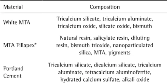
The following materials were tested: White MTA Angelus® (Angelus; Londrina, PR, Brazil), MTA Fillapex (Angelus) and PC (Cia. de Cimento Portland Itaú, Itaú de Minas, MG, Brazil). The main components of each material are described in Table 1. Materials were mixed according to the manufacturers’ instructions. Preparation of Portland cement followed the same proportions as employed for MTA. Materials were inserted into a round metal appliance designed for production of discs measuring 5 mm wide and 3 mm high. Materials were allowed to set for 24 h in a humid atmosphere and aseptic conditions.
After setting, each specimen was immersed into 1 mL of DMEM supplemented with 10% FBS and incubated for 72 h (13). The specimens were then discarded and the eluate extracts were filtered by 0.22-μm pore size membranes (Millipore; Billerica, MA, USA).
Cell Stimulation with Material Extracts
Prior to the insertion in cell cultures, the extracts were serially two-fold diluted in DMEM supplemented with 10% FBS. Cells plated in 96-well plates were incubated with
100µL of dilution or medium only, in triplicate. After 24, 48 and 72 h, the supernatant was collected and the cells were subjected to the (3-[4,5-dimethylthiazol-2-yl]-2,5-diphenyltetrazolium bromide (MTT) assay for cytotoxicity assessment (14). Negative controls were performed in empty (not cell-containing) wells.
MTT Assay
After collection of cell supernatants, 20 µL of an MTT solution (Sigma-Aldrich, St. Louis, MO, USA) (5 mg/mL) in phosphate buffered saline (PBS) were added to the wells followed by 180 µL of 10% FBS DMEM. Fibroblasts were incubated at 37ºC for 4 h protected from light. MTT solution was discarded and replaced by 100 µL of isopropanol (Merck; Whitehouse Station, NJ, USA). The plate was kept under continuous agitation for 30 min. The optical densities were measured at 570 nm in a spectrophotometer (FLUOstar Optima; BMG Labtech, Ortenberg, Germany).
Nitric Oxide (NO) Measurement
The concentration of released NO by cell supernatants was measured based on nitrite (NO2-) content, which is one of two primary stable breakdown products of NO. For this purpose, the Griess method was employed (12). Aliquots of 50 µL of each cell supernatant were added to a 96-well plate in duplicate. As negative controls, 50 µL of the extracts and DMEM supplemented with 10% FBS alone were also tested. Twenty-five microliter of 2% sulfanilamide (Sigma-Aldrich) in 5% phosphoric acid (Merck) were added to each well, followed by 25 µL of 0.2% N-(1-naphthyl)ethylenediaminedihydrochloride (NED) (Sigma-Aldrich) in distilled water added 10 min later. Samples were kept protected from light for additional 10 min, and the absorbance was measured at 530 nm. Nitrite quantification was performed based on a standard curve of a NaNO2 solution (Sigma-Aldrich), two-fold serially diluted in 10% FBS DMEM, with 200 µM as the starting point.
Data Analysis
Statistical analysis was performed using GraphPad
Table 1. Major components of the tested materials
Material Composition
White MTA Tricalcium silicate, tricalcium aluminate, tricalcium oxide, silicate oxide, bismuth
MTA Fillapex®
Natural resin, salicylate resin, diluting resin, bismuth trioxide, nanoparticulated
silica, MTA, pigments
Portland Cement
Tricalcium silicate, dicalcium silicate, tricalcium aluminate, tetracalcium aluminoferrite,
Cytoto
xic study of endodontic materials
Prism 5 (GraphPad Software, La Jolla, CA, USA). Results were subjected to one-way analysis of variance followed by Tukey's post-test for comparison between pairs of groups. The significance level was set at p<0.05.
Results
Cytotoxicity Evaluation
The cell viability of human periodontal ligament fibroblasts in contact with the extracts was evaluated by means of MTT assay.
At 24 h of incubation, a statistically significant viability reduction in comparison with control (DMEM only) was observed for MTA Fillapex extracts when pure and half-diluted, and also for pure MTA extracts (Fig. 1A) (p<0.0001).
As expected, extract dilution in DMEM decreased the material’s cytotoxicity. Compared with extracts of the other tested materials at the same dilution (1 and ½), MTA Fillapex presented a higher toxicity to the periodontal ligament fibroblasts (p<0.0001).
At 48 h of incubation, MTA Fillapex presented a higher toxicity when pure, ½ and ¼ diluted in comparison with MTA and PC (Fig. 1B) (p<0.0001). At this time point, no significant differences in cell viability were observed for MTA and PC.
At 72 h of incubation, pure MTA extract displayed significant toxicity in comparison to control (Fig. 1C). At this time point, MTA Fillapexpresented the highest rates of toxicity when pure, ½ and ¼ diluted (p<0.0001). Alterations in cell viability were not observed for PC extracts.
114
P
. Y
oshino et al.
NO Production
NO was not detected in the supernatants of fibroblasts treated with DMEM only. Nevertheless, increased NO levels were detected in the extract of treated cells supernatants for the three materials. It is worth mentioning, however, that for all tested materials, similar nitrite levels were detected in the extracts themselves. Detected amounts ranged from 1.42 to 5.49 μM for MTA, 3.71-4.17 μM for MTA Fillapex and 0.28-3.51 μM for PC. Therefore, deducting the amount of nitrite present in the material extracts, low levels of NO (less than 2μM) were barely detected in cell supernatants.
Discussion
An important issue to consider during the experimental design for in vitro biocompatibility studies is the cell type. Osteoblasts (15), mouse fibroblasts (16) and human fibroblasts (1) are some of the cell types usually employed in such investigations. Human fibroblasts were selected based on their relevance to clinical conditions. If a tested material is proven to be cytotoxic to cultured cells in vitro, one may assume a similar behavior in vivo. Additionally, human cells can be conveniently cultured with a low number of passages, resulting in minimal cell changes due to cell culture manipulation (13). Choosing human periodontal ligament fibroblasts presents the additional advantage of reducing bias concerning species origin and non-tissue specific cell lines (17). Therefore, the choice for cultured human periodontal ligament fibroblasts for the present study is justified by the close relation of endodontic sealers and cements to the periapical tissues (18).
The choice for sealer/cements extracts was performed based on the fact that, when placed into the root canals or as root-end fillings, sealers and cements release soluble components that may be diluted by tissue fluids and then carried to the surrounding cells and tissues. The eluates can also be experimentally subjected to serial dilutions allowing the evaluation of a possible dose-response effect in a similar manner to the increased dilution occurring in vivo (1,19,20). It may also be assumed that the eluate dilution might lead to a more accurate comparison of cytotoxicity among different materials.
MTA was developed to seal off the internal and external tooth surfaces, which means that it is a material designed for surgical and repair purposes (4). A huge number of scientific reports describe the properties of MTA, highlighting especially the biocompatibility of this cement in comparison with any other developed endodontic sealer or cement (5-6,21). Despite this fact, in this study, MTA extracts presented some cellular toxicity. Fibroblast viability was significantly reduced when cells were in contact with the pure extract for 24 and 72 h. However, cell population was reduced to less than 50% at 24 h and less than 75%
at 72 h. These data may suggest the occurrence of cell death and alteration of cell growth rates. Ma et al. (20) also observed a dose-response effect of MTA extracts on cell toxicity in vitro at 3 days.
In the presence of water, calcium oxide, one of MTA’s major components, is converted into calcium hydroxide, which in turn elevates the surrounding pH (21). Alkaline pH has a destructive effect on protein structures and may promote enzyme denaturation and also cell membrane damage (22). Intriguingly, the occurrence of chemical irritation by endodontic materials, such as calcium hydroxide, may not be so detrimental after all. The presence of a non-extensive inflammatory process on the underlying tissue may lead to the stimulation of tissue repair. A thin necrotic zone initiates inflammation, which in turn results in cell migration, proliferation and in new collagen deposition (23). As demonstrated in vivo by Cunha et al. (6), necrotic areas were observed at early stages after MTA subcutaneous insertion. However, longer experimental periods demonstrate presence of connective tissue and absence of inflammatory infiltrate.
In the present study, MTA Fillapex displayed the highest cytotoxic rates in a dose-dependent manner. An increase in MTA Fillapex cytotoxicity is observed over time, since ¼ diluted extracts displayed higher cytotoxic levels at 72 h than at 24 and 48 h. MTA Fillapex was significantly more cytotoxic than MTA and PC (Fig. 1). Viability decrease caused by MTA Fillapex exceeded 75% when pure and ½ diluted. The addition of components in MTA Fillapex, such as resins (8), bismuth oxide (9) and other pigments, may have increased the toxic effect of this sealer compared to its precursor, MTA. In a recent study using osteoblast culture, Scelza et al. (15) also observed significant cytotoxic levels for MTA Fillapexextracts, when prepared 1 or 7 days after the sealer manipulation. Bin et al. (19), described higher cytotoxic levels of MTA Fillapex in comparison with MTA on Chinese hamster fibroblasts. However, in this study the fibroblasts were in contact with the extracts for 24 h only and the culture medium was in contact with materials after 24, 48 or 72 h of setting time. In human osteoblast cultures, MTA Fillapex also compromised cell viability at a significant rate. In spite of that, MTA Fillapex led to increased alkaline phosphatase activity in comparison with the control (24).
Cytoto
xic study of endodontic materials
remarkable differences between these cements is the presence of bismuth oxide in MTA as an improvement to radiopacity. Min et al. (9) reported that bismuth oxide added-PC led to increased NO release by cells in comparison with pure PC. Scelza et al. (15) also observed better results for PC than MTA Fillapex for the extracts prepared 7 days after sealer/cement manipulation.
In addition to cell viability, we sought to investigate in the present study the release of NO by periodontal ligament fibroblasts treated with MTA, MTA Fillapex and PC extracts. In agreement with the previous report of Min et al. (9), nitrite was detected in cell supernatants of extract-treated cells. However, the assessment of nitrite content of the extracts used as negative controls allowed us to observe that the origin of the nitrite content detected on the supernatants were the materials themselves. Based on these findings, it seems unreasonable to assume an important NO production by periodontal ligament fibroblasts per se in consequence to the treatment with sealer/cements extracts. Special care must be taken on negative controls when performing in vitro studies to test the biological effects of dental materials, since an uncountable number of chemicals may be released during the experimental procedures, thus leading to unpredictable interferences to the assay reagents and resulting in important biased results.
Interestingly, the release of nitrite by sealers or cements to periapical tissues may be of some biological importance. It is well known that the presence of •NO
and •NO-derived species, such as •NO
2 and oxidants such as O2•-and CO3•- yields the formation of peroxynitrite, a potent pro-oxidant. In consequence, proteins may suffer nitration, a biological process that may impair function or either improve it (25). It may be speculated that nitrite release by endodontic materials may contribute to protein nitration at periapical tissues resulting in some alteration of proteins and enzyme functions. The real occurrence and the biological importance of this phenomenon is an issue that requires further investigations.
In conclusion, among the tested materials, MTA Fillapex displayed higher cytotoxic levels to human periodontal ligament fibroblasts in vitro in comparison to its chemical precursor, MTA. PC, in turn, did not show cytotoxic activity over cultured periodontal ligament fibroblasts.
Resumo
O objetivo deste estudo foi comparar a citotoxicidade in vitro de agregado trióxido mineral (MTA) branco, MTA Fillapex® e cimento Portland (PC) em cultura de fibroblastos de ligamento periodontal humano. A cultura de fibroblastos de ligamento periodontal foi estabelecida e as células foram utilizadas para os testes citotóxicos após a quarta passagem. A densidade celular foi ajustada em 1,25x10⁴ células/poço em placas de
96 poços. Extratos dos materiais endodônticos foram preparados por meio da inserção de corpos de prova dos cimentos (5 x 3 mm) em 1 mL de meio de cultura durante 72 h. Os extratos foram diluídos serialmente
na razão de ½ e inseridos aos poços contendo as células por 24, 48 e 72 h. Ensaio de MTT foi realizado para a avaliação da viabilidade celular. O sobrenadante das células foi testado em relação à presença de óxido nítrico utilizando o sistema de reagentes de Griess. O MTA apresentou efeito citotóxico quando o extrato era aplicado sem diluição durante 24 e 72 h. O MTA Fillapex apresentou os maiores níveis de citotoxicidade com importante redução da viabilidade celular quando o extrato foi aplicado puro e em diluições de ½ e ¼. Neste estudo, PC não induziu alterações na viabilidade de fibroblastos. Óxido nítrico foi detectado no sobrenadante de células tratadas com os extratos e ainda nos extratos somente, o que sugere a presença de nitrito no conteúdo solúvel dos materiais testados. No presente estudo, MTA Fillapex foi o material que demonstrou o maior efeito citotóxico sobre fibroblastos de ligamento periodontal seguido do MTA branco e do PC.
Acknowledgements
This research had financial support from The São Paulo Research Foundation (FAPESP) by means of a Research Grant to C.F. Santos (Process #2005/60167-0) and a Doctorate Scholarship to C.R. Sipert (Process #2007/00306-1).
References
1. Keiser K, Johnson CC, Tipton DA. Cytotoxicity of mineral trioxide aggregate using human periodontal ligament fibroblasts. J Endod 2000;26:288-291.
2. Torabinejad M, Hong CU, Pitt Ford TR, Kaiyawasam SP. Tissue reaction to implanted super-EBA and mineral trioxide aggregate in the mandible of guinea pigs: a preliminary report. J Endod 1995;21:569-571. 3. Huang FM, Tai KW, Chou MY, Chang YC. Cytotoxicity of resin-, zinc
oxide-eugenol-, and calcium hydroxide-based root canal sealers on human periodontal ligament cells and permanent V79 cells. Int Endod J 2002;35:153-158.
4. Torabinejad M, Hong CU, McDonald F, Pitt Ford TR. Physical and chemical properties of a new root-end filling material. J Endod 1995;21:349-353.
5. Koh ET, McDonald F, Pitt Ford TR, Torabinejad M. Cellular response to mineral trioxide aggregate. J Endod 1998;24:543-547.
6. Cunha SA, Rached FJ, Jr., Alfredo E, Leon JE, Perez DE. Biocompatibility of sealers used in apical surgery: a histological study in rat subcutaneous tissue. Braz Dent J 2011;22:299-305.
7. Oliveira MG, Xavier CB, Demarco FF, Pinheiro AL, Costa AT, Pozza DH. Comparative chemical study of MTA and Portland cements. Braz Dent J 2007;18:3-7.
8. Hanks CT, Strawn SE, Wataha JC, Craig RG. Cytotoxic effects of resin components on cultured mammalian fibroblasts. J Dent Res 1991;70:1450-1455.
9. Min KS, Chang HS, Bae JM, Park SH, Hong CU, Kim EC. The induction of heme oxygenase-1 modulates bismuth oxide-induced cytotoxicity in human dental pulp cells. J Endodont 2007;33:1342-1346. 10. Nathan C. Nitric oxide as a secretory product of mammalian cells.
FASEB J 1992;6:3051-3064.
11. Morandini AC, Chaves Souza PP, Ramos-Junior ES, Brozoski DT, Sipert CR, Souza Costa CA, et al.. Toll-like receptor 2 knockdown modulates interleukin (IL)-6 and IL-8 but not stromal derived factor-1 (SDF-1/ CXCL12) in human periodontal ligament and gingival fibroblasts. J Periodontol 2013;84:535-544.
12. Sipert CR, Moraes IG, Bernardinelli N, Garcia RB, Bramante CM, Gasparoto TH, et al.. Heat-killed Enterococcus faecalis alters nitric oxide and CXCL12 production but not CXCL8 and CCL3 production by cultured human dental pulp fibroblasts. J Endod 2010;36:91-94. 13. Karimjee CK, Koka S, Rallis DM, Gound TG. Cellular toxicity of mineral
trioxide aggregate mixed with an alternative delivery vehicle. Oral Surg Oral Med Oral Pathol Oral Radiol Endod 2006;102:E115-E120. 14. Mosmann T. Rapid colorimetric assay for cellular growth and survival:
application to proliferation and cytotoxicity assays. J Immunol Methods 1983;65:55-63.
116
P
. Y
oshino et al.
multiparametric assay to compare the cytotoxicity of endodontic sealers with primary human osteoblasts. Int Endod J 2012;45:12-18. 16. Haglund R, He J, Jarvis J, Safavi KE, Spangberg LS, Zhu Q. Effects of
root-end filling materials on fibroblasts and macrophages in vitro. Oral Surg Oral Med Oral Pathol Oral Radiol Endod 2003;95:739-745. 17. Huang FM, Chang YC. Cytotoxicity of resin-based restorative materials
on human pulp cell cultures. Oral Surg Oral Med Oral Pathol Oral Radiol Endod 2002;94:361-365.
18. Economides N, Pantelidou O, Kokkas A, Tziafas D. Short-term periradicular tissue response to mineral trioxide aggregate (MTA) as root-end filling material. Int Endod J 2003;36:44-48.
19. Bin CV, Valera MC, Camargo SE, Rabelo SB, Silva GO, Balducci I, et al.. Cytotoxicity and genotoxicity of root canal sealers based on mineral trioxide aggregate. J Endod 2012;38:495-500.
20. Ma J, Shen Y, Stojicic S, Haapasalo M. Biocompatibility of two novel root repair materials. J Endod 2011;37:793-798.
21. Holland R, de Souza V, Nery MJ, Otoboni Filho JA, Bernabe PF, Dezan Junior E. Reaction of rat connective tissue to implanted dentin tubes
filled with mineral trioxide aggregate or calcium hydroxide. J Endod 1999;25:161-166.
22. Nelson DL, Cox MM. Lehninger Principles of Biochemistry 5th. W.H. Freeman; Gordonsville, 2008.
23. Schroder U. Effects of calcium hydroxide-containing pulp-capping agents on pulp cell migration, proliferation, and differentiation. J Dent Res 1985;64 Spec No:541-548.
24. Salles LP, Gomes-Cornelio AL, Guimaraes FC, Herrera BS, Bao SN, Rossa-Junior C, et al.. Mineral trioxide aggregate-based endodontic sealer stimulates hydroxyapatite nucleation in human osteoblast-like cell culture. J Endod 2012;38:971-976.
25. Radi R, Peluffo G, Alvarez MN, Naviliat M, Cayota A. Unraveling peroxynitrite formation in biological systems. Free Radic Biol Med 2001;30:463-488.