UNIVERSIDADE DE LISBOA
FACULDADE DE MEDICINA VETERINÁRIA
NOROVIRUS OUTBREAKS IN THE PORTUGUESE ARMY
ANTÓNIO EDUARDO BRUNO LOPES JOÃO
Orientadores: Professora Doutora Maria de São José Garcia Alexandre Professor Doutor Carlos Penha Gonçalves
Tese especialmente elaborada para obtenção do grau de Doutor em Ciências
Veterinárias na especialidade de Segurança Alimentar
Júri
Presidente: Professor Doutor Luís Filipe Lopes da Costa
Vogais:
- Professora Doutora Maria de São José Garcia Alexandre
- Professora Doutora Paula Cristina Maia Teixeira
- Professor Doutor Miguel Agostinho Sousa Pinto Torres Fevereiro
- Professora Doutora Marília Catarina Leal Fazeres Ferreira
Author Declaration
Declaração relativa às condições de reprodução da tese
Nome:
Título da Tese: Norovirus Outbreaks in the Portuguese Army Ano de conclusão: 2019
Designação do curso de Doutoramento: Ciências Veterinárias Área científica em que melhor se enquadra (assinale uma):
Clínica Produção Animal e Segurança Alimentar Morfologia e Função Sanidade Animal
Declaro sobre compromisso de honra que a tese ou dissertação agora entregue corresponde à que foi aprovada pelo júri constituído pela Faculdade de Medicina Veterinária da ULISBOA.
Declaro que concedo à Faculdade de Medicina Veterinária e aos seus agentes uma licença não-exclusiva para arquivar e tornar acessível, nomeadamente através do seu repositório institucional, nas condições abaixo indicadas, a minha tese ou dissertação, no todo ou em parte, em suporte digital.
Declaro que autorizo a Faculdade de Medicina Veterinária a arquivar mais de uma cópia da tese ou dissertação e a, sem alterar o seu conteúdo, converter o documento entregue, para qualquer formato de ficheiro, meio ou suporte, para efeitos de preservação e acesso.
Retenho todos os direitos de autor relativos à tese ou dissertação, e o direito de a usar em trabalhos futuros (como artigos ou livros).
Concordo que a minha tese ou dissertação seja colocada no repositório da Faculdade de Medicina Veterinária com o seguinte estatuto (assinale um):
1. Disponibilização imediata do conjunto do trabalho para acesso mundial;
2. Disponibilização do conjunto do trabalho para acesso exclusivo na Faculdade de Medicina Veterinária durante o período de 6 meses, 12 meses, sendo que após o tempo assinalado autorizo o acesso mundial*;
* Indique o motivo do embargo (OBRIGATÓRIO)
É AUTORIZADA A REPRODUÇÃO INTEGRAL DESTA TESE/TRABALHO APENAS PARA EFEITOS DE INVESTIGAÇÃO, MEDIANTE DECLARAÇÃO ESCRITA DO INTERESSADO, QUE A TAL SE COMPROMETE.
Faculdade de Medicina Veterinária da Universidade de Lisboa, ____ de ____________________ de 20___
____________________________________________________Acknowledgments
I want to acknowledge all the people that directly or indirectly have contributed
to the accomplishment of this work. I feel deeply honored and grateful to all those that
have helped me in this long and hard journey. It is a unique moment in my professional
life and it was made possible by all persons with whom I have worked and it is with a
feeling of great respect and gratitude that I want to address them here.
First of all, I would like to thank Professor Maria de São José, for all the support,
the patience and the inspiration in the development of this thesis. It was following a
lecture by Professor Maria de São José that the idea came out. The passionate way
the lecture was given was really motivational and inspiring. Professor Maria de São
José was paramount for all the work that followed, with a special note for the
publication of the papers. I wish to thank deeply to Colonel Veterinary Penha
Gonçalves, my former chief, for all the stimulus for pursuing this academic study and
for the example of well doing, knowledge, intelligence and wisdom. It was for me, really
a privilege to take part on the working team of Colonel Veterinary Penha Gonçalves.
My truly thanks for Professor João Mesquita who was tireless in the processes of
writing and submission of the papers, always with a gentle and good mood and for
Professor Ana Duarte for the kindness and motivating words that always gave me.
I feel a special sense of gratitude to Doctor Mónica Oleastro for the work of
genotyping of the viral strains, for the wise advising, the intelligent remarks, the good
conversations and the prompt and friendly response to my requests. I also want to
thank in a particular way Doctor Rita Sousa for the work of genotyping, the kindness
and the noroviruses genetic diversity book who was a source of inspiration in the
writing process and to MSc Inês Costa for the contribution in the writing of the papers.
For me it has been a special honor and privilege to work during all these years
with the team of persons of the Portuguese Army, my Comrades of Arms, that were
very important in this process, so to Major Med Vet Júlio Carvalho, Major Med Vet José
Freitas, Major Med Vet Pedro Silva, Major Med Vet Wilson Antunes, Major Med Vet
Carlos Alonso and Captain Med Vet Inês Gomes I thank you all very much. My true
gratitude to First Lieutenant Susana Rodrigues, Second Lieutenant Joana Fernandes,
Second Lieutenant Octávio Barbosa, Second Lieutenant Nuno Bem, Sargent Vanda
Queimada, Sargent Carina Gomes, Sargent Ricardo Vieira, Soldier Tiago Pereira and
MSc Verónica Ribeiro.
I thank all the other personnel of the Laboratorial Military Unit, namely
Lieutenant Colonel Farm Vânia Tira-Picos, Major Farm Miguel Cabrita, Captain Farm
Tiago Gonçalves, Captain Farm Pedro Neto, Captain Farm Paula Lopes, Second
Lieutenant Luís Carvalho, Sargent Licínia Gomes, Soldier João Castela and MSc
Joana Saramago.
To all the people who directly or indirectly helped me in the work of collecting
epidemiologic data I am really grateful: Lieutenant Colonel Med Vet Isabel Gabriel,
Lieutenant Colonel Med Vet Daniel Simões, Lieutenant Colonel Med Vet Ana Silva,
Major Med Vet António Coimbra, Major Med Vet Samuel Miguéis, Captain Med Vet
João Machado, Second Lieutenant Med Vet João Cabrita, Sargent-Chief José Simões,
MSc Cristina Orrico and Soldier André Assis.
With the risk of forgetting the names of many persons in the Army that
contributed to this study to the doctors, nurses, pharmacists and health technicians in
the health centers and in the hospitals, I thank you all very much.
A special thanks to Doctor Ana Rita Henriques, my doctorate colleague and
close friend for the support and the stimuli to write this thesis.
To my close friends for all the patience and unconditional support. It is my
privilege to have you nearby.
Finally, to the best Family a person can have my infinite gratitude.
___________________________________Financial Support Acknowledgements
The Present work was partially supported by the Portuguese Army.The author wants to acknowledge the Portuguese Army for the conditions provided for the experimental work, the development of the thesis and the financial support.
______________________________________________________Resumo
Surtos de gastrenterite por norovírus no Exército Português
As infeções gastrointestinais encontram-se entre as doenças mais frequentemente reportadas como causa de diminuição da capacidade operacional e da prontidão de uma força militar. Os norovírus são os agentes mais vezes implicados na doença aguda gastrointestinal em teatros de operações, marítimos e terrestres, e em forças estacionadas ou centros de treino militares das Forças Armadas de Países Ocidentais.
Esta tese teve como principal objetivo avaliar a relevância da gastrenterite aguda causada por norovírus no Exército Português. Foi estabelecido um sistema de vigilância no Exército, de modo que todos os surtos de gastroenterite são reportados ao Laboratório de Bromatologia e Defesa Biológica. Assim, verificou-se que entre 2013 e 2017 foram registados 14 episódios, que afetaram cerca de 410 militares, tendo os norovírus sido responsáveis pela maioria dos surtos (8/14) e dos casos (210/410). Os surtos ocorreram em exercícios militares e em bases do Exército com impacto na capacidade operacional e funcionamento regular das Unidades. Embora a origem destes tenha sido difícil de confirmar, acredita-se que 5 (63%) tiveram origem alimentar (incluindo os manipuladores de alimentos e a água) e que em 3 (37%) predominou a transmissão pessoa a pessoa. Os surtos foram causados por norovírus dos genogrupos (G) I e II, contudo o G II foi responsável pelo maior número de surtos (6/8) e casos (145/210). O genogrupo I esteve associado à água e o genogrupo II à transmissão pessoa a pessoa ou aos alimentos. Cada surto foi causado por um genótipo distinto, refletindo a grande diversidade genética dos norovírus que se encontram em circulação. Três dos genótipos identificados (GI.9, GII.17 e GII.16-GII.2) foram reportados pela primeira vez em Portugal.
Ao identificar o norovírus como o mais importante agente etiológico de surtos de gastroenterite aguda e de doença com origem nos alimentos e na água no Exército Português, o presente trabalho contribuiu para a alteração de medidas preventivas de doenças de origem alimentar e para o reforço de ações de controlo dos surtos causados por este agente.
Palavras-chave:
Norovírus, Exército Português, surtos de gastroenterite, sistema de vigilância e doença de origem alimentar_______________________________________________________Abstract
Norovirus outbreaks in the Portuguese Army
Gastrointestinal infections have consistently been among the most frequent diseases and non-battle injuries, degrading operational effectiveness and force readiness in the military. Noroviruses appear as the most frequent agents causing acute gastrointestinal illness both in maritime and land theatres of operations, as well as in stationed troops or military training centers in many western countries.
Nevertheless, little is known on the burden of norovirus gastroenteritis in the Portuguese military. In this work we set out to establish a gastroenteritis outbreak surveillance system (GOSS) to report disease cases to the Bromatology and Biologic Defense Laboratory during the five-year period, 2013-2017. During this period 14 gastroenteritis outbreaks were registered that affected a total of 410 military. Noroviruses were responsible for the majority of the outbreaks (8/14) and disease cases (210/410). Norovirus outbreaks occurred either in military exercises or in military bases and showed to have an impact on force readiness and operational effectiveness. In most cases the origin of the outbreaks was difficult to confirm but five (63%) were likely foodborne (including food handlers) or waterborne and three (37%) had predominant person-to-person transmission. Outbreaks were caused by both genogroup (G) I and II, but G II clearly outnumber those caused by G I (6/8) and case numbers (145/210). Norovirus GI was associated with waterborne outbreaks while GII was associated to foodborne and person-to-person transmission. Each outbreak was caused by a different genotype highlighting the high genetic diversity of the circulating noroviruses. Three of the identified genotypes (GI.9, GII.17 and GII.16-GII.2) were reported for the first time in Portugal.
In conclusion, the present thesis identified norovirus as the most important etiologic agent of acute gastroenteritis outbreaks and the most frequent cause of food- and waterborne illness in the Portuguese Army. This work had contributed to change preventive measures and allowed the reinforcement of control actions that minimized the impact of norovirus outbreaks.
keywords:
Norovirus, Portuguese Army, gastroenteritis outbreak, surveillance system, foodborne disease
___________________________________________________List of Publications
The results presented in this thesis have been published:Papers in International peer-reviewed journals
Lopes-João A, Mesquita JR, de Sousa R, Oleastro M, Penha-Gonçalves C, Nascimento MS. (2019). A simultaneous norovirus outbreak in three Portuguese Army bases in the Lisbon region, December 2017. J R Army Med Corps. 0:1-4.
Lopes-João A, Mesquita JR, de Sousa R, Oleastro M, Penha-Gonçalves C, Nascimento MS. (2018) Country-wide surveillance of norovirus outbreaks in the Portuguese Army, 2015-2017. J R Army Med Corps. 164 (6):419-422.
Lopes-João A, Mesquita JR, de Sousa R, Oleastro M, Penha-Gonçalves C, Nascimento MS. (2017) Acute gastroenteritis outbreak associated to norovirus GI.9 in a Portuguese Army base. J Med Virol. 89 (5):922-925.
Lopes-João A, Costa I, Mesquita JR, Oleastro M, Penha-Gonçalves C, Nascimento MSJ. (2015) Multiple enteropathogenic viruses in a gastroenteritis outbreak in a military exercise of the Portuguese Army. J Clin Virol. 68:73-5.
Papers in national journals
Lopes-João A. (2013) Surtos de Toxinfecção Alimentar no Exército Português 2006-2011. Revista Portuguesa de Ciências Veterinárias. 108 (587-588) 154-160.
______________________Communications in congress and scientific meetings
Oral communications
Lopes-João A, Mesquita JR, de Sousa R, Oleastro M, Penha-Gonçalves C, Nascimento MS. A countrywide surveillance of norovirus outbreaks in the Portuguese Army (2018) in “Food Defense International Symposium”, 11 September, Academia Militar, Amadora, Portugal. Lopes-João A, Mesquita JR, de Sousa R, Oleastro M, Penha-Gonçalves C, Nascimento MS. Toxinfeções alimentares no Exército, a experiência do Laboratório de Bromatologia e de Defesa Biológica (2018) in “Doenças de transmissão alimentar e saúde dos manipuladores”, 22 February, Centro de Saúde Militar de Coimbra, Portugal.
Lopes-João A, Mesquita JR, de Sousa R, Oleastro M, Penha-Gonçalves C, Nascimento MS. Toxinfecçoes alimentares no Exército Português (2015) in VI Seminário de Alimentação das Forças Armadas, Defesa e Cultura Alimentar, 28 October, Rio de Janeiro, Brazil.
Lopes João A, Mesquita JR, Costa I, Oleastro M, Nascimento MS.Surto de gastroenterite por norovirus num Exercício Militar do Exército Português (2014) in VI Congresso da Sociedade Portuguesa de Ciências veterinárias, about veterinary sciences: praxis and future, 4 April, Instituto Nacional de Investigação Agrária e Veterinária, Oeiras, Portugal.
Lopes João A, Mesquita JR, Costa I, Oleastro M, Nascimento MS. Norovirus, um agente emergente (2013) in VIII Congresso Científico AEFFUP, about infectious emergent disease, 27 November, Faculdade de Farmácia da Universidade do Porto, Porto, Portugal.
Posters
Lopes João A, Mesquita JR, Costa I; Oleastro M, Nascimento MS (2014) Toxinfeções alimentares no Exército Português 2006-2013. VI Congresso da Sociedade Portuguesa de Ciências veterinárias about veterinary sciences: praxis and future”, Instituto Nacional de Investigação Agrária e Veterinária, Oeiras, Portugal 3-5 April.
Lopes João A, Mesquita JR, Costa I; Oleastro M, Nascimento MS (2013) Norovirus gastroenteritis in a nurse attending an acute gastroenteritis outbreak in a care facility of the Portuguese Armed Forces. IV Congresso Nacional de Virologia e VIII Encontro Nacional da Sociedade Portuguesa de Virologia, Lisboa, Portugal 25-26 November.
___________________________________________________________Table of Contents
Author Declaration... ii
Resumo ... ix
Abstract ... x
List of Publications ... xi
Papers in International peer-reviewed journals ... xi
Papers in national journals ... xi
Communications in congress and scientific meetings ... xii
Oral communications... ... xii
Posters... ... xii
List of Figures ... xv
List of Tables... xvi
Abbreviations & Acronyms ... xvii
I. INTRODUCTION ... 1
1. Norovirus ... 3
1.1. The discovery of norovirus ... 3
1.2. Virus taxonomy ... 4
1.3. Virus characteristics and life cycle... 5
1.4. Genogroups and genotypes of norovirus... 7
1.5. Cultivation of human norovirus ... 9
1.6. Environmental resistance ... 10
1.7. Epidemiology of norovirus infection ... 10
1.7.1. Disease impact worldwide ... 10
1.7.2. Molecular epidemiology of norovirus ... 12
1.7.3. Incidence seasonality of norovirus infection ... 13
1.7.4. Transmission of norovirus ... 14
1.7.5. Norovirus and foodborne and waterborne illness ... 16
1.8. Human norovirus infection... 20
1.8.1. Pathogenesis of human norovirus infection. ...20
1.8.3. Immunologic response to norovirus...22
1.8.4. Immune evasion and lack of vaccine against norovirus... 23
1.9. Laboratory detection of norovirus ... 24
1.9.1. Molecular methods to detect norovirus ... 24
1.9.2. Immunological methods ... 26
1.9.3. Detection of norovirus in environmental and food samples ... 27
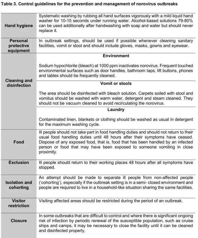
1.10. Prevention and control of norovirus outbreaks ... 28
2. Foodborne Disease Surveillance Systems... 31
2.1. Principles underlying the surveillance and management of foodborne disease outbreaks proposed by WHO ... 32
3. Norovirus in the Military ... 33
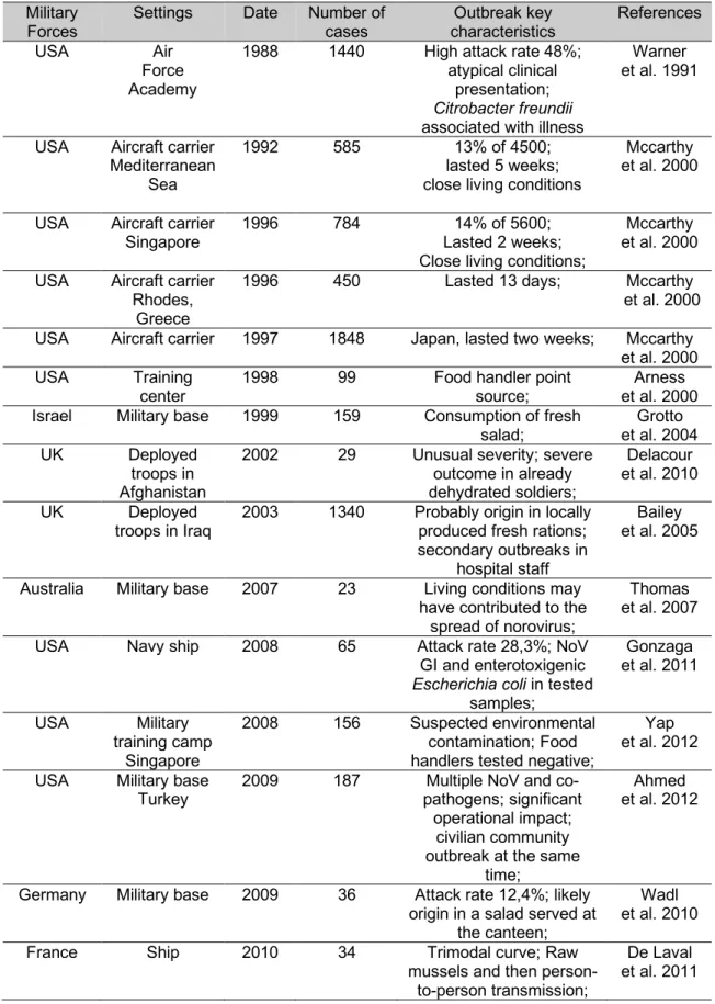
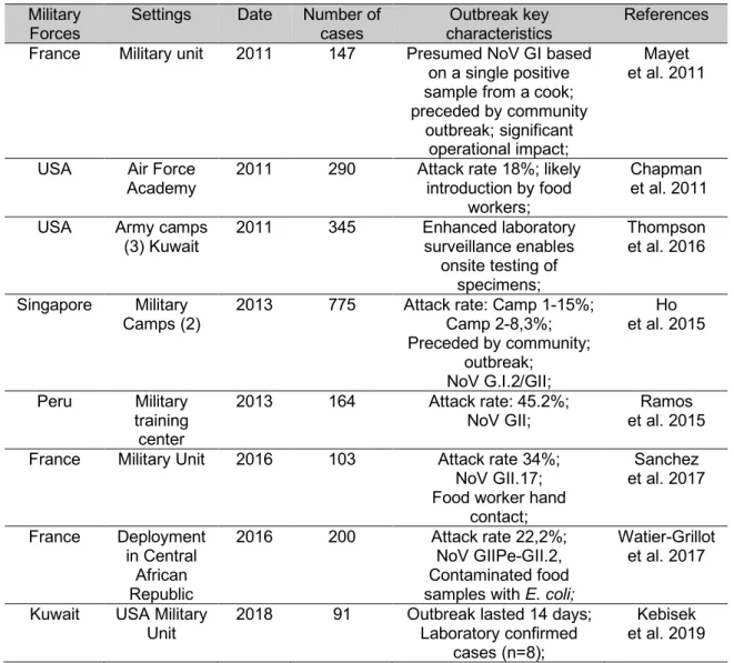
3.1. Epidemiologic data and norovirus disease burden ... 35
3.2. Noroviruses as biologic warfare agents ... 38
3.3. Portuguese Army “Gastroenteritis Outbreak Surveillance System”... 39
3.3.1. Organization ... 39 3.3.2. Clinical Investigation ... 40 3.3.3. Epidemiological Investigation ... 40 3.3.4. Laboratory Investigation... 41 II. OBJECTIVES ... 43 III. RESULTS ... 47
1. Multiple enteropathogenic viruses in a gastroenteritis outbreak of the Portuguese Army ... 49
2. Acute Gastroenteritis Outbreak Associated to Norovirus GI.9 in a Portuguese Army Base ... 55
3. A Country-wide surveillance of norovirus outbreaks in the Portuguese Army ... 61
4. Simultaneous norovirus outbreak in three Portuguese Army Bases in Lisbon Region December 2017 ... 67
5. Characterization of the norovirus outbreaks reported in the Portuguese Army ... 73
5.1. Summary of the norovirus outbreaks ... 75
5.2. Modes of transmission and key features of the Portuguese Army norovirus outbreaks ... 80
5.3. Sporadic norovirus gastroenteritis in the Portuguese Army ... 82
5.4. Norovirus genogroups/genotypes involved in the Portuguese Army outbreaks .. 83
IV. DISCUSSION ... 85
V. CONCLUSIONS ... 95
VI. REFERENCES ... 99
APPENDIX ... 121
_____________________________________________________________List of Figures Figure 1. Human norovirus genomic organization and structure. ... 6
Figure 2. The composition and life cycle of human noroviruses ... 7
Figure 3. Genetic classification based on VP1 amino acid sequences ... 8
Figure 4. Transmission routes of norovirus in the USA . ... 18
Figure 5. Genome regions used for genotyping of noroviruses. ... 25
Figure 6. Organigram of the Portuguese Army Health System units and subunits involved in foodborne disease investigations. ... 40
Figure 7. Actions taken in case of a gastrointestinal disease outbreak in the Portuguese Army. ... 41
Figure 8. Number of gastroenteritis outbreaks caused by norovirus versus other causes, reported in the Portuguese Army, 2013-2017. ... 75
Figure 9. Number of cases reported in the 14 gastroenteritis outbreaks, caused by noroviruses versus other causes, reported in the Portuguese Army, 2013-2017. ... 76
Figure 10. Number of cases of gastroenteritis caused by noroviruses versus other causes and the temporal distribution of the outbreaks, reported in the Portuguese Army, 2013-2017. ... 77
Figure 11. Estimated attack rates of the 14 gastroenteritis outbreaks, reported in the Portuguese Army, 2013-2017. ... 78
______________________________________________________________List of Tables
Table 1. Caliciviridae family classification including genus and type virus of mammal hosts ... 4 Table 2. Foodborne illness of norovirus origin ... 17 Table 3. Control guidelines for the prevention and management of norovirus
outbreaks ... 30 Table 4. Norovirus gastroenteritis outbreaks in the military 1988-2010 ... 36 Table 5. Norovirus gastroenteritis outbreaks in the military 2011-2016 ... 37 Table 6. Clinical details of the norovirus outbreaks, reported in the Portuguese Army,
2013 to 2017 ... 79 Table 7. Annual incidence rate of norovirus gastroenteritis in the active Army military,
2013-2017 ... 80 Table 8. Point of origin, main transmission way and key features of the norovirus
outbreaks, reported in the Portuguese Army, 2013-2017 ... 81 Table 9. Number of cases and genogroups/genotypes involved in the norovirus
_____________________________________________Abbreviations
&Acronyms
ABHR Alcohol-based hand rub
AHC Army Health Center
AHD Army Health Direction
BBDL Bromatology and Biologic Defense Laboratory
BCDLU Biologic and Chemical Defense Laboratory Unit
CD4 Cluster of differentiation 4
CD8 Cluster of differentiation 8
CDC Center for Disease Control and prevention
CEN European Committee for Standardization
cfu Colony Forming Unit
E. coli Escherichia coli
ECDC European Center for Disease Control and Prevention
EFSA European Food Safety Authority
EIA Enzyme immunoassay
EST Epidemiologic Surveillance Team
FTA Flinders Technology Associates
FUT Fucosiltransferase
GEC Genomic Equivalent Copies
GOSS Gastroenteritis Outbreak Surveillance System
GI Genogroup I
GII Genogroup II
HACCP Hazzard Analysis and Critical control Points
HBGA Histoblood group antigens
HNORS Hospital norovirus outbreak reporting scheme
IFN Interferon
IL Interleukin
ISO International Organization for Standardization
MVU Military Veterinary Unit
NGS Next generation sequencing
Norostat Norovirus Sentinel Testing and Tracking
NORS National Outbreak Report System
NoV Norovirus
nm nanometer
P22 Protein 22
P48 Protein 48
P-domain Protruding domain
PCR Polymerase chain reaction
POL Polymerase
ppm Parts per million
PRO Protease
RdRp RNA-dependent RNA polymerase
RNA Ribonucleic acid
RT-PCR Reverse transcription polymerase chain reaction
RT-qPCR Reverse transcription quantitative polymerase chain reaction
S-domain Shell-domain
SRSV Small round structured virus
spp. species
STEC Shigatoxinogenic Escherichia coli
TNF-a Tumor necrosis factor alfa
UK United Kingdom
USA United States of America
VLP Virus-like particle
VPg Viral protein genome-linked
VP1 Major capsid protein
VP2 Minor capsid protein
I. INTRODUCTION
_____________________________________________________________________
1. Norovirus
Human noroviruses are the leading cause of epidemic and sporadic gastroenteritis across all age groups (Glass et al. 2009; Robilotti et al. 2015). Noroviruses are ubiquitous, associated with 18% of acute gastroenteritis cases worldwide, with similar proportions of disease in high-, middle-, and low- income settings (Ahmed et al. 2014). Noroviruses are estimated to cause approximately 200.000 deaths annually worldwide, with about 70,000– 100,000 fatal cases among children in developing countries (Ahmed et al. 2014; Lopman et al. 2016). Noroviruses are highly contagious and humans are the only known reservoir for human noroviruses (Green 2013). Transmission is fecal-oral and occurs via direct person-to-person, foodborne, waterborne or through fecal contamination or environmental fomites (Green 2013).
1.1. The discovery of norovirus
In 1929, Zahorsky was the first that described, what is thought to be norovirus gastroenteritis, “hyperemesis hemis” or “winter vomiting disease”, an illness characterized by the sudden onset of self-limited vomiting and diarrhea that typically peaked during the colder months (Patel et al. 2009).
During the 1940-1950s, several studies demonstrated the transmission of this “gastroenteric disease” to healthy volunteers by oral administration of a bacteria-free stool filtrate (Kapikian 2000). Because the infectious agent was transmissible after filtration, a viral etiology was at that time suspected (Kapikian 2000). In 1972, Kapikian identified a 27-nm virus-like particle, by immuno-electron microscopy, in an infectious stool filtrate from an outbreak of gastroenteritis that occurred in 1968 in an elementary school in Norwalk, Ohio (Kapikian et al. 1972). This allowed the recognition and identification of a virus-like particle that did not have a distinctive morphology and was among the smallest viruses known as the cause of the Norwalk outbreak (Kapikian 2000).
Over the two decades following the discovery of Norwalk virus, viruses similar to Norwalk virus in morphology and associated with similar clinical manifestations, but antigenically distinct, were discovered and provisionally classified as “small round structured viruses” (SRSV) (Dolin et al. 1982; Caul 1996).
In the early 1990s, the Norwalk virus genome was cloned and sequenced, establishing this virus as member of the family Caliciviridae (Green 2013). Later molecular studies allowed
classification of the virus into a new genus termed “Norwalk-like viruses” and more recently (2007) called genus Norovirus (Green 2013).
1.2. Virus taxonomy
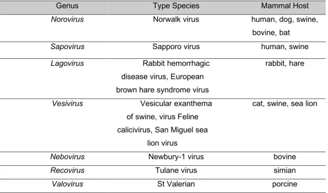
Norovirus belongs to the Caliciviridae family, in reference to the cup-like (calyx) depressions on the virus surface (Green 2013). This family encompasses seven genera that infect mammals, Norovirus, Sapovirus Vesivirus, Lagovirus, Nebovirus, Recovirus and
Valovirus, two genera infect birds, Bavovirus and Nacovirus and two fishes, Minovirus and Salovirus. Caliciviruses cause species-specific infection (Vinjé et al. 2019). The genus Norovirus is the most frequent cause of disease in humans, among the members of the Caliciviridae family (Vinjé et al. 2019). In this family, there are important animal pathogens
namely the feline calicivirus, (genus Vesivirus) and the rabbit hemorrhagic disease virus (genus Lagovirus). Sapovirus mainly cause mild gastroenteritis in children up to 5 years of age, while norovirus is pathogenic for humans in all age groups (Svraka et al. 2010; Ahmed et al. 2014). In the next Table are the established genera, type virus and mammal hosts of the Caliciviridae family.
Table 1. Caliciviridae family classification including genus, type virus of mammal hosts
Genus Type Species Mammal Host
Norovirus Norwalk virus human, dog, swine,
bovine, bat
Sapovirus Sapporo virus human, swine
Lagovirus Rabbit hemorrhagic disease virus, European brown hare syndrome virus
rabbit, hare
Vesivirus Vesicular exanthema of swine, virus Feline calicivirus, San Miguel sea
lion virus
cat, swine, sea lion
Nebovirus Newbury-1 virus bovine
Recovirus Tulane virus simian
Valovirus St Valerian porcine
1.3. Virus characteristics and life cycle
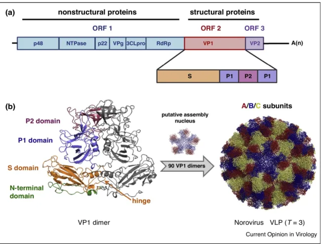
Noroviruses are single-strand RNA viruses of positive sense, non-enveloped with icosahedral symmetry nucleocapsid, with 27-37nm in diameter and have a 7.5-7.7kilobase genome (Green 2013). The genome serves as mRNA and as a template for a complementary negative strand being transcribed into genome RNA through the viral polymerase. The genome is organized in three open reading frames (ORF) (Figure 1). ORF1 encodes a 194-kDa polyprotein which is cleaved by the virus protease into six proteins, including the RNA-dependent RNA polymerase (RdRp) and other non-structural proteins responsible for transcription and replication. ORF2 encodes the major capsid protein VP1 a structural 64 k-Da protein, which forms the virus capsid and ORF3, considered the most variable region in the genome, encodes the minor capsid protein VP2 a 23-kDa protein that interacts with the genome RNA when the virion formation occurs. (Green 2013).
The VP1 protein is formed by the internal N-terminal shell (S) domain as well as a protruding (P) domain comprised of a P2 subdomain that is the most exposed part of the virus; the P1 subdomain lies below P2 (closer to the S domain). The viral capsid is formed by 180 copies (90 dimers) of VP1 symmetrically arranged (Pogan et al. 2018). The S domain surrounds the viral RNA, and the P domain, which is linked to the S domain through a flexible hinge, correspond to the C-terminal part of the VP1 protein. The highly variable P2 subdomain contains the putative neutralization sites and interacts with histoblood group antigens (HBGAs) (Vongpunsawad et al. 2013). VP2 protein is located inside the nucleocapsid and is most likely involved in capsid assembly and genome encapsidation (Vongpunsawad et al. 2013) VP2 is thought also to play a role in capsid stability and viral entry (Pogan et al. 2018; Gaziano et al. 2019).
Figure 1. Human norovirus genomic organization and structure.
(a) norovirus have three ORF, ORF1, ORF 2 and ORF 3, respectively. A polyprotein which includes nonstructural proteins is encoded by ORF1. The two structural proteins, the major (VP1) and minor capsid protein (VP2), are encoded by ORF2 and ORF3, respectively. The VP1 protein is formed by the shell (S) and protruding (P) domains. (b) 90 dimers of the VP1 assemble into icosahedral T=3 norovirus like particles (VLP). The S domain of the VP1 monomers build a shell that surround the viral RNA in form of a scaffold. The more flexible P domain is subdivided into P1 and P2 and connect to S via a hinge. The domains are highlighted in the VP1 dimer structure (left) and the three quasiequivalent subunits (A/B/C) forming the capsids are shown in the VLP structure (Pogan et al. 2018).
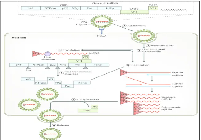
Norovirus attach to host cells via a carbohydrate receptor of the histoblood group antigens (HBGAs) and probably another receptor entering into cells through clathrin and caveolin independent endocytosis (Green 2013; Ushijima et al. 2014). Bile salts and HBGAs are key mediators of norovirus entry; however, the molecular mechanisms by which these molecules promote infection and the identity of a potential human norovirus receptor remains unknown (Graziano et al. 2019). Inside the cell the virus is uncoated and the viral genomic RNA is released into the cytoplasm. The genomic RNA functions as a messenger RNA and it codes for the three ORFs. Translation is mediated by host translation factors that are recruited by non-structural protein VPg, which covalently binds to the 5´end of the genome (Green 2013; Ushijima et al. 2014). Viral genome-linked protein is then removed and viral RNA is translated into a processed ORF 1 polyprotein to yield the replication proteins. This polyprotein is cleaved
by the virus encoded protease (Pro) to produce six proteins: p48, nucleoside triphosphatase, p22, VPg, Pro and RdRp (RNA-dependent RNA polymerase). These proteins work in the replication process to copy the (+) sense genomic RNA into a (-) sense copy that is used as a template to produce a (+) sense sub genomic RNA and new genomic (+) sense RNA. The subgenomic RNA, which contains only ORF2 and ORF3, is translated to produce the major capsid protein (VP1) and a few molecules of a second capsid protein (VP2) that can self-assemble into virus like particles when these two proteins are expressed alone. At the end, new virus particles are assembled and release by cell lysis (Green 2013; Ushijima et al. 2014).
Figure 2. The composition and life cycle of human noroviruses (Graaf et al. 2016).
1.4. Genogroups and genotypes of norovirus
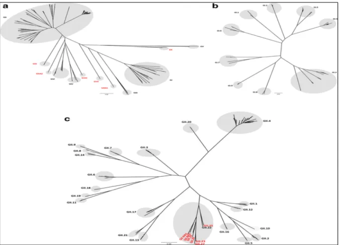
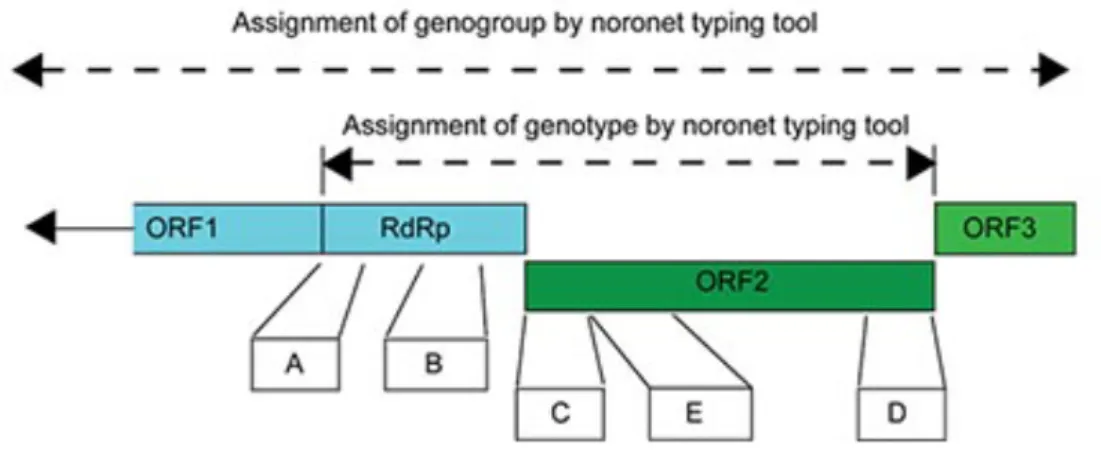
Noroviruses are genetically diverse viruses and are classified into different genogroups as well as polymerase (P) groups and further divided into genotypes and P-types based on aminoacid diversity of the complete VP1 gene and the nucleotide diversity of the RNA-dependent RNA polymerase region of ORF1, respectively (Chhabra et al. 2019).
Currently, noroviruses are divided in 10 genogroups (GI-GX) based on VP1 amino acid sequence diversity, in 49 genotypes (9 GI, 27 GII, 3 GIII, 2 GIV, 2 GV, 2 GVI and one genotype each for GVII, GVIII, GIX and GX), eight P-groups and 60 P-types based on partial nucleotide sequences of RdRp regions (Chhabra et al. 2019).
Figure 3. Genetic classification based on VP1 amino acid sequences into ten genogroups and two nonassigned genogroups (a), GI genotypes (b) and GII genotypes (Chhabra et al. 2019). The scale bar shows the number of nucleotide substitutions per site.
Norovirus within the same genogroup show 45-61% similarity in nucleotide sequence in the VP1 region and within the same genotype a similarity of 80 % (Zheng et al. 2006). New genotypes have been assigned when the VP1 amino acid sequence differs by more than 20% compared to other genotypes (Vinjé et al. 2000). In recent years, with the rapid accumulation of more norovirus sequence data, the cutoff threshold of 20 % needed adjustment due to increasing within-genotype diversity (Kroneman et al. 2013). So, within a genotype the amino acid divergence was changed to 14.1 % and a minimum of 15 % pairwise difference between the next-nearest genotype (Zheng et al. 2006).
Until 2019, a dual-nomenclature system was used, with the P-type designation linked to the corresponding VP1 genotype of the strain (Chhabra et al. 2019) and when no VP1 sequence was known, orphan P-types that only were seen in combination with established ORF2 genotypes, instead of a unique ORF2 genotype of their own, were designated by a letter, such as GII.Pc, GII.Pe and GII.Pg (Kroneman et al. 2013). As recombination in the ORF1-ORF2 junction region is common and some capsid genotypes seem to be more prone to recombination than others (Kroneman et al. 2013). A norovirus with a GII.4 polymerase and
capsid type were designated GII.P4-GII.4 and a recombinant GII.3 polymerase and a GII.4 capsid will be designated GII.3-GII.4 (Kroneman et al. 2013).
Classification and nomenclature updates in the future will be based on complete genome sequences (Chhabra et al. 2019).
1.5. Cultivation of human norovirus
Despite almost 50 years of attempts to develop in vitro culture methods this remains the major limitation to study human norovirus biology and the development of effective antiviral strategies (Estes et al. 2019). Routine cell cultures failed to yield replication of human noroviruses and attempts to develop a method for their cultivation were unsuccessful (Duizer et al. 2004). The noroviruses cell tropism has been an enigma for a long time, but it was assumed that human norovirus infected intestinal epithelial cells. Recent data support a more complex cell tropism of epithelial and nonepithelial cell types (Wobus 2018).The discovery of murine norovirus in 2003 and that it could replicate successfully in a murine macrophage line
in vitro and primary immune cells in vivo, suggested that immune cells may also support
replication of human norovirus (Wobus et al. 2004). Recently it was reported that a human B cell line supported the replication of a norovirus GII.4 strain in the presence of enteric bacteria (Jones et al. 2014), but its usage for in vitro replication of norovirus still awaits further developments (Jones et al. 2015). The human intestinal enteroids cultures reproduce the complexity and cell diversity of the gastrointestinal tract in relatively the same proportions as in the intestine itself (Zachos et al. 2016) and successfully support the replication of human norovirus (Costantini et al. 2018). These studies that confirmed enterocytes as the preferential site for human norovirus replication, support the genetic basis of host restriction and identify a role of bile as a strain-specific requisite or enhancer in virus infectivity (Ettayebi et al. 2016; Zou et al. 2017). Replication of human norovirus in Zebrafish and in organoids, made from pluripotent stem cells, has been recently reported enabling new studies on human norovirus biology (Van Dycke et al. 2019; Sato et al. 2019).
Successful cultivation was based on the discovery of genetically encoded host factors required for infection, knowledge of the site of infection in humans, and advances in the cultivation of human intestinal epithelium cells achieved by developmental and stem cells biologists (Estes et al. 2019).
1.6. Environmental resistance
Noroviruses are extremely stable in the environment, lasting weeks to years, depending on environmental conditions such as temperature and relative humidity (Seitz et al. 2011). They resist to nearly all of the active compounds of cleaning products, sanitizers, and disinfectants commonly used in food production and processing, including quaternary ammonium compounds, detergents, alcohols, and even chlorine at regulated concentrations (Hoezler et al. 2013). They can also survive to food processing and preservation methods such as heat, ionizing radiation, organic acids, preservatives, and manipulation of pH or water activity (Moore et al. 2015). Thus, human noroviruses are seen as the near-perfect foodborne pathogen, except for the fact that it cannot multiply (but does persist) in foods and the environment (Moore et al. 2015). Norovirus in various water sources are highly resistant to environmental degradation and long-term infectivity has been reported for groundwater which when seeded with the prototype norovirus (GI.1 Norwalk virus), in a clinical trial, was infectious for at least 61 days (Seitz et al. 2011).
Norovirus contamination of drinking water can be controlled by adequate free chlorine disinfection practices if proper pre-treatment processes are applied before chlorination (Shin and Sobsey 2008). In the environment, norovirus can be found in water that comes into contact with human stool samples which can also lead to contaminated crops (irrigation) and shellfish (growing waters) (Glass et al. 2009; Barclay et al. 2014; Katayama and Vinjé 2017). Norovirus also showed significant persistence in abiotic environments as it was reported that two carpet fitters who removed a carpet in a ward where a norovirus outbreak had occurred 3 weeks earlier, developed norovirus gastroenteritis symptoms (Cheesbrough et al. 1997).
In the presence of bacteria human norovirus was found to be more stable to acute heat stress, suggesting that bacteria may increase norovirus stability in the environment (Li et al. 2015; Graaf et al. 2017).
1.7. Epidemiology of norovirus infection
1.7.1.
Disease impact worldwide
Norovirus is estimated to cause approximately 699 million illness cases and 219,000 deaths worldwide resulting in $4.2 billion in health system costs and $60.3 billion in societal costs annually (Bartsch et al. 2016). As most persons with acute gastroenteritis do not seek medical care and therefore do not incur healthcare costs, overlooking productivity losses would
severely underestimate the true cost of norovirus illness (Bartsch et al. 2016). Total cost estimates were most sensitive to hospitalization rates, probability of missing productive days, and care seeking rates (Bartsch et al., 2016).
Acute gastroenteritis causes the second greatest burden of all infectious diseases worldwide (Ahmed et al. 2014). Noroviruses are a leading cause of sporadic cases and outbreaks of acute gastroenteritis across all age groups, accounting for 18% of acute gastroenteritis cases worldwide (Ahmed et al. 2014). Norovirus prevalence, based on studies that used PCR based diagnosis, in patients with acute gastroenteritis, tends to be higher in cases in the community (24%) and outpatient settings (20%) compared with inpatient settings (17%) (Ahmed et al. 2014). Prevalence is also higher in low-mortality developing (19%) and developed countries (20%) compared with high-mortality developing countries (14%) (Ahmed et al. 2014). This may be explained by the fact that low-income countries have higher prevalence of other pathogens that cause acute gastroenteritis so the proportion of acute gastroenteritis caused by norovirus in these countries is relatively lower (Ahmed et al. 2014). Also, it should be noted in low-income countries medical services have lower coverage and the rate of underreported norovirus cases is probably higher (Nguyen et al. 2017). Another study estimates a norovirus prevalence of 17% in patients with acute gastroenteritis in developing countries (Nguyen et al., 2017). By age, prevalence was similar in patients with acute gastroenteritis under 5 years, 5 years and over, and of mixed ages (Ahmed et al. 2014; Nguyen et al. 2017). By country income, prevalence decreased as income decreased (Nguyen et al. 2017).
Although recognized as the leading cause of epidemic acute gastroenteritis across all age groups, norovirus has remained poorly characterized with respect to its endemic disease incidence. In the United States, norovirus causes an average of 570–800 deaths, 56,000– 71,000 hospitalizations, 400,000 emergency department visits, 1.7–1.9 million outpatient visits, and 19–21 million total illnesses per year (Hall et al. 2013). People over 65 years of age are at greatest risk for norovirus-associated death while children under 5 years of age have the highest rates of norovirus associated medical care visits (Hall et al. 2013). In the US norovirus incidence is estimated at 700 illnesses/10,000 population, in the United Kingdom 450 illnesses/10,000 population, in the Netherlands 380 illnesses/10,000 population and in Canada 1,040 illnesses/10,000 population (Phillips et al. 2010; Tam et al. 2012; Hall et al. 2013; Verhoef et al. 2013).
Following the introduction of surveillance of outbreaks of gastrointestinal infection in England and Wales in 1992, norovirus was found responsible for more than 1800 outbreaks that affected more than 45,000 patients and hospital staff (Harris et al. 2014). Since 2009 another hospital-based surveillance system was implemented in England, the Hospital Norovirus Outbreak Reporting Scheme (HNORS)
(
https://www.gov.uk/government/publications/reported-norovirus-outbreaks-suspected-and-lab-confirmed-in-hospitals-2019). In the first 3 years (2009–2011) of the HNORS surveillance scheme, 4,000 outbreaks of norovirus were reported in England, affecting 40,000 patients and 10,000 staff (Harris 2016). Annually, on average these outbreaks affected 13 000 patients and 3400 staff, with 15 000 lost bed-days (Harris 2016).
In the aftermath of the hurricane Katrina a large norovirus outbreak affected more than 11,000 evacuees who were sheltered in the Reliant Park Complex in Houston, over an 11 day period (Yee et al. 2007). Future disaster preparedness and response planning should anticipate outbreaks caused by norovirus among evacuees, better prepared shelters with appropriate materials to educate about disease transmission and the importance of surveillance and prevention, and provide the proper policies, procedures, and resources to ensure good personal hygiene and sanitation for all involved (Yee et al. 2007).
In Portugal, information on prevalence of norovirus infection is scarce, but infections should occur frequently as antibodies against norovirus GII.4 were detected in 70% of a Portuguese cohort (Mesquita and Nascimento 2014). The underreporting of norovirus infections could be due to either the lack of knowledge among clinicians or limited availability of norovirus diagnostic testing in routine clinical laboratories (Mesquita and Nascimento 2014). In a two year hospital-based study performed in Portugal between 2011 and 2013, norovirus GII.4 (strain Sydney 2012) was detected in 11,6% of stool from patients hospitalized with acute diarrhea in 13 hospitals from Portugal (Costa et al. 2015).
1.7.2. Molecular epidemiology of norovirus
Noroviruses that infect humans have been associated only to genogroups GI, GII and GIV (Vinjé 2015). Noroviruses belonging to the genogroup GI and GII are responsible for the majority of disease cases in humans, whereas noroviruses from genogroup GIV are rarely detected as the cause of epidemic or sporadic gastroenteritis (Lindesmith et al. 2008; Robilotti et al. 2015). In humans, GII noroviruses are the predominant genotype detected (70%-89%), whereas noroviruses GI, which include virus of the GI.1 prototype Norwalk virus strain, cause approximately 11% of the outbreaks (Vinjé 2015; Lee et al. 2015). Within GII noroviruses genotype 4 (GII.4) are responsible for the majority of outbreaks and sporadic cases worldwide (Siebenga et al. 2009; Allen et al. 2016; Jung et al. 2017; Qin et al. 2017). In fact, GII.4 norovirus have caused all the six major pandemics of acute gastroenteritis in the last two decades (White 2014). These six pandemic GII.4 variants include US 96, which caused a pandemic in the late 1990s, Farmington Hills 2002, Hunter 2004, Den Haag 2006b, New
Orleans 2009 and Sydney 2012 (Leshem et al. 2013; Lee et al. 2015). These variants emerged in a periodicity of 2 or 3 years by genetic drift until 2012 (Van Beek et al. 2018).
An epidemiological study reported that in USA norovirus GII caused 72% of the outbreaks, 94% of each were either GII.4 New Orleans or GII.4 Sydney (Vega et al. 2014). The GII.4 Sydney seems to persist though recombination, as a norovirus recombinant of GII.p16-GII.4 Sydney 2012 variant was reported in Asia and Europe (van Beek et al. 2018). In China and Japan the genotype GII.4 was displaced by GII.17, which became the predominant genotype since 2014 (Qin et al. 2017).
Several non-GII.4 genotypes were significantly more associated with foodborne transmission and the cyclic emergence of new non-GII.4 norovirus strains and genotypes are also more often associated with foodborne outbreaks (Vega et al. 2014).
A study in Denmark investigated the genotype distribution in relation to age and setting and observed that norovirus GII.4 predominated in patients with 60 or more years and in health care centers while in children norovirus GII.P21 and GII.3 were more prevalent than in adults (Franck et al. 2014). It has been hypothesized that viral genotypes with lower antigenic variation, such as GII.3, more frequently infect children who do not have established immune-associated protection from previous norovirus infections (Franck et al. 2014). Moreover GII.4 strains are associated with more severe outcomes, including mortality, than infections during outbreaks of non-GII.4 strains (Desai et al. 2012).
1.7.3. Incidence seasonality of norovirus infection
Endemic norovirus disease occurs year-round but exhibits a pronounced winter peak and increases by around 50% during years in which pandemic strains emerge (Hall et al. 2013). In regions of temperate climate, norovirus occurrence is seasonal with most infections observed during the winter months (Katayama et al. 2008; Cannon et al. 2017). This annual fluctuation is likely caused by biological, environmental and behavioral factors that influence viral transmission, virulence and persistence in the host population (Rohayem 2009). Several studies have found associations between norovirus seasonality and climatic weather phenomena and specifically abundant rainfall and low temperatures seem to enhance viral persistence in water environments (Greer et al. 2009; Bruggink and Marshall 2010; Ahmed et al. 2014) The seasonality of norovirus gastroenteritis is also known to be influenced by the host behavior, in particular, crowding and prolonged sharing of indoors spaces are possible factors increasing human-to-human transmission of these viruses during winter (Mounts et al. 2000; Lindesmith et al. 2012; Zhou et al. 2016). As seasonal fluctuations modulate host cellular and humoral immune function, it is has been speculated that during wintertime diminished UV-radiation reduces vitamin D synthesis impairing immune response against
norovirus, as vitamin D is an important regulator of phagocyte function and is associated with the antiviral response to influenza virus infection by immune cells (Rohayem 2009).
In Europe norovirus seasonality coincides with northern hemisphere winter season, but GII.Pe/GII.P4-GII.4 strains show the clearest winter seasonal patterns while GI and other GII strains are more continuously present throughout the year (van Beek et al. 2018). China and Japan show increased norovirus incidence in the northern hemisphere winter season with the peak in November, two months earlier compared to Europe (van Beek et al. 2018). New Zealand shows highest incidence in October and November, and South Africa in September to November in southern hemisphere spring (van Beek et al., 2018). In the Middle east and North Africa countries peaks were observed during colder months, although norovirus infections were reported all year round (Kreidieh et al., 2017).
Climate change may affect norovirus seasonality with subsequent impact on norovirus transmission, host susceptibility to norovirus infection, resistance of norovirus to environmental conditions and change the interaction of norovirus with their host (Rohayem 2009). Human migration may become significantly altered as the result of climate change and as a consequence of floods or droughts, massive displacement of populations and crowding in refugee camps may facilitate the introduction of noroviruses into immunologically naïve populations, resulting in epidemics and the emergence of new norovirus strains (Rohayem 2009). In this context, norovirus evolution may be modulated by periods of elevated or facilitated transmission and evolutionary bottlenecks through rapid mutation or recombination events, which may in turn cause larger oscillations in the prevalence of the disease than are currently observed (Rohayem 2009).
1.7.4. Transmission of norovirus
Norovirus enter the human organism through the mouth. Virions are shed in large amounts in stools and in lower numbers in vomitus (Kilgore et al. 1996; Lee et al. 2007; Aoki et al. 2010). Virus expelling is highest during the acute phase of the disease, although shedding can last 9-56 days, even in asymptomatic persons (Atmar et al. 2008; Aoki et al. 2010). Peak shedding (1011 viral particles per gram of stool) occurs in the first days following infection but
can persist for over three weeks, especially in young children (Atmar et al. 2008; Siebenga et al. 2008). In asymptomatic children shedding can last for 100 days and in immunocompromised individuals can be prolonged for several years (Murata et al. 2007; Ludwig et al. 2008; Schorn et al. 2010). Immunodeficient patients may become chronic symptomatic shedders although there is still no evidence that these individuals can transmit the virus and cause an infection (Koopmans 2005; van Beek et al. 2017). In human challenges
et al. 2008; Kirby et al. 2014). Infected people who are asymptomatic may shed similar amounts of virus as ill persons (Koopmans 2005; Newman et al. 2016).
Patients shedding low levels of norovirus RNA may not be infectious, as a recent study seemed to demonstrate with norovirus GII.pe-GII.4 Sydney in a human intestinal enteroid model (Chan et al. 2019).
Norovirus can be transmitted by different ways such as through food, persons, water, and even the environment (Verhoef et al. 2015). The most important mode of transmission is the fecal-oral spread although infectious vomit, either by mechanical transmission from environmental surfaces (hand/mouth contact) or by aerosolization might account for the rapid and extensive spread of disease outbreaks, especially in closed settings (Patel et al. 2009).
Norovirus are infectious by oral route at very low doses (Glass et al. 2009), with an, estimated dose of 18 to 1000 of viral particles (Teunis et al. 2008).
Sometimes it is difficult to determine the transmission of norovirus, as more than a route can occur in a single outbreak. After primary introduction of the norovirus through food, secondary person-to-person and environmental transmission can rapidly take over, making it hard to trace the disease back to contaminated food. Another complexity is that foodborne transmission can follow different routes as well (Verhoef et al. 2015).
Person-to-person transmission is thought to account for 62–84% of all reported outbreaks (Moore et al. 2015). A study of norovirus outbreaks in Europe revealed that primary transmission occurs through the fecal-oral route, by person-to-person spread in 88%, ingestion of norovirus contaminated food in 10% or water in 2% (Kroneman et al. 2008).
Using data from the CaliciNet surveillance system from 2009 to 2013 in USA the estimated transmission in norovirus outbreaks was 83.7% for person-to-person and 16.1% for foodborne (Vega et al. 2014). In the same study, 62.5% of the outbreaks occurred in long-term care facilities, 9.8% in restaurants and 5.8% in schools. Applying the profiles in surveillance database, the proportion of outbreaks attributed to foodborne transmission varied slightly with a global estimate of 13.7% (Verhoef et al. 2015). More recently in another European study, 77,4% of the transmission was considered person-to-person, 19,9% foodborne, 2,1% waterborne and 0,7% from other modes of transmission (van Beek et al. 2018).
The epidemiologic curves are usually distinct between outbreaks involving person-to-person or food-borne transmission. In the person-to-person-to-person-to-person transmission the numbers of patients increase and then decrease gradually, on the other hand in the foodborne transmission, infection occur at once and intensively, since the cohort is mostly exposed to the contaminated food at the same time (Ushijima et al. 2014). Norovirus GII.4 is relatively more often associated to person-to-person transmission than other genotypes (van Beek et al. 2018).
Animal noroviruses has not yet been found in humans, however detection of human noroviruses in animals and simultaneous animal and human viruses in bivalve mollusks suggests a risk of transmission (Bank-Wolf et al. 2010). Human noroviruses have been detected in stool of dogs, swine, cattle, wild birds and rodents (Mattison et al. 2007; Caddy et al. 2015; Summa et al. 2018). Canine seroprevalence to different human norovirus genotypes resembles the seroprevalence in the human population (Caddy et al. 2015). Serum antibodies against bovine and canine noroviruses have also been detected in humans, with higher level in veterinarians than in the general population (Widdowson, Rockx, et al. 2005; Mesquita et al. 2013). A recent study found more evidence for human noroviruses in animals than the reverse, suggesting that human noroviruses could be a reverse zoonosis, although it is still too early to consider norovirus a zoonotic or reverse zoonotic pathogen (Villabruna et al. 2019). Zoonotic transmission was never proved. Pet dogs and wildlife (birds and rodents) may play a role in the spreading of human noroviruses in the environment (Summa et al. 2012; Summa et al. 2018).
1.7.5. Norovirus and foodborne and waterborne illness
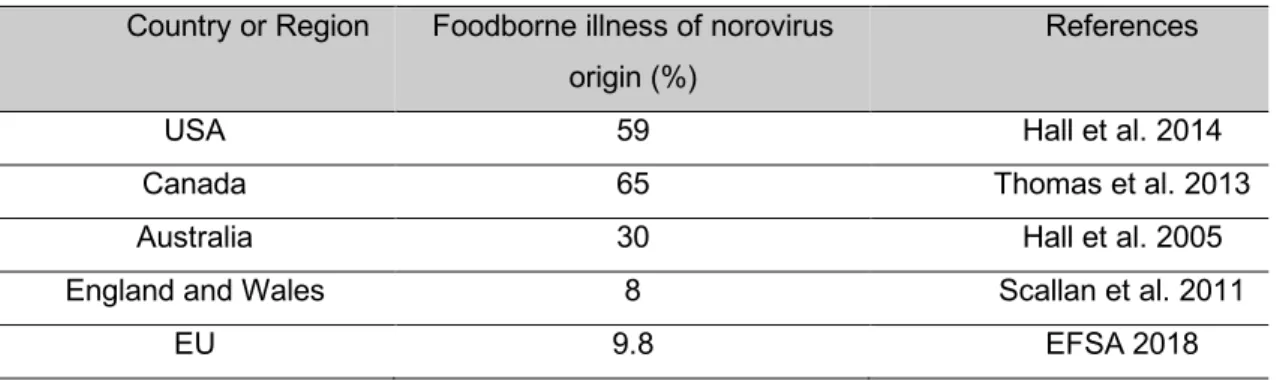
Norovirus is the leading cause of acute gastroenteritis and foodborne disease outbreaks in the United States of America (USA) (Hall et al. 2014). It is estimated that in the USA viruses account for 59% of the foodborne diseases and that norovirus account for 99% (5.5 million) of all viral foodborne illness incidents per year (Scallan et al. 2011) . Norovirus is also responsible for 26% of hospitalization and 11% of death related to foodborne diseases (Scallan et al. 2011). In Canada, norovirus is also considered the leading cause of foodborne illness, accounting for 65% of known illnesses (Thomas et al. 2013). In Australia norovirus is the leading cause of foodborne illness, accounting for 30% of illnesses caused by known pathogens (Hall et al. 2005). In England and Wales, norovirus accounted for only 8% of known foodborne illnesses (Scallan et al. 2011). Foodborne illness of norovirus origin by country or region is summarized in Table 2.
As reported by the European Food Safety Authority (EFSA), in 2017, in the European Union (EU) viruses accounted overall for the 9.8% of total foodborne and waterborne outbreaks in 2016, which is comparable with 2015. At the EU-level, no trends in the outbreak number reported were observed for calicivirus (including norovirus). During the 2010–2016 period, Denmark, Lithuania and Sweden reported a statistically significant decreasing trend in the number of outbreaks of calicivirus including norovirus, while France and the Netherlands reported a statistically significant increase. Three other European countries (Finland, Germany
and United Kingdom) reported a 2016 increase in the number of outbreaks by calicivirus including norovirus of over 50% as compared with the previous year (EFSA 2018).
Table 2. Foodborne illness of norovirus origin
Country or Region Foodborne illness of norovirus origin (%)
References
USA 59 Hall et al. 2014
Canada 65 Thomas et al. 2013
Australia 30 Hall et al. 2005
England and Wales 8 Scallan et al. 2011
EU 9.8 EFSA 2018
Foodborne norovirus outbreaks are difficult to identify and even more difficult to trace back to a common food source as most foodborne norovirus outbreaks are small, associated with a retail setting (and hence investigated locally or regionally) and often with secondary person-to-person spread (propagated outbreaks) (Moore et al. 2015). Contamination of food frequently happens at the end of the production-logistic chain with typical examples of a food handler with poor hand hygiene or persons who were sick inside a room where food was prepared or stored (Koopmans 2005). Larger foodborne outbreaks affecting vast geographic regions have also been detected (Koopmans and Duizer 2004; Koopmans 2005; Bernard et al. 2014; Morgan et al. 2019). In this case it is thought that the contamination event is early in the food chain, for instance with berries that are harvested under poor hygienic conditions, or that were irrigated or washed with contaminated water (Koopmans and Duizer 2004; Koopmans 2005).
The majority of norovirus outbreaks were due to person-to-person transmission, although foodborne transmission is also significant (Moore et al. 2015). Complex, prepared, ready-to-eat foods are overrepresented in outbreaks, and the most common settings for these are restaurants, delicatessens and catering facilities, implicating food handling as the likely contamination source (Moore et al. 2015). In the case of simple foods, vegetables, fruits, and nuts are the most likely culprits, but it is difficult to discern how relevant was pre-harvest contamination in these products (Moore et al. 2015).
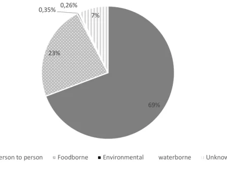
In a study performed in USA foodborne transmission was observed in 23% norovirus outbreaks during the 4 year study period, while person-to-person transmission accounted for 69%, environmental for 0,35%, waterborne for 0,26% and unknown transmission for 7% (Figure 3) (Hall et al. 2014). In a study, in China, from 2014 to 2017, the majority of the
outbreaks (63%) were due to person-to-person transmission, followed by multiple modes of transmission (11%), foodborne (5%) and waterborne (3%) transmission (Lian et al. 2019).
Figure 4. Transmission routes of norovirus in the USA (adapted from Hall et al. 2014).
Of the norovirus outbreaks that could be attributed to a single location, restaurants and delicatessens were the most common (63–64%), followed by catering and banquet halls (11– 17%) and private homes (4–6%) (Hall et al. 2014). Food workers were implicated as the source in 70% of the outbreaks with an identified cause. Most outbreaks with a demonstrated implicated food resulted from food contamination during preparation (92%) and food consumed raw (75%). The most frequently food categories implicated in the outbreaks were vegetable row crops (e.g., leafy vegetables) (30%), fruits (21%), and mollusks (19%) (Hall et al. 2014).
In the EU according to EFSA report of 2017, zoonotic agents and food-borne outbreaks in 2016, caused by norovirus ranked first among the causative agents of outbreaks caused by ‘fish and fishery products’ (51.4%) and was also reported in high proportions in outbreaks by ‘other foods’ (22.8%), ‘mixed food and buffet meals’ (22.3%) and ‘vegetables, fruits, cereals, sprouted seeds, herbs and spices and their products’ (26.4%) (EFSA 2017).
In another study on a global scale it was estimated that 14% of norovirus outbreaks were attributed to food (Verhoef et al. 2015). The proportion of norovirus outbreaks attributed to foodborne transmission is in the same order of magnitude as the 17% found in a study from the Netherlands and 11% in the United Kingdom as estimated from outbreak surveillance data (Adak et al. 2002; Havelaar et al. 2012; Verhoef et al. 2015). Interventions to reduce the frequency of foodborne norovirus outbreaks should focus on food workers, food production and shellfish as most foods are likely contaminated during preparation and service, except for
69% 23%
0,35% 0,26% 7%
mollusks, and occasionally contaminated during production and processing (Hall, Eisenbart, et al. 2012).
The number of foodborne illnesses caused by enteric bacteria have in generally declined over the past two decades (Center for Disease Control and Prevention [CDC] 2006) and possible reasons for this trend include better public awareness of safe cooking and hygiene food handling practices, improved standards in industrial food processing and better refrigeration and storage (Widdowson, Sulka, et al. 2005). On the other hand, the number of foodborne outbreaks caused by norovirus have remained unchanged or have increased and were estimated at 30-50% of all foodborne outbreaks (CDC 2006). It is likely that control measures in the food industry and public health awareness campaigns aimed at reducing foodborne bacterial illness have done little to reduce norovirus gastroenteritis (Cannon 2008).
Norovirus genotype profiles can be used to differentiate foodborne outbreaks caused by food contamination early in the food chain from those caused by food handlers contaminating food (Verhoef et al. 2010). In a study in Denmark, norovirus GI was more frequently observed in food-borne outbreaks than in outbreaks involving person-to-person transmission and was also more frequent in foodborne outbreaks than in outbreaks in health care and community settings (Franck et al. 2014). In outbreaks in community settings, GII infections outnumber GI infections, as GII strains have been found to be associated to 80% or more of outbreaks with the epidemic strains belonging to GII.4 being responsible for the vast majority of cases, particularly those associated with person-to-person transmission (Matthews et al. 2012; Vega et al. 2014). In the USA, 72% of the outbreaks reported from 2009 to 2013, were caused by GII.4 strains and non-GII.4 strains were significantly more associated with foodborne transmission (Vega et al. 2014).
Norviruses strains detected in foodborne outbreaks showed a genotype profile similar to those in bivalve mollusk monitoring (predominance of GI strains) and dissimilar to the profile detected in human stools with respect to the frequently seen genotypes which could reflect the ability of these genotypes to survive outside humans or their diminished ability to spread or replicate within the human population (Verhoef et al. 2010).
The genotype profiles and proportions may be helpful for estimating the number of outbreaks with potential of geographic dissemination. Early detection should enable containment of viral foodborne infection and thus prevent further spread to large numbers of human infectionsas the identification and investigation of such outbreaks provides insight into effective prevention measures during the production process (Verhoef et al. 2010).
1.8. Human norovirus infection
1.8.1. Pathogenesis of human norovirus infection
Norovirus recognize HBGA in a strain-specific manner (Chen et al. 2011). All three major families of HBGA, the AB0, Lewis and secretor families are involved in binding noroviruses (Huang et al. 2003; Tan and Jiang 2005). Persons carrying a functional fucosyltransferase 2 (encoded by FUT2 gene) are termed secretors and express HBGAs, whereas homozygous individuals, called non- secretors, are almost completely protected from GI.1 and GII.4 norovirus infections (Jin et al. 2013). However, polymorphisms in the FUT2 genes vary considerably depending on ethnicity and non-secretors can be infected by other norovirus genotypes (Jin et al. 2013). Because GII.4 viruses can bind a wider range of HBGAs than other genotypes, they are able to infect a larger susceptible population (Vinjé 2015).
After entering the gut lumen noroviruses gain access to inflammatory cells through microfold cells (M cells),a specialized subset of enterocytes that overlie Peyer´s patches and isolated lymphoid follicles and are highly efficient at sampling and transporting luminal material to the underlying immune aggregates (Karst and Tibbetts 2016). Noroviruses can infect both innate (macrophages and dendritic cells) and adaptive (B cells) immune cells along the intestinal tract, and infection of these cell types is a crucial determinant of viral pathogenesis (Karst and Tibbetts 2016). It seems that commensal bacteria enhance norovirus infection and facilitate infection persistence by direct mechanisms like the expression of H type HBGAs that stimulate norovirus adherence to permissive B cells and also by indirect mechanisms in the form of immunomodulation (suppression of type III IFN for example) (Karst and Tibbetts 2016). The intestinal mucosa appears to remain intact during human norovirus infection, but there are histopathological changes in the epithelium, like the broadening and blunting of the villi (Karst 2010). Crypt cell hyperplasia has also been reported following norovirus infection (Karst 2010). It is thought that these changes can impair the absorption of D-xylose, fat and lactose and the changes of secretory and/or absorptive processes are thought to be in the origin of the diarrhea (Karst et al. 2015). The pathophysiology of the vomit is thought to be related to a marked delay in gastric emptying, possible due to abnormal gastric motor function (Karst et al. 2015) .
1.8.2. Norovirus gastroenteritis
Typically, the incubation period of norovirus gastroenteritis is 12-48 hours (Katayama and Vinjé 2017), although it can vary from 10 to 51 hours (Glass et al. 2009).