http://dx.doi.org/10.1590/S1678-9946201860051
1Universidade Federal de Sergipe, Departamento de Morfologia, Laboratório de Entomologia e Parasitologia Tropical, São Cristóvão, Sergipe, Brazil
2Fundação Oswaldo Cruz, Instituto Oswaldo Cruz, Laboratório de Referência Nacional em Esquistossomose – Malacologia, Rio de Janeiro, Rio de Janeiro, Brazil
3Fundação Oswaldo Cruz, Instituto Oswaldo Cruz, Laboratório de Biologia e Parasitologia de Mamíferos Silvestres Reservatórios, Rio de Janeiro, Rio de Janeiro, Brazil
Correspondence to: Silvio Santana Dolabella
Universidade Federal de Sergipe, Departamento de Morfologia, Laboratório de Entomologia e Parasitologia Tropical, Av. Marechal Rondon, s/n, Jd. Rosa Elze, CEP 49100-000, São Cristóvão, SE, Brazil
E-mail: dolabella@ufs.br
Received: 16 April 2018
Accepted: 16 August 2018
First records of molluscs naturally infected with
Angiostrongylus cantonensis (Nematoda: Metastrongyloidea) in Sergipe State, Northeastern Brazil, including new global
records of natural intermediate hosts
Jucicleide Ramos-de-Souza1, Silvana Carvalho Thiengo2, Monica Ammon
Fernandez2, Suzete Rodrigues Gomes2, Jéssica Corrêa-Antônio2, Marianna
de Carvalho Clímaco1, Juberlan Silva Garcia3, Arnaldo Maldonado-Junior3,
Luciene Barbosa1, Silvio Santana Dolabella1
ABSTRACT
Human neural angiostrongyliasis is an emerging infectious disease caused by nematode Angiostrongylus cantonensis. The present study investigated the presence of Angiostrongylus spp. in terrestrial molluscs collected from the following areas in the Metropolitan Region of Aracaju, Sergipe State, Brazil: Barra dos Coqueiros, Nossa Senhora do Socorro, Sao Cristovao and Aracaju. In total, 703 specimens representing 13 mollusc species were screened for Angiostrongylus spp. Larvae of Angiostrongylus spp. were found in three species. Larvae recovered from Achatina fulica were used for experimental infection in Wistar rats (Rattus norvegicus). For specific identification of nematodes, the mitochondrial cytochrome c oxidase subunit I (COI) was sequenced from both larvae and adults recovered from molluscs and rats, respectively. Infection with A. cantonensis was detected in all municipalities and in the following three host species: Bulimulus tenuissimus, Cyclodontina fasciata (Barra dos Coqueiros), and A. fulica (Aracaju, Nossa Senhora do Socorro and Sao Cristovao). Co-infections were also found with Caenorhabditis sp. and Strongyluris sp. larvae. This is the first study of the helminth fauna associated with the terrestrial malacofauna in Sergipe State, and confirms that these three snail species are involved in the transmission of A. cantonensis in the state. In addition, B. tenuissimus and C. fasciata are newly reported natural hosts of the parasite.
KEYWORDS: Eosinophilic meningitis. Angiostrongylus.Achatina fulica.Bulimulus
tenuissimus.Cyclodontina fasciata. Land snails.
INTRODUCTION
Achatina fulica Bowdich, 1822 is listed among 100 of the worst invasive
species in the world and is present in almost all Brazilian states1,2. This species’
rapid proliferation is the main reason it is considered an agricultural pest. It can also act as an intermediate host of Angiostrongylus cantonensis (Chen, 1935) and
Angiostrongylus costaricensis Morera and Céspedes, 1971. These two species are
the etiological agents of human neural angiostrongyliasis and human abdominal angiostrongyliasis, respectively2-4.
for its intermediate hosts, which means that many mollusc species have already been found to be naturally infected globally8,9. However, A. fulica is an important intermediate
host for A. cantonensis4,8,10-12.
Human neural angiostrongyliasis occurs primarily by ingestion of snails that are infected with L3 larvae13. These
larvae reach the brain where they mature into L4 and L5 but are unable to migrate from the brain to the heart (as they do in the definitive rat hosts) but move around in the brain causing neurological damage and, especially when they die, cause intense inflammatory reactions that result in additional neurological damage and symptoms5,7,14.
More than 2,800 human cases of eosinophilic meningitis caused by A. cantonensis have been reported in more than 30 countries11 since it was first reported in Taiwan in 194515.
In Brazil, out of 84 suspected cases, there have been 34 confirmed cases of A. cantonensis infections in humans as well as the confirmation of definitive hosts and infected intermediate hosts in different regions of the country10.
In Sergipe State, 19 municipalities have reported infestation of A. fulica (Comissao de Combate ao Caramujo Africano, personal communication) in addition to Barra dos Coqueiros municipality8. The objective of this study
was to verify the infection rate by nematode larvae in A.
fulica and other terrestrial molluscs collected in the Aracaju
Metropolitan Region, Sergipe State, Northeastern Brazil.
MATERIALS AND METHODS
This study was undertaken in urban environments of four municipalities that constitute the Metropolitan Region of Aracaju, Sergipe State (Figure 1): Aracaju, Barra dos Coqueiros, Nossa Senhora do Socorro and Sao Cristovao (Table 1).
Each point of collection was georeferenced, followed by manual random sampling of terrestrial molluscs using forceps
and gloves. The collections were carried out in the following three seasons: April (autumn), July (winter) and October (spring) of 2016, at three locations in each municipality. The samples were sent to the National Reference Laboratory for Schistosomiasis - Malacology (LRNEM) of the Instituto Oswaldo Cruz, Fundação Oswaldo Cruz (IOC/Fiocruz, Rio de Janeiro), where they were analyzed. The identification of the molluscs species was done based on conchological characteristics compared to photos and descriptions from catalogues16-19. We also compared our material with material
deposited in the Instituto Oswaldo Cruz. Samples of each species and from each collection locality were deposited in the Mollusca Collection of the Instituto Oswaldo Cruz (CMIOC 10103 - 10134, 11206).
To collect the nematode larvae, the molluscs were artificially digested by a modified Wallace and Rosen technique20. The recovered material was observed
under a stereoscopic microscope following the LRNEM Identification Guide and based on the literature4,21. The
Angiostrongylus sp. (Nematoda: Metastrongylidae) larvae
were identified under a compound microscope according to Thiengo et al.4. Ten Angiostrongylus sp. larvae from
each sample were collected and cryopreserved at -20 °C until DNA extraction for subsequent molecular analysis. Some rhabditiform larvae were observed and prepared for DNA extraction. In addition, Strongyluris sp. was morphologically identified, based on Thiengo21.
To assess the viability of the parasites, Angiostrongylus
sp. larvae recovered from A. fulica from the municipality of Aracaju were used to infect two specimens of Rattus
norvegicus through an orogastric tube at a concentration
of 50 L3 larvae per rodent. Fifty days after infection, the animals were killed and necropsied to confirm the presence of adult A. cantonensis22. This procedure was carried out
following the Ethics Commission on Animal Use of the Oswaldo Cruz Foundation (LW-47/14).
Molecular diagnosis of nematode larvae
DNA was extracted from 10 Angiostrongylus sp. larvae obtained from each mollusc in which larvae had been found and resuspended in 30 µL of PCR Buffer solution (Thermo Fisher Scientific, Massachusetts, USA). The polymerase chain reaction (PCR) mixtures were prepared in a volume of 20 µL containing 8 µL of ultrapure water, 5 µL of 10% trehalose, 2.5 µL of 10x PCR Reaction Buffer, 2 µL of 2.5 mM dNTPs, 1.25 µL 50 mM MgCl2, 0.5 µL each of 5 µM forward and reverse primer (Nem 3 from Prosser et al.23),
and 0.25 µL of recombinant Taq DNA polymerase (Thermo Fisher Scientific). A total of 5 µL of the DNA sample was added to the mixture, producing a final volume of 25 µL for each reaction. For all the reactions, ultrapure water was used as a negative control template, and the positive control was performed with genomic DNA of A. cantonensis as template. The 700 bp fragment of the Angiostrongylus sp. larvae mitochondrial cytochrome c oxidase subunit I (COI) gene was amplified using the following PCR conditions: initial denaturation at 94 °C for 1 min, five cycles at 94 °C for 40 s, 45 °C for 40 s, and 72 °C for 1 min, followed by 30 cycles at 94 °C for 40 s, 51 °C for 40 s, and 72 °C for 1 min and a final extension at 72 °C for 1 min.
For rhabditiform larvae, individual DNA from some isolates was used to amplify the 480 bp ribosomal 18S region24.
The PCR products were purified using the Illustra GFX PCR DNA and Gel Band Purification Kit (GE Healthcare, Little Chalfont, UK) following the manufacturer’s protocol. Purified products after amplification were bidirectionally sequenced using BigDye Terminator v3.1 Cycle Sequencing kit (Applied Biosystems, California, USA) according
to the manufacturer’s instructions. Chromatograms of the sequences obtained were analyzed and edited using Geneious version R9 software (http://www.geneious. com), resulting in a consensus sequence (contig). Then, a comparative similarity search was performed on GenBank (www.ncbi.nlm.nih.gov/genbank/)25 using BLAST (Basic
Local Alignment Search Tool) to identify the closest match. The obtained sequences are available in GenBank under the access Nº MH511539 - MH511542, and MH547424.
RESULTS
In total, 703 terrestrial gastropods were analyzed (190 in Aracaju, 250 in Barra dos Coqueiros, 159 in Nossa Senhora do Socorro, and 104 in São Cristovão) and identified as the following 13 species: Achatina
fulica (Bowdich, 1822), Allopeas gracile (Hutton, 1834),
Subulina octona (Bruguière, 1789), Leptinaria unilamellata
(d’Orbigny, 1835), Bulimulus tenuissimus (d’Orbigny, 1835), Cyclodontina fasciata (Potiez & Michaud, 1838),
Latipes erinaceus (Colosi, 1921), Sarasinula linguaeformis
(Semper, 1885), Streptartemon cookeanus (Baker, 1914),
Streptartemon quixadensis (Baker, 1914), Tamayoa
banghaasi (Thiele, 1927), Helicina sp. and Omalonyx sp.
Angiostrongylus sp. larvae were found in specimens
of A. fulica, B. tenuissimus and C. fasciata and were later
confirmed by molecular analysis as A. cantonensis (Table 2).
Achatina fulica was found in all four municipalities, with
snails harboring infection by A. cantonensis in three of them (Aracaju, Sao Cristovao and Nossa Senhora do Socorro).
Bulimulus tenuissimus occurred in all municipalities, while
specimens of C. fasciata were found only in Aracaju and Barra dos Coqueiros. Both B. tenuissimus and C. fasciata
Table 1 - Geographical coordinates by location of collection of terrestrial molluscs
Municipalities Description of locality Geographical coordinates
Barra dos Coqueiros
Square / open area 10°49'30.1"S 36°56'46.7"W
Outer clothing area 10°49'42.6"S 36°56'08.2"W
School backyard / Rubble 10°49'12.2"S 36°56'58.9"W
Sao Cristovao
Street / close to houses 10°55'57.8"S 37°06'26.6"W
Street / close to houses 10°55'27.7"S 37°06'43.2"W
Street and house backyard 10°55'43.1"S 37°07'16.2"W
Aracaju
Street / close to houses 10°58'29.7"S 37°06'16.7"W
Ground / adjacent to a commercial establishment 11°00'11.9"S 37°05'03.2"W
Close to sewer-line 10°54'24.1"S 37°05'23.3"W
Nossa Senhora do Socorro
Garden and houses backyard 10°50'19.0"S 37°03'15.0"W
Houses backyard 10°51'02.2"S 37°05'38.1"W
were infected by A. cantonensis in the municipality of Barra dos Coqueiros, near the port area of Sergipe State.
Co-infections were observed between A. cantonensis
and other nematodes, such as Strongyluris sp. and rhabditiform larvae (Table 3). The rhabditiform larvae were analyzed molecularly and were found to be 99% similar to a sequence from GenBank identified as Caenorhabditis sp.
Infections of Caenorhabditis sp. were observed
in A. fulica, B. tenuissimus, C. fasciata, S. octona, L.
unilamellata, S. linguaeformis and Omalonyx sp. Infections
by Strongyluris sp. were observed in A. fulica, C. fasciata
and S. linguaeformis.
Two rats that were experimentally infected with
Angiostrongylus sp. isolates displayed symptoms 50 days
after infection. Forty A. cantonensis adult worms were recovered in the first rat (37.5% males and 62.5% females), and 42 adult worms were recovered in the second (40.4% males and 59.5% females).
DISCUSSION
The snail A. fulica has already been reported as being parasitized with A. cantonensis by several authors2,4,8,10,19,26.
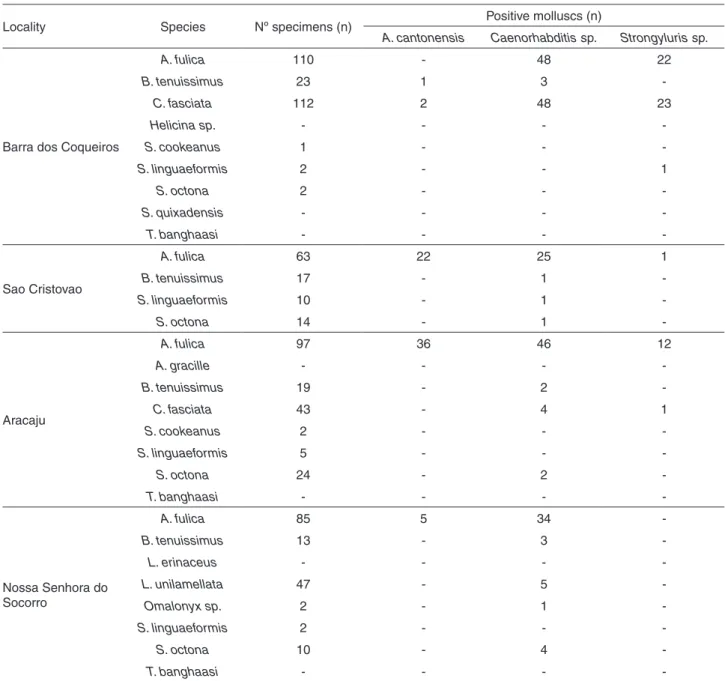
In Brazil, the species is considered one of the main Table 2 - Prevalence of nematodes in terrestrial molluscs from the Metropolitan Region of Aracaju, Sergipe State, Northeast Brazil, from April to October 2016
Locality Species Nº specimens (n) Positive molluscs (n)
A. cantonensis Caenorhabditis sp. Strongyluris sp.
Barra dos Coqueiros
A. fulica 110 - 48 22
B. tenuissimus 23 1 3
-C. fasciata 112 2 48 23
Helicina sp. - - -
-S. cookeanus 1 - -
-S. linguaeformis 2 - - 1
S. octona 2 - -
-S. quixadensis - - -
-T. banghaasi - - -
-Sao Cristovao
A. fulica 63 22 25 1
B. tenuissimus 17 - 1
-S. linguaeformis 10 - 1
-S. octona 14 - 1
-Aracaju
A. fulica 97 36 46 12
A. gracille - - -
-B. tenuissimus 19 - 2
-C. fasciata 43 - 4 1
S. cookeanus 2 - -
-S. linguaeformis 5 - -
-S. octona 24 - 2
-T. banghaasi - - -
-Nossa Senhora do Socorro
A. fulica 85 5 34
-B. tenuissimus 13 - 3
-L. erinaceus - - -
-L. unilamellata 47 - 5
-Omalonyx sp. 2 - 1
-S. linguaeformis 2 - -
-S. octona 10 - 4
-potential transmitters of human neural angiostrongyliasis, considering their widespread distribution, high population densities and proximity to humans10,26.
Despite knowledge of the presence of the giant African snail in Sergipe State since 200627,28, only one previous
investigative study has been carried out in this region8. Ten
years after the first detection of A. cantonensis in Brazil, this study records its presence in Sergipe State for the first time; it is also the first report of C. fasciata and B. tenuissimus
as natural intermediate hosts of the parasite. In addition, we observed co-infection with other nematodes, which is consistent with other studies12,26,28.
Since 2007, when the first Brazilian case of eosinophilic meningitis caused by A. cantonensis was recorded, in Espirito Santo State29, reports of the disease have increased. Currently,
the parasite has been found in 12 states (Figure 2)2,7,10,29-31.
The first known occurrence of A. fulica naturally infected by A. cantonensis in the northeast region of Brazil was reported in 2008, with human cases of eosinophilic meningitis and naturally infected intermediate hosts in the municipalities of Escada and Olinda, Pernambuco State4.
A study carried out in Brazilian port areas, including the port of Sergipe State, reported the presence of terrestrial and aquatic molluscs that, although previously negative for
A. cantonensis, tested positive in the bordering Bahia State8
and in Ceara State10.
Several other terrestrial molluscs have also been found to be naturally infected with this nematode in Brazil2,3,8,19
and in other countries9,13,14. Among these species are S.
linguaeformis and S. octona2,3,8,9, which, however, were not
found to be infected in the present study. A possible reason for this is the lower abundance of these species in the sampled localities. The three species found infected by A. cantonensis
were also those that were more abundant in the environment. The present study identifies, for the first time, the following two species as natural hosts of the parasite:
C. fasciata, which had not yet been described in the
literature as an intermediate host for A. cantonensis or for other nematodes of human and veterinary interest, and
B. tenuissimus, described as a host of metacercariae of the
genus Postharmostomum32. This interaction between exotic
and native species has become a new concern in the ecology of the disease and may result in increased parasitic spread as well as pose a threat to native species9.
Studies indicate the need to implement control measures against molluscs associated with public health, particularly the exotic species A. fulica. However, passive geographic dispersal is the main form of spread of the giant African snail, which hinders its control33.
The present study reinforces the need to investigate the current situation of A. cantonensis dispersal in the Northeastern region of Brazil, since there are reports of Table 3 - Prevalence of co-infection between Angiostrongylus cantonensis, Caenorhabditis sp. and Strongyluris sp. in land molluscs from the Aracaju Metropolitan Region, Sergipe State, Northeast Brazil, from April to October 2016
Locality Species
Specimens analyzed
(n)
Positive specimens
for A. cantonensis Co-infection (n)
(n) (%) A. cantonensis +
Strongyluris sp.
A. cantonensis +
Caenorhabditis sp.
A. cantonensis +
Caenorhabditis sp. +
Strongyluris sp.
Barra dos Coqueiros
B. tenuissimus 23 1 4.3 - 1
-C. fasciata 109 2 1.8 - -
-São Cristovao A. fulica 63 22 34.9 - 6
-Aracaju A. fulica 99 36 36.4 3 10 7
Nossa Senhora
do Socorro A. fulica 85 5 5.9 - -
-Figure 2 - Distribution in Brazil of Angiostrongylus cantonensis
in natural vertebrate and invertebrate hosts, and in humans. Adapted from: Thiengo et al.5,Carvalho et al.8, Morassutti et
its presence in four of its nine states, i.e., Bahia, Ceara, Pernambuco and Sergipe7,8, as well as the evident dispersal
of A. fulica in several municipalities in the Sergipe State.
ACKNOWLEDGMENTS
The authors are grateful to the Genomic Platform DNA Sequencing - RPT01A (Rede de Plataformas Tecnologicas FIOCRUZ) and for the technical support of the LRNEM/ IOC/Fiocruz team, Eduardo Cinilha for help with map preparation, and the CAPES for the scholarship to Jucicleide R. Souza.
CONFLICT OF INTERESTS
The authors declare that there are no conflicts of interest.
REFERENCES
1. Lowe S, Browne M, Boudjelas S, De Poorter M. 100 of the world’s worst invasive alien species: a selection from the Global Invasive Species Database. Auckland: Invasive Species Specialist Group; 2000.
2. Thiengo SC, Fernandez, MA. Gastrópodes neotropicais continentais de importância médica. In: Coura JR, editor. Dinâmica das doenças infecciosas e parasitárias. 2a ed. Rio
de Janeiro: Guanabara Koogan; 2018. p.131-53.
3. Laitano AC, Genro JP, Fontoura R, Branco SS, Maurer RL, Graeff-Teixeira C, et al. Report on the occurrence ofAngiostrongylus costaricensisin southern Brazil, in a new intermediate host from the genusSarasinula(Veronicellidae, Gastropoda). Rev Soc Bras Med Trop. 2001;34:95-7.
4. Thiengo SC, Maldonado Júnior A, Mota EM, Torres EJ, Caldeira R, Carvalho OS, et al. The giant African snail Achatina fulica as natural intermediate host of Angiostrongylus cantonensis in Pernambuco, northeast Brazil. Acta Trop. 2010;115:194-9.
5. Thiengo SC, Simões RO, Fernandez M, Maldonado Júnior A. Angiostrongylus cantonensis and rat lung worm disease in Brazil. Hawaii J Med Public Health. 2013;72 Suppl 2:18-22.
6. Graeff-Teixeira C, Silva AC, Yoshimura K. Update on eosinophilic meningoencephalitis and its clinical relevance. Clin Microbiol Rev. 2009;22:322-48.
7. Cowie RH. Biology, systematics, life cycle, and distribution of angiostrongylus cantonensis, the cause of rat lungworm disease. Hawaii J Med Public Health. 2013;72 Suppl 2:6-9.
8. Carvalho OS, Scholte RG, Mendonça CL, Passos LK, Caldeira RL. Angiostrongylus cantonensis (Nematode: Metastrongyloidea) in molluscs from harbour areas in Brazil. Mem Inst Oswaldo Cruz. 2012;107:740-6.
9. Kim JR, Hayes KA, Yeung NW, Cowie RH. Diverse gastropod hosts of angiostrongylus cantonensis, the rat lungworm,
globally and with a focus on the Hawaiian Islands. PloS One. 2014;9:e94969.
10. Morassutti AL, Thiengo SC, Fernandez M, Sawanyawisuth K, Graeff-Teixeira C. Eosinophilic meningitis caused by Angiostrongylus cantonensis: an emergent disease in Brazil. Mem Inst Oswaldo Cruz. 2014;109:399-407.
11. Wang QP, Lai DH, Zhu XQ, Chen XG, Lun ZR. Human
angiostrongyliasis. Lancet Infect Dis. 2008;8:621-30.
12. Maldonado Júnior A, Simões RO, Oliveira AP, Motta
EM, Fernandez MA, Pereira ZM, et al. First report of Angiostrongylus cantonensis (Nematoda: Metastrongylidae) in Achatina fulica (Mollusca: Gastropoda) from Southeast and South Brazil. Mem Inst Oswaldo Cruz. 2010;105:938-41.
13. Vitta A, Polsut W, Fukruksa C, Yimthin T, Thanwisai A, Dekumyoy P. Levels of infection with the lungworm Angiostrongylus cantonensis in terrestrial snails from Thailand, with Cryptozona siamensis as a new intermediate host. J Helminthol. 2016;90:737-41.
14. Maldonado Júnior A, Simões R, Thiengo SC. Angiostrongyliasis in the Americas. In: Morales-Lorenzo J, editor. Zoonosis. London: IntechOpen; 2012. p.303-20.
15. Beaver PC, Rosen L. Memorandum on the first report of Angiostrongylus in Man, by Nomura and Lin, 1945. Am J Trop Med Hyg. 1964;13:589-90.
16. Thomé JW. Estado atual da sistemática dos Veronicellidae (Mollusca, Gastropoda) americanos, com comentários sobre sua importância econômica, ambiental e na saúde. Biociências. 1993;1:61-75.
17. Salgado NC, Coelho AC. Moluscos terrestres do Brasil (Gastrópodes operculados ou não, exclusive Veronicellidae, Milacidae e Limacidae). Rev Biol Trop. 2003;5 Suppl 3:149-89.
18. Simone LR. Land and freshwater molluscs of Brazil: an illustrated inventory on the Brazilian malacofauna, including neighbor regions of the South America, respect to the terrestrial and freshwater ecosystems. São Paulo: EGB; 2006.
19. Ohlweiler FP, Takahashi FY, Guimarães MC, Gomes SR, Kawano T. Manual de gastrópodes límnicos e terrestres do Estado de São Paulo associados às helmintoses. Porto Alegre: Redes; 2010.
20. Wallace GD, Rosen L. Techniques for recovering and identifying larvae of Angiostrongylus cantonensis from molluscs. Malacologia. 1969;7:427-38.
21. Thiengo SC. Presence of Strongyluris-like larvae (Nematoda) in some terrestrial molluscs in Brazil. Mem Inst Oswaldo Cruz. 1995;90:619-20.
23. Prosser SW, Velarde-Aguilar MG, León-Règagnon V, Hebert PDN. Advancing nematode barcoding: a primer cocktail for the cytochrome c oxidase subunit I gene from vertebrate parasitic nematodes. Mol Ecol Resour. 2013;13:1108-15.
24. Blaxter ML, De Ley P, Garey JR, Liu LX, Scheldeman P, Vierstraete A, et al. A molecular evolutionary framework for the phylum Nematoda. Nature. 1998;392:71-5.
25. Kearse M, Moir R, Wilson A,Stones-Havas S, Cheung M, Sturrock S,et al. Geneious Basic: an integrated and extendable desktop software platform for the organization and analysis of sequence data. Bioinformatics. 2012;28:1647-9.
26. Oliveira AP, Gentile R, Maldonado Júnior A, Torres EJ, Thiengo SC. Angiostrongylus cantonensis infection in molluscs in the municipality of São Gonçalo, a metropolitan area of Rio de Janeiro, Brazil: role of the invasive species Achatina fulica in parasite transmission dynamics. Mem Inst Oswaldo Cruz. 2015;110:739-44.
27. Ramos-de-Souza J, Thiengo SC, Gomes SR, Fernandez MA, Clímaco MC, Jain S, et al. Native and alien terrestrial snails in urban areas of Sergipe, Northeast Brasil. Tentacle. 2017;25:9-13.
28. Thiengo SC. Helmintoses de interesse médico-veterinário transmitidas por moluscos no Brasil. In: Santos SB, Pimenta AD, Thiengo SC, Fernandez MA, Absalão RS, organizadores.
Tópicos em malacologia: ecos do XVIII EBRAM. Rio de Janeiro: Sociedade Brasileira de Malacologia; 2007. p.287-94.
29. Caldeira RL, Mendonça CL, Goveia CO, Lenzi HL, Graeff-Teixeira C, Lima WS, et al. First record of molluscs naturally infected with Angiostrongylus cantonensis (Chen, 1935) (Nematoda: Metastrongylidae) in Brazil. Mem Inst Oswaldo Cruz. 2007;102:887-9.
30. Espírito-Santo MC, Pinto PL, Mota DJ, Gryschek RC. The first case of Angiostrongylus cantonensis eosinophilic meningitis diagnosed in the city of São Paulo, Brazil.Rev Inst Med Trop Sao Paulo. 2013;55:129–32.
31. Moreira VL, Giese EG, Melo FT, Simões RO, Thiengo SC, Maldonado Júnior A, et al. Endemic angiostrongyliasis in the Brazilian Amazon: natural parasitism of Angiostrongylus cantonensis in Rattus rattus and R. norvegicus, and sympatric giant African land snails, Achatina fulica. Acta Trop. 2013;125:90-7.
32. Thiengo SC, Amato SB. Phyllocaulis variegatus (Mollusca: Veronicellidae), a new intermediate host for Brachylaima sp. (Digenea: Brachylaimatidae). Mem Inst Oswaldo Cruz, 1995;90:621-2.
33. Colley E, Fisher ML. Avaliação dos problemas enfrentados no manejo do caramujo gigante africano Achatina fulica (Gastropoda: Pulmonata) no Brasil. Zoologia. 2009;26:674-83.
ERRATUM
Page 1 Previous Title:
“First records of molluscs naturally infected with Angiostrongylus cantonensis (Nematoda: Metastrongyloidea) in Northeastern Brazil, including new global records of natural intermediate hosts”
New title:
“First records of molluscs naturally infected with Angiostrongylus cantonensis (Nematoda: Metastrongyloidea) in Sergipe State, Northeastern Brazil, including new global records of natural intermediate hosts”
Author name
Jéssica Corrêa Antônio change to Jéssica Corrêa-Antônio