Genetic variations in circadian rhythm genes and susceptibility for myocardial
infarction
Ivana Škrlec
1,3, Jakov Milic
1, Marija Heffer
1, Borut Peterlin
2and Jasenka Wagner
1,3 1Department of Medical Biology and Genetics, Faculty of Medicine, J. J. Strossmayer University of Osijek,
Croatia
2
Clinical Institute of Medical Genetics, University Medical Center Ljubljana, Slovenia.
3
Faculty of Dental Medicine and Health, J. J. Strossmayer University of Osijek, Croatia.
Abstract
Disruption of endogenous circadian rhythms has been shown to increase the risk of developing myocardial infarction (MI), suggesting that circadian genes might play a role in determining disease susceptibility. We conducted a case-control study on 200 patients hospitalized due to MI and 200 healthy controls, investigating the association be-tween MI and single nucleotide polymorphisms (SNPs) in four circadian genes (ARNTL, CLOCK, CRY2, and PER2). The variants of all four genes were chosen based on their previously reported association with cardiovascular risk factors, which have a major influence on the occurrence of myocardial infarction. Statistically significant differences, assessed through Chi-square analysis, were found in genotype distribution between cases and controls of thePER2 gene rs35333999 (p=0.024) and the CRY2 gene rs2292912 (p=0.028); the corresponding unadjusted odds ratios, also significant, were respectively OR=0.49 (95% CI 0.26-0.91) and OR=0.32 (95% CI 0.11-0.89). Our data suggest that genetic variability in theCRY2 and PER2 genes might be associated with myocardial infarction.
Keywords: cardiovascular diseases, circadian rhythm, myocardial infarction, polymorphisms. Received: May 23, 2017; Accepted: October 22, 2017.
Cardiovascular diseases (CVD) are the world’s lead-ing cause of death (WHO, 2015), and myocardial infarction (MI) is the third leading cause of mortality in Croatia (Hrabak Zerjavicet al., 2010). In the last few years,
ge-nome-wide association studies (GWAS) have identified many genetic variants that contribute to a higher risk of MI (Erdmann et al., 2010). Despite numerous studies
con-ducted on MI, its etiology is still largely unknown. Human physiological activities and diseases are un-der the control of circadian rhythm. Several physiological factors can cause MI, and several of these factors are known to oscillate with circadian rhythm (Kanth et al., 2013).
Some of those are blood pressure (Woon et al., 2007;
Englundet al., 2009; Dashtiet al., 2014), glucose
homeo-stasis (Dashtiet al., 2014; Lipkovaet al., 2014), vascular endothelial function, myocardial contractile function and metabolism (Ebisawaet al., 2001; Martino and Sole, 2009;
Bonneyet al., 2013a,b).
Epidemiological studies found daylight to be the leading regulator of the human circadian rhythm, and sun-light cycles are crucial for keeping a healthy cardiovascular
system (Bonneyet al.,2013a,b). In humans, relevant
rela-tionships exist between circadian clocks and the metabolic syndrome (Englundet al., 2009).
There is increasing evidence that circadian rhythms have an important role in preserving homeostasis and ap-propriate body function including cardiac metabolism (Woonet al., 2007; Englund et al., 2009; Bonneyet al., 2013a,b; Corellaet al., 2016). In the mutant mouse models
is emphasized that mutations in thePER2gene were
associ-ated with protection from myocardial ischemia (Bonneyet al., 2013a,b).
Circadian clock network consists of molecular com-ponents whereARNTL, CLOCK, CRY2and PER2genes
represent the central node in the network (Corellaet al.,
2016). Those core clock genes establish the internal clock and constitute negative and positive transcriptional and translational feedback loops. Heterodimers of the ARNTL/CLOCK proteins initiate the transcription of the
CRY2, PER2, and other clock-related genes. Heterodimers of the CRY2/PER2 proteins assemble the negative feed-back loop and inhibit the transcriptional activity of the
ARNTLandCLOCKgenes (Takeda and Maemura, 2010, 2016).
The aim of this study was to explore a possibility of association of the genetic variability of the ARNTL, CLOCK, CRY2andPER2genes with myocardial infarction
DOI: http://dx.doi.org/10.1590/1678-4685-GMB-2017-0147
Send correspondence to Ivana Škrlec. Department of Medical Biol-ogy and Genetics, Faculty of Medicine, Josipa Huttlera 4, 31000 Osijek, Croatia. E-mail: [email protected].
in humans. We implemented a case-control study on a pop-ulation of patients with myocardial infarction in compari-son with a control population. The study was conducted from August 2012 to December 2013. Patients with myo-cardial infarction were hospitalized at the Clinical Depart-ment of Cardiovascular Diseases and Intensive Care at the University Hospital Osijek, Croatia.
Myocardial infarction was defined as the presence of at least two of the following: typical increase of biochemi-cal marker of myocardial necrosis – cardiac troponin T (above the 99thpercentile), ischemic chest pain symptoms
lasting more than 30 minutes, and electrocardiography changes (ECG) indicative of ischemia (ST-segment eleva-tion or depression) (Thygesenet al., 2012). Thirty-eight
pa-tients were excluded from this study because they did not meet the criteria as mentioned above or they had a per-cutaneous coronary intervention or coronary artery bypass grafting. Fifty-three patients refused to participate in the study, and nine patients drop out of the study.
The control group consisted of 200 healthy sex- and age-matched participants, whose medical documentation did not show any history of cardiovascular diseases. Their primary care physician chose them in an ambulatory. We excluded any patient’s relatives from the control group be-cause of complex heritability of cardiovascular risk factors showed in monozygotic twins (Elderet al., 2009).
Systematic information on the medical history was collected from all participants. The questionnaire included questions about age, history of smoking, hypertension, dys-lipidemia, respiratory diseases, diabetes mellitus, kidney and liver diseases. All given information was checked in patient’s medical record.
This study was approved by the Ethics Committee of the Faculty of Medicine, the Josip Juraj Strossmayer Uni-versity of Osijek (No. 2158-61-07-12-21) and by the Ethics Committee of the University Hospital Osijek (No. 25-1:3160-3/2012). The study was conducted according to the Declaration of Helsinki and its amendments. Written in-formed consent was obtained from all participants in the study.
In this study, genetic variants were genotyped in four key circadian rhythm regulating genes,ARNTL,CLOCK, CRY2, andPER2. Ten SNPs previously associated with cardiovascular risk factors were investigated. Six single nu-cleotide polymorphisms (SNPs) were chosen fromARNTL
and CLOCK genes. Of these, three SNPs, rs3789327, rs4757144, and rs12363415 in theARNTLgene, and three
SNPs, rs11932595, rs6811520, and rs13124436 were se-lected in the CLOCK gene. Two SNPs, rs2292912 and
rs10838524 in theCRY2gene, and two SNPs, rs35333999, and rs934945 were selected in thePER2gene.
Genomic DNA was extracted from the peripheral blood lymphocyte using standard procedures (QIAamp DNA Blood Mini Kit, Qiagen, Hilden, Germany). Geno-typing was carried out by real-time PCR method performed on 7500 Real-Time PCR System (Applied Biosystems,
Foster City, CA, USA) using TaqMan SNP genotyping assays. The PCR reaction mix of 6.25mL final volume con-sisted of 6 ng of genomic DNA, 3.13mL of TaqMan Uni-versal PCR Master Mix 2X, 0.15mL Assay Mix 40X, and 2.17mL ddH2O. The protocol for PCR amplification was:
initial denaturation step at 95 °C for 10 min, then 40 cycles of denaturation at 92 °C for 15 s, followed by 60 °C for 1 min, and a final extension at 60 °C for 1 min. The allelic discrimination analysis was performed using SDS 7500 Software Version 2.3 (Applied Biosystems, Foster City, CA, USA).
Chi-square tests (c2) on contingency tables were used to compare allelic and genotype frequencies in controls and cases. To further assess the presence of associations, we calculated, for the indicated genetic risk factors, the odds ratios (OR) and their respective 95% confidence intervals (CI). Analyses were performed using SHEsis web tool (Shi and He, 2005; Liet al., 2009). An additional level of
geno-typing quality control was performed using Chi-Square goodness-of-fit test, by comparing our genotype distribu-tion with those predicted by Hardy-Weinberg equilibrium. Associations were considered significant when they reached thep-value of equal to or less than 0.05. Appropri-ate corrections of significance values were also applied us-ing the Benjamini-Hochberg correction method (false-dis-covery rate – FDR values) because of multiple SNPs were investigated. Theqvalues of less than 0.05 were considered
to be significant. As the participants of the study were not related, haplotypes and the pairwise linkage disequilibrium (LD) were estimated using SNPStats web tool (Soléet al.,
2006).
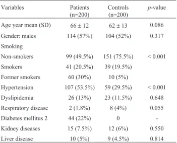
The prevalence of cardiovascular risk factors among all participants included in the study sample is summarized in Table 1. The mean age of the study population was 64±
Table 1- Prevalence of cardiovascular risk factors among all participants
included in the study by groups.
Variables Patients
(n=200)
Controls (n=200)
p-value
Age year mean (SD) 66±12 62±13 0.086
Gender: males 114 (57%) 104 (52%) 0.317
Smoking
Non-smokers 99 (49.5%) 151 (75.5%) < 0.001
Smokers 41 (20.5%) 39 (19.5%)
Former smokers 60 (30%) 10 (5%)
Hypertension 107 (53.5%) 59 (29.5%) < 0.001
Dyslipidemia 26 (13%) 23 (11.5%) 0.648
Respiratory disease 2 (1.8%) 8 (4%) 0.055
Diabetes mellitus 2 44 (22%) 0
-Kidney diseases 15 (7.5%) 12 (6%) 0.550
Liver disease 10 (5%) 9 (4.5%) 0.814
13 years, and 54.5% were males. Genotype frequencies of investigated polymorphisms were predicted by the Hardy-Weinberg equilibrium in the study and the control group (p
> 0.05), except for rs6811520, which was excluded from further analyses. Minor allele frequencies of the almost all investigated SNPs are consistent with a reference popula-tion of HapMap phase 3, CEU; the exceppopula-tion was rs35333999 in the PER2 gene and rs6811520 in the
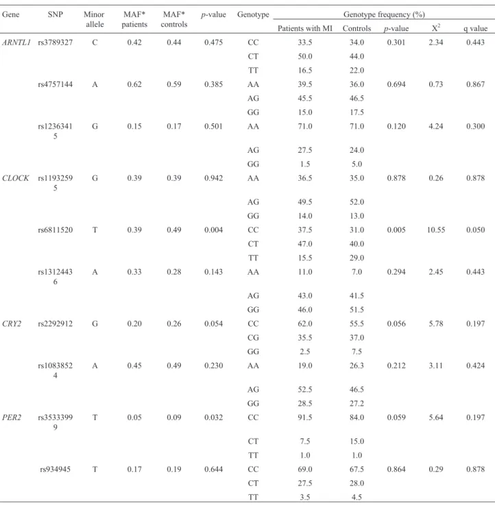
CLOCKgene (Table 2). Genotype and allelic distribution of theARNTL,CLOCK,CRY2andPER2polymorphisms of the 200 MI patients and 200 healthy controls are shown in Table 2.
We did not find any significant associations between rs3789327, rs4757144, and rs12363415 in theARNTLgene
and MI, and there was no significant difference comparing the frequencies of four the most frequent haplotypes for the
Table 2 -Allele and genotype distribution and frequencies of theARNTL,CLOCK,CRY2andPER2polymorphisms.
Gene SNP Minor
allele
MAF* patients
MAF* controls
p-value Genotype Genotype frequency (%)
Patients with MI Controls p-value X2 q value
ARNTL1 rs3789327 C 0.42 0.44 0.475 CC 33.5 34.0 0.301 2.34 0.443
CT 50.0 44.0
TT 16.5 22.0
rs4757144 A 0.62 0.59 0.385 AA 39.5 36.0 0.694 0.73 0.867
AG 45.5 46.5
GG 15.0 17.5
rs1236341 5
G 0.15 0.17 0.501 AA 71.0 71.0 0.120 4.24 0.300
AG 27.5 24.0
GG 1.5 5.0
CLOCK rs1193259 5
G 0.39 0.39 0.942 AA 36.5 35.0 0.878 0.26 0.878
AG 49.5 52.0
GG 14.0 13.0
rs6811520 T 0.39 0.49 0.004 CC 37.5 31.0 0.005 10.55 0.050
CT 47.0 40.0
TT 15.5 29.0
rs1312443 6
A 0.33 0.28 0.143 AA 11.0 7.0 0.294 2.45 0.443
AG 43.0 41.5
GG 46.0 51.5
CRY2 rs2292912 G 0.20 0.26 0.054 CC 62.0 55.5 0.056 5.78 0.197
CG 35.5 37.0
GG 2.5 7.5
rs1083852 4
A 0.45 0.49 0.230 AA 19.0 26.3 0.212 3.11 0.424
AG 52.5 46.5
GG 28.5 27.2
PER2 rs3533399 9
T 0.05 0.09 0.032 CC 91.5 84.0 0.059 5.64 0.197
CT 7.5 15.0
TT 1.0 1.0
rs934945 T 0.17 0.19 0.644 CC 69.0 67.5 0.864 0.29 0.878
CT 27.5 28.0
TT 3.5 4.5
*MAF –minor allele frequency Pearson Chi-square test
three analyzed SNPs in theARNTLgene in the patients and control groups.
The SNPs in the CLOCK gene, rs11932595, and
rs13124436 did not show significant association of geno-type or allelic distribution between the patients and control groups. Accordingly, we did not find any significant differ-ence when comparing the frequencies of four most frequent haplotypes for the two analyzed SNPs in theCLOCKgene
in the study and control groups.
No significant associations were found between the rs2292912 and rs10838524 SNPs of theCRY2gene and MI.
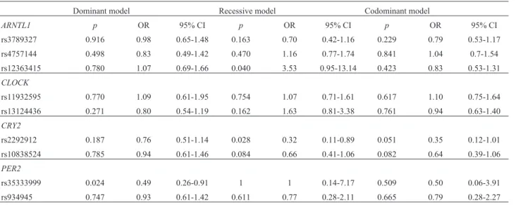
Under the recessive genotype model (GG versus CC + CG), OR of 0.32 was estimated for theCRY2gene polymorphism
rs2292912 (p=0.028, OR=0.32 with 95% CI 0.11-0.89) (Table 3). We did not find any significant difference when comparing the frequencies of the three most frequent haplotypes for the two analyzed SNPs in theCRY2gene in the patients and control groups.
A statistically significant difference was seen in the allelic distribution of rs35333999 (p=0.033) in thePER2
gene. However, we did not find any significant association between the rs934945 polymorphism of thePER2gene and MI. Under the dominant genotype model (TT + CT versus CC), an OR of 0.49 was estimated for thePER2gene
poly-morphism rs35333999 (p=0.024, OR=0.49 with 95% CI 0.26-0.91) (Table 3).
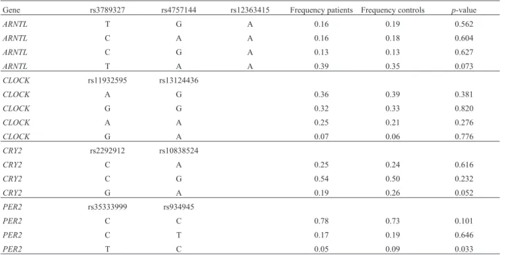
We analyzed the completed haplotypes in the four in-vestigated genes. Table 4 shows the frequencies of pre-dicted haplotypes in the patients and the control group. A statistically significant difference in haplotype distribu-tions was confirmed at thePER2gene locus when
compar-ing the frequency of haplotype TC (p=0.033) between
participants with MI and control group.
Linkage disequilibrium calculated for the CLOCK
gene SNPs (rs11932595 and rs13124436) were D’=0.06, r2=0.001. SNPs in the
CRY2 gene were in LD (D’=0.97,
r2=0.31). There was no LD between SNPs in theARNTL
gene, LD between rs4757144 and rs12363415 was D’=0.15, r2=0.001, between rs3789327 and rs4757144 was
D’=0.10, r2=0.01, and between rs3789327 and rs12363415 was D’=0.61, r2=0.09. LD for SNPs in the
PER2 gene
(rs35333999 and rs934945) was D’=0.001, r2=0.01.
In this case-control study we found an association be-tween MI and gene variants of theCRY2andPER2gene in
a sample of 400 participants. The circadian clock is a 24-hour internal system that allows an organism to main-tain environmental changes and acclimate to them. There-fore, circadian rhythms handle a broad diversity of physio-logical and metabolic functions, and any interruption of these rhythms may influence on human health.
Two feedback loops, ARNTL/CLOCK and
CRY/PER control expression of downstream transcription factors which regulate downstream target genes involved in different biochemical pathways, such as metabolism of glu-cose and lipids, synthesis of cholesterol, and others (Staels, 2006). A small number of studies have considered the role of the circadian rhythm in MI. One suggested that gene ex-pression of the cardiomyocyte circadian clock influences myocardial contractile function, metabolism and gene ex-pression (Brayet al., 2008). Another showed that in the
cardiomyocyte-specific circadian clock mutant mice, the clock is a direct regulator of triglyceride metabolism in the heart (Tsaiet al., 2010), while deletion ofARNTLin mice
adipocyte resuled in obesity (Paschoset al., 2012). PER2is involved in the regulation of fatty acid
me-tabolism with increased oxygen consumption (Grimaldiet al., 2010). Lipolysis was markedly attenuate in circadian clock mutant mice hearts, and there is a potential explana-tion for accelerated metabolic pathologies, such as athero-sclerosis which might lead to MI in patients (Tsaiet al.,
2010).PER2knock-out mice had larger infarct sizes, and
the cardiacPER2have an important role in fatty acid
me-Table 3- Genotype models of theARNTL,CLOCK,CRY2andPER2polymorphisms.
Dominant model Recessive model Codominant model
ARNTL1 p OR 95% CI p OR 95% CI p OR 95% CI
rs3789327 0.916 0.98 0.65-1.48 0.163 0.70 0.42-1.16 0.229 0.79 0.53-1.17
rs4757144 0.498 0.83 0.49-1.42 0.470 1.16 0.77-1.74 0.841 1.04 0.7-1.54
rs12363415 0.780 1.07 0.69-1.66 0.040 3.53 0.95-13.14 0.423 0.83 0.53-1.31
CLOCK
rs11932595 0.770 1.09 0.61-1.95 0.754 1.07 0.71-1.61 0.617 1.10 0.75-1.64
rs13124436 0.271 0.80 0.54-1.19 0.162 1.63 0.81-3.38 0.761 0.94 0.63-1.40
CRY2
rs2292912 0.187 0.76 0.51-1.14 0.028 0.32 0.11-0.89 0.051 0.35 0.12-1.01
rs10838524 0.785 0.94 0.61-1.46 0.084 0.66 0.41-1.06 0.082 0.64 0.39-1.06
PER2
rs35333999 0.024 0.49 0.26-0.91 1 1 0.14-7.17 0.509 0.50 0.06-3.91
rs934945 0.747 0.93 0.61-1.42 0.611 0.77 0.28-2.11 0.665 0.79 0.28-2.27
tabolism and inflammation during myocardial ischemia and reperfusion (Bonneyet al., 2013b).
Depletion of glycogen stores leads to increased in-farct sizes inPER2mutated mice because of reduced
gly-colysis during myocardial ischemia (Eckleet al., 2012). It has been shown that the protein PER2 has a cardioprotec-tive role during myocardial ischemia in mice (Bonneyet al., 2013a), and mutation of thePER2gene is associated with a shorter circadian period during constant darkness (Vukolicet al., 2010). The study of Suarez-Barrientoset al.
(2011) found that infarct size was larger in the early morn-ing, what is similar to the findings that light-dependent sta-bilization ofPER2had cardioprotection role in ischemia
(Eckleet al., 2012).
Genetic variations in thePER2gene are associated
with abdominal obesity (Garauletet al., 2010) and meta-bolic syndrome (Garcia-Rioset al., 2012) due to its part in
the lipid metabolism.PER2activation during ischemia
reg-ulates fatty acid beta-oxidation during ischemia and in-flammation during reperfusion by increasing inflammatory cytokines, metabolism and inflammation are connected, and inflammation can be a consequence of pathologic me-tabolism (Bonney et al., 2013b). Thereby, patients who have metabolic syndrome and higher inflammatory mark-ers are at greater risk to develop CVD (Haffner, 2006). Al-though the genetic variation rs35333999 in thePER2gene and rs2292912 in theCRY2gene were associated with MI
in this study, they are not precise because of the broad 95% CI values.
Disruption of the circadian clock has been implicated in the pathogenesis of cardiovascular disease, for which hy-pertension is a major factor (Kovanenet al., 2015). Aortic
endothelial dysfunction with decreased production of nitric
oxide was found in the mice with the mutatedPER2gene,
as well as, decreased vasodilatory prostaglandins and ele-vated the release of cyclooxygenase-1-derived vasocon-strictors (Scott, 2015). In endothelial hemostatic function
CLOCK, thrombomodulin, and plasminogen activator
in-hibitor-1 are involved. A circadian clock controls those genes in endothelial cells (Scott, 2015), and disruption of those genes might lead to atherosclerosis and MI. Some ge-netic variants of theCLOCK gene are related to obesity
(Bandínet al., 2013; Garcia-Rioset al., 2012), metabolic syndrome, and CVD (Garcia-Rioset al., 2012).
The links from genetic variants to physiologic func-tions are most likely less than predicted, a study identified increased weight and obesity and features of metabolic syn-drome as characteristics ofCLOCK-deficient mice (Turek
et al., 2005). Studies onCLOCKmutant mouse indicate an
important role of myocardialCLOCKgene in energy
me-tabolism, myocardial contractility, and in the diurnal heart rate control (Scott, 2015). In response to a high-fat diet, mu-tations inBMAL1andCLOCKgenes adjust circadian
varia-tion in glucose and triglycerides levels and affect the progress of insulin resistance (Scott, 2015).
Circadian rhythm has a significant role in regulating glucose metabolism, and cryptochromes are critical com-ponents of the circadian system in regulating glucose ho-meostasis (Kellyet al., 2012; Lipkovaet al., 2014),
dys-regulation of which can lead to diabetes mellitus type 2. Genetic variations of theBMAL1gene, the mouse analog of
the human ARNTL gene, are associated with diabetes
mellitus type 2 and hypertension, providing evidence for the role ofARNTLvariants in the pathology of the
meta-bolic syndrome in human (Woonet al., 2007).
Table 4- Frequencies and distribution of probable haplotypes in the patients and the control groups.
Gene rs3789327 rs4757144 rs12363415 Frequency patients Frequency controls p-value
ARNTL T G A 0.16 0.19 0.562
ARNTL C A A 0.16 0.18 0.604
ARNTL C G A 0.13 0.13 0.627
ARNTL T A A 0.39 0.35 0.073
CLOCK rs11932595 rs13124436
CLOCK A G 0.36 0.39 0.381
CLOCK G G 0.32 0.33 0.820
CLOCK A A 0.25 0.21 0.276
CLOCK G A 0.07 0.06 0.776
CRY2 rs2292912 rs10838524
CRY2 C A 0.25 0.24 0.616
CRY2 C G 0.54 0.50 0.232
CRY2 G A 0.19 0.26 0.052
PER2 rs35333999 rs934945
PER2 C C 0.78 0.73 0.101
PER2 C T 0.17 0.19 0.646
Human studies have identified genetic variants and expression patterns of circadian clock genes, such as
ARNTL,CLOCK,CRY2,NPAS2orPER2, that are
associ-ated with metabolic syndrome, hypertension or diabetes mellitus type 2 (Ohkuraet al., 2006; Scott, 2015).
Circa-dian clock genes play a major role in hemostatic balance by regulating the fibrinolytic systems, andCLOCKandCRY
genes are directly involved in this activity (Ohkuraet al.,
2006), and therefore increase the risk for CVD. A role of the circadian rhythm in cardiovascular function is firmly supported in all those studies, but our study found the con-nection of myocardial infarction and some of the circadian rhythm genes SNPs.
Although some of the obtained results are significant, they are hardly suitable for diagnosis and prognosis pur-poses. A limitation of our study is its sample size. The sam-ple size was relatively small and could yield false positive results. Furthermore, the high ORs and broad 95% CIs, as well as the low frequency of some genotypes does not allow adjusting for clinical and demographic confounders (i.e., multiple regression analysis), and low frequency of some genotypes may have resulted in insufficient statistical power to detect a positive association. For participants in control groups, there is a risk of developing some of the CVD.
In conclusion, we provide data indicating that genetic variability in theCRY2andPER2genes may be associated with MI. This suggests a role for the circadian rhythm in the development of myocardial infarction, but genetic varia-tions inARNTLandCLOCKgenes are not directly associ-ated with MI. Further verification and mechanistic analysis of the circadian system in MI are possible.
Acknowledgments
This work was supported by the Josip Juraj Stros-smayer University of Osijek, Croatia – UNIOS project ‘Variation in circadian rhythm genes in patients with myo-cardial infarction’ under grant number IZIP-2014-150.
References
Bandín C, Martinez-Nicolas A, Ordovás JM, Ros Lucas JA, Castell P, Silvente T, Madrid JA and Garaulet M (2013) Dif-ferences in circadian rhythmicity in CLOCK 3111T/C ge-netic variants in moderate obese women as assessed by ther-mometry, actimetry and body position. Int J Obes 37:1044–50.
Bonney S, Hughes K, Harter, PN, Mittelbronn M, Walker L and Eckle T (2013a) Cardiac period 2 in myocardial ischemia: Clinical implications of a light dependent protein. Int J Biochem Cell Biol 45:667–71.
Bonney S, Kominsky D, Brodsky K, Eltzschig H, Walker L and Eckle T (2013b) Cardiac Per2 functions as novel link be-tween fatty acid metabolism and myocardial inflammation during ischemia and reperfusion injury of the heart. PloS One 8:e71493.
Bray MS, Shaw CA, Moore MWS, Garcia RAP, Zanquetta MM, Durgan DJ, Jeong WJ, Tsai JY, Bugger H, Zhang D,et al. (2008) Disruption of the circadian clock within the cardio-myocyte influences myocardial contractile function, metab-olism, and gene expression. Am J Physiol Heart Circ Physiol 294:H1036–H1047.
Corella D, Asensio EM, Coltell O, Sorlí JV, Estruch R, Martínez-González MÁ, Salas-Salvado J, Castaner O, Aros F, Laperta J,et al.(2016) CLOCK gene variation is associated with in-cidence of type-2 diabetes and cardiovascular diseases in type-2 diabetic subjects: dietary modulation in the PREDIMED randomized trial. Cardiovasc Diabetol 15:4. Dashti HS, Smith CE, Lee YC, Parnell LD, Lai CQ, Arnett DK,
Ordovas JM and Garaulet M (2014) CRY1 circadian gene variant interacts with carbohydrate intake for insulin resis-tance in two independent populations: Mediterranean and North American. Chronobiol Int 31:660–7.
Ebisawa T, Uchiyama M, Kajimura N, Mishima K, Kamei Y, Katoh M, Watanabe T, Sekimoto M, Shibui K, Kim K,et al. (2001) Association of structural polymorphisms in the hu-man period3 gene with delayed sleep phase syndrome. EMBO Rep 2:342–6.
Eckle T, Hartmann K, Bonney S, Reithel S, Mittelbronn M, Walker LA, Lowes BD, Han J, Borchers CH, Buttrick PM, et al.(2012) Adora2b-elicited Per2 stabilization promotes a HIF-dependent metabolic switch crucial for myocardial ad-aptation to ischemia. Nat Med 18:774–82.
Elder SJ, Lichtenstein AH, Pittas AG, Roberts SB, Fuss PJ, Greenberg AS, McCrory MA, Bouchard TJ Jr, Saltzman E and Neale MC (2009) Genetic and environmental influences on factors associated with cardiovascular disease and the metabolic syndrome. J Lipid Res 50:1917–1926.
Englund A, Kovanen L, Saarikoski ST, Haukka J, Reunanen A, Aromaa A, Lonngvist J and Partonen T (2009) NPAS2 and PER2 are linked to risk factors of the metabolic syndrome. J Circadian Rhythms 7:5.
Erdmann J, Linsel-Nitschke P and Schunkert H (2010) Genetic causes of myocardial infarction: new insights from geno-me-wide association studies. Dtsch Ärztebl Int 107:694–9. Garaulet M, Corbalán-Tutau MD, Madrid JA, Baraza JC, Parnell
LD, Lee YC and Ordovas JM (2010) PERIOD2 variants are associated with abdominal obesity, psycho-behavioral fac-tors, and attrition in the dietary treatment of obesity. J Am Diet Assoc 110:917–21.
Garcia-Rios A, Perez-Martinez P, Delgado-Lista J, Phillips CM, Gjelstad IMF, Wright JW, Karlstrom B, Kiec-Wilk B, van Hess AM, Helal O,et al.(2012) A Period 2 genetic variant interacts with plasma SFA to modify plasma lipid concen-trations in adults with metabolic syndrome. J Nutr 142:1213–8.
Grimaldi B, Bellet MM, Katada S, Astarita G, Hirayama J, Amin RH, Granneman JG, Piomelli D, Leff T and Sassone-Corsi P. (2010) PER2 controls lipid metabolism by direct regula-tion of PPARg. Cell Metab 12:509–520.
Haffner SM. (2006) The metabolic syndrome: Inflammation, dia-betes mellitus, and cardiovascular disease. Am J Cardiol 97:3–11.
Kanth R, Ittaman S and Rezkalla S (2013) Circadian patterns of ST elevation myocardial infarction in the new millennium. Clin Med Res 11:66–72.
Kelly MA, Rees SD, Hydrie MZI, Shera AS, Bellary S, O’Hare JP, Kumar S, Taheri S, Basit A, Barnett AH,et al.(2012) Circadian gene variants and susceptibility to type 2 diabetes: a pilot study. PloS One 7:e32670.
Kovanen L, Donner K, Kaunisto M and Partonen T (2015) CRY1, CRY2 and PRKCDBP genetic variants in metabolic syn-drome. Hypertens Res 38:186–192.
Li Z, Zhang Z, He Z, Tang W, Li T, Zeng Z, He L and Shi Y (2009) A partition-ligation-combination-subdivision EM al-gorithm for haplotype inference with multiallelic markers: update of the SHEsis (http://analysis.bio-x.cn) Cell Res 19:519–23.
Lipkova J, Splichal Z, Bienertova-Vasku JA, Jurajda M, Parenica J, Vasku A and Goldbergova M P (2014) Period3 VNTR polymorphism influences the time-of-day pain onset of acute myocardial infarction with ST elevation. Chronobiol Int, 31(8), 878–90.
Martino TA and Sole MJ (2009) Molecular time: An often over-looked dimension to cardiovascular disease. Circ Res 105:1047–61.
Ohkura N, Oishi K, Fukushima N, Kasamatsu M, Atsumi GI, Ishida N, Horie S and Matsuda J (2006) Circadian clock molecules CLOCK and CRYs modulate fibrinolytic activity by regulating the PAI-1 gene expression. J Thromb Haemost 4:2478–2485.
Paschos GK, Ibrahim S, Song WL, Kunieda T, Grant G, Reyes TM, Bradfield CA, Vaughan CH, Eiden M, Masoodi M,et al.(2012) Obesity in mice with adipocyte-specific deletion of clock component Arntl. Nat Med 18:1768–77.
Scott EM (2015) Circadian clocks, obesity and cardiometabolic function. Diabetes Obes Metab 17:84–89.
Shi YY and He L (2005) SHEsis, a powerful software platform for analyses of linkage disequilibrium, haplotype construction, and genetic association at polymorphism loci. Cell Res 15:97–8.
Solé X, Guinó E, Valls J, Iniesta R and Moreno V (2006) SNPStats: a web tool for the analysis of association studies. Bioinformatics 22:1928–9.
Staels B. (2006) When the clock stops ticking, metabolic syn-drome explodes. Nat Med 12:54–55.
Suarez-Barrientos A, Lopez-Romero P, Vivas D, Castro-Ferreira F, Nunez-Gil I, Franco E, Ruiz-Mateos B, Garcia-Rubira JC, Fernandez-Ortiz A, Macaya C,et al.(2011) Circadian varia-tions of infarct size in acute myocardial infarction. Heart 97:970–976.
Takeda N and Maemura K (2010) Circadian clock and vascular disease. Hypertens Res 33:645–51.
Takeda N and Maemura K (2016) Circadian clock and the onset of cardiovascular events. Hypertens Res 39:383–390. Thygesen K, Alpert JS, Jaffe AS, Simoons ML, Chaitman BR and
White HD (2012) Third universal definition of myocardial infarction. Nat Rev Cardiol 9:620–33.
Tsai JY, Kienesberger PC, Pulinilkunnil T, Sailors MH, Durgan DJ, Villegas-Montoya C, Jahoor A, Gonzalez R, Garvey ME, Boland B,et al.(2010) Direct Regulation of Myocar-dial Triglyceride Metabolism by the Cardiomyocyte Circa-dian Clock. J Biol Chem 285:2918–2929.
Turek FW, Joshu C, Kohsaka A, Lin E, Ivanova G, McDearmon E, Laposky A, Losee-Olson S, Easton A, Jensen DR,et al. (2005) Obesity and metabolic syndrome in circadian clock mutant mice. Science 308:1043–1045.
Vukolic A, Antic V, Van Vliet BN, Yang Z, Albrecht U and Montani J (2010) Role of mutation of the circadian clock gene Per2 in cardiovascular circadian rhythms. Am J Physiol Regul Integr Comp Physiol 298:627–634.
WHO (2015) Global status report on noncommunicable diseases 2014. WHO, Geneva, 280 p.
Woon PY, Kaisaki PJ, Bragança J, Bihoreau MT, Levy JC, Farrall M and Gauguier D (2007) Aryl hydrocarbon receptor nu-clear translocator-like (BMAL1) is associated with suscepti-bility to hypertension and type 2 diabetes. Proc Natl Acad Sci U S A 104:14412–7.
Associate Editor: Angela M. Vianna-Morgante