RAPID COMMUNICATION
DEFB1 gene 5
9
untranslated region (UTR)
polymorphisms in inflammatory bowel diseases
Valentina Zanin,I Ludovica Segat,IIAnna Monica Bianco,II Lara Padovan,II Nathalia de Alencar Cunha Tavares,IIISergio CrovellaIVIUniversity of Trieste, Institute for Maternal and Child Health – IRCCS ‘‘Burlo Garofolo’’, Trieste, Italy.IIInstitute for Maternal and Child Health – IRCCS ‘‘Burlo Garofolo’’- Trieste, Italy.IIIFederal University of Pernambuco, Laboratory of Immunopathology Keizo Asami (LIKA), Recife/PE, Brazil.IVInstitute for Maternal and Child Health - IRCCS ‘‘Burlo Garofolo’’, Trieste, University of Trieste, Italy.
Email: [email protected] Tel.: 39 0403785422
INTRODUCTION
Inflammatory bowel diseases (IBDs) are multifactorial disorders resulting from an abnormal immune response driven by the presence of normal luminal flora. IBDs are associated with the production of nonspecific mediators of inflammation that initiate inflammatory processes and tissue destruction (1-3). Crohn’s disease (CD) and ulcerative colitis (UC) are the two main forms of IBDs.
An accepted hypothesis for IBD pathogenesis is that an uncorrected balance between the host defenses and the commensal microbiota in the gut of CD and UC patients might promote the disease by causing bacterial invasion, inflammation and loss of tolerance (4). A key role in the host defense is performed by the intestinal epithelium, which acts as a physical barrier that limits the access of enteric microbes that are able to produce endogenous antimicrobial peptides (AMPs) (5).
Human defensins, which are classified asa-defensins (HDs) and b-defensins (HBDs) based on the arrangement of three disulfide bridges (6), are antimicrobial peptides that represent a wide spectrum of activity against pathogens. Human b -defensin-1 (hBD-1), the first describedb-defensin, is character-ized by antimicrobial, chemotactic and immuno-enhancing activities (7-9). The hBD-1 protein, which is encoded by the DEFB1 gene (8p23.1), is constitutively expressed by epithelial cells of a wide variety of tissues, but its expression can vary between individuals and can be modified during the inflammatory process. A decrease in hBD-1 expression was reported in the mucosa of CD and UC patients (10-12).
Impaired production of defensins appears to contribute to the pathogenesis of IBDs (13,14), and a correlation between DEFB1 expression and single-nucleotide polymorphisms (SNPs) present in the regulatory region of the gene has been reported (15,16). Therefore, we analyzed the possible association of 59 untranslated region (UTR) DEFB1 SNPs, namely c.-52G.A (rs1799946) c.-44C.G (rs1800972) and c.-20G.A (rs11362), with the susceptibility to inflammatory
bowel diseases in a group of Italian IBD patients (CD and UC) and healthy control patients.
MATERIAL AND METHODS
Patients and controls
We enrolled 145 patients with inflammatory bowel disease (93 males, mean age at diagnosis 37.18¡14.11; 52 females, mean age at diagnosis 37.36¡16.35). Among the IBD
patients, 108 suffered from Crohn’s disease (mean age at diagnosis 36.52¡14.01) and 37 suffered from ulcerative colitis (mean age at diagnosis 38.24¡15.66). A total of 130
healthy adult blood donors (63 M/67F, mean age 31,3¡12.36)
with no history of IBDs and from the same ethnic origin as the IBD patients were recruited and used as controls.
The IRCCS Burlo Garofolo Ethical Committee approved the study (prot n. CIB 15/07, 03/03/2008).
The diagnosis of Crohn’s or UC disease was established by following the European Crohn’s and Colitis Organisation (ECCO) Consensus guidelines for IBDs (https://www. ecco-ibd.eu/) (17,18) and the protocols of the Inflammatory Bowel Disease and Functional Bowel Disorders Review Group (http://www.cochrane.uottawa.ca/ibd/default.htm) (19).
DEFB1 SNP genotyping
Genomic DNA was extracted from peripheral whole blood with the EZ1 DNA blood kit (Qiagen, Milan, Italy) following the manufacturer’s instructions. The three polymorphisms (rs1799946, rs1800972 and rs11362) in the 59UTR region of the DEFB1 gene were genotyped by direct sequencing with Big Dye Terminator chemistry and an ABI 3130 DNA Sequencer (Applied Biosystems, Foster City, CA) using primers and protocols previously described in the literature(20,21). The sequences were analyzed with the 4peaks software (http:// www.mekentosj.com/science/4peaks).
Statistical analyses
The allele and genotype frequencies were calculated by direct gene counting and compared by the Fisher exact test using 2X2 and 3X2 contingency tables. The haplotype frequencies were obtained according to the EM algorithm using the Arlequin software (22) (version 3.11). The odds ratios (O.R.) and 95% confidence intervals (C.I.) were also calculated. The Bonferroni correction for multiple tests was applied when required, and only corrected p,0.05 were
considered to be statistically significant. The R package (www.r-project.org) was used for all the statistical analyses. Copyrightß2012CLINICS– This is an Open Access article distributed under
the terms of the Creative Commons Attribution Non-Commercial License (http:// creativecommons.org/licenses/by-nc/3.0/) which permits unrestricted non-commercial use, distribution, and reproduction in any medium, provided the original work is properly cited.
No potential conflict of interest was reported.
CLINICS 2012;67(4):395-398 DOI:10.6061/clinics/2012(04)14
RESULTS
No statistically significant differences were observed in the distribution of the genotypic or allelic frequencies of the DEFB1 c.-52G.A and c.-44C.G SNPs among the CD, UC and healthy control subjects (Table 1). For the DEFB1 c.-20G.A SNP, no significant differences in the allelic or genotype frequencies were observed between the UC patients and the healthy controls. A slight difference was observed in the allelic distribution with a higher frequency of the c.-20G.A G allele in the healthy subjects than in the CD patients (62% vs. 53%; uncorrectedp= 0.04; O.R. = 0.676;
95% C.I. = 0.461-0.992). Smaller differences were also observed for the other genotypes. These differences, how-ever, did not retain statistical significance after Bonferroni correction. The three DEFB1 SNPs were observed to be in linkage disequilibrium (p,0.05, D’.0.7) in the CD, UC or
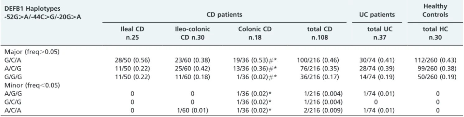
control subjects and combined into three major haplotypes (G/C/A; A/C/G; G/G/G) with a frequency .0.05 and three minor haplotypes (A/G/G; G/C/G; A/C/A) with a frequency ,0.05 (Table 2). No statistically significant differences were observed when comparing the CD and UC patients with the controls.
For 73 of the 108 CD patients, a classification of the disease according to the bowel-disease anatomical localization was
also possible: 25 patients (34%) presented ileal CD, 30 (41%) presented ileocolonic CD and 18 (25%) presented colonic CD. The DEFB1 c.-52G.A G allele was more frequent in the patients with ileal localization than in the healthy control patients (78% vs. 58%; uncorrectedp= 0.010, O.R. = 2.552; 95%
C.I. = 1.215-5.781), which was also observed with the GG genotype (64% versus 30%, uncorrectedp= 0.002; O.R. = 4.106;
95% C.I. = 1.554-11.521).
The C allele of the DEFB1 c.-44C.G SNP was more frequent in the patients with colonic localization than in the healthy subjects (94% vs. 78%; uncorrected p= 0.037;
O.R. = 4.773; 95% C.I. = 1.113-20.473), which was also observed with the CC genotype (89% vs. 59%; uncorrected
p= 0.017; O.R. = 5.633; 95% C.I. = 1.242-52.507).
The DEFB1 c.-20G.A G allele was more frequent in the control patients (62%) than in the colonic patients, (44%) (uncorrectedp= 0.046; O.R. = 0.485; C.I. = 0.224-1.038), which
was also observed with the GG genotype (p= 0.03, O.R. = 0.222;
95% C.I. = 0.024-1.013).
However, the statistical significances of all the compar-isons were lost after applying Bonferroni’s correction.
When analyzing the haplotype frequencies, we observed a different distribution of the three major haplotypes between CD colonic patients and healthy control patients (uncorrected p= 0.039). This difference was even greater
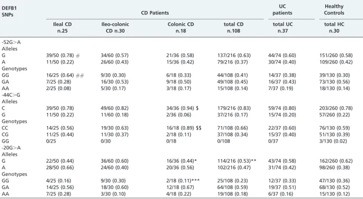
Table 1 -Allele and genotype count (and frequencies) of 59UTR DEFB1 single-nucleotide polymorphisms in Crohn’s disease patients, ulcerative colitis patients and healthy controls. The Crohn’s disease patients are also stratified
according to the anatomical localization of the disease. Thep-values,0.05 (uncorrected), odds ratios (ORs) and 95% CIs
are also reported.
DEFB1
SNPs CD Patients
UC patients
Healthy Controls
Ileal CD n.25
Ileo-colonic CD n.30
Colonic CD n.18
total CD n.108
total UC n.37
total HC n.30
-52G.A Alleles
G 39/50 (0.78)# 34/60 (0.57) 21/36 (0.58) 137/216 (0.63) 44/74 (0.60) 151/260 (0.58) A 11/50 (0.22) 26/60 (0.43) 15/36 (0.42) 79/216 (0.37) 30/74 (0.40) 109/260 (0.42) Genotypes
GG 16/25 (0.64)## 9/30 (0.30) 6/18 (0.33) 44/108 (0.41) 14/37 (0.38) 39/130 (0.30) GA 7/25 (0.28) 16/30 (0.53) 9/18 (0.50) 49/108 (0.45) 16/37 (0.43) 73/130 (0.56)
AA 2/25 (0.08) 5/30 (0.17) 3/18 (0.17) 15/108 (0.14) 7/37 (0.19) 18/130 (0.14)
-44C.G Alleles
C 39/50 (0.78) 49/60 (0.82) 34/36 (0.94) $ 179/216 (0.83) 59/74 (0.80) 203/260 (0.78) G 11/50 (0.22) 11/60 (0.18) 2/36 (0.06) 37/216 (0.17) 15/74 (0.20) 57/260 (0.22) Genotypes
CC 14/25 (0.56) 19/30 (0.63) 16/18 (0.89) $$ 71/108 (0.66) 22/37 (0.60) 76/130 (0.59) CG 11/25 (0.44) 11/30 (0.37) 2/18 (0.11) 37/108 (0.34) 15/37 (0.40) 51/130 (0.39)
GG 0/25 0/30 0/18 0/108 0/37 3/130 (0.02)
-20G.A Alleles
G 22/50 (0.44) 36/60 (0.60) 16/36 (0.44)* 114/216 (0.53)** 43/74 (0.58) 162/260 (0.62) A 28/50 (0.66) 24/60 (0.40) 20/36 (0.56) 102/216 (0.47) 31/74 (0.42) 98/260 (0.38) Genotypes
GG 4/25 (0.16) 9/30 (0.30) 2/18 (0.11)*** 25/108 (0.23) 12/37 (0.33) 47/130 (0.36) GA 14/25 (0.56) 18/30 (0.60) 12/18 (0.67) 64/108 (0.59) 19/37 (0.51) 68/130 (0.52)
AA 7/25 (0.28) 3/30 (0.10) 4/18 (0.22) 19/108 (0.18) 6/37 (0.16) 15/130 (0.12)
#Ileal CD vs. HC,p= 0.01, O.R. = 2.552 (1.215-5.781);
##Ileal CD vs. HC,p =0.002, O.R. = 4.106 (1.554-11.521); $ Colonic CD vs. HC,p =0.037, O.R. = 4.773 (1.113-20.473); $$ Colonic CD vs. HC,p =0.017, O.R. = 5.633 (1.242-52.507); *Colonic CD vs. HC,
p =0.046, O.R. = 0.485 (0.224-1.038); **CD vs. HC,
p =0.04, O.R. = 0.676 (0.461-0.992); ***Colonic CD vs. HC,
p =0.03, O.R. = 0.222 (0.024-1.013).
DEFB1 polymorphisms in inflammatory bowel diseases
Zanin V et al. CLINICS 2012;67(4):395-398
when the minor haplotypes were also included in the analysis (uncorrectedp,0.001). (Table 2).
DISCUSSION
To our knowledge, only one study has previously investi-gated the role of DEFB1 SNPs in Crohn’s disease (23). The frequencies of the DEFB1 SNPs in CD patients reported by Kocsis et al. are comparable to the frequencies reported by our study. Moreover, similar to our study, the authors failed to observe any association between the c.-52G.A and c.-20G.A DEFB1 SNPs and the overall susceptibility to CD. For the c.-44C.G SNP, they observed a different distribution between the CD and control patients with an increased frequency of the c.-44C.G GG genotype in the control patients (12%) com-pared with the CD patients (4%). This difference was not observed in our study; however, none of the CD patients in our study presented the c.-44C.G GG genotype, whereas three (2%) of the control patients, which were matched for sex, age and ethnicity, presented the genotype.
In addition, Kocsis et al. (23) reported several significant associations between the DEFB1 SNPs and the anatomical localization of Crohn’s disease (ileal, ileocolonic or colonic). For the DEFB1 c.-44C.G SNP, Kocsis et al. observed a significant increase in the CC genotype in ileocolonic patients and to a lesser extent in colonic but not ileal patients compared with healthy control patients. Our results indicate an increased frequency of the CC genotype in the colonic patients (89% vs. 59% in control patients). Additionally, a slight increase in the CC genotype frequency was observed in the ileocolonic patients (63% vs. 59% in control patients), but not in ileal.
Kocsis et al. (23) also showed that the GG genotype (c.-20G.A SNP) was more frequent in control subjects than in patients with colonic CD. Our results appear to confirm these findings. Because colonic CD localization has been associated with impaired hBD-1 expression and because the c.-20G.A A allele appears to be associated with reduced levels of DEFB1 expression, it can be hypothesized that this polymorphism might cause lower hBD-1 expression in colonic epithelial cells.
For c.-52G.A, Kocsis et al. (23) did not report any difference between patients with different localization of CD and controls. Conversely, we observed that the GG
genotype was more frequent in the ileal CD patients (64%) than in the controls (30%), but the difference was not statistically significant.
The constitutive expression of hBD-1, which is known to be a constitutively expressed peptide, has already been reported in CD and UC patients. Moreover, stressful events could regulate DEFB1 mRNA levels, (24-27) which could be relevant for the intestinal mucosa. The presence of 59UTR DEFB1 SNPs may be able to explain interindividual changes in DEFB1 expression, and these differences may be able to account for different susceptibility to diseases (such as CD). However, the mechanism by which the same DEFB1 SNP can be associated with the development of the disease in a specific anatomical region and not in others remains unknown. Variations in hBD-1 production among different intestinal traits of CD and UC patients are more likely to result from the actions of other genes or factors involved in the regional regulation of the hBD-1 peptide (24-27) rather than as a direct consequence of a single SNP or a combination of SNPs to form haplotypes.
We are aware that the loss of statistical significance of the
p-values after correcting for multiple tests comparisons,
which is possibly due to the limited number of samples analyzed, is likely the major weakness of this study. However, it is noteworthy that our results confirm the findings previously reported by another research group (23), which indicate that DEFB1 may a contributing factor to susceptibility to Crohn’s disease.
ACKNOWLEDGMENTS
Grants RC03/04 from IRCCS Burlo Garofolo Trieste (Italy) and APQ-1130-2.02/08 (FACEPE, Recife, Brazil) supported this study. SC is the recipient of a fellowship grant from the European Project ‘‘Talents for an International House’’ within the framework of the 7th Research & Development Framework Programme PEOPLE – Marie Curie Actions – COFUND (Co-Funding of Regional, National and International Programmes).
AUTHOR CONTRIBUTIONS
Zanin V, Bianco AM and Padovan L collected the biological samples and extracted the DNA. Crovella S designed the study. Crovella S and Segat L redacted the manuscript and performed statistical analysis. Tavares CN and Zanin V performed the DEFB1 genotyping.
Table 2 -Haplotype counts (and frequencies) of 5’UTR DEFB1 polymorphisms in Crohn’s disease patients, ulcerative colitis patients and healthy controls. The Crohn’s disease patients are also stratified according to the anatomical
localization of the disease. Thep-values (uncorrected),0.05 are also indicated.
DEFB1 Haplotypes
-52G.A/-44C.G/-20G.A CD patients UC patients
Healthy Controls
Ileal CD n.25
Ileo-colonic CD n.30
Colonic CD n.18
total CD n.108
total UC n.37
total HC n.30
Major (freq.0.05)
G/C/A 28/50 (0.56) 23/60 (0.38) 19/36 (0.53)#* 100/216 (0.46) 30/74 (0.41) 112/260 (0.43) A/C/G 11/50 (0.22) 25/60 (0.42) 13/36 (0.36)#* 76/216 (0.35) 28/74 (0.39) 99/260 (0.38) G/G/G 11/50 (0.22) 11/60 (0.18) 1/36 (0.02)#* 36/216 (0.17) 14/74 (0.19) 50/260 (0.19) Minor (freq,0.05)
A/G/G 0 0 1/36 (0.02)* 1/216 (0.004) 1/74 (0.01) 0
G/C/G 0 0 1/36 (0.02)* 1/216 (0.004) 0 0
A/C/A 0 1/60 (0.01) 1/36 (0.02)* 2/216 (0.009) 1/74 (0.01) 0
#Colonic CD vs. HC major haplotypes,p =0.039. *Colonic CD vs. HC major
+minor haplotypes,p =0.0004595.
CLINICS 2012;67(4):395-398 DEFB1 polymorphisms in inflammatory bowel diseases
Zanin V et al.
REFERENCES
1. Papp M, Norman GL, Altorjai I, Lakatos PL. Utility of serological markers in inflammatory bowel diseases: gadget or magic? World J Gastroenterol. 2007;13(14):2028-36.
2. Swidsinski A, Ladhoff A, Pernthaler A, Swidsinski S, Loening-Baucke V, Ortner M, et al. Mucosal flora in inflammatory bowel disease. Gastroenterology. 2002;122(1):44-54, http://dx.doi.org/10.1053/ gast.2002.30294.
3. Fiocchi C. Inflammatory bowel disease: etiology and pathogenesis. Gastroenterology. 1998;115(1):182-205, http://dx.doi.org/10.1016/ S0016-5085(98)70381-6.
4. Balfour Sartor R. Bacteria in Crohn’s disease: mechanisms of inflamma-tion and therapeutic implicainflamma-tions. J Clin Gastroenterol. 2007;41 Suppl:S37-43, http://dx.doi.org/10.1097/MCG.0b013e31802db364 5. Bals R. Epithelial antimicrobial peptides in host defense against
infection. Respir Res. 2000;1:141-50, http://dx.doi.org/10.1186/rr25. 6. Klotman ME, Chang TL. Defensins in innate antiviral immunity. Nat Rev
Immunol. 2006;6(6):447-56, http://dx.doi.org/10.1038/nri1860. 7. Chertov O, Michiel DF, Xu L, Wang JM, Tani K, Murphy WJ, et al.
Identification of defensin-1, defensin-2, and CAP37/azurocidin as T-cell chemoattractant proteins released from interleukin-8-stimulated neutro-phils. J Biol Chem. 1996;271(6):2935-40.
8. Yang D, Chertov O, Bykovskaia SN, Chen Q, Buffo MJ, Shogan J, et al. Beta-defensins: linking innate and adaptive immunity through dendritic and T cell CCR6. Science. 1999;286(5439):525-8, http://dx.doi.org/10.1126/ science.286.5439.525.
9. Niyonsaba F, Ogawa H, Nagaoka I. Human beta-defensin-2 functions as a chemotactic agent for tumour necrosis factor-alpha-treated human neutrophils. Immunology. 2004;111(3):273-81, http://dx.doi.org/ 10.1111/j.0019-2805.2004.01816.x.
10. Arijs I, De Hertogh G, Lemaire K, Quintens R, Van Lommel L, Van Steen K, et al. Mucosal Gene Expression of antimicrobial peptides in inflamamatory bowel disease before and after first Infliximab treatment. PloS One. 2009;4(11):e7984, http://dx.doi.org/10.1371/journal.pone. 0007984.
11. Bajaj-Elliott M, Fedeli P, Smith GV, Domizio P, Maher L, Ali RS, et al. Modulation of host antimicrobial peptide (b-defensins 1 and 2) expression during gastritis. Gut. 2002;51(3):356-61, http://dx.doi.org/ 10.1136/gut.51.3.356.
12. Wehkamp J, Harder J, Weichenthal M, Mueller O, Herrlinger KR, Fellermann K, et al. Inducible and costitutive-defensis are differentially expressed in Crohn’s disease and ulcerative colitis. Inflammatory Bowel Disease. 2003;9(4):215-23.
13. Ramasundara M, Leach ST, Lemberg DA, Day AS. Defensins and inflammation: the role of defensins in inflammatory bowel disease. J Gastroenterol Hepatol. 2009;24(2):202-8, http://dx.doi.org/10.1111/j. 1440-1746.2008.05772.x.
14. Fellermann K, Wehkamp J, Herrlinger KR, Stange EF. Crohn’s disease: a defensin deficiency syndrome? Eur J Gastroenterol Hepatol. 2003;15(6):27-634, http://dx.doi.org/10.1097/01.meg.0000059151.68845.88.
15. Milanese M, Segat L, Crovella S. Transcriptional effect of DEFB1 gene 59
untranslated region polymorphisms. Cancer Res. 2007;67(12):5997, http://dx.doi.org/10.1158/0008-5472.CAN-06-3544.
16. Sun CQ, Arnold R, Fernandez-Golarz C, Parrish AB, Almekinder T, He J, et al. Humanb-defensin 1, a potential chromosome 8p tumor suppressor: control of transcription and induction of apoptosis in renal cell carcinoma. Cancer Res. 2006;66(17):8542-9, http://dx.doi.org/10.1158/ 0008-5472.CAN-06-0294.
17. Stange EF, Travis SPL, Vermeire S, Beglinger C, Kupcinskas L, Geboes K, et al. European evidence based consensus on the diagnosis and management of Crohn’s disease: definitions and diagnosis. Gut. 2006;55 suppl:i1-i15, http://dx.doi.org/10.1136/gut.2005.081950a. 18. Stange EF, Travis SPL, Vermeire S, Reinisch W, Geboes K, Barakauskiene
A, et al. European evidence-based Consensus on the diagnosis and management of ulcerative colitis: Definitions and diagnosis. Journal of Crohn’s and Colitis. 2008;2(1):1-23, http://dx.doi.org/10.1016/j.crohns. 2007.11.001.
19. Chande N, MacDonald JK, McDonald JW. Interventions for treating microscopic colitis: a Cochrane Inflammatory Bowel Disease and Functional Bowel Disorders Review Group systematic review of randomized trials. Am J Gastroenterol. 2009;104(1):235-41; quiz 234, 242, http://dx.doi.org/10.1038/ajg.2008.16.
20. Braida L, Boniotto M, Pontillo A, Tovo PA, Amoroso A, Crovella S. A single-nucleotide polymorphism in the human beta-defensin 1 gene is associated with hiv-1 infection in italian children. AIDS. 2004; 18(11):1598-600, http://dx.doi.org/10.1097/01.aids.0000131363.82951. fb.
21. Jurevic RJ, Bai M, Chadwick RB, White TC, Dale BA. Single-nucleotide polymorphisms (snps) in human beta-defensin 1: High-throughput snp assays and association with candida carriage in type i diabetics and nondiabetic controls. J Clin Microbiol. 2003;41(1):90-6, http://dx. doi.org/10.1128/JCM.41.1.90-96.2003.
22. Excoffier L, Laval G, Schneider S. Arlequin ver 3.0: an integrated software package for population genetics data analysis. Evolutionary Bioinformatics Online. 2005;1:47-50.
23. Kocsis AK, Lakatos PL, Somogyva´ri F, Fuszek P, Papp J, Fischer S, et al. Association of beta-defensin 1 single nucleotide polymorphisms with Crohn’s disease. Scandinavian Journal of Gastroenterology. 2008; 43(3):299-307, http://dx.doi.org/10.1080/00365520701682615.
24. Prado-Montes de Oca E, Garcı´a-Vargas A, Lozano-Inocencio R, Gallegos-Arreola MP, Sandoval-Ramı´rez L, Da´valos-Rodrı´guez NO, et al. Association of beta-defensin 1 single nucleotide polymorphisms with atopic dermatitis. Int Arch Allergy Immunol. 2007;142(3):211-18, http:// dx.doi.org/10.1159/000097023.
25. Chakraborty K, Ghosh S, Koley H, Mukhopadhyay AK, Ramamurthy T, Saha DR, et al. Bacterial exotoxins downregulate cathelicidin (hCAP18/ LL37) and human beta-defensin 1 (HBD-1) expression in the intestinal epithelial cells. Cell Microbiol. 2008;10(10):2520-37, http://dx.doi.org/ 10.1111/j.1462-5822.2008.01227.x.
26. Sherman H, Chapnik N, Froy O. Albumin and amino acids upregulate the expression of human beta-defensin 1. Mol Immunol. 2006;43(10):1617-23, http://dx.doi.org/10.1016/j.molimm.2005.09.013.
27. Sherman H, Froy O. Expression of human beta-defensin 1 is regulated via c-Myc and the biological clock. Mol Immunol. 2008;45(11):3163-67, http://dx.doi.org/10.1016/j.molimm.2008.03.004.
DEFB1 polymorphisms in inflammatory bowel diseases
Zanin V et al. CLINICS 2012;67(4):395-398