Edited by: Yoann Rombouts, UMR5089 Institut de Pharmacologie et de Biologie Structurale (IPBS), France Reviewed by: Michael Hickey, Monash University, Australia Luciana Barros Arruda, Universidade Federal do Rio de Janeiro, Brazil *Correspondence: Paula A. Videira p.videira@fct.unl.pt; Robert Sackstein rsackstein@rics.bwh.harvard.edu †Co-first authorship. Specialty section: This article was submitted to Microbial Immunology, a section of the journal Frontiers in Immunology Received: 30 September 2017 Accepted: 08 December 2017 Published: 19 January 2018 Citation: Silva M, Videira PA and Sackstein R (2018) E-Selectin Ligands in the Human Mononuclear Phagocyte System: Implications for Infection, Inflammation, and Immunotherapy. Front. Immunol. 8:1878. doi: 10.3389/fimmu.2017.01878
e-Selectin Ligands in the Human
Mononuclear Phagocyte System:
implications for infection,
inflammation, and immunotherapy
Mariana Silva
1,2†, Paula A. Videira
3,4*
†and Robert Sackstein
1,2,5*
1 Department of Dermatology, Harvard Skin Disease Research Center, Brigham and Women’s Hospital, Harvard Medical School, Boston, MA, United States, 2 Program of Excellence in Glycosciences, Harvard Medical School, Boston, MA, United States, 3 UCIBIO, Departamento Ciências da Vida, Faculdade de Ciências e Tecnologia, Universidade NOVA de Lisboa, Lisboa, Portugal, 4 Professionals and Patient Associations International Network (CDG & Allies – PPAIN), Faculdade de Ciências e Tecnologia, Universidade NOVA de Lisboa, Lisboa, Portugal, 5 Department of Medicine, Brigham and Women’s Hospital, Harvard Medical School, Boston, MA, United States
The mononuclear phagocyte system comprises a network of circulating monocytes
and dendritic cells (DCs), and “histiocytes” (tissue-resident macrophages and DCs) that
are derived in part from blood-borne monocytes and DCs. The capacity of
circulat-ing monocytes and DCs to function as the body’s first-line defense against offendcirculat-ing
pathogens greatly depends on their ability to egress the bloodstream and infiltrate
inflammatory sites. Extravasation involves a sequence of coordinated molecular events
and is initiated by E-selectin-mediated deceleration of the circulating leukocytes onto
microvascular endothelial cells of the target tissue. E-selectin is inducibly expressed by
cytokines (tumor necrosis factor-
α and IL-1β) on inflamed endothelium, and binds to
sialofucosylated glycan determinants displayed on protein and lipid scaffolds of blood
cells. Efficient extravasation of circulating monocytes and DCs to inflamed tissues is
crucial in facilitating an effective immune response, but also fuels the immunopathology
of several inflammatory disorders. Thus, insights into the structural and functional
prop-erties of the E-selectin ligands expressed by different monocyte and DC populations
is key to understanding the biology of protective immunity and the pathobiology of
several acute and chronic inflammatory diseases. This review will address the role of
E-selectin in recruitment of human circulating monocytes and DCs to sites of tissue
injury/inflammation, the structural biology of the E-selectin ligands expressed by these
cells, and the molecular effectors that shape E-selectin ligand cell-specific display. In
addition, therapeutic approaches targeting E-selectin receptor/ligand interactions, which
can be used to boost host defense or, conversely, to dampen pathological inflammatory
conditions, will also be discussed.
Keywords: mononuclear phagocyte, HCeLL, e-selectin ligand, cell migration, e-selectin, sialyl Lewis X
iNTRODUCTiON
The mononuclear phagocyte system (MPS) comprises monocytes, dendritic cells (DC), and
tissue-resident macrophages. MPS cells have specialized phagocytic capabilities, and antigen
pro-cessing and presenting functions, thereby initiating the immune response and linking innate and
adaptive immune systems (
1
). In addition to their role as key sentinels and regulators of immunity,
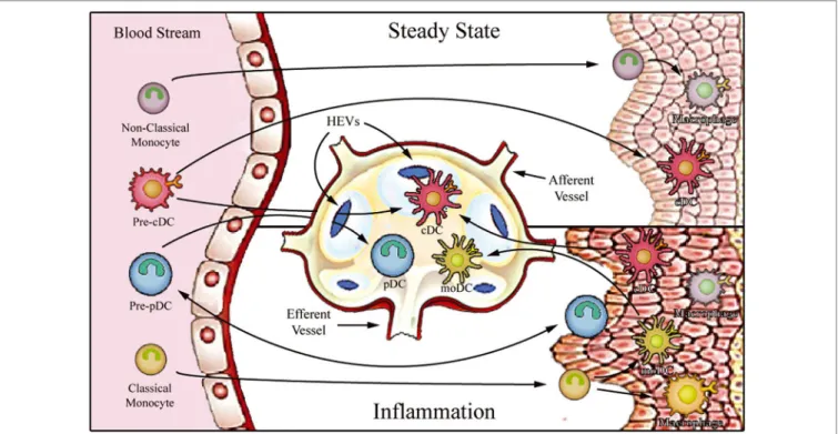
FigURe 1 | Proposed model for migration of human monocytes and dendritic cell (DC) progenitors into tissues in steady-state and inflammatory conditions. After
differentiation in the bone barrow, precursors of DCs and monocytes enter the blood stream and are distributed to lymphoid organs [through high endothelial venules (HEV)] and to various peripheral tissues. In steady state, non-classical monocytes are preferentially recruited into the resting vasculature, where they patrol the endothelium and may contribute to the maintenance of tissue-resident macrophage and DC populations. Conventional DCs (cDCs) recirculate between peripheral tissues and lymphoid organs (migratory cDCs), participating in the induction of peripheral tolerance, or reside in the lymphoid organs (lymphoid-resident cDCs). By contrast, plasmacytoid DCs (pDCs) mostly populate lymphoid tissues (lymphoid-resident pDCs) and lack migratory ability under steady-state conditions. Upon inflammation, classical monocytes, cDCs, and pDCs are recruited to affected tissues. After antigen uptake and differentiation into fully functional mature DCs, monocyte-derived DCs (moDCs), and cDCs enter draining lymph nodes via afferent lymphatics. pDCs can only access reactive lymph nodes from the blood stream via HEVs.
mononuclear phagocytes are also involved in several
pathologi-cal inflammatory conditions, including autoimmune diseases,
infection, cancer, and abnormal wound healing processes (
2
).
To access inflammatory sites, circulating monocytes and DCs
must first engage the vascular endothelial barrier against the
prevailing forces of hemodynamic shear, a process that occurs
via adhesive interactions between vascular E-selectin and its
glycan counter-receptors (E-selectin ligands) on the
circulat-ing cells (
3
). This initial contact results in tethering and slow
rolling of the cells along the endothelial surface at velocities
well below that of blood flow (
4
). E-selectin-mediated slow
roll-ing is a vital step in this cascade of events as it allows intimate
contact between MPS cells and the inflamed endothelium, and
the recognition of inflammatory molecules within the milieu
(
3
). Consequently, a greater knowledge of how E-selectin ligand
display is elaborated by different types of circulating
mono-cytes and DCs is key to understanding the physiological and
pathological events associated with the MPS. In this review, we
will provide information on the structural biology and
opera-tion of the wide variety of E-selectin-binding glycoconjugates
expressed by circulating MPS cells (i.e., blood monocyte and
non-tissue-resident DC populations) in light of their impact on
pathology and potential therapies. Furthermore, we will discuss
the molecular basis of the biosynthesis of these glycoconjugates,
and how such knowledge can frame novel strategies to inhibit
or enforce trafficking of MPS cells.
MONONUCLeAR PHAgOCYTe FAMiLY:
HeTeROgeNeiTY AND MigRATORY
CAPABiLiTieS
Monocytes
Monocytes constitute a heterogeneous cell population,
compris-ing approximately 5–10% of total peripheral blood leukocytes.
These cells arise from granulocyte–macrophage progenitors in
the bone marrow and are subsequently released into peripheral
blood, where they circulate for several days (
5
). At steady state
(i.e., without any inflammatory cue), monocytes can enter
non-lymphoid tissues, and there they either retain their blood
monocytic behavior (
6
), or generate the immediate precursors
of “monocyte-derived macrophages and DCs,” which constitute
a small portion of tissue-resident macrophage and DC
popula-tions (
7
–
9
). On the other hand, under inflammatory conditions,
monocytes transmigrate into injured tissues, where they then
directly mediate antimicrobial activity or, depending on the local
biochemical milieu, differentiate into inflammatory macrophages
or monocyte-derived DCs (moDCs) (
10
) (Figure 1). Circulating
monocytes, thus, function as a systemic reservoir of
tissue-resident myeloid cells (
11
,
12
).
There are three subsets of human monocytes, each of which
dis-play different functional and migratory abilities and can be
distin-guished based on their expression of specific chemokine receptors,
CD14 [the lipopolysaccharide (LPS) receptor], and CD16 (Fcγ
RIII) (
13
). “Classical” monocytes (CD14
++CD16
−), which account
for about 90% of circulating monocytes in healthy individuals,
express high levels of the C-C chemokine receptor type 2 (CCR2),
display high phagocytic and myeloperoxidase activities, generate
reactive oxygen species, and produce inflammatory cytokines,
such as interleukin (IL)-1β, IL-6, and tumor necrosis factor
(TNF)-α (
14
). On the other hand, the “non-classical” monocytes
(CD14
+CD16
++) comprise a population that exhibits low
phago-cytic and myeloperoxidase activities (
15
,
16
). Importantly, while
classical monocytes are recruited preferentially to distressed tissues
(
17
), non-classical monocytes are recruited to non-inflamed areas,
where they patrol the microvasculature via the CX3C chemokine
receptor 1 (CX3CR1) and leukocyte function-associated antigen
(LFA)-1, monitoring the luminal surface of resting endothelium
for signs of tissue damage or infection (
18
–
20
). In addition,
non-classical monocytes are mainly responsive to virus-associated
signals, via toll-like receptors (TLRs) 7 and 8, whereas classical
monocytes respond mostly to bacteria-associated signals (
21
). An
intermediate subset of monocytes, characterized as CD14
++CD16
+,
is viewed as being a transitional population between classical and
non-classical monocyte subsets, displaying significant production
of TNF-α and IL-1β, but low peroxidase activity (
16
,
22
). While
the migratory ability of the intermediate subset is controversial,
they express the chemokine receptor CCR2, a feature supporting
their ability to infiltrate sites of inflammation (
23
). Still, overall,
the intermediate monocyte population reportedly displays weaker
ability to migrate across resting endothelium compared to the
other two monocytic subsets (
24
).
Dendritic Cells
Dendritic cells are the antigen-presenting cells par excellence,
showing a unique capacity to initiate immune responses. These
specialized antigen-presenting cells constitute a unique leukocyte
population that display high morphological and functional
het-erogeneity (
25
). DCs can be originated from common myeloid
or lymphoid precursors and are divided into two main groups:
conventional DCs (cDCs) and plasmacytoid DCs (pDCs) (
26
).
After being released into the bloodstream, they are distributed
to lymphoid organs (lymph nodes, spleen, and thymus) and
various peripheral tissues. DC function is intrinsically related
to their anatomical localization, and, therefore, a stringent
DC functional-anatomical classification needs to be defined
(Figure 1). At steady state, DCs are found to be immature (as
indicated by high phagocytic and endocytic capacity and low
expression of MHC and costimulatory molecules) and can be
classified as either migratory or lymphoid-resident DCs (
27
,
28
).
Migratory DCs serve as immune sentinels screening peripheral
tissues for signals of danger. They can also capture apoptotic cells
or self-antigens in non-inflamed tissues and, after entering lymph
nodes via afferent lymphatics, present these to T cells in the lymph
nodes, thus playing a key role in antigen-mediated peripheral
tolerance (
29
–
31
). On the other hand, lymphoid-resident DCs
differentiate within lymphoid organs directly from blood DC
precursors, and they function to continuously survey blood or
lymph (
27
,
32
). Both cDC and pDC hematopoietic progenitors
contribute to the lymphoid-resident DC pool, whereas most
migratory DCs arise from blood cDCs (
33
). Under infection or
sterile inflammatory circumstances, both circulating cDCs and
classical monocytes enter inflamed tissues, where they capture
antigens and differentiate into highly functional mature DCs. The
mature DCs migrate to the lymph nodes via the afferent lymph,
initiating T cell-mediated immune responses.
In addition to cDCs and pDCs, a distinct subset of DCs are
derived from monocytes (known as “moDCs”) which are
consid-ered to be “inflammatory DCs”; these cells prominently produce
TNF-α, nitric oxide, and IL-23, and are potent inducers of TH17
cells (
34
–
37
). Interestingly, although pDCs are believed to be
absent from peripheral tissues under steady-state conditions, a
number of recent publications reported pDC extravasation into
some inflamed tissues, where they secrete large amounts of type I
interferon (
38
–
42
). In contrast to cDCs, pDCs do not enter
reac-tive secondary lymphoid organs after trafficking from peripheral
tissue via afferent lymphatics; instead, they apparently migrate
directly from the bloodstream via high endothelial venules
(HEVs) by an E-selectin-dependent mechanism (
43
–
47
).
Macrophages
Macrophages are a heterogeneous and versatile population of
tissue-resident cells, mostly originating from self-renewing
embryo-derived progenitors and from blood monocytes that
have colonized tissues (
48
,
49
). They exist virtually in every
tissue throughout the body, where they survey for potential
signs of infection/danger and perform phagocytic clearance of
dying cells (
50
). In addition, macrophages play a role in
adap-tive immunity through antigen presentation and production of
cytokines (
51
,
52
).
There are two main macrophage subsets, the M1 and the M2
macrophages, with distinct responses to environmental signals.
The M1 subset produces high amounts of pro-inflammatory
cytokines and reactive oxygen and nitrogen species, thus
play-ing a crucial role in Th1 polarization and promotion of cellular
immunity. M2 macrophages are characterized by their ability to
stimulate humoral immune responses, fight extracellular parasite
infections, and promote tissue repair, angiogenesis, and tumor
pro-gression (
53
,
54
). Whereas the major function of macrophages is
to fight infections and kill target cells, they do not typically display
hematogenous migration, nor leave sites of tissue injury (
11
,
55
).
MPS extravasation Cascade:
The Multistep Model
Recruitment of circulating cells from blood to inflamed
tis-sue involves a sequential and coordinated series of molecular
actions mediated by adhesive interactions between circulating
sentinels and endothelial cells in post-capillary venules (
56
).
Here, we review the molecular effectors that regulate the initial
phagocyte–endothelial binding interactions, which are essential
for transendothelial migration of blood monocytes and DCs to
sites of injury.
FigURe 3 | The selectin family. Selectins are a family of three
carbohydrate-binding proteins: P-selectin, expressed on activated platelets and endothelial cells, E-selectin expressed on activated endothelial cells, and L-selectin expressed on leukocytes. The figure represents the five domains shared by selectins: C-type lectin domain, epidermal growth factor-like domain (EGF), a varying number of short consensus repeats having homology to complement regulatory proteins, a transmembrane region, and a cytoplasmatic domain.
FigURe 2 | Multistep model of circulating blood cell adhesion and migration along the vascular endothelium. Cells make adhesive contacts onto the inflamed
endothelial surface through engagement of their sialofucosylated glycan determinants to vascular E-selectin (Step 1—tethering and rolling). Subsequent engagement of chemokine receptors leads to integrin activation (Step 2) and firm adhesion of leukocytes to endothelium (Step 3), allowing their transmigration (Step 4).
To initiate the extravasation process, circulating phagocytes
establish low-affinity and reversible interactions (tethering) on
target endothelial cells, achieving low velocity “rolling” adhesive
interactions (Step 1, Figure 2). Rolling exposes these cells to
chemokines that are immobilized by glycosaminoglycans on
the endothelial surface, and, in turn, facilitates engagement of
G-protein-coupled chemokine receptors (GPCRs) expressed on
the mononuclear phagocyte cell surface (Step 2, Figure 2), with
resultant G-protein-driven integrin activation (
3
,
57
). Activated
integrins on phagocytes, principally very late activation protein
4 and LFA-1, bind to their respective endothelial receptors
vascular cell adhesion molecule-1 and intercellular adhesion
molecule-1 (ICAM-1), leading to firm adhesion of MPS cells
on the endothelium (Step 3, Figure 2) (
3
,
57
). The binding of
activated integrins then allows diapedesis into the tissue (Step
4, Figure 2). Two distinct mechanisms enable diapedesis: (1)
transient dismantling of endothelial junctions (paracellular
migration) or (2) migration through individual endothelial cells
(transcellular migration) (
58
,
59
).
Although several cell-associated proteins are specialized
at mediating the first step of cell migration, the selectins and
their ligands are the most potent effectors of tethering and
rolling adhesive interactions. These molecules are responsible
for the initial low-affinity binding interactions of leukocytes on
endothelial layer (
60
), a property related to the unique biophysics
of lectin–carbohydrate interactions under fluid shear conditions.
SeLeCTiNS AND THeiR
gLYCOCONJUgATe LigANDS
The Selectin Family
The selectins are a family of three carbohydrate-binding
pro-teins that can be expressed on endothelial cells, leukocytes and
platelets (Figure 3). Due to their requirement of calcium ions
for binding, all three selectins, E-selectin (CD62E), P-selectin
(CD62P), and L-selectin (CD62L), belong to the C-type lectin
family (
61
). Selectins share a common structure of five
differ-ent domains: an N-terminal carbohydrate recognition domain
(CRD), an epidermal growth factor-like domain (EGF), a
varying number of short consensus repeats that have
homol-ogy to complement regulatory domains (“CRs” of which there
are 2, 6, and 9 within L-, E-, and P-selectin, respectively), a
transmembrane region, and a C-terminal cytoplasmatic domain
(Figure 3) (
62
–
64
). While the CRD and EGF domains are
highly homologous between the three selectins, the structure
of the transmembrane and cytoplasmic portions, as well as the
extracellular CR domains are not conserved across the selectins,
resulting in structural diversity and varying molecular weights
between selectins (
61
,
65
).
Despite sharing common elements, the three selectins have
different functions in diverse pathological and physiological
processes and vary in their distribution and binding kinetics.
The biology of L-selectin was first elucidated by use of an in vitro
assay in which suspensions of lymphocytes were overlaid onto
lymph node sections (
66
). This assay then allowed for creation of
mAb that could interrupt this binding, such as the mAb known
as “MEL-14” described by Gallatin and coworkers (
67
) in 1983,
and thereafter led investigators to cloning of this structure (
62
).
L-selectin is highly expressed on hematopoietic stem cells and
mature leukocytes, including all myeloid cells, subsets of natural
killer cells, naïve T and B cells, and central memory T cells. When
leukocytes are activated, cell surface levels of L-selectin are
down-regulated by proteolytic cleavage via metalloprotease-dependent
shedding of the extracellular domain (
61
,
68
,
69
).
P-selectin was described in 1984 by McEver and
cowork-ers (
70
,
71
) and Furie and coworkers (
72
) as a glycoprotein
expressed on the cell surface of activated platelets. P-selectin is
constitutively expressed by circulating platelets and endothelial
cells, where it is stored in α-granules and Weibel–Palade bodies,
respectively. Because it can be expressed on endothelial cells,
P-selectin together with E-selectin (described below) are known
as the “vascular selectins.” Following pro-inflammatory stimulus
by molecules such as thrombin or histamine, P-selectin is rapidly
translocated from the granules to the cell surface by fusion of
intracellular storage compartments with the plasma membrane.
In murine endothelial cells, inflammatory mediators, such as
TNF-α, IL-1β, and LPS, induce P-selectin mRNA transcription,
which requires the cooperative binding of the nuclear factor
κ-light chain-enhancer of activated B cells (NF-κB) and
activat-ing transcription factor-2 (ATF-2) to their response elements
within the P-selectin promoter (
73
–
75
). However, importantly,
the promoter of P-selectin in humans and other primates lacks
binding sites for NF-κB and ATF-2 (
76
). For this reason, in human
endothelial cells, the only vascular selectin inducibly expressed by
TNF-α, LPS, and IL-1β is E-selectin (
77
).
E-selectin was first reported by Bevilacqua and coworkers
(
63
,
78
) in 1980s as a leukocyte adhesion molecule on
acti-vated endothelial cells. Skin and bone marrow microvessels
express E-selectin constitutively (
79
), however, in other
tis-sues, endothelial cells do not constitutively express E-selectin
but its expression is strongly upregulated by inflammatory
cytokines, such as TNF-α and IL-1β. These cytokines potently
induce transient transcription (within hours of exposure) of
E-selectin mRNA in both human and mouse endothelial cells
(
80
). Cytokine-dependent activation of E-selectin is mediated
by NF-κB binding to regulatory domains in the E-selectin
pro-moter (
81
). Functionally, E-selectin slows leukocyte rolling to
much lower velocities than do either L- or P-selectin, favoring
subsequent leukocyte arrest (
4
,
82
). This capacity, along with
the inability of human endothelial cells to upregulate P-selectin
in the presence of IL-1β and TNF-α, is why E-selectin is
con-sidered to be the most important selectin for cell trafficking
to sites of inflammation in humans, and it plays a critical role
in the recruitment of immune effectors to target inflammatory
sites.
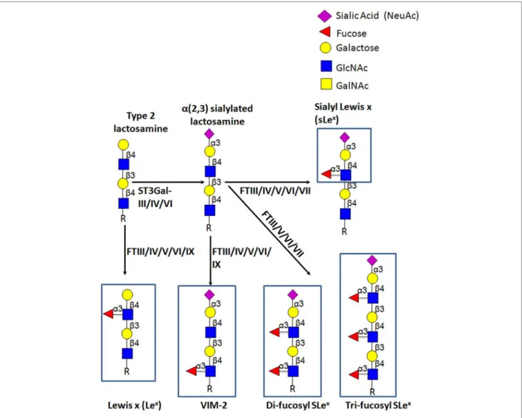
The Carbohydrate e-Selectin Ligands
E-selectin recognizes a range of structurally diverse glycan
epitopes expressed by human leukocytes that typically contain
α(1,3)-fucose (Fuc) and α(2,3)-sialic acid (Sia) modification(s)
on a lactosamine backbone [consisting of galactose (Gal) linked
to N-acetylglucosamine (GlcNAc)], as shown in Figure 4 (
79
).
The terminal tetrasaccharide known as sialyl Lewis X (sLe
x—
Siaα2-3Galβ1-4(Fucα1-3)GlcNAc) is the prototypical
E-selectin-binding determinant (
83
–
85
). Some sLe
x-variant structures can
also exhibit E-selectin binding activity, namely an internally
fucosylated sLe
x-variant (VIM-2) and other polylactosamine
structures, in which Fuc modifications occur at more than one
GlcNAc residue along the polylactosamine chain
(tri-fucosyl-sia-lyl Lewis
xand di-fucosyl-sialyl Lewis
x) (
86
–
88
). In addition, other
glycan structures that are not natively expressed on leukocytes
exhibit E-selectin binding activity, namely the sLe
xisomer, sialyl
Lewis a (sLe
a—Siaα2-3Galβ1-3(Fucα1-4)GlcNAc) (
89
), some
sulfated derivatives of Le
xand Le
a(3′-sulfo-Le
xand 3′-sulfo-Le
a,
respectively) (
90
,
91
), and a fucosylated glycoform of LacdiNac
that displays a terminal N-acetylgalactosamine (GalNAc) instead
of Sia (GalNAc-Lewis x) (
92
).
glycosyltransferases involved in the
Biosynthesis of
Selectin–Carbohydrate-Binding Determinants
E-selectin binding determinants are typically displayed at the
end of O-glycans, N-glycans, or glycolipid precursor structures,
and require the coordinated and sequential action of specific
glycosyltransferases localized within the lumen of the Golgi
apparatus. Assembly of sLe
xis driven by the terminal addition
of Sia (to Gal) and of Fuc (to GlcNAc) through the action of
α(2,3)-sialyltransferases and α(1,3)-fucosyltransferases (FTs),
respectively, on type 2 lactosamine (LacNAc) chains (i.e., Gal
connected to GlcNAc through a β(1,4)-linkage) (Figure 4) (
93
,
94
). The sialylated forms of Lewis antigens are synthesized by
the action of the α(2,3)-sialyltransferases (ST3Gal isoenzymes).
These enzymes transfer Sia residues to the Gal on the LacNAc
chain, exclusively acting prior to fucosylation (
95
,
96
). There are
six members of the α(2,3)-sialyltransferase family (ST3Gal-I–
ST3Gal-VI), but only ST3Gal-III, ST3Gal-IV, and ST3Gal-VI
are reported to sialylate lactosamine chains (
97
). Importantly,
ST3Gal-III exhibits preference for type 1 lactosamine chain
acceptors (wherein Gal is connected to GlcNAc through a
β(1,3)-linkage), whereas ST3Gal-IV and ST3Gal-VI preferentially act on
type 2 polylactosamine chains (
98
–
100
). When Type 1
lactosa-mines are decorated with Sia in α(2,3)-linkage to Gal and with
Fuc in β(1,3)-linkage to GlcNAc, this tetrasaccharide is known as
sialyl Lewis A (sLe
A).
So far, six human FTs have been found to catalyze the
addition of Fuc at α(1,3) linkage to GlcNAc with a type 2
lactosamine—FTIII, FTIV, FTV, FTVI, FTVII, and FTIX.
Each enzyme exhibits specificity for acceptor substrates and,
FigURe 4 | Schematic representation of biosynthesis of the E-selectin ligand determinants. The α(2,3)-sialyltransferases, ST3Gal-III, -IV, and -VI, terminate the elongation of both O- and N-glycans by creating sialylated type 2 lactosamine chains. These can be further fucosylated by the action of the specific fucosyltransferases, yielding different Lewis-related structures that display E-selectin binding activity.
therefore, has the ability to generate distinct fucosylated
structures (
101
,
102
). Particularly, FTIII and FTV are unique
in that they exhibit both α(1,3) and α(1,4) FT activity on both
sialylated and unsialylated type 2 and type 1 lactosamines
thereby creating (s)Le
xand (s)Le
aepitopes, respectively (
103
,
104
). On the other hand, FTIV and FTVI fucosylate both
sialylated and unsialylated type 2 lactosamine chains, with
FTIV creating VIM-2 and Le
x(
105
,
106
) and, modestly, sLe
xdeterminants (
107
,
108
), and FTVI creating these structures
as well as di-fucosyl-sLe
x(
108
–
110
). Uniquely, FTVII can only
act on sialylated type 2 lactosamines, yielding sLe
xand
di/tri-fucosyl-sLe
x-structures (
111
,
112
), whereas FTIX is known to
synthesize mostly Le
x(
101
,
106
).
Most of the reports that assess the role of the different
glyco-syltransferases involved in selectin ligand biosynthesis in
leu-kocytes have been performed using knock-out mouse models,
with a small proportion of these studies using human leukocytes
or human hematopoietic cell lines. Concerning the role of the
α(2,3)-sialyltransferases, murine studies suggest that ST3Gal-IV
and ST3Gal-VI collaborate together in murine E-selectin ligand
biosynthesis, with ST3Gal-IV having an important role in the
regulation of E-selectin-dependent rolling velocity (
113
,
114
).
Interestingly, ST3Gal-III does not seem to contribute to the
synthesis of murine E-selectin ligand moieties, since deficiency
of this enzyme did not affect E-selectin ligand expression or
activity on murine leukocytes (
113
). Surprisingly, ST3Gal-IV
is reportedly the only human α(2,3)-sialyltransferase involved
in the biosynthesis of E-selectin ligands in human myeloid
leukocytes, since ST3Gal-IV-silenced HL-60 cells (a human
promyelocytic cell line), and neutrophils derived from stable
ST3Gal-IV knockdown hematopoietic stem cells fail to engage
in tethering and rolling interactions on E-selectin-bearing
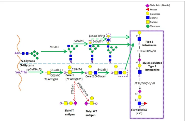
FigURe 5 | Schematic representation of the biosynthetic pathways leading to glycoprotein synthesis: O-linked and N-linked glycosylation. The O-glycosylation
process is characterized by a stepwise sugar addition that occurs in the Golgi apparatus and involves a broad array of enzymes. This synthesis is initiated by one of the N-acetylgalactosaminyltransferase (ppGalNAcTs) family members, forming the Tn antigen. After the first sugar [N-acetylgalactosamine (GalNAc)] addition, Tn is typically elongated by Core 1 β(1,3)galactosyltransferase (Core1GalT or T synthase, whose Golgi expression requires the activity of its chaperone COSMC), creating the “Core 1” O-glycan (also known as “T antigen”). Core 1 is then further lengthened by C2GnT-I, which adds an N-acetylglucosamine (GlcNAc) to the GalNAc, forming the “Core 2” O-glycan structure. Alternatively, Core 1 sialylation, by ST3Gal-I or ST6GalNAc-II [forming sialyl-T (sT) or sialyl-6T (s6T) antigens, respectively] stops Core 2 formation. In contrast to O-glycosylation, the N-glycosylation process requires the production of an oligosaccharide precursor (GlcNAc2Man5) in the cytoplasmic face of the endoplasmic reticulum (ER) membrane. This glycan flipped in the ER lumen and is then transferred en block from a lipid donor to the Asn residue of a newly synthesized protein within the ER lumen, and then further processed in the Golgi compartment. Biosynthesis of hybrid and complex glycans is initiated by the action of MGAT-I, which adds a GlcNAc residue to the mannose (Man) present on the α(1,3)-arm of the Man5GlcNAc2 structure. Repetitive additions of galactose (Gal) and GlcNAc by β(1/4)GalT and β(1,3)GnT enzymes, respectively, can further elongate Core 2 O-glycan and hybrid- and complex-type N-glycan structures, creating the lactosaminyl type 2 chains that serve as acceptors for terminal sialofucosylation reactions.
substrates (
115
). In case of α(1,3)-FTs, studies demonstrate that
mostly FTVII, and to a lesser extent FTIV, are the key murine
α(1,3) FTs that mediate leukocyte selectin ligand biosynthesis.
In fact, murine leukocytes lacking FTVII show poor adhesive
contacts with E- and P-selectin, indicating that this FT plays
a prominent role in murine E-selectin ligand biosynthesis
(
116
,
117
). However, others reported that FTIV is crucial for
slow murine leukocyte rolling velocity (
118
,
119
). Importantly,
E-selectin binding activity conferred by murine FTIV, but not
by FTVII, apparently occurs mainly on glycolipids rather than
glycoproteins (
120
). Conversely, in human leukocytes, there is
evidence that FTVII, FTIV, and FTIX could each act in synthesis
of E-selectin ligand determinants (
121
). Notably, the mouse
genome encodes only FTIV, FTVII, and FTIX (
122
), whereas
primates possess an additional three FT gene products—FTIII,
FTV, and FTIV. These additional FTs provide for a much wider
capacity to create sLe
X; in addition, the expression of FTIII and
FTV in primates uniquely drives creation of sLe
Adeterminants.
Human circulating monocytes express all the α(1,3)-FTs, with
the exception of FTV, heightening the potential for variability
in glycoconjugates bearing sLe
xamong human and mouse cells
(
123
). Notably, sLe
Ais not expressed on any primate leukocytes
as these cells do not synthesize Type 1 lactosamines (
3
).
Other glycosyltransferases involved in the biosynthesis
of sLe
xhave also been studied for their relevance in
generat-ing functional selectin ligands (Figure 5). Regardgenerat-ing sLe
xpresentation on O-glycans, one study reported that leukocytes
from mice deficient in the enzyme required for initiation of
O-glycosylation, ppGalNAcT-1, showed impaired recruitment
during inflammation due to a significant reduction in E- and
P-selectin ligand levels (
124
). Mice lacking the O-glycan core
1
β3galactosyltransferase (C1GalT-I) showed dramatic loss
of leukocyte rolling on E-selectin and, consequently, these
leukocytes did not transmigrate into inflamed tissues (
125
).
Transgenic mouse studies, where the O-glycan core 2
β6-N-acetylglucosaminyltransferase-I (C2GnT-I) was knocked out,
also showed reduced E-selectin and P-selectin binding
activ-ity of leukocytes under static and shear-based rolling assays,
with impaired leukocyte recruitment to sites of inflammation
(
126
–
128
). In agreement, in human moDCs, the
downregula-tion of C2GnT-I, with concurrent upreguladownregula-tion of ST3Gal-I and
GalNAc α(2,6)sialyltransferase (ST6GalNAc)-II, results in a loss
of the core 2 structures required for O-glycan display of sLe
x(Figure 5) (
129
). Furthermore, studies using HL-60 cells have
revealed that the ST6GalNAc-II overexpression abrogates sLe
xcell surface display and reduces the number of adherent cells
to E-selectin under flow conditions, reinforcing the notion that
there exists a competition between ST6GalNAc-II and C2GnT-I
for core 1 acceptors, affecting the biosynthesis of sLe
x-bearing
core 2-O-glycan structures (Figure 5) (
130
). Interestingly, in
mice, knockout of one of the β(1,4)galactosyltransferases (of
the family of five isoenzymes) involved in Type 2 lactosamine
synthesis,
β(1,4)galactosyltransferase-I (β(1,4)GalT-I), showed
reduced inflammatory responses and impaired P-selectin
binding activity; however, the contribution of this enzyme in
the synthesis of E-selectin counter-receptors remains to be
elucidated (
131
).
One study has recently evaluated the contributions of N-glycans,
O-glycans and glycosphingolipids (GSLs) to E-selectin binding by
human myeloid cells under physiological flow conditions (
132
).
To address this issue, O-glycan and GSL synthesis was abolished
by, respectively, knocking-out the core 1 Gal transferase
chaper-one, i.e., the C1GalT-I-specific Molecular Chaperone (COSMC),
β1,2 GlcNAc-transferase (MGAT-I), and UDP-glucose ceramide
glucosyltransferase (UGCG). Notably, these studies indicate that
while O-glycans are indispensable for myeloid cell binding to
L- and P-selectins, N-glycans play the major role in the initial
myeloid cell recruitment into E-selectin-bearing substrates, with
O-glycans playing a more modest role. In addition, both
glycolip-ids and N-glycans are responsible for the slowing down of rolling
velocities that precede firm arrest (
132
).
Most studies that have assessed biologic modulators of
E-selectin ligands in leukocytes have been performed using
human and murine T cells. An array of cytokines has been
shown to regulate E-selectin ligand expression via upregulation
or downregulation of specific glycosyltrasferases that control sLe
xexpression. Specifically, IL-2, IL-7, IL-15, and IL-12 increase the
expression of glycosyltransferases involved in the biosynthesis
of E-selectin ligand determinants, whereas IL-4 has the
oppo-site effect (
133
). This selectin ligand upregulation in T cells in
response to cytokine signaling was shown to be dependent on
Th1 transcription factor T-bet (
134
) and on STAT4-mediated
pathways (
135
). Interestingly, human myeloid cells treated with
granulocyte-colony stimulating factor (G-CSF) show increased
cell surface expression of E-selectin ligands associated with
sig-nificant increases in gene expression of the glycosyltransferases
ST3Gal-IV, FTIV, and FTVII (
136
).
e-SeLeCTiN LigAND ACTiviTY
DiSPLAYeD BY CiRCULATiNg MPS
SUBSeTS
Among the cells of MPS, human circulating monocytes and, to a
lesser extent, human blood cDCs and moDCs are the most
com-prehensively analyzed group in terms of E-selectin ligand
activ-ity. In our studies, human classical monocytes (CD14
++CD16
−)
showed significantly higher levels of sLe
xdeterminants as
compared to intermediate monocytes (CD14
++CD16
+),
whereas non-classical monocytes (CD14
+CD16
++) were almost
devoid of sLe
xexpression (
123
). Another study compared the
trafficking capacity of human monocyte subsets by
analyz-ing their ability to bind to activated endothelial monolayers,
and commensurately, classical monocytes showed noticeably
higher capability of adhering to reactive endothelium than did
non-classical/intermediate monocytes (
137
). In agreement
with human studies, murine classical monocytes (Ly-6C
hi)
exhibit greater binding to E-selectin under flow conditions and
express higher levels of the scaffolds that bear sLe
xdeterminants
compared to non-classical monocytes (Ly-6C
lo) (
138
,
139
). This
differential pattern of E-selectin ligand display is in agreement
with the specific migratory requirements among the monocyte
subsets: classical monocytes are typically recruited to inflamed
lesions (
138
,
139
), whereas non-classical monocytes migrate to
non-inflamed endothelium (
14
) upon which they patrol healthy
tissues in a LFA-1-dependent manner (
19
). Indeed, although
the first observations of non-classical monocytes were made in
non-inflamed skin blood vessels (
19
), these cells were further
described in the microvasculature of kidney under steady-state
conditions (
20
). The patrolling profile that these cells exhibit is
independent of the activation state of the endothelium, since
non-classical monocytes constitutively scavenge the luminal side
of non-reactive endothelium (
18
). Therefore, their ability to bind
to endothelium seems to be independent of E-selectin receptor/
ligand interactions, but, instead, appears critically regulated by
LFA-1 expression and its interaction with endothelial ICAM (
19
,
20
). Notably, although selectins play a major role in the initial
adhesive contacts with endothelium surfaces, integrins can also
support tethering and rolling events under flow conditions,
albeit with less potency than do selectins (
140
,
141
).
Multiple adhesion molecules are involved in monocyte
attachment to endothelium. While E-selectin receptor/ligand
interactions prominently mediate Step 1 events in transmigration
for all leukocytes, L-selectin-dependent binding interaction have
also been observed to potently mediate human peripheral blood
monocyte binding to activated vascular endothelium under shear
stress (
142
–
144
). Thus, even though the majority of the reports
indicates that initial monocyte adhesion to activated endothelial
cells is most critically dependent on E-selectin receptor/ligand
interactions (
123
,
145
–
150
), distinct interactions were also
reported by other authors. The differences have to do with
dif-ferences in the leukocyte populations under study, variations in
the assay conditions employed (i.e., shear stress levels employed,
rotatory shear versus fluid shear conditions, temperature, etc.),
differences in the adhesion metrics (i.e., number of adhered
cells, number of rolling cells, rolling velocity measurements,
etc) altogether compounded by the innate biologic differences
between mice and human cells, could alternatively emphasize the
contribution(s) of other adhesion molecules.
Concerning DCs, human blood cDCs express high levels
of sLe
xdeterminants, which allow them to tether and roll on
E-selectin under flow conditions (
151
,
152
). Importantly, in in vivo
intravital microscopy studies, human blood cDCs adoptively
transferred into mice were observed to roll along resting murine
skin endothelium and extravasate at sites of inflammation (
151
).
Notably, human moDCs significantly express sLe
x, especially on
O-glycan structures. Upon maturation of moDCs with the TLR4
ligand, LPS, sLe
xexpression is downregulated due to decreased
C2GnT-I expression and upregulation of ST6GalNAc-II and
ST3Gal-I (
129
). Biologically, these observations suggest that
sLe
xis less relevant for transendothelial migration of
TLR4-induced-mature moDCs, or that maturation is a step that follows
transendothelial migration. By contrast, IFN-γ-induced
matura-tion of moDCs leads to an upregulamatura-tion of C2GnT-I, resulting
in increased expression of core 2 O-glycan substrates for sLe
xdecoration (
153
). These features suggest that sLe
x-bearing core
1-derived (or core 2) O-glycans are required for human moDC
migration and are modulated according to specific maturation
stimuli. Human pDCs also express sLe
x, allowing their
recruit-ment to some inflamed tissues (
38
,
39
,
154
) and to reactive lymph
nodes (
43
), a process believed to be mediated by the expression of
E-selectin in HEVs (
45
,
47
).
gLYCOCONJUgATe STRUCTUReS THAT
DiSPLAY e-SeLeCTiN LigAND
DeTeRMiNANTS iN CiRCULATiNg MPS
CeLLS
Several diverse and structurally singular glycostructures with
E-selectin binding activity have been identified on human
classi-cal monocytes or human blood DCs. Human classiclassi-cal monocytes
greatly display sLe
xdecorations on an array of protein scaffolds,
consisting of P-selectin glycoprotein ligand-1 (PSGL-1), CD43
and CD44, and GSLs (
123
), whereas human circulating DCs
appear to display sLe
xsolely on PSGL-1 (
129
,
155
).
Cutaneous Lymphocyte Antigen
The cutaneous lymphocyte antigen (CLA) is the
E-selectin-reactive glycoform of PSGL-1. PSGL-1 is a transmembrane
240-kDa homodimeric, mucin-like glycoprotein expressed
on leukocytes (and, reportedly, on some activated endothelial
cells) that plays a crucial role in the homing of leukocytes into
inflamed tissue (
156
,
157
). E-selectin binding activity of PSGL-1
is conferred by sialylated and fucosylated core 2-based-O-glycans
that are cluster-distributed along the stalk region of the PSGL-1
extracellular domain (
158
). A number of studies have identified
PSGL-1 as one of the several scaffolds expressed by human
clas-sical monocytes displaying E-selectin-binding activity (
123
,
155
).
On the other hand, PSGL-1 is the only known scaffold that
pre-sents sLe
xdeterminants on human circulating cDCs (
151
,
155
)
and moDCs (
129
). Yet, Silva et al. observed that although PSGL-1
is essential for P- and L-selectin recognition by human moDCs
under fluid shear conditions, it is not mandatory for tethering to
E-selectin (
153
). Moreover, there are reports that extravasation
of murine immature DC to inflamed tissues requires both E- and
P-selectin, but not PSGL-1 (
159
). Together, these data suggest
the expression of ligands for E-selectin in addition to CLA by
human and murine DCs. Still, PSGL-1 is the dominant ligand for
P- and L- selectin and is the only known glycoprotein that binds
all three selectins (
160
,
161
). Accordingly, circulating monocytes
that have already bound to E-selectin on inflamed endothelium
can also interact with L-selectin expressed by other circulating
monocytes/DCs via PSGL-1 and support their secondary
cap-ture, potentiating mononuclear phagocyte recruitment to sites of
inflammation (
162
).
Hematopoietic Cell e- and L-Selectin
Ligand
HCELL is a sialofucosylated glycoform of CD44 that exhibits
potent E-selectin (and L-selectin) binding activity. CD44 is a
transmembrane protein that exists in a wide variety of protein
isoforms due to alternative splicing and extensive
post-transla-tional modifications (with molecular weight ranging from 80 to
220 kDa). CD44 is expressed by most mammalian cells, where it
serves as the principal receptor for hyaloronic acid and
partici-pates in a broad range of cellular activities, including lymphocyte
activation, leukocyte trafficking, hematopoiesis, cell growth and
survival, and tumor dissemination (
163
). Post-translational
modifications along with extensive alternative splicing allow
the formation of multiple protein isoforms, expressed in a
tissue-specific manner (
164
,
165
). The standard protein isoform
of CD44 (CD44s or CD44H) is encoded by mRNA transcripts
comprising exons 1–5 and 16–20 (“s1–s5 and s6–s10”). CD44s is
ubiquitously expressed by mammalian cells and is the form most
often displayed by hematopoietic-lineage cells. In addition to
CD44s, non-hematopoietic cells characteristically display CD44
variant isoforms contain peptide products of variant exons (exons
“v2–v10”) in addition to the standard exon peptide products.
CD44 post-translational modifications include the addition
of different glycan structures, namely glycosaminoglycans, and
N- and O-glycan substitutions (
166
). While previous studies
indicated that HCELL was only expressed by human
hemat-opoietic stem and progenitor cells (HSPCs) (
167
,
168
), and some
hematologic (
167
,
169
) and solid malignancies (
170
), HCELL was
recently reported to be expressed by classical human monocytes
(
123
). Importantly, for human HSPCs, the sLe
xdeterminant is
exclusively displayed on N-glycan lactosamines on CD44s, but
classical monocytes express sLe
xon O-glycans of CD44s (
123
).
Because of its ability to engage E- and L-selectin under relatively
high fluid shear conditions (i.e., in excess of 20 dynes/cm
2shear
stress), HCELL is considered the most potent L- and E-selectin
ligand expressed on mammalian cells (
167
,
171
).
CD43e
CD43, also known as sialophorin or leukosialin, is a cell surface
glycoprotein expressed by nearly all hematopoietic cells and is
involved in several important processes, including cell
develop-ment, activation, survival, and migration (
172
–
176
). Glycosylation
of CD43 molecules with either core 1 or core 2 O-glycan structures
produces the ~115 and ~135 kDa glycoforms, respectively (
177
).
Recently, we reported that human classical monocytes display
E-selectin binding activity on CD43 (
123
). CD43 that displays
sLe
xand binds E-selectin is known as “CD43E” and this protein
harbors sLe
xon O-glycans (
178
).
glycosphingolipids
In contrast to results in mouse leukocytes, reports using human
cells have suggested that sialofucosylated determinants displayed
on lipid scaffolds can mediate adhesion to E-selectin under
static and flow conditions. In native human leukocytes and in
the myelocytic leukemia cell line HL-60, the glycolipid structures
that display ability to bind to E-selectin consist of sialylated
lactosylceramides decorated with internal fucosylated GlcNAc
structures (myeloglycans) (
86
,
87
,
179
). It has been reported that
the disruption of myeloglycan biosynthesis via knockdown of
ceramide glucosyltransferase (UGCG) in HL-60 cells disturbed
stable cell rolling and impaired transmigration across inflamed
endothelial cells (
180
). Finally, human classical monocytes
treated with a broadly active protease did not show a complete
abrogation of sLe
xstaining, suggesting that although the majority
of sialofucosylated moieties required for E-selectin binding are
preferentially expressed on proteins rather than lipids, a
consid-erable amount of sialofucosylated determinants are present on
glycolipids (
123
).
PATHOPHYSiOLOgiCAL iMPORTANCe
OF THe SeLeCTiNS
Due to its crucial role in the transmigration of specific myeloid
populations to sites of inflammation, the selectin/selectin–ligand
axis is involved in the development of many acute and chronic
inflammatory conditions (
181
,
182
). In fact, cutaneous
inflam-matory disorders, such as allergic contact dermatitis (
183
),
atopic dermatitis (
184
–
188
), and psoriasis (
183
,
184
,
189
–
191
),
are known to be promoted by the upregulation of selectins on
dermal microvasculature. Specifically, P- and E-selectins are
upregulated in skin lesions of cutaneous inflammatory patients,
which enables inflammatory mononuclear phagocyte
infiltra-tion with subsequent release of soluble cytotoxic mediators,
T cell activation, and destruction of the dermal layer of skin
(
154
,
192
–
194
). Atherosclerosis is another chronic inflammatory
disease in which the recruitment of monocytes into
selectin-expressing endothelial beds constitutes a key molecular event
in the pathogenesis of the disease (
195
). Both E-and P-selectins
are expressed on human arterial luminal endothelial cells of
atherosclerotic plaques (
195
,
196
), and mouse studies have
shown that E-selectin and/or P-selectin deficiency substantially
reduces the formation of atherosclerotic plaques, suggesting an
overlapping function of these two selectins in the development
of atherosclerotic lesions (
197
,
198
). Murine classical monocytes
(Ly6C
hi) preferentially migrate into activated endothelium and
infiltrate developing atheromas to become atherosclerotic
macrophages, inflammatory DCs, or foam cells (
139
). Indeed,
murine classical monocytes display high levels of PSGL-1 and
higher binding affinity to E-/P-selectin expressing cells than do
the non-classical patrolling monocytes, which might explain
why they are preferentially recruited to sites of endothelial
inflammation and thrombosis (
138
).
Inflammatory bowel disease (
199
–
202
), multiple sclerosis
(
203
,
204
), rheumatoid arthritis (
205
–
207
), and type 1 diabetes
(
208
,
209
) are other examples of inflammatory diseases in which
upregulated E-/P-selectin expression in tissue microvasculature
drives myeloid cell recruitment crucial for the development of
the disease. Increased E-selectin expression and mononuclear
phagocyte infiltration have been similarly observed in cases of
transplantation rejection, including human renal (
210
,
211
),
lung (
212
–
214
), and cardiac (
215
–
217
) rejection and in acute
graft versus host disease (
218
–
221
). Recruitment of
inflam-matory classical monocytes (
222
,
223
) and pDCs (
38
,
41
,
224
)
to tumor-cell-activated endothelium has also been reported
to be dependent on E-selectin expression, which can lead to
an inhibition of the tumor-specific immune defense response
and induction of tolerance (
225
,
226
). Some studies have also
indicated that inflammatory monocytes license extravasation of
tumor cells via the induction of E-selectin-dependent adhesive
interactions (
222
).
THeRAPeUTiC STRATegieS TARgeTiNg
THe SeLeCTiN/SeLeCTiN–LigAND AXiS
The critical role of selectins and their ligands in the pathogenesis
of many inflammatory conditions makes them potential
molecu-lar targets for therapy and, therefore, several strategies have been
developed to interfere with this biology (
182
,
227
,
228
). One
strategy relies on the development of pan-selectin competitive
inhibitors that inhibit leukocyte/endothelial cell interaction and,
therefore, cell recruitment to affected inflammatory or metastatic
tissues. One example is the molecule Bimosiamose (Encysive
Pharmaceuticals), a sLe
xmimetic that showed clinical efficacy
in both asthma and psoriasis (
229
,
230
). Other examples of
pan-selectin inhibitors include sLe
x-peptides (
231
), sLe
x-bearing
liposomes (
232
) or heparin oligosaccharides (
233
,
234
). Also,
GMI-127 and GMI-1271, E-selectin antagonists developed by
GlycoMimetics based on the bioactive conformation of sLe
xin
the carbohydrate-binding domain of E-selectin, have been used
in treatment of sickle cell crisis (ClinicalTrials.gov Identifier:
NCT00911495) and as an adjuvant for chemotherapy of
hema-tological malignancies, including multiple myeloma and acute
myeloid leukemia (NCT02811822, NCT02306291). In addition,
an alternative approach involves the inhibition of the selectin
ligand synthesis, either by interfering with the expression of key
glycosyltransferases involved in sLe
xbiosynthesis (
235
), or by
using fluorinated analogs of Sia and/or Fuc residues (
232
,
236
)
that inhibit sLe
xsynthesis. Finally, competing antibodies that bind
to vascular E- and P-selectins and/or to selectin ligands expressed
on the leukocyte cell surface constitute alternative methods
that have been explored to prevent inflammatory exacerbation
(
182
,
228
). Indeed, a soluble form of PSGL-1 linked to the Fc
portion of human IgG1 (PSGL-1-Ig) has been shown to inhibit
leukocyte rolling in several disease models in mice (
237
–
240
).
On the other hand, the disruption of the selectin–leukocyte
interaction has been reported to trigger severe immune-deficiency
by disruption of immunosurveillance (
241
). Leukocyte adhesion
deficiency Type II is a rare genetic disorder characterized by
defec-tive neutrophil and monocyte migration caused by a mutation in
a GDP-Fuc transporter gene (
242
). This mutation leads to the
formation of glycans that lack fucosylation, resulting in impaired
leukocyte rolling and consequent leukocytosis and recurrent
infections (
242
,
243
). To overcome deficient cellular rolling, our
lab developed a FT-driven sLe
xbiosynthesis approach to enforce
cell surface expression of E-selectin ligands while preserving cell
viability, called glycosyltransferase-programmed
stereosubstitu-tion (GPS) (
244
,
245
). This platform uses optimized reaction
conditions, which enables the efficient α(1,3)-fucosylation of
underfucosylatated sialylated type 2 lactosamine acceptors
(Siaα2-3Galβ1-4GlcNAc), via a soluble α(1,3) FT, installing
expression of sLe
x(Siaα2-3Galβ1-4GlcNAcα1-3Fuc) (
3
). GPS has
been used to improve the recruitment of a variety of human and
mouse cells into E-selectin-bearing endothelial beds, driving cell
migration into bone marrow and inflammatory sites (
123
,
178
,
208
,
244
,
246
–
248
).
CONCLUDiNg ReMARKS
Specific migratory routes and distinct localization in steady and
inflammatory conditions of the different human MPS subsets
suggests that they play differential roles in immunity. These
cells are operational in multiple beneficial processes, including
immediate antimicrobial host defense, activation of the adaptive
immune system, and tissue healing processes. However, they
may also contribute to the pathobiology of several
inflamma-tory conditions. A better understanding of the molecular basis
of glycosylation-dependent creation of E-selectin ligands could
yield the development of novel therapeutic approaches for
inflammatory diseases, or, alternatively, could yield enhanced
ability to infiltrate sites where immunity is needed (e.g., tumors
or infection). To our knowledge, only a limited number of studies
have analyzed the functional and structural biology of the full
spectrum of E-selectin ligands expressed by different circulating
human MPS subsets. Future work will be required to address this
issue and to elucidate how custom-modified expression of these
homing receptors can be achieved to preferentially influence the
specific migratory routes of the different subsets of circulating
MPS cells.
AUTHOR CONTRiBUTiONS
All authors listed have made a substantial, direct, and intellectual
contribution to the work and approved it for publication.
ACKNOwLeDgMeNTS
We thank Dr. Nandini Mondal and Kyle Martin for their helpful
review of the manuscript.
FUNDiNg
This work was supported by the National Institutes of Health
National Heart Lung Blood Institute grant PO1 HL107146
(Program of Excellence in Glycosciences) (RS), and by the Team
Jobie Fund (RS) and the Faye Geronemus Leukemia Research
Fund (RS). INclude Fulbright Commission fellowship (to PAV).
ReFeReNCeS
1. Haniffa M, Bigley V, Collin M. Human mononuclear phagocyte system reunited. Semin Cell Dev Biol (2015) 41:59–69. doi:10.1016/j.semcdb.2015.05.004 2. Chow A, Brown BD, Merad M. Studying the mononuclear phagocyte system
in the molecular age. Nat Rev Immunol (2011) 11(11):788–98. doi:10.1038/ nri3087
3. Sackstein R. Glycosyltransferase-programmed stereosubstitution (GPS) to create HCELL: engineering a roadmap for cell migration. Immunol Rev (2009) 230(1):51–74. doi:10.1111/j.1600-065X.2009.00792.x
4. Kunkel EJ, Ley K. Distinct phenotype of E-selectin-deficient mice. E-selectin is required for slow leukocyte rolling in vivo. Circ Res (1996) 79(6):1196–204. doi:10.1161/01.RES.79.6.1196
5. van Furth R, Cohn ZA. The origin and kinetics of mononuclear phagocytes. J Exp Med (1968) 128(3):415–35. doi:10.1084/jem.128.3.415
6. Jakubzick C, Gautier EL, Gibbings SL, Sojka DK, Schlitzer A, Johnson TE, et al. Minimal differentiation of classical monocytes as they survey steady-state tis-sues and transport antigen to lymph nodes. Immunity (2013) 39(3):599–610. doi:10.1016/j.immuni.2013.08.007
7. Epelman S, Lavine KJ, Beaudin AE, Sojka DK, Carrero JA, Calderon B, et al. Embryonic and adult-derived resident cardiac macrophages are maintained through distinct mechanisms at steady state and during inflammation. Immunity (2014) 40(1):91–104. doi:10.1016/j.immuni.2013.11.019
8. Bain CC, Scott CL, Uronen-Hansson H, Gudjonsson S, Jansson O, Grip O, et al. Resident and pro-inflammatory macrophages in the colon represent alternative context-dependent fates of the same Ly6Chi monocyte precursors. Mucosal Immunol (2013) 6(3):498–510. doi:10.1038/mi.2012.89
9. Rivollier A, He J, Kole A, Valatas V, Kelsall BL. Inflammation switches the differentiation program of Ly6Chi monocytes from antiinflammatory
macrophages to inflammatory dendritic cells in the colon. J Exp Med (2012) 209(1):139–55. doi:10.1084/jem.20101387
10. Serbina NV, Jia T, Hohl TM, Pamer EG. Monocyte-mediated defense against microbial pathogens. Annu Rev Immunol (2008) 26:421–52. doi:10.1146/ annurev.immunol.26.021607.090326
11. Gordon S, Taylor PR. Monocyte and macrophage heterogeneity. Nat Rev Immunol (2005) 5(12):953–64. doi:10.1038/nri1733
12. Shi C, Pamer EG. Monocyte recruitment during infection and inflammation. Nat Rev Immunol (2011) 11(11):762–74. doi:10.1038/nri3070
13. Stansfield BK, Ingram DA. Clinical significance of monocyte heterogeneity. Clin Transl Med (2015) 4:5. doi:10.1186/s40169-014-0040-3
14. Geissmann F, Jung S, Littman DR. Blood monocytes consist of two principal subsets with distinct migratory properties. Immunity (2003) 19(1):71–82. doi:10.1016/S1074-7613(03)00174-2
15. Ziegler-Heitbrock L, Ancuta P, Crowe S, Dalod M, Grau V, Hart DN, et al. Nomenclature of monocytes and dendritic cells in blood. Blood (2010) 116(16):e74–80. doi:10.1182/blood-2010-02-258558
16. Kratofil RM, Kubes P, Deniset JF. Monocyte conversion during inflammation and injury. Arterioscler Thromb Vasc Biol (2016) 37(1):35–42. doi:10.1161/ ATVBAHA.116.308198
17. Ziegler-Heitbrock L, Hofer TP. Toward a refined definition of monocyte subsets. Front Immunol (2013) 4:23. doi:10.3389/fimmu.2013.00023 18. Thomas G, Tacke R, Hedrick CC, Hanna RN. Nonclassical patrolling
monocyte function in the vasculature. Arterioscler Thromb Vasc Biol (2015) 35(6):1306–16. doi:10.1161/ATVBAHA.114.304650
19. Auffray C, Fogg D, Garfa M, Elain G, Join-Lambert O, Kayal S, et al. Monitoring of blood vessels and tissues by a population of monocytes with patrolling behavior. Science (2007) 317(5838):666–70. doi:10.1126/ science.1142883