UNIVERSIDADE DE LISBOA
FACULDADE DE CIÊNCIAS
DEPARTAMENTO DE BIOLOGIA VEGETAL
Genetic deletion of adenosine A
2A
receptor in mice
model of Alzheimer’s disease: impact on synaptic
plasticity
Ana Carolina Amaral Peixoto
Mestrado em Biologia Molecular e Genética
Dissertação orientada por:
Luísa Lopes
Rui Gomes
Agradecimentos
Gostaria de começar por agradecer ao David Blum (INSERM,Lille) e à Catherine Ledent por disponibilizarem os ratinhos APP/PS1 e THY-Tau22, tornando possível a realização deste trabalho.
À Luísa Lopes, um obrigado por me dar a oportunidade de desenvolver a minha dissertação de mestrado no seu grupo de trabalho.
Ao Rui, um obrigada por ter aceitado ser o meu orientador interno e por ter demonstrado sempre disponibilidade e interesse no decorrer deste ano.
Agradeço a todos os Lopez por toda a ajuda e apoio e acima de tudo por proporcionarem um ambiente magnífico e por não fazerem do laboratório apenas um local de trabalho! Gostaria de dirigir um agradecimento especial à Joana e à Mariana por toda a disponibilidade, paciên-cia, ajuda, ensinamentos e companheirismo que facilitaram este trabalho e o tornaram mais divertido.
Resumo
A percentagem de população envelhecida a nível mundial está a aumentar consideravel-mente. Com este aumento de população idosa, há também um aumento de doenças ligadas à idade avançada, tais como doenças cardiovasculares e neurodegenerativas [1]. O envelheci-mento é caracterizado pelo auenvelheci-mento de défices cognitivos assim como pelas alterações a nível da estrutura e funcionalidade do hipocampo, que apresenta um papel fundamental na memória. O envelhecimento e a doença de Alzheimer estão relacionados de forma direta, sendo que o envelhecimento é o maior fator de risco para a doença de Alzheimer [1].
A doença de Alzheimer (DA) é uma doença neurodegenerativa, sendo a forma de demência mais comum. É caracterizada pela presença de placas senis e novelos neurofibrilares [2]. At-ualmente, existem duas teorias que são globalmente aceites: a teoria da cascata β amiloide e a teoria da hiperfosforilação da proteína tau [3]. De acordo com a teoria da cascata β amilóide, as placas senis são caracterizadas pela acumulação extracelular do péptido β amilóide (Aβ). Isto ocorre devido a falhas no processamento da proteína precursora amilóide (APP) [2]. Aβ é pro-duzida nos indivíduos saudáveis. No entanto, sob certas circunstâncias, esta molécula agrega-se e leva ao desenvolvimento da doença de Alzheimer. Este processo ocorre quando duas enzimas proteolíticas atuam juntas, secretase-β e -γ, reguladas por recetores acoplados à proteína G [4]. Foram feitos vários estudos que suportam esta teoria nomeadamente estudos que mostram que a acumulação de Aβ é a causa primária da AD e que a progressão da doença, incluindo a for-mação de novelos de tau, é devida às alterações na Aβ [5]. A maior objeção a esta teoria é que o número de depósitos de Aβ não é proporcional ao grau de défices cognitivos [5]. Segundo a teoria da hiperfosforilação da proteína tau, os novelos neurofibrilares são formados devido à acumulação da proteína tau nos neurónios. Tau é uma proteína neuronal presente nos axónios e é essencial para a organização e dinâmica dos microtúbulos. Alterações na fosforilação desta proteína levam à sua agregação. Quando a proteína tau se torna hiperfosforilada, separa-se dos microtúbulos, agrega-se e forma os novelos de proteína tau [2, 6].
Existem vários modelos animais da DA que podem ser usados para estudar as duas teorias supramencionadas assim como alterações na transmissão sináptica e comportamentais [7]. Os dois modelos de ratinhos usados neste trabalho foram: APP/PS1 e THY-Tau22. Ratinhos APP (apresentam APP sobre-expresso com mutação em um ou mais locais) foram cruzados com ratinhos PS1 (sobre-expressam o gene humano presenilina–PS1 que está envolvido no proces-samento da APP) a fim de obter ratinhos transgénicos APP/PS1 [7]. Ratinhos APP/PS1 apre-sentam um aumento de Aβ com os primeiros depósitos observados aos 5 meses. Este modelo animal é uma ferramenta fundamental para estudar o impacto da acumulação da proteína Aβ nos défices de memória[8, 9]. Ratinhos THY-Tau22 são um modelo animal para agregação da proteína tau presente na DA assim como em taupatias. Este modelo animal apresenta a proteína tau hiperfosforilada com progressão associada à idade: leve aos 3 meses e severa aos 7 meses. Este modelo é fundamental para estudar a correlação da presença da proteína tau com a DA [10].
Adenosina é um neuromodulador muito importante responsável por diversos processos tanto a nível fisiológico como patológico. É uma molécula sinalizadora extracelular que influencia
a transmissão sináptica [11]. Quando esta molécula se liga a recetores acoplados a proteína G pode atuar a nível pré-sináptico (inibindo ou facilitando a libertação de neurotransmissores) ou a nível pós-sináptico (afetando a ação de recetores) [12]. Quando a molécula de adenosina se encontra na membrana celular pode ativar quatro subtipos de recetores acoplados à proteína-G: A1, A2A, A2Be A3; apresentando maior afinidade para os recetores A1, e A2A[13]. Os recetores
A1 são mais abundantes no cérebro, são responsáveis pela diminuição da libertação de
glutam-ato tendo um papel de neuroprotecção. Os recetores A2Aestão localizados nas sinapses, sendo
considerados recetores excitatórios na medida em que facilitam a libertação de neurotransmis-sores [13].
Durante o processo de envelhecimento há um aumento da expressão do recetor de Adenosina A2A(A2AR) nas zonas corticais, estando este associado ao aumento dos défices cognitivos [14].
Os A2AR apresentam um papel importante para a presença de danos sinápticos e cognitivos
presentes na população envelhecida e nos doentes de Alzheimer. O bloqueio destes recetores ou a sua deleção previne alguns dos fenótipos da doença de Alzheimer, nomeadamente défices na LTD, toxicidade sináptica e neuroinflamação, em modelos animais [15, 16]. A cafeína é a droga psicoativa mais usada no mundo sendo capaz de interagir com neurotransmissores em diferentes partes do cérebro e funcionando como um antagonista não específico dos recetores A2A. Dados epidemiológicos mostram que o consumo de cafeína atrasa o aparecimento de
demência, reduz o risco de Alzheimer e atrasa o aparecimento de défices cognitivos em adultos de idade avançada [2, 16].
O hipocampo é uma estrutura localizada no cérebro humano responsável pela consolidação de memória e aprendizagem [17]. Esta estrutura é considerada um bom exemplo de estudo da plasticidade neuronal através da potenciação de longa duração (LTP) e depressão de longa duração (LTD), que são fundamentais para aprendizagem e memória. A LTP consiste num aumento na transmissão sináptica e a LTD numa diminuição [18]. Ambos são considerados como bons mecanismos de estudo do armazenamento de memória e podem ser induzidos ar-tificialmente através de certos protocolos de estimulação eléctrica [18]. Alterações na LTP e LTD no hipocampo foram descritos de estarem presentes em modelos de ratinho da doença de Alzheimer [19].
O nosso objectivo com este trabalho foi comparar o impacto da deleção com o bloqueio farmacológico do receptor A2Anos modelos de Alzheimer (APP/PS1 e THY-Tau22) na
poten-ciação de longa duração (LTP) e depressão de longa duração (LTD).
Para isso, registamos potenciais excitatórios pós-sinápticos de campo (fEPSP) a partir da zona Schaffer collaterals – CA1 das fatias de hipocampo de dois grupos de animais transgéni-cos, modelos da doença de Alzheimer - APP/PS1 e THY-Tau22. No que diz respeito aos rat-inhos APP/PS1, usamos 4 grupos diferentes de animais com 11-12 meses de idade que foram previamente submetidos a testes comportamentais. Os 4 grupos são: wild type (WT), A2A
dele-tado (A2AR-/-), transgénico (APP/PS1) e transgénico com A2AR deletado (APP PS1/A2AR-/-).
Induzimos LTP usando os protocolos de estímulos de alta frequência (HFS) (4 trains de 100Hz) e theta (TBS) (10 trains de 4 pulsos) e também a curva input/output (I/O) de forma a avaliar alterações na plasticidade sináptica. A partir dos resultados obtidos, conseguimos ver que em resposta ao estímulo TBS, o declive fEPSP era menor no grupo transgenico APP/PS1
compara-tivamente ao WT. Vimos também que este grupo, no que diz respeito à I/O, apresentou também um défice apresentando um declive da fEPSP menor para a mesma intensidade de estímulo. Não observámos nenhum efeito de reversão significativo da deleção dos A2AR nas respostas
anteriormente mencionadas. Para além da LTP foi também induzida LTD usando estímulos de baixa frequência (3 trains de 1200 pulsos, 2Hz, 10 minutos de intervalo) de forma a estudar alterações na plasticidade sináptica. Conseguimos observar que ratinhos APP/PS1 apresentam alterações no declive da fEPSP, apresentando potenciação quando induzidos por estímulos de baixa frequência. Sob este estímulo, tanto o antagonista específico dos A2AR (SCH-58261)
como a deleção dos A2AR reverteram a potenciação observada nos ratinhos transgénicos. No
entanto, quando foi usado o antagonista específico, este reverteu totalmente a potenciação, ap-resentando novamente uma depressão similar à observada nos ratinhos WT. No que diz respeito aos ratinhos transgénicos THY-Tau22, usámos um protocolo de indução de LTD (3 trains de 1200 pulsos, 2Hz, 10 minutos de intervalo). Verificou-se que estes ratinhos transgénicos ap-resentam uma potenciação quando induzidos por um estímulo de baixa frequência. Quando o antagonista específico SCH-58261 foi adicionado verificou-se que este reverteu a potenciação observada nos THY-Tau22.
Em suma, ratinhos transgénicos APP/PS1 apresentaram uma diminuição na LTP, que está associada aos défices de memória observados nos pacientes de Alzheimer. No entanto a deleção do receptor A2AR não melhorou ou reverteu estes danos na LTP induzidos por estímulos de
fre-quência theta especificamente. No que diz respeito à LTD, conseguimos verificar que tanto o bloqueio dos A2AR como a sua deleção reverte a alteração na transmissão sináptica
obser-vada neste modelo, sendo que o bloqueio foi mais eficiente. Quanto aos ratinhos THY-Tau22, podemos verificar que estes apresentam uma alteração na LTD e que esta é revertida totalmente quando adicionamos o antagonista específico SCH-58261.
Estes resultados parecem sugerir que a LTD é mais sensível a alterações induzidas pelo receptor A2A presente na sinapse Schaffer collaterals – CA1 do hipocampo. Mais estudos são
necessários para se perceber o mecanismo subjacente a estas alterações.
Palavras chave: Doença de Alzheimer; Receptores A2A; Eletrofisiologia; APP/PS1;
Abstract
Aging is a process characterized by cognitive decline, structural and functional alterations in the hippocampus, being the main risk for Alzheimer’s disease (AD) [1].
During the aging process, there is an upsurge of adenosine A2Areceptor (A2AR) expression
in cortical areas which is associated to cognitive defects [14]. A2AR are G-protein
coupled-receptors that have an important role in the establishment of synaptic/cognitive impairments throughout ageing and in neurodegeneration, particularly in AD. Blockade of A2AR or genetic
deletion prevents some of the AD phenotypes in animal models; namely LTD defects, synaptic toxicity or neuroinflammation [15, 16]
Our main goal was to compare genetic deletion and pharmacological blockade of A2A
re-ceptor as a driving force to Alzheimer’s disease, performing a knockout of A2AR in an APP/PS1
and blocking A2AR in THY-Tau22 transgenic AD mice model and to assess changes in synaptic
plasticity and basal transmission.
We recorded field excitatory postsynaptic potentials (fEPSP) from Schaffer collaterals -CA1 hippocampal synapse from the two AD mice model. Regarding APP/PS1 mice, we used 4 groups of mice previously subjected to cognitive testing: wild type (WT), A2Areceptor
knock-out (A2AR-/-), APP/PS1 (APP/PS1) and APP/PS1 x A2A receptor KO (APP/PS1/A2AR -/-).
We elicited LTP using theta burst stimulus (TBS) (10 trains of 4 pulses, 100Hz) and assessed input/output (I/O) response to evaluate changes in synaptic plasticity. We observed that in re-sponse to TBS, the fEPSP slope was decreased in APP/PS1 mice compared to WT. This group also showed a shift to the right in the I/O curve. We could not observe any significant effect of knocking-out A2AR in those responses. We also elicited LTD using a LFS protocol (3 trains
of 1200 pulses, 2Hz, 10 minutes interval). We observed that APP/PS1 transgenic mice showed a LTD-to-LTP shift. When A2AR was deleted on APP/PS1 mice model, the potentiation
ob-served was abolish, but LTD was not restored. However, when we applied the selective A2AR
antagonist, SCH-58261, to the slices of APP/PS1 mice, LTD impairment observed was fully rescued.
Concerning THY-Tau22 mice we elicited LTD (3 trains of 1200 pulses, 2Hz, 10 minutes interval). We observed that fEPSP slope was increased in THY-Tau22, showing a potentiation in response to a low frequency stimulus. When SCH-58261 (A2AR antagonist) was infused in
the electrophysiology chamber, the LTD impairment was reverted.
Altogether, APP PS1 transgenic mice show LTP impairments, which are correlated with memory changes, as observed in AD. However, the deletion of A2AR did not improve or reverse
LTP induced by this particular stimulation protocol. THY-Tau22 transgenic mice show LTD impairment and SCH-58621 rescued LTD impairment in THY-Tau22 mice.
Hence, the A2AR effects in CA3-CA1 synapse in the hippocampus are more significant in
LTD which may give more clues to the molecular mechanism to be explored in the future. Key words: Alzheimer’s disease; A2A Receptors; Electrophysiology; APP/PS1;
Contents
Agradecimentos ii
Resumo iii
Abstract vi
List of Figures viii
Abbreviations ix
1 Introduction 1
1.1 Alzheimer’s Disease . . . 1
1.1.1 Animal Models . . . 4
1.2 Adenosine . . . 4
1.2.1 Memory and Synaptic Plasticity . . . 6
1.3 Aim . . . 7
2 Methodology 8 2.1 Animal Models . . . 8
2.2 Electrophysiology recordings . . . 9
2.2.1 Hippocampal slice preparation . . . 9
2.2.2 Extracellular recordings . . . 10
2.2.3 Statistical analysis . . . 12
3 Results 13 3.1 APP/PS1 mice . . . 13
3.1.1 Input/Output curve . . . 13
3.1.2 Long Term Potentiation - LTP . . . 14
3.1.3 Long Term Depression - LTD . . . 17
3.2 THY-Tau22 mice . . . 19
3.2.1 Long Term Depression - LTD . . . 19
4 Discussion 21 4.1 APP/PS1 . . . 21
4.2 THY-Tau22 . . . 22
List of Figures
1.1 Morphological differences between normal and AD brain . . . 1
1.2 APP processing and Aβ accumulation . . . 2
1.3 Tau in healthy neurons and in tauopathies . . . 3
1.4 Schematic representation o the distribution of adenosine receptors . . . 5
1.5 The hippocampal circuitry . . . 6
2.1 Triple transgenic mice APP/PS1 A2AR KO breeding generation . . . 8
2.2 Schematic drawing of hippocampal slices . . . 10
2.3 Representative schemes of the induction protocols . . . 11
2.4 Representative fEPSP waveform . . . 11
3.1 Input/Output curve (I/O) . . . 13
3.2 LTP induced by HFS . . . 15
3.3 LTP induced by TBS . . . 16
3.4 LTD induced by LFS in APP/PS1 mice model . . . 18
Abbreviations
A1R - Adenosine A1 receptor
A2AR - Adenosine A2Areceptor
A2BR - Adenosine A2Breceptor
A3R - Adenosine A3 receptor
Aβ - Amyloid β-peptide AD - Alzheimer’s disease
APP - Amyloid percursor protein AR - Adenosine receptors
ATP - Adenosine triphosphate DG - Dentate gyrus
fEPSP - Field excitatory postsynaptic potential GABA - γ-Aminobutyric acid
HFS - High frequency stimulus I/O - Input/output curve
KO - Knockout
LFS - Low frequency stimulus LTD - Long term depression LTP - Long term potentiation NFT - Neurofibrillary tangles PS1 - Presinilin gene
TBS - Theta burst stimulus WT - wild type
1
Introduction
1.1
Alzheimer’s Disease
Aging of the population poses a challenge to the healthcare as it is associated with an in-crease number of age-related conditions such as cardiovascular and neurodegenerative diseases. Among those, dementia is the one that has more impact on people’s quality of life. With in-creasing longevity occurs an increased prevalence of cognitive deficits, especially in brain areas related to memory, namely the hippocampus. [1, 20]. Aging is associated with a decline in cognitive function that can, in part, be explained by changes in neuronal plasticity or cellular alterations that directly affect mechanisms of plasticity [1]. Plasticity is a fundamental property of the brain and it consists on the ability to change in response to experience and use. Plasticity allows the brain to learn and remember patterns in the sensory world [21].
Alzheimer’s disease (AD) is the most frequent form of dementia and appears as a sporadic multifactorial disease resulting from the interaction of different environmental, epigenetic and genetic factors [2]. Aging is the main risk factor for AD being both associated with cognitive decline and structural and functional alterations in the brain [22].
Figure 1.1: Morphological differences between a normal brain and a brain affected by Alzheimer’s disease. Amyloid plaques build up between nerve cells and neurofibrillary tangles inside neurons [23].
AD is a neurodegenerative disease characterized by the presence of two main neuropatho-logical hallmarks, senile plaques and neurofibrillary tangles [2] (Figure 1.1). During the past decade, many hypotheses have been put forward for AD pathogenesis. Among them, the β-amyloid (Aβ) cascade and the tau hyperphosphorylation are the theories that have widely been accepted [3].
According to the amyloid hypothesis, senile plaques are characterized by the extracellular accumulation of Aβ (amyloid β - peptide) due to an abnormal processing of the amyloid pre-cursor protein (APP) [2]. Although Aβ is produced in normal individuals under certain circum-stances, this molecule may aggregate and start disease progression playing a major role in neu-ronal dysfunction (Figure 1.2) [4]. APP can be proteolytically cleaved by non-amyloidogenic or
amyloidogenic pathways [24]. The non-amyloidogenic APP processing pathway is initiated by α–secretases, leading to the formation of α-APPs that play a role in synaptic plasticity, neuro-protection and synaptogenesis [22]. The amyloidogenic pathway comprises the sequential APP cleavages by β-secretase and γ-secretase that are responsible for Aβ production [22] (Figure 1.2). Aβ normally has a propensity for self-aggregation, resulting in Aβ fibril formation which ultimately form senile plaques extracellulary, causing neuronal damage and synaptic dysfunc-tion [25]. The strongest support for this hypothesis comes from numerous studies showing Aβ to be neurotoxic [26]. There are other observations that also support this theory showing that Aβ accumulation is the primary influence in AD and the rest of the disease progress, including tau tangle formation, results from alterations in Aβ. Alterations in tau protein cause frontotem-poral dementia with parkinsonism [5]. This neurodegenerative disorder is characterized by the presence of neurofibrillary tangles in the brain, but no deposition of amyloid protein plaques. This suggests that even the most severe consequences of tau alterations are not sufficient to induce amyloid plaques characteristic of AD. Thus, tau tangles are likely to be deposited after changes in Aβ metabolism. The most voiced objection to this hypothesis is that the number of amyloid deposits does not correlate with the degree of cognitive impairment. Indeed, some humans without symptoms of AD have deposits of cortical Aβ [5].
Figure 1.2: APP processing and Aβ accumulation. Mature APP (center, inside dashed box) is metab-olized by 2 competing pathways, the α-secretase pathway and the β-secretase pathway. Aβ aggregates into small multimers (dimers, trimers, etc.) known as oligomers. Oligomers appear to be the most potent neurotoxins, while the end stage senile plaque is relatively inert [27].
On the other hand, several studies report that neurofibrillary degeneration is due to the patho-logical accumulation in the neurons of the naturally present tau protein. Tau is a neuronal pro-tein located within the axonal compartment, essential for the organization, stabilization and dy-namics of microtubules [28]. Moreover, tau has additional important neuronal functions at the dendritic and nuclear levels. The physiological and pathological functions of tau are regulated by post-translational modifications, such as phosphorylation [2]. Changes in tau phosphory-lation may affect multiple tau functions and facilitate tau aggregation [6]. When tau becames
hyperphosphorylated, it detaches from the microtubule and aggregates and forms tangles of accumulated tau [29] (Figure 1.3). Importantly, spreading of the neurofibrillary lesions in the cortex (first entorhinal cortex, then hippocampus and lastly neocortex), corresponds to the pro-gression of the symptoms [6]. This supports the hypothesis that tau pathology drives cognitive alterations as stressed by observations that it impairs various forms of synaptic plasticity and cognitive tasks in mouse models [30, 2].
Figure 1.3: Tau in healthy neurons and in tauopathies: a) Tau facilitates microtubule stabilization within cells and is particularly abundant in neurons. b) Tau hyperphosphorylation reduces the bind-ing of tau to microtubules and the sequestration of hyperphosphorylated tau into neurofibrillary tangles (NFTs) reduces the amount of tau that is available to bind microtubules. The loss of tau function leads to microtubule instability and reduced axonal transport, which could then contribute to neuropathology [31].
Finally, neuroinflammatory processes are considered a third pathological component in AD, as recently pointed out by several genetics studies [2]. Although their respective contribution to the amyloid and tau sides of AD remain unclear so far, astrogliosis and neuroinflammatory events, especially those mediated by microglial cells, have an instrumental role in AD [2].
1.1.1 Animal Models
There are several AD mice models that are used to study both theories explained above.These transgenic mice models are used to investigate mechanisms underlying disease pathology such as synaptic transmission, space memory and cognitive deficits and identify therapeutic strate-gies [7].
APP-derived mice models over-express the human form of APP, mutated in one or more sites. With aging these mice exhibit Aβ accumulation and plaques but do not display neurophib-rillary tangles [7]. PS1-derived mice models over-express the human presenilin gene (PS1) that is implicated in the proteolysis of APP. Both APP/PS1-derived and PS1-derived mouse models were crossed in order to increase the brain Aβ accumulation and plaque load. This double trans-genic model harbored the human APP Swedish mutation with the PS1δE9 transgene (Human presenilin 1 gene with deletion of exon 9) [7]. APP/PS1 mice exhibit progressive increase of Aβ load with first plaques observed within 5 months together with increased activated microglia and astrocytes surrounding deposits [8, 9] and exhibit mild hippocampal plasticity impairments [32, 33] but still lack neurofibrillary tangles [7]. These mice are thus a valuable tool to study modifiers that will impact on Aβ levels and memory deficits.
THY-Tau22 mice are a model for tau aggregation as well as numerous tauopathies. With age these mice develop a variety of tau-related neuropathological changes, including tau hyper-phosphorylation, neurofibrillary-like tau inclusions, and ghost tangles (tangles outside of cells where the neuron has died) [10, 34]. Tau pathology is generally mild at three to four months of age, moderate at six to seven months, and extensive at nine months and beyond. Hence, tau transgenic mice are valuable models to investigate the role of tau protein in Alzheimer’s disease and other tauopathies [10].
1.2
Adenosine
Among the diversity of neuromodulators in the brain, adenosine and ATP have important roles as finetuners. ATP is degraded at synaptic cleft by a cascade of ectoenzymes being a relevant source of extracellular adenosine [35].
Adenosine is a neuromodulator which takes part in a variety of processes in both physio-logical and pathophysio-logical conditions [11]. Adenosine itself behaves as an extracellular signal-ing molecule that influences synaptic transmission. Ussignal-ing G-protein coupled receptors mech-anisms, adenosine modulates neuronal actvity presynaptically (inhibiting or facilitating neuro-transmitter release), postsynaptically (affecting the action of other neuroneuro-transmitters) and non-synaptically (hyperpolarizing or depolarizing neurons) [12]. Adenosine on cell surface can activate four subtypes of G protein-coupled adenosine receptors (ARs): A1, A2A, A2B and A3,
showing the highest affinity to A1 and A2A receptors. The role of A2BR and A3 has received
less attention because of their low abundance in the brain [13].
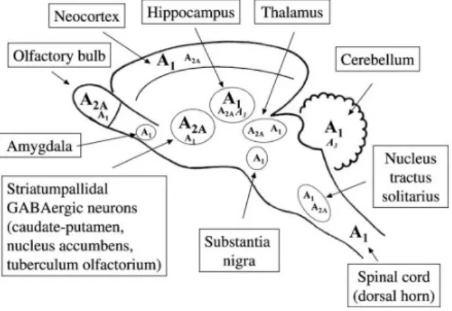
The adenosine A1 receptor is highly expressed in brain cortex, cerebellum, hippocampus
and dorsal horn of the spinal cord [35] (Figure 1.4). Most mammalian cell types are equipped with inhibitory adenosine A1 receptors (A1Rs). These receptors are the most abundant and
release and hyperpolarize neurons [12]. A1Rs are more abundant in neurons, in synapses, but
they are also present in astrocytes, microglia and oligodendrocytes although with much lower density [13].
Figure 1.4: Schematic representation o the distribution of adenosine receptors (A1, A2Aand A3) in the
main regions of the central nervous system. High levels of expression are indicated by bigger alphabets [12].
The adenosine A2Areceptor (A2AR) is highly expressed in the striatum and olfactory bulb
whereas in the neocortex and hippocampus they are present at very low levels [35] (Figure 1.4). Cortical A2AR are predominantly located in synapses, in particular in the presynaptic active
zone which is in agreement with the role of these receptors in controlling the neurotransmitter release [12]. These receptors have been shown to facilitate the release of most neurotransmitter types (glutamate, GABA) [13].
Previous studies found compelling evidence of cortical and hippocampal upsurge of A2AR
expression associated to cognitive deficits [36]. A2AR play an important role in both aging and
AD, its expression is increased with age and in the brain of AD patients [37]. Blockade of this receptors in experimental models mimicking aging or AD prevents or even reverts hippocampal-related impairments such as synaptic defects, memory deficits, amyloid burden and tau phos-phorylation [37]. Furthermore, previous studies show that A2AR deletion, in a mouse model of
AD, is sufficient to prevent memory defects, hippocampal plasticity impairments as well as Tau hyperphosphorylation [16, 38].
The methylxanthine caffeine is the world’s most popular psychoactive drug being able to in-teract with neurotransmission in different regions of the brain, promoting behavioural functions such as vigilance, attention as well as improvement of cognition [2]. Caffeine is a non-selective adenosine A2AR antagonist [16]. Chronic treatment with caffeine in AD transgenic mice models
improves memory and mitigates accumulation of amyloid peptides and hyperphosphorylated tau proteins, the two neuropathological hallmarks of this disorder [39, 16]. Epidemiological data support that caffeine consumption is able to delay dementia onset, reduce AD risk and slow down cognitive decline in elderly [16, 2].
1.2.1 Memory and Synaptic Plasticity
Hippocampus is widely regarded as being in the center of a brain network supporting encod-ing, consolidation and retrieval of memory [17]. Being central to the study of human memory, the hippocampus has been implicated in episodic and semantic long-term memory, novelty de-tection, sleep-dependent memory consolidation, pattern discrimination, spatial navigation and the binding of temporally and spatially distributed representations. Beyond these cognitive functions, the hippocampus is also involved in the regulation of the emotion, fear, anxiety and stress [18].
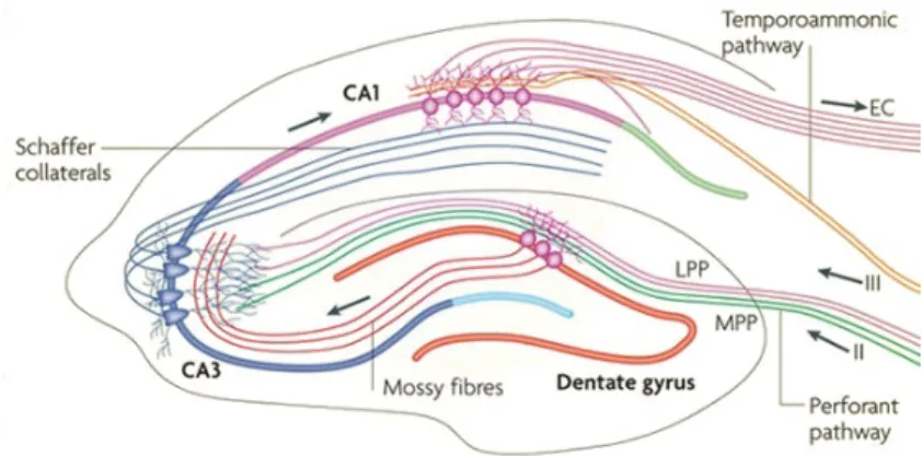
The hippocampus is a three-layered allocortical structure that is reciprocally connected to other cortical and subcortical areas. The principal neurons of the hippocampus are organized in layers and receive unidirectional input. The pathway from the DG to CA3 via mossy fibers and onward to CA1 via Schaffer collaterals is the principal feed-forward circuit involved in the processing of information through the hippocampus. The regions CA1–CA3 are separated into three layers (stratum oriens, stratum pyramidal, stratum radiatum) [17] (Figure 1.5). Learning and memory processes within hippocampal circuits are regulated by synaptic plasticity mecha-nisms that require activation of specific molecular cascades [18].
Figure 1.5: The hippocampal circuitry. The traditional excitatory pathway is depicted by solid arrows. The axons of layer II neurons in the entorhinal cortex project to the dentate gyrus through the perforant pathway. The dentate gyrus sends projections to the pyramidal cells in CA3 through mossy fibres. CA3 pyramidal neurons relay the information to CA1 pyramidal neurons through Schaffer collaterals. CA1 pyramidal neurons send back-projections into deep-layer neurons of the entorhinal cortex (EC) [17].
The hippocampus has long been considered as a classic example for the study of functional neuroplasticity as many mechanisms of synaptic plasticity such as long-term potentiation (LTP) and long-term depression (LTD) have been observed in hippocampal circuits and are thought to be fundamental to learning and memory [18].
Long-term potentiation (LTP) corresponds to an increase in synaptic transmission, whereas lasting decrease in synaptic transmission is referred to as long-term depression (LTD). Both are considered as mechanisms for memory storage at cellular level and can be artificially induced by certain protocols of electrical stimulation [18, 19]. LTP is a prime tool for the detection
of synaptic deficits in AD mouse models because it represents a sensitive indicator for early-onset pathology [19]. Impairments on LTP in brain regions such as the hippocampus were described to precede neurodegenerative changes that are typical for clinical AD [17]. LTD is a complementary mechanism for memory storage and is likely to be affected by the progression of neurodegenerative diseases, both in humans and in mice models [19].
1.3
Aim
This study aimed to compare the impact of global knock out of A2AR with pharmacological
2
Methodology
2.1
Animal Models
APP/PS1APP/PS1 mice contain human transgenes for both APP bearing the Swedish mutation and PSEN1 containing deletion of exon 9, both under the control of the prion promoter. Amyloid plaque deposition starts at approximately six weeks of age in the neocortex [40]. Deposits appear in the hippocampus at about three to four months, and in the striatum, thalamus, and brainstem at four to five months. A publication from Radde group characterizing these mice reported that they exhibited impaired learning on a spatial maze task at eight months of age [40]. Others have subsequently reported earlier observations of cognitive impairment, including deficits in the Morris Water maze at seven months of age [41].
For this work, we studied the impact of Knocking out A2A receptor in a slow-progressing
AD model, the APPsw/PS1d9 strain [(B6C3-Tg(APPswe,PSEN1δE9)85Dbo/J [42, 9])]. The 4 groups of mice with 11-12 months old used were: wild type (WT/A2AR +/+), wild type
with knockout of A2Areceptor (WT/A2AR -/-), transgenic mice APP/PS1 A2AR +/+; transgenic
mice APP/PS1 A2AR KO (-/-). The triple transgenic mice APP/PS1 A2AR KO were obtained
by crossing double transgenic model APP/PS1dE9 with A2AR heterozygous mice as explained
on Figure 2.1 [43].
Figure 2.1: Triple transgenic mice APP/PS1 A2AR KO breeding generation (courtesy of David Blum,
THY-Tau22
7-month-old THY-Tau22 mice were also studied to evaluate synaptic plasticity. These mice are a model for tau aggregation, a pathological hallmark of AD as well as numerous tauopathies. In these mice levels of human tau increase with age [10]. These mice develop a variety of tau-related neuropathological changes, including tau hyperphosphorylation, neurofibrillary-like tau inclusions, and ghost tangles. Tau pathology is generally mild at three to four months of age, moderate at six to seven months, and extensive at nine months [44]. Behaviorally, these mice display learning and memory deficits [10].
The neuropathology and behavioral changes coincide with changes in hippocampal synap-tic plassynap-ticity. This mouse model (THY-Tau22) expresses human 4-repeat tau mutated at sites G272V and P301S under a Thy1.2-promotor, displaying tau pathology in the absence of any motor dysfunction that may interfere with behavioral testing [10].
2.2
Electrophysiology recordings
In order to evaluate changes in synaptic plasticity and how is it correlated to memory changes we performed electrophysiology and monitored extracellular field recordings in hip-pocampal slices, recording field Excitatory Postsynaptic Potentials (fEPSP) from Schaffer fibers-CA1 dendrites. Long-term potentiation (LTP) and long-term depression (LTD) induction proto-cols evaluated changes in synaptic plasticity, the input/output curves assessed changes in basal synaptic transmission. [45].
Concerning APP/PS1, we used two protocols for long-term potentiation that were previous described by literature: High frequency Stimulus and Theta burst stimulus [7]; and one protocol for long-term depression: Low frequency stimulus. Previous studies show that these mice have impaired LTP at 8 and 15 months of age [46]. Regarding THY-Tau22 mice, we used a protocol to study LTD changes. Previous studies show that LTP appears to be intact and that at nine to ten months these mice exhibit impaired LTD compared with wild-type [44].
2.2.1 Hippocampal slice preparation
The preparation of hippocampal slice for electrophysiology studies started by sacrificing the animal by cervical dislocation followed by the extraction of the brain and dissection of the two hippocampi in ice-cold Krebs solution, which is composed of (mM): NaCl 124; KCl 3; NaH2PO41.25; NaHCO326; MgSO41; CaCl22 and D-glucose 10, previously gassed with 95%
O2 and 5% CO2, pH 7.4. This solution mimics osmolarity, oxygenation and ionic composition of the in vivo cerebrospinal fluid. Transverse hippocampal slices (400 µm) were obtained with a McIIwain tissue shopper and kept in a resting chamber in Krebs solution, oxygenated, for 1 hour to ensure the energetic and functional recovery of the tissue [47].
2.2.2 Extracellular recordings
After this period, the slices were carefully deposited in the recording chamber where they were continually superfused with Krebs solution at a constant flow (3ml/min) and temperature (32oC). SCH-58261 was prepared from a stock solution (5mM) and dissolved in 500mL of krebs solution to a final concentration of 50nM. The slice was in the chamber with SCH-58261 for 30 minutes prior to induction of the LTD protocol. The recording and stimulation electrodes were placed under visual guidance of magnifying lens as illustrated by figure 2.2. The recording electrode was positioned in stratum radiatum of CA1 area. Two stimulation electrodes were used, one was positioned in stratum radiatum of CA2 area and the other was positioned in stratum radiatum of CA1 area (Figure 2.2). Responses were recorded through an amplifier coupled to the computer, using WinLTP as data acquisition software [48].
Figure 2.2: Schematic drawing of hippocampal slices. Schaffer collaterals projecting from CA3 to CA1 were stimulated by two stimulation electrodes that were positioned in the stratum radiatum. For field potential recordings, the pipette was positioned in stratum radiatum of the CA1 region of the hip-pocampus [49].
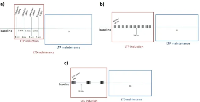
Field excitatory postsynaptic potentials (fEPSPs) were recorded in stratum radiatum of the CA1 area. Two protocols for long term potentiation (LTP) were used: High Frequency Stim-ulus (HFS) (4 trains of 100 Hz, 1s, 5-minutes interval, induced at 0.5mV/ms; applied to the Schaffer collaterals at the test stimulus intensity (50% half-maximal fEPSP)) (Figure 2.3 a)) and Theta Burst Stimulus (TBS) (10 trains of 4 pulses, 100Hz, induced at 0.5mV/ms; applied to the Schaffer collaterals at the test stimulus intensity (50% half-maximal fEPSP)) (Figure 2.3 b)). For long term depression (LTD) the protocol used consisted in a low frequency stimulation (LFS) paradigm (3 trains of 1200 pulses, 2Hz, 10 minutes interval) (Figure 2.3 c)). Finally, to evaluate alterations in basal transmission we used Input/Output curve protocol (I/O) that con-sisted in fEPSP evoked by increasing stimulation intensities between 80-360 µA.
Figure 2.3: Representative schemes of the induction protocols used: a) HFS protocol characterized by 4 trains of 100Hz; b) TBS protocol characterized by 10 trains of 4 pulses 100Hz; c) LFS protocol characterized by 3 trains of 1200 pulses 2Hz.
In control conditions we can distinguish two main components in the evoked potentials recorded extracellularly form CA1 area: the presynaptic volley (number 1, Figure 2.4) and the field synaptic potential (number 2, Figure 2.4). In order to access changes in LTP or LTD we measure the slope of the waveform (blue line from Figure 2.4). Slope is calculated by taking all the waveform points of the slope and calculating a linear regression line through these points. The peak amplitude (red line from 2.4) corresponds to the difference between the prestimulus baseline value and the calculated peak [50].
Figure 2.4: Representative fEPSP recorded from CA1 area. 1) presynaptic volley; 2) field synaptic potential; 3) slope of the fEPSP; 4) peak amplitude.
2.2.3 Statistical analysis
The data were collected on a computer using winLTP software and all statistical analyses were performed after using GraphPad Prism version 5.00 for Windows, GraphPad Software, San Diego California USA, www.graphpad.com. Statistical comparisons included F test (one-way analysis of variance used to assess weather the expected values differ from each other ), two-sided unpaired t test (used to compare the mean of two independent groups), one-way ANOVA (used to compare means of two or more samples) and two-way ANOVA (used to compare two independent variables) followed by Bonferroni’s multiple comparisons post hoc test.
3
Results
3.1
APP/PS1 mice
Previous studies showed that AD mice models are characterized by an upsurge of A2AR in
the hippocampus that is associated with cognitive and synaptic plasticity deficits [36]. In order to assess changes in synaptic plasticity in APP/PS1 transgenic mice and study the impact of deleting the A2AR in the hippocampus we used different types of induction protocols: two to
induce long-term potentiation - HFS and TBS - and one to induce long-term depression - LFS. We also assessed changes in basal transmission by recording Input/Output curves.
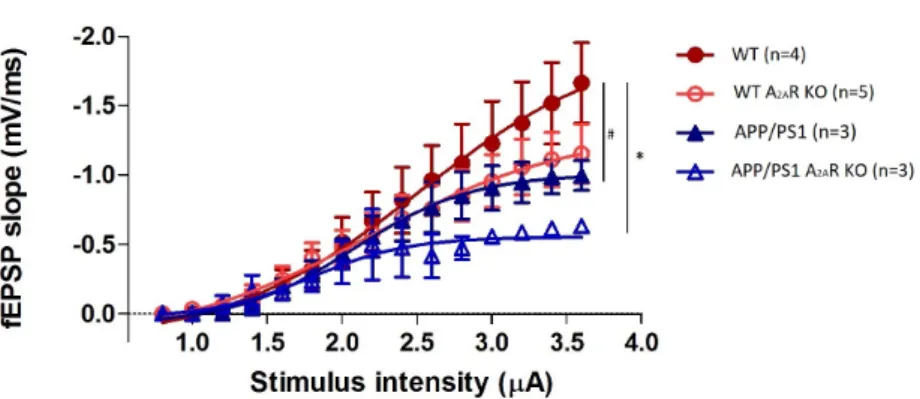
3.1.1 Input/Output curve
Firstly, in order to assess differences in basal transmission we performed Input/Output curves where we evoked fEPSP by increasing stimulation intensities between 80-360µA (Fig-ure 3.1). We can observe a change on basal transmission on APP/PS1 transgenic mice when compared to WT. This is represented by a shift to the right of the I/O curve in APP/PS1 mice, meaning that, for the same stimulus, these mice respond with a smaller fEPSP than WT mice. Deletion of A2A receptor resulted in a smaller fEPSP slope for the same stimulus intensity in
both WT A2AR KO and APP/PS1 A2AR KO when compared to their respective controls WT
and APP/PS1.
Figure 3.1: Input/Output curve (I/O). fEPSP slope evoked by different stimulation intensities (#p<0.05, APP/PS1 comparing to WT ; *p<0.05, APP/PS1 A2AR KO comparing to WT; F test. All values are mean
3.1.2 Long Term Potentiation - LTP
HFS (High-frequency stimulation)
Using high frequency stimulus, we compared potentiation of the different genotypes used. As we can see on Figure 3.2, after a stable baseline, HFS stimulation produced a long-lasting potentiation (at least 1h). We can observe that there are no significant differences between WT and APP/PS1 transgenic mice using this particular protocol (Figure 3.2 a) ). Furthermore, A2AR
deletion does not have an impairment on fEPSP slope percentage in APP/PS1 mice (Figure 3.2 b) ).
LTP magnitudes of genotypes are represented on Figure 3.2 c). We may observe no signifi-cant differences on percentage of change of LTP magnitude between genotypes.
TBS (Theta-burst stimulation)
We next tested whether using a weaker protocol that more closely mimics physiological theta-wave frequency could unmask LTP differences in these animals, since it is described that HFS can lead to LTP saturation [51]. For that, we used a different protocol, theta-burst stimu-lation. We found that APP/PS1 transgenic mice show a tendency for a diminished potentiation when compared to WT mice (Figure 3.3). When we compared APP/PS1 with APP/PS1 AA2R
KO, we can observe a lower potentiation when A2AR was deleted (Figure 3.3 b) ).
If we compare all genotypes, we observed that the percentage change was LTP magnitude is lower when A2AR is deleted, both in WT and APP/PS1 transgenic mice (Figure 3.3 c) ).
a)
b)
c)
Figure 3.2: LTP induced by HFS. Changes in fEPSP slope induced by high-frequency stimulation recorded from: a) WT (n=4) and APP/PS1 transgenic mice hippocampal slices (n=5). b) APP/PS1 (n=5) and APP/PS1 A2AR KO (n=3) hippocampal slices. Right panels are representative tracings prior (1)
and 1h after (2) LTP induction in each condition. c) LTP magnitude percentage, comparison between genotypes (p>0.05, two-way ANOVA followed by Bonferroni’s multiple comparison test). All values are mean ± SEM.
b) a)
c)
Figure 3.3: LTP induced by TBS. Changes in fEPSP slope induced by theta-burst stimulation recorded from: a) WT (n=4) and APP/PS1 transgenic mice hippocampal slices (n=4). b) APP/PS1 (n=4) and APP/PS1 A2AR KO (n=3) hippocampal slices. Right panels are representative tracings prior (1) and 1h
after (2) LTP induction in each condition. c) LTP magnitude percentage, comparison between genotypes (*p<0.05, APP/PS1 comparing to WT, unpaired t test; p>0.05, two-way ANOVA followed by Bonfer-roni’s multiple comparison test). All values are mean ± SEM.
3.1.3 Long Term Depression - LTD
We studied LTD using low frequency stimulus (LFS) protocol in order to assess changes in synaptic plasticity on these transgenic mice. APP/PS1 mice showed an impaired LTD (Figure 3.4) that resulted in potentiation under a LFS protocol instead of depression.
Figure 3.4 b) depicts the effect of deletion of A2AR in potentiation in transgenic mice
un-der LFS protocol. We can observe that when A2AR was deleted on APP/PS1 mice model, the
potentiation observed was abolish, but LTD was not restored. However, when we applied the se-lective A2AR antagonist, SCH-58261 (100nM) to the slices of APP/PS1 mice, LTD impairment
observed was fully rescued and we obtained a similar percentage of change of fEPSP slope of that found in WT (Figure 3.4 c)).
The percentage of change of LTD magnitude between genotypes is represented on Figure 3.4 d).
a)
b) c)
d)
Figure 3.4: LTD induced by LFS. Changes in fEPSP slope induced by low-frequency stimulation recorded from: a) WT (n=4) and APP/PS1 transgenic mice hippocampal slices (n=4). Right panels are representative tracings prior (1) and 1h after (2) LTD induction. b) APP/PS1 (n=4) and APP/PS1 A2AR
KO (n=4) hippocampal slices. c) APP/PS1 (n=4) and APP/PS1 with SCH 100nM (n=3). Above panels are representative tracings prior (1) and 1h after (2) LTD induction in each condition. d) LTD magnitude percentage, comparison between genotypes (*p<0.05, APP/PS1 comparing to WT; *#p<0.05, APP/PS1 A2AR KO comparing to APP/PS1; #p<0.05, APP/PS1 SCH 100nM comparing to APP/PS1; one-way
ANOVA followed by Bonferroni’s multiple comparison test). All values are mean ± SEM. (experiments made with Mariana Temido)
3.2
THY-Tau22 mice
3.2.1 Long Term Depression - LTD
Regarding THY-Tau22 transgenic mice, in order to observe changes on synaptic plasticity, we performed LFS induction protocol. As we can see on Figure 3.5, transgenic mice show a LTD impairment, by having a potentiation under a low frequency stimulus, consistent with the results observed in APP/PS1 mice.
Following, we compared THY-Tau22 transgenic mice before and after we superfused SCH-58261. As we can see on Figure 3.5 b), SCH-58261 application to THY-Tau22 mice slices abolished the potentiation observed in THY-Tau22 mice, but did not fully restored LTD.
Finally, Figure 3.5 c) depicts all genotypes, WT and THY-Tau22 mice, both with and with-out superfusion of A2AR antagonist, SCH-58261. We can observe once again that transgenic
mice presents an impairment on LTD magnitude and that this impairment was rescued by SCH-58261, being the percentage of change of LTD magnitude of WT and THY-Tau22+SCH 50nM similar.
b) 1 1 2 2 a) * ** * ** WT SCH 50nM WT THY-Tau22 THY-Tau22 SCH 50nM c)
Figure 3.5: LTD induced by LFS. Changes in fEPSP slope induced by low-frequency stimulation recorded from: a) WT (n=3) and THY-Tau22 transgenic mice hippocampal slices (n=2). b) THY-Tau22 (n=2) and THY-Tau22 with SCH 50nM (n=3) hippocampal slices. Right panels are representative trac-ings prior (1) and 1h after (2) LTD induction in each condition. c) LTD magnitude percentage, com-parison between genotypes (*p<0.05, THY-Tau22 comparing to WT;**p<0.05, THY-Tau22 SCH 50nM comparing to THY-Tau22; two-way ANOVA followed by Bonferroni’s multiple comparison test). All values are mean ± SEM.
4
Discussion
4.1
APP/PS1
Here we show that global deletion of A2AR in APP/PS1 mice model of AD affects basal
transmission and synaptic plasticity in CA1 area synapses. With aging these mice exhibit Aβ accumulation and plaques formation (observed within 5 months), but do not display neurophib-rillary tangles [7]. Furthermore, at 8 and 15 months of age, these mice exhibit an impaired LTP under high frequency stimulation protocol (1 train of 100 stimuli, 200 Hz) [46].
In order to assess changes in basal transmission we performed I/O curves (Figure 3.1). We observed that APP/PS1 transgenic mice showed an impaired basal transmission since under the same intensity of stimulus these mice exhibit a lower slope of fEPSP than WT mice. When we compared APP/PS1 transgenic mice with APP/PS1 A2AR KO we saw that the deletion of A2AR
did not rescue this impairment observed.
Moreover, we described that LTP induced by different protocols with different frequencies of stimulation resulted in different responses. We show here that LTP induced by HFS was not sensitive and we could not see differences between genotypes (Figure 3.2). However, when LTP was induced by TBS (Figure 3.3), we could observe differences in percentage of change of fEPSP between genotypes. We saw that under this particular stimulus, APP/PS1 transgenic mice showed a diminished potentiation when compared to WT. Finally, we also observed that the percentage of change in LTP magnitude was lower when A2AR was deleted, both in WT
and APP/PS1. This disparity between responses under the two different protocols may happen due to the type of frequency of the stimulus. HFS stimulus albeit being effective, it does not resemble physiological patterns of activity [52]. On the other hand, TBS protocol that has a lower frequency mimics endogenous theta frequency activity and approximate physiological pattern of excitability [51]. Hence, this weaker LTP induction protocol prevent saturation and thus allowed to detect impairments on LTP magnitude in these mice.
Our results are consistent with previous studies that show that APP/PS1 mice have amyloid deposition that results in LTP impairment and consequently memory deficits [38]. Aβ has an essential role at the synapse and in synaptic structure-functional plasticity critical to learning and memory [53]. Other studies reported that LTP in APP/PS1 mice model was abolished in CA3 pyramidal cells area and that acute pharmacological inhibition of A2AR restored CA3 LTP
[38]. Our results are consistent with these previous findings in the way that showed an impaired LTP on APP/PS1 transgenic mice, however in CA1 area synapses.
Hence, we may postulate that these impairments observed in synaptic plasticity assessed by I/O curves and LTP are related to Aβ deposits, since a previous study showed that in animals models of AD there was a clear correlation between the higher concentration of Aβ and synaptic plasticity dysfunctions [53]. When we studied transgenic mice with global deletion of A2AR
we did not observed a rescue. This may happened since A2AR was deleted and so A2AR-A1R
cross-talk was loss. Hence, adenosine may activate A1R reducing neurotransmitter release at
LTD has been largely neglected when analyzing hippocampal function in AD mice model [7]. Only two studies have so far been reported concerning LTD analysis [55, 56]. Chang group analyzed LTD on an APP/PS1 mice model with a different mutation (APPNhL/PS1P246L). They reported, using LFS protocol (900 pulses, 1Hz), an impairment on LTD expression in CA1 area in these transgenic mice [55].
Here we described that APP/PS1 mice present deficits in synaptic plasticity leading to a LTD-to-LTP shift. As we can observe on Figure 3.4 a), APP/PS1 mice shown potentiation un-der a LFS instead of a depression. APP/PS1 mice with global deletion of A2AR reverse the
potentiation shown on APP/PS1 mice (Figure 3.4 b) ), however, it did not fully rescued LTD. When hippocampal slices of APP/PS1 transgenic mice were superfused with a specific antago-nist of A2AR, SCH-58261 (100nM), LTD was fully rescued and normalized to WT magnitude
(Figure 3.4 c) ).
With these evidences, we may see that the blockade of A2AR with a specific antagonist is
more effective than the global deletion. This may have happened due to the fact that triple transgenic mice (APP/PS1 A2AR KO) may have established compensatory mechanisms during
aging.
Altogether, APP/PS1 transgenic mice presents LTP and LTD impairments, which are corre-lated with synaptic plasticity and memory changes observed in AD. However, deletion of A2AR
did not reverse LTP impairment under TBS protocol. Finally, LTD impairments observed in APP/PS1 were fully rescued using an acute pharmacological antagonist of A2AR.
4.2
THY-Tau22
Here we reveal that the LTD-to-LTP shift observed in APP/PS1 mice model also occurs in the hippocampus of THY-Tau22 transgenic mice. These mice are a model for tau aggregation and with age the levels of tau protein increases and they start to develop neuropathological changes including tau hyperphosphorylation [10]. Furthermore, these mice exhibit impaired maintenance of LTD compared to WT at 9-10 month old but normal LTP [20, 10].
Previous studies have shown learning and memory deficits and changes in synaptic plasticity in the hippocampus in THY-Tau22 mice that are consistent with the learning impairments seen in AD patients [57]. Another study reported that THY-Tau22 transgenic mice have an LTD impairment which was rescued by genetic deletion of A2AR [16]. Our findings are consistent
with this work in the way that, as we can observe on Figure 3.5 a), THY-Tau22 transgenic mice show LTD-to-LTP shift since they exhibit a potentiation under a LFS stimulus. When we superfused a THY-Tau22 hippocampal mouse slice with SCH-58261, a selective antagonist of A2AR, we observed a complete rescue of the LTD impairment (Figure 3.5). We found no
significant differences between LTD magnitude of WT and THY-Tau22 transgenic mice with SCH-58261.
In sum, we know by previous studies that A2AR expression in the hippocampus of AD mice
model is increased [38]. In this work, we described that upon A2AR blockade using a specific
antagonist occurs a complete rescue of LTD impairments in both AD models. Hence, we can postulate that A2AR is contributing for the synaptic plasticity deficits, namely LTD, underlying
in the progression of Alzheimer’s disease.
The fact that A2AR effects in CA3-CA1 synapse the hippocampus are more significant in
References
[1] Sara N Burke and Carol A Barnes. Neural plasticity in the ageing brain. Nature Reviews Neuroscience, 7(1):30–40, 2006.
[2] Vanessa Flaten, Cyril Laurent, Joana E Coelho, Ursula Sandau, Vânia L Batalha, Sylvie Burnouf, Malika Hamdane, Sandrine Humez, Detlev Boison, Luísa V Lopes, et al. From epidemiology to pathophysiology: what about caffeine in alzheimer’s disease?, 2014.
[3] Yang Hong-Qi, Sun Zhi-Kun, and Chen Sheng-Di. Current advances in the treatment of alzheimer’s disease: focused on considerations targeting aβ and tau. Translational neurodegeneration, 1(1):21, 2012.
[4] Amantha Thathiah and Bart De Strooper. The role of g protein-coupled receptors in the pathology of
alzheimer’s disease. Nature Reviews Neuroscience, 12(2):73–87, 2011.
[5] John Hardy and Dennis J Selkoe. The amyloid hypothesis of alzheimer’s disease: progress and problems on the road to therapeutics. science, 297(5580):353–356, 2002.
[6] Valérie Vingtdeux, Nicolas Sergeant, and Luc Buée. Potential contribution of exosomes to the prion-like propagation of lesions in alzheimer’s disease. Frontiers in physiology, 3:229, 2012.
[7] Cristina Marchetti and Hélène Marie. Hippocampal synaptic plasticity in alzheimer’s disease: what have we learned so far from transgenic models? Reviews in the neurosciences, 22(4):373–402, 2011.
[8] Braydon L Burgess, Sean A McIsaac, Kathryn E Naus, Jeniffer Y Chan, Gavin HK Tansley, Jing Yang, Fudan Miao, Colin JD Ross, Miranda van Eck, Michael R Hayden, et al. Elevated plasma triglyceride levels precede amyloid deposition in alzheimer’s disease mouse models with abundant aβ in plasma. Neurobiology of disease, 24(1):114–127, 2006.
[9] Joanna L Jankowsky, Hilda H Slunt, Victoria Gonzales, Nancy A Jenkins, Neal G Copeland, and David R Borchelt. App processing and amyloid deposition in mice haplo-insufficient for presenilin 1. Neurobiology of aging, 25(7):885–892, 2004.
[10] Katharina Schindowski, Alexis Bretteville, Karelle Leroy, Séverine Bégard, Jean-Pierre Brion, Malika Ham-dane, and Luc Buée. Alzheimer’s disease-like tau neuropathology leads to memory deficits and loss of functional synapses in a novel mutated tau transgenic mouse without any motor deficits. The American journal of pathology, 169(2):599–616, 2006.
[11] Malika El Yacoubi, Catherine Ledent, Marc Parmentier, Rosalia Bertorelli, Ennio Ongini, Jean Costentin, and Jean-Marie Vaugeois. Adenosine a2a receptor antagonists are potential antidepressants: evidence based on pharmacology and a2a receptor knockout mice. British journal of pharmacology, 134(1):68–77, 2001. [12] JA Ribeiro, AM Sebastiao, and A De Mendonca. Adenosine receptors in the nervous system:
pathophysio-logical implications. Progress in neurobiology, 68(6):377–392, 2002.
[13] Rodrigo A Cunha. Neuroprotection by adenosine in the brain: from a1 receptor activation to a2a receptor blockade. Purinergic signalling, 1(2):111–134, 2005.
[14] Rodrigo A Cunha, M Dolores Constantino, Ana M Sebastião, and J Alexandre Ribeiro. Modification of ai and a2a adenosine receptor binding in aged striatum, hippocampus and cortex of the rat. Neuroreport, 6(11):1583, 1995.
[15] Paula M Canas, Lisiane O Porciúncula, Geanne MA Cunha, Carla G Silva, Nuno J Machado, Jorge MA Oliveira, Catarina R Oliveira, and Rodrigo A Cunha. Adenosine a2a receptor blockade prevents synapto-toxicity and memory dysfunction caused by β-amyloid peptides via p38 mitogen-activated protein kinase pathway. Journal of Neuroscience, 29(47):14741–14751, 2009.
[16] C Laurent, S Burnouf, B Ferry, VL Batalha, JE Coelho, Y Baqi, E Malik, E Mariciniak, S Parrot, Anneke Van der Jeugd, et al. A2a adenosine receptor deletion is protective in a mouse model of tauopathy. Molecular psychiatry, 21(1):97–107, 2016.
[17] Wei Deng, James B Aimone, and Fred H Gage. New neurons and new memories: how does adult hippocam-pal neurogenesis affect learning and memory? Nature reviews. Neuroscience, 11(5):339, 2010.
[18] T Bartsch and P Wulff. The hippocampus in aging and disease: from plasticity to vulnerability, 2015. [19] Tariq Ahmed, David Blum, Sylvie Burnouf, Dominique Demeyer, Valérie Buée-Scherrer, Rudi D’Hooge,
Luc Buée, and Detlef Balschun. Rescue of impaired late–phase long-term depression in a tau transgenic mouse model. Neurobiology of aging, 36(2):730–739, 2015.
[20] Heather D VanGuilder, Julie A Farley, Han Yan, Colleen A Van Kirk, Matthew Mitschelen, William E Son-ntag, and Willard M Freeman. Hippocampal dysregulation of synaptic plasticity-associated proteins with age-related cognitive decline. Neurobiology of disease, 43(1):201–212, 2011.
[21] Daniel E Feldman. Synaptic mechanisms for plasticity in neocortex. Annual review of neuroscience, 32:33– 55, 2009.
[22] Karine Bertoldi, Laura Reck Cechinel, Bruna Schallenberger, Carla Basso, Gisele Agustini Lovatel, Lisiane Bernardi, Marcelo Lazzaron Lamers, and Ionara Rodrigues Siqueira. Aging process alters hippocampal and cortical secretase activities of wistar rats. Behavioural brain research, 317:374–381, 2017.
[23] N Pescosolido, A Pascarella, G Buomprisco, and D Rusciano. Critical review on the relationship between glaucoma and alzheimer’s disease. Adv Ophthalmol Vis Syst, 1(4):00024, 2014.
[24] Vivian W Chow, Mark P Mattson, Philip C Wong, and Marc Gleichmann. An overview of app processing enzymes and products. Neuromolecular medicine, 12(1):1–12, 2010.
[25] Sandeep Kumar Singh, Saurabh Srivastav, Amarish Kumar Yadav, Saripella Srikrishna, and George Perry. Overview of alzheimer’s disease and some therapeutic approaches targeting aβ by using several synthetic and herbal compounds. Oxidative medicine and cellular longevity, 2016, 2015.
[26] J Carter and CF Lippa. β amyloid, neuronal death and alzheimer’s disease. Current molecular medicine, 1(6):733–737, 2001.
[27] Sam Gandy. The role of cerebral amyloid β accumulation in common forms of alzheimer disease. Journal of Clinical Investigation, 115(5):1121, 2005.
[28] Goran Šimi´c, Mirjana Babi´c Leko, Selina Wray, Charles Harrington, Ivana Delalle, Nataša Jovanov-Miloševi´c, Danira Bažadona, Luc Buée, Rohan De Silva, Giuseppe Di Giovanni, et al. Tau protein hyper-phosphorylation and aggregation in alzheimer’s disease and other tauopathies, and possible neuroprotective strategies. Biomolecules, 6(1):6, 2016.
[29] C-X Gong and K Iqbal. Hyperphosphorylation of microtubule-associated protein tau: a promising therapeutic target for alzheimer disease. Current medicinal chemistry, 15(23):2321–2328, 2008.
[30] Adrian C Lo, Emilia Iscru, David Blum, Ina Tesseur, Zsuzsanna Callaerts-Vegh, Luc Buée, Bart De Strooper, Detlef Balschun, and Rudi D’Hooge. Amyloid and tau neuropathology differentially affect prefrontal synap-tic plassynap-ticity and cognitive performance in mouse models of alzheimer’s disease. Journal of Alzheimer’s Disease, 37(1):109–125, 2013.
[31] Kurt R Brunden, John Q Trojanowski, and Virginia M-Y Lee. Advances in tau-focused drug discovery for alzheimer’s disease and related tauopathies. Nature reviews. Drug discovery, 8(10):783, 2009.
[32] Arturas Volianskis, Rasmus Køstner, Morten Mølgaard, Susanne Hass, and Morten S Jensen. Episodic memory deficits are not related to altered glutamatergic synaptic transmission and plasticity in the ca1 hip-pocampus of the appswe/ps1δe9-deleted transgenic mice model of β-amyloidosis. Neurobiology of aging, 31(7):1173–1187, 2010.
[33] Hui-li Liu, Gang Zhao, Kui Cai, Hai-hua Zhao, et al. Treadmill exercise prevents decline in spatial learn-ing and memory in app/ps1 transgenic mice through improvement of hippocampal long-term potentiation. Behavioural brain research, 218(2):308–314, 2011.
[34] Alberto Serrano-Pozo, Matthew P Frosch, Eliezer Masliah, and Bradley T Hyman. Neuropathological alter-ations in alzheimer disease. Cold Spring Harbor perspectives in medicine, 1(1):a006189, 2011.
[35] JA Ribeiro and AM Sebastiao. Modulation and metamodulation of synapses by adenosine. Acta physiologica, 199(2):161–169, 2010.
[36] Luísa V Lopes, Rodrigo A Cunha, and JA Ribeiro. Cross talk between a1 and a2a adenosine receptors in the hippocampus and cortex of young adult and old rats. Journal of Neurophysiology, 82(6):3196–3203, 1999. [37] VL Batalha, José M Pêgo, BM Fontinha, AR Costenla, JS Valadas, Y Baqi, H Radjainia, CE Müller, AM
Se-bastião, and LV Lopes. Adenosine a2a receptor blockade reverts hippocampal stress-induced deficits and restores corticosterone circadian oscillation. Molecular psychiatry, 18(3):320–331, 2013.
[38] Silvia Viana Da Silva, Matthias Georg Haberl, Pei Zhang, Philipp Bethge, Cristina Lemos, Nélio Gonçalves, Adam Gorlewicz, Meryl Malezieux, Francisco Q Gonçalves, Noëlle Grosjean, et al. Early synaptic deficits in the app/ps1 mouse model of alzheimer/’s disease involve neuronal adenosine a2a receptors. Nature com-munications, 7, 2016.
[39] GW Arendash, W Schleif, K Rezai-Zadeh, EK Jackson, LC Zacharia, JR Cracchiolo, D Shippy, and J Tan. Caffeine protects alzheimer’s mice against cognitive impairment and reduces brain β-amyloid production. Neuroscience, 142(4):941–952, 2006.
[40] Rebecca Radde, Tristan Bolmont, Stephan A Kaeser, Janaky Coomaraswamy, Dennis Lindau, Lars Stoltze, Michael E Calhoun, Fabienne Jäggi, Hartwig Wolburg, Simon Gengler, et al. Aβ42-driven cerebral amyloi-dosis in transgenic mice reveals early and robust pathology. EMBO reports, 7(9):940–946, 2006.
[41] Lutgarde Serneels, Jérôme Van Biervliet, Katleen Craessaerts, Tim Dejaegere, Katrien Horré, Tine Van Houtvin, Hermann Esselmann, Sabine Paul, Martin K Schäfer, Oksana Berezovska, et al. γ-secretase heterogeneity in the aph1 subunit: Relevance for alzheimer’s disease. Science, 324(5927):639–642, 2009. [42] Joanna L Jankowsky, Hilda H Slunt, Tamara Ratovitski, Nancy A Jenkins, Neal G Copeland, and David R
Borchelt. Co-expression of multiple transgenes in mouse cns: a comparison of strategies. Biomolecular engineering, 17(6):157–165, 2001.
[43] Catherine Ledent, Jean-Marie Vaugeois, Serge N Schiffmann, Thierry Pedrazzini, Malika El Yacoubi, Jean-Jacques Vanderhaeghen, Jean Costentin, John K Heath, Gilbert Vassart, and Marc Parmentier. Ag-gressiveness, hypoalgesia and high blood pressure in mice lacking the adenosine a2a receptor. Nature, 388(6643):674–678, 1997.
[44] Ann Van der Jeugd, Ben Vermaercke, Maxime Derisbourg, Adrian C Lo, Malika Hamdane, David Blum, Luc Buée, and Rudi D’Hooge. Progressive age-related cognitive decline in tau mice. Journal of Alzheimer’s Disease, 37(4):777–788, 2013.
[45] Maria J Diógenes, Ana R Costenla, Luísa V Lopes, André Jerónimo-Santos, Vasco C Sousa, Bruno M Fontinha, Joaquim A Ribeiro, and Ana M Sebastião. Enhancement of ltp in aged rats is dependent on endogenous bdnf. Neuropsychopharmacology, 36(9):1823–1836, 2011.
[46] Simon Gengler, Alison Hamilton, and Christian Hölscher. Synaptic plasticity in the hippocampus of a app/ps1 mouse model of alzheimer’s disease is impaired in old but not young mice. PLoS One, 5(3):e9764, 2010. [47] Bertil B Fredholm, Thomas V Dunwiddie, Birgit Bergman, Karin Lindstro, et al. Levels of adenosine and
adenine nucleotides in slices of rat hippocampus. Brain research, 295(1):127–136, 1984.
[48] William W Anderson and Graham L Collingridge. Capabilities of the winltp data acquisition program ex-tending beyond basic ltp experimental functions. Journal of neuroscience methods, 162(1):346–356, 2007. [49] Stephan Kratzer, Corinna Mattusch, Eberhard Kochs, Matthias Eder, Rainer Haseneder, and Gerhard
Rammes. Xenon attenuates hippocampal long-term potentiation by diminishing synaptic and extrasynaptic n-methyl-d-aspartate receptor currents. The Journal of the American Society of Anesthesiologists, 116(3):673– 682, 2012.
[50] William W Anderson and Graham L Collingridge. The ltp program: a data acquisition program for on-line analysis of long-term potentiation and other synaptic events. Journal of neuroscience methods, 108(1):71–83, 2001.
[51] Ruben V Hernandez, Mary M Navarro, Ward A Rodriguez, Joe L Martinez, and Richard G LeBaron. Dif-ferences in the magnitude of long-term potentiation produced by theta burst and high frequency stimulation protocols matched in stimulus number. Brain research protocols, 15(1):6–13, 2005.
[52] Lawrence M Grover, Eunyoung Kim, Jennifer D Cooke, and William R Holmes. Ltp in hippocampal area ca1 is induced by burst stimulation over a broad frequency range centered around delta. Learning & memory, 16(1):69–81, 2009.
[53] Mordhwaj S Parihar and Gregory J Brewer. Amyloid-β as a modulator of synaptic plasticity. Journal of Alzheimer’s Disease, 22(3):741–763, 2010.
[54] Hai-Ying Shen and Jiang-Fan Chen. Adenosine a2a receptors in psychopharmacology: modulators of behav-ior, mood and cognition. Current neuropharmacology, 7(3):195–206, 2009.
[55] Eric H Chang, Mary J Savage, Dorothy G Flood, Justin M Thomas, Robert B Levy, Veeravan Mahadom-rongkul, Tomoaki Shirao, Chiye Aoki, and Patricio T Huerta. Ampa receptor downscaling at the onset of alzheimer’s disease pathology in double knockin mice. Proceedings of the National Academy of Sciences of the United States of America, 103(9):3410–3415, 2006.
[56] Marcello D’amelio, Virve Cavallucci, Silvia Middei, Cristina Marchetti, Simone Pacioni, Alberto Ferri, Adamo Diamantini, Daniela De Zio, Paolo Carrara, Luca Battistini, et al. Caspase-3 triggers early synaptic dysfunction in a mouse model of alzheimer’s disease. Nature neuroscience, 14(1):69–76, 2011.
[57] Ann Van der Jeugd, David Blum, Sylvie Raison, Sabiha Eddarkaoui, Luc Buée, and Rudi D’Hooge. Ob-servations in thy-tau22 mice that resemble behavioral and psychological signs and symptoms of dementia. Behavioural brain research, 242:34–39, 2013.