Cinnamon Ameliorates Experimental Allergic
Encephalomyelitis in Mice via Regulatory T
Cells: Implications for Multiple Sclerosis
Therapy
Susanta Mondal, Kalipada Pahan*
Department of Neurological Sciences, Rush University Medical Center, Chicago, IL, United States of America
Abstract
Upregulation and/or maintenance of regulatory T cells (Tregs) during an autoimmune insult may have therapeutic efficacy in autoimmune diseases. Although several immunomodula-tory drugs and molecules are available, most present significant side effects over long-term use. Cinnamon is a commonly used natural spice and flavoring material used for centuries throughout the world. Here, we have explored a novel use of cinnamon powder in protecting Tregs and treating the disease process of experimental allergic encephalomyelitis (EAE), an animal model of MS. Oral feeding of cinnamon (Cinnamonum verum) powder sup-presses clinical symptoms of relapsing-remitting EAE in female PLP-TCR transgenic mice and adoptive transfer mouse model. Cinnamon also inhibited clinical symptoms of chronic EAE in male C57/BL6 mice. Dose-dependent study shows that cinnamon powder at a dose of 50 mg/kg body wt/d or higher significantly suppresses clinical symptoms of EAE in mice. Accordingly, oral administration of cinnamon also inhibited perivascular cuffing, maintained the integrity of blood-brain barrier and blood-spinal cord barrier, suppressed inflammation, normalized the expression of myelin genes, and blocked demyelination in the central ner-vous system of EAE mice. Interestingly, cinnamon treatment upregulated Tregs via reduc-tion of nitric oxide producreduc-tion. Furthermore, we demonstrate that blocking of Tregs by neutralizing antibodies against CD25 abrogates cinnamon-mediated protection of EAE. Taken together, our results suggest that oral administration of cinnamon powder may be beneficial in MS patients and that no other existing anti-MS therapies could be so economi-cal and trouble-free as this approach.
Introduction
Regulatory T cells (Tregs) are regarded as the master regulator of immune responses because this cell type maintains the homeostasis between immune activation and immune suppression OPEN ACCESS
Citation:Mondal S, Pahan K (2015) Cinnamon Ame-liorates Experimental Allergic Encephalomyelitis in Mice via Regulatory T Cells: Implications for Multiple Sclerosis Therapy. PLoS ONE 10(1): e0116566. doi:10.1371/journal.pone.0116566
Academic Editor:Robyn Klein, Washington Univer-sity, UNITED STATES
Received:July 7, 2014
Accepted:December 11, 2014
Published:January 8, 2015
Copyright:© 2015 Mondal, Pahan. This is an open access article distributed under the terms of the Creative Commons Attribution License, which permits unrestricted use, distribution, and reproduction in any medium, provided the original author and source are credited.
Data Availability Statement:All relevant data are within the paper.
Funding:This work was supported by NIH AT6681. The funder had no role in study design, data collec-tion and analysis, decision to publish, or preparacollec-tion of the manuscript.
[1,2]. Tregs suppress activation and proliferation of self-reactive T cells and thereby inhibit im-mune response of self-reactive T cells against self-antigens [1,2]. Tregs are characterized by the presence of transcription factor forkhead box p3 (Foxp3) and therefore, Foxp3+CD4+CD25+T cells are considered as the most common phenotype of Tregs [1,3]. Under normal physiologi-cal conditions, Tregs are able to suppress self-reactive T cells. However, during autoimmune pathogenesis, the immune system is dysregulated, resulting in a substantial decrease in the ac-tivity and the number of Tregs, and thereby leading to proliferation of self-reactive T cells and subsequent autoimmune attack.
It is increasingly becoming clear that Tregs play important roles in multiple sclerosis (MS) and its animal model experimental autoimmune encephalomyelitis (EAE). MS is associated with deficiency of Treg numbers and function [4,5]. It has been shown that Tregs play a critical role in protection and recovery from EAE [6]. Although the exact mechanism of protection by Tregs is not clearly understood, it is suspected that Tregs exert protection by increasing the Th2 phenotype and decreasing the homing of autoreactive T cells [6]. Depletion of
CD4+CD25+cells inhibits natural recovery from EAE, whereas transfer of these cells to recipi-ent mice reduces disease severity [7]. These observations imply that regulation of Tregs might play a decisive role in susceptibility to EAE. Recent studies suggest that the expression of Foxp3 and the numbers of peripheral CD4+CD25+Foxp3+T cells are significantly reduced in relaps-ing-remitting MS patients compared with those in control subjects [8]. Therefore, upregulation and/or maintenance of Tregs may be beneficial for MS.
Although there are some immunomodulatory compounds [9,10], here we have tested a novel approach to achieve immunomodulation. Cinnamon is a commonly used natural spice and flavoring material used for centuries throughout the world. Here we delineate that cinna-mon treatment increased the expression of Foxp3 and enriched Tregs in EAE mice via suppres-sion of nitric oxide production. Accordingly, cinnamon treatment suppressed Th1 and Th17 responses and augmented Th2 responsein vivoin EAE mice. Finally, cinnamon treatment was capable of ameliorating the disease process of relapsing-remitting EAE in two different models in female mice and chronic EAE in male mice. Furthermore, abrogation of the cinnamon-me-diated protection from EAE by anti-CD25 antibody suggests that cinnamon protects against EAE via Tregs. These results suggest that cinnamon may be used to control autoimmune pa-thologies in MS via upregulation/maintenance of Tregs.
Materials and Methods
Animal maintaining and experiments were in accordance with National Institute of Health guidelines and were approved by the Institutional Animal Care and Use committee
(IACUC#11–005) of the Rush University of Medical Center, Chicago, IL. Animals exhibiting paralysis were kept on soft bed and fed and watered through animal feeding needles. However, if any mouse came to the moribund stage, it was decapitated after anesthesia with ketamine/ xylazine injectables.
Reagents
Screening of PLP-TCR transgenic (Tg) mice
PLP139–151-specific 5B6 TCR Tg mice were obtained from Prof. Vijay Kuchroo (Harvard Medi-cal School, Boston, MA). These mice were genotyped by flow cytometry. Briefly, a drop of blood was collected from tail bleed into 200 µl PBS in a 96 well plate. Samples were spun, RBCs were lyzed and cells were stained with Thy1.1, CD4 and Vβ6. When gated on CD4+ cells, the homozygotes were positive Thy1.1 and 90% or greater cells were positive for Vβ6.
Induction of EAE
Adoptively-transferred EAE.It was performed as described previously by us [11,12,13,14]. Briefly, 4–5 weeks old female SJL/J mice were purchased from Harlan Sprague-Dawley (India-napolis, IN). Donor mice were immunized s.c. with 400 µg bovine MBP and 60 µgM. tubercu-losisin IFA [11,12,13,14]. Animals were killed 10–12 days postimmunization, and the draining lymph nodes were harvested and single cell suspensions were cultured in RPMI 1640 supplemented with 10% FBS, 50 µg/mL MBP, 50 µM 2-ME, 2 mM L-glutamine, 100 U/mL penicillin, and 100 µg/ml streptomycin. On day 4, cells were harvested and resuspended in HBSS. A total of 2 × 10⁷viable cells in a volume of 200 µL were injected into the tail vein of naive mice. Pertussis toxin (150 ng/mouse; Sigma-Aldrich) was injected once via i.p. route on 0 day post-transfer (dpt) of cells. Animals were observed daily for clinical symptoms. Six mice were used in each group. Female mice (4–5 week old) were randomly selected for any group. Experimental animals were scored by a masked investigator, as follows: 0, no clinical disease; 0.5, piloerection; 1, tail weakness; 1.5, tail paralysis; 2, hind limb weakness; 3, hind limb paraly-sis; 3.5, forelimb weakness; 4, forelimb paralyparaly-sis; 5, moribund or death.
A mouse was considered moribund when it showed any of the following criteria. Conditions for moribund were as follows: Prolonged inappetance; Evidence of muscle atrophy; Central nervous system disturbance (Head tilt, Seizures, Tremors, Circling, Spasticity, and Paresis); Chronic diarrhea or constipation; Rough coat and distended abdomen; Spreading area of alo-pecia caused by disease; Coughing, rales, wheezing and nasal discharge; Distinct jaundice and/ or paleness (anemia); Markedly discolored urine, polyuria or anuria; Inability to remain up-right; Frank bleeding from any orifice; Persistent self-induced trauma.
Relapsing EAE in 5B6 PLP-TCR Tg mice.Female Tg mice (4–5 weeks old) were immu-nized with 10 or 25 µg of PLP139–151 inM. tuberculosisin IFA as described above. Mice also received pertussis toxin (150 ng/mouse) once on 0 day post-immunization (dpi). In the EAE group (Fig. 1B), where female PLP-TCR transgenic mice were immunized with 25μg PLP139–151, two mice died without humane intervention on 17 days post-immunization (dpi) and four moribund mice were decapitated after anesthesia. However, according to the disease scale, all six mice in this group received a score of 5.
Chronic EAE.C57BL/6 mice were immunized with 100μg of MOG35–55 as described above. Mice also received two doses of pertussis toxin (150 ng/mouse) on 0 and 2 dpi.
Cinnamon treatment
Cinnamon (Cinnamonum verum) powder was mixed in 0.5% methylcellulose (MC) and EAE mice were gavaged 100 µL cinnamon-mixed MC powder once daily using gavage needle as de-scribed [15,16]. Therefore, control EAE mice received only MC as vehicle.
Histological microscopy
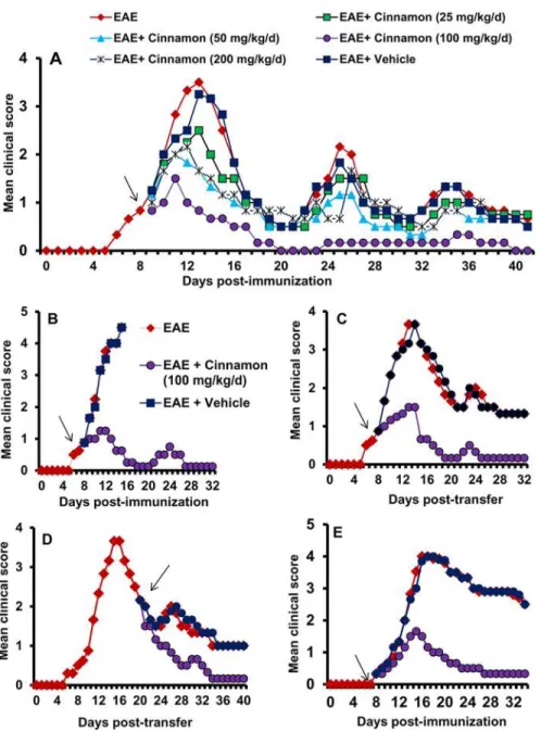
Figure 1. Oral administration of ground cinnamon suppresses clinical symptoms of EAE in female PLP-TCR transgenic (Tg) mice, adoptive transfer model in female SJL/J mice and chronic model in male C57/BL6 mice.A) PLP-TCR Tg mice were immunized with 10μg of PLP139–151, and from 8 days post- immunization (dpi) mice were treated with different doses of ground cinnamon or vehicle
(0.1% methylcellulose) via gavage. Mice (n= 6 in each group) were scored daily until 41 dpi. B) PLP-TCR Tg mice were immunized with 25μg of PLP139–151, and from 8 dpi mice were treated with ground cinnamon (100 mg/kg body wt/d) via gavage. Mice (n= 6 in each group) were scored daily until 32 dpi. C) EAE was induced in female SJL/J recipient mice by adoptive transfer of MBP-primed T cells. From 8 dpt, mice were treated with either cinnamon (100 mg/kg body wt/d) or vehicle via gavage. Mice (n= 6 in each group) were scored daily until 32 dpt. D) In this adoptive transfer model, mice were also treated with either cinnamon or vehicle from 20 dpt. Mice (n= 6 in each group) were scored daily until 40 dpt. E) C57BL/6 mice were immunized with 100μg of MOG35–55, and from 8 dpi, mice were treated with either cinnamon or vehicle. Mice (n= 6 in each group) were scored daily until 34 dpi.
then with 4% (w/v) paraformaldehyde solution in PBS, cerebellum and whole spinal cord was dissected out from each mouse. The tissues were further fixed and then divided into halves: one-half was used for histological staining whereas the other half was used for myelin staining as described earlier [11,12,13,14]. For histological analysis, routine histology was performed to obtain perivascular cuffing and morphological details of CNS tissues of EAE mice. Parafor-maldehyde-fixed tissues were embedded in paraffin, and serial sections (4 µm) were cut. Sec-tions were stained with conventional H&E staining method. Digital images were collected under bright-field setting using an x40 objective. Slides were assessed in a blinded fashion by three examiners for inflammation in different anatomical compartments (meninges and paren-chyma). Inflammation was scored using the following scale as described: for meninges and pa-renchyma: 0, no infiltrating cells; 1, few infiltrating cells; 2, numerous infiltrating cells; and 3, widespread infiltration. For vessels: 0, no cuffed vessel; 1, one or two cuffed vessels per section; 2, three to five cuffed vessels per section and 3, more than five cuffed vessels per section. At least six serial sections of each spinal cord from each of five mice per group were scored and statistically analyzed by ANOVA.
Staining for myelin
Sections were stained with Luxol fast blue for myelin as described earlier [13,14]. Slides were assessed in a blinded fashion for demyelination by three examiners using the following scale: 0, normal white matter; 1, rare foci; 2, a few areas of demyelination; and 3, large areas of demy-elination. At least six serial sections of each spinal cord from each of five mice per group were scored and statistically analyzed by ANOVA.
Semi-quantitative RT-PCR analysis
Total RNA was isolated from splenic T cells and spinal cord by using the RNeasy mini kit (Qiagen, Valencia, CA) and from spleen and cerebellum by using the Ultraspec-II RNA reagent (Biotecx laboratories, Inc, Houston, TX) following manufacturer’s protocol. To remove any contaminating genomic DNA, total RNA was digested with DNase. Semi-quantitative RT-PCR was carried out as described earlier [13,14] using a RT-PCR kit from Clonetech (Mountain View, CA). Briefly, 1 µg of total RNA was reverse transcribed using oligo(dT)12–18as primer and MMLV reverse transcriptase (Clontech) in a 20 µL reaction mixture. The resulting cDNA was appropriately-diluted, and diluted cDNA was amplified using Titanium Taq DNA poly-merase and following primers. Amplified products were electrophoresed on a 1.8% agarose gels and visualized by ethidium bromide staining.
Foxp3: Sense, 5'-CAG CTG CCT ACA GTG CCC CTAG-3'
Antisense, 5'-CAT TTG CCA GCA GTG GGT AG-3’
CD25: Sense, 5'-AGC CAA GTA GGG TGT CTC TCA ACC-3'
Antisense, 5'-GCC CAG GAT ACA CAG TGA AGA ACG-3'
CD4: Sense, 5’- CCA ACA AGA GCT CAA GGA GAC CAC-3’
Antisense, 5’- CGT ACC CTC TTT CCT AGC AAA GGA-3’
CD62L: Sense, 5’- AGC CTC TTG CCA GCC AGG GT-3’
Antisense, 5’- CCA GCC CCG AGA ATG CGG TG-3’
Antisense, 5’- CCC GTT GCC CAT GCC CAC AA-3’
IFN-γ: Sense, 5’- GCTGTTACTGCCACGGCACA-3’
Antisense, 5’- GGACCACTCGGATGAGCTCA-3’
T-bet: Sense, 5’- GGAGCGGACCAACAGCATC-3’
Antisense, 5’- CCACGGTGAAGGACAGGAAT-3’
IL-10: Sense, 5’- GCACTGCTATGCTGCCTGCT-3’
Antisense, 5’- CCGATAAGGCTTGGCAACCC-3’
GATA3: Sense, 5’- TCTGGAGGAGGAACGCTAATGG-3’
Antisense, 5’- GAACTCTTCGCACACTTGGAGACTC-3’
IL-17: Sense, 5’- GCTGACCCCTAAGAAACCCC-3’
Antisense, 5’- GAAGCAGTTTGGGACCCCTT-3’
iNOS: Sense: 5’-CCCTTCCGAAGTTTCTGGCAGCAGC-3’
Antisense: 5’-GGCTGTCAGAGCCTCGTGGCTTTGG3’
IL-1β: Sense: 5’-CTCCATGAGCTTTGTACAAGG-3’
Antisense: 5’-TGCTGATGTACCAGTTGGGG-3’
MBP: Sense: 5’-TGGAGAGATTCACCGAGGAGA-3’
Antisense: 5’-TGAAGCTCGTCGGACTCTGAG-3’
CNPase: Sense: 5’-CTACCCTCCACGAGTGCAAGA-3’
Antisense: 5’-AGTCTAGTCGCCACGCTGTCT-3’
GAPDH: Sense: 5'-GGTGAAGGTCGGTGTGAACG3'
Antisense: 5'-TTGGCTCCACCCTTCAAGTG-3'
The relative expression of each gene with respect to GAPDH was measured after scanning the bands with a Fluor Chem 8800 Imaging System (Alpha Innotech, San Leandro, CA).
Real-time PCR analysis
It was performed using the ABI-Prism7700 sequence detection system (Applied Biosystems, Foster City, CA) as described earlier [13,14]. Briefly, reactions were performed in a 96-well op-tical reaction plates on cDNA equivalent to 50 ng DNase-digested RNA in a volume of 25 µL, containing 12.5 µL TaqMan Universal Master mix and optimized concentrations of FAM-la-beled probe, forward and reverse primers following the manufacturer’s protocol. All primers and FAM-labeled probes for mouse genes and GAPDH were obtained from Applied Biosys-tems. The mRNA expressions of respective genes were normalized to the level of GAPDH mRNA. Data were processed by the ABI Sequence Detection System 1.6 software and analyzed by ANOVA.
Assay for NO synthesis
Flow cytometry
Two-color flow cytometry was performed as described previously [17,18]. Briefly, 1 × 10⁶ lymph node cells (LNC) or splenocytes suspended in flow staining buffer were incubated at 4°C with appropriately diluted FITC-labeled Ab to CD4 for 30 min, washed, and resuspended in fixation and permeabilization solution. Following incubation in dark for 30 min, cells were washed, blocked with test Fc block (anti-mouse CD16/32) in permeabilization buffer, and sub-sequently incubated with appropriately diluted PE-labeled Abs to T-bet, IFN-γ, GATA3, IL-4, IL-17, RORγT, or Foxp3 at 4°C in the dark. After incubation, the cell suspension was centri-fuged, washed thrice, and resuspended in flow staining buffer. The cells then were analyzed through FACS (BD Biosciences, San Jose, CA). Cells were gated based on morphological char-acteristics. Apoptotic and necrotic cells were not accepted for FACS analysis.
Assay of suppressive activity of Tregs
Because cells from tomato red transgenic (B6.129(Cg)-Gt(ROSA)26Sortm4(ACTB-tdTomato,-EGFP) Luo/J) mice exhibit red color, we used these mice (Jackson Laboratories, Bar Harbor, ME) for
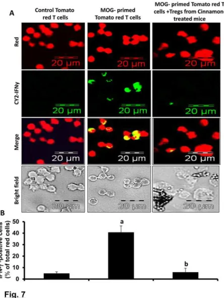
clear visualization of the suppressive activity of Tregs. These mice were immunized with MOG (100 µg/mouse) suspended in IFA containing 60 µgM. tuberculosisand 12 d after immuniza-tion, splenocytes were isolated and re-primed with MOG (10 µg/ml) for 2 d. These MOG-primed tomato red T cells expressed Th1 and Th17 cytokines (data not shown). In a parallel experiment, B6.129 mice were also immunized with MOG and treated with cinnamon for 2d followed by purification of CD4+CD25+ Tregs. Then these cinnamon-induced Tregs were added to MOG-primed splenocytes isolated from tomato red transgenic mice at a ratio of 2:1 of tomato red T cell:cinnamon-induced Tregs and the suppressive activity of Tregs was moni-tored by the inhibitory effect on the IFN-γexpression by MOG-primed tomato red T cells. Therefore, after 24 h of incubation, CD3+ T cells were purified and immunostained for IFN-γ (green). IFN-γ-expressing red cells were counted and expressed as percent of total red cells.
Results
Cinnamon inhibits clinical symptoms and disease severity of EAE in
female PLP-TCR transgenic mice
EAE with a mean clinical score of 1.42 + 0.49. However, as reported [20], immunization with low dose (10μg/mouse) of PLP139–151 strongly induced clinical symptoms of EAE (Fig. 1A). EAE mice were treated with different doses of cinnamon powder from 8 days post immuniza-tion (dpi) when these mice exhibited a clinical score of 0.5 or higher. An addiimmuniza-tional group of mice was treated with vehicle (Fig. 1A). Since the relapsing-remitting type of EAE is associated with multiple chronic phase peaks following the acute phase peak, we continued our observa-tions until 42 dpt. Even at a dose of 25 mg/kg body wt/d, cinnamon significantly inhibited clin-ical symptoms (Fig. 1A&Table-1) with no decrease in disease incidence considering a clinical symptom of 1 or higher as disease incidence. On the other hand, at a dose of 100 mg/kg body wt/d, a dramatic inhibition of clinical symptoms and a significant reduction in disease inci-dence were observed in acute as well as chronic phases of EAE (Fig. 1A&Table-1). Vehicle (0.1% methyl cellulose) remained unable to inhibit the clinical symptoms of EAE (Fig. 1A& Table-1), suggesting the specificity of the effect. However, at a dose of 200 mg/kg body wt/d, cinnamon was less potent than either 50 or 100 mg/kg body wt/d in suppressing clinical symp-toms (Fig. 1A&Table 1), suggesting that at higher dose, it may be toxic for EAE mice. We in-duced severe disease by immunizing these PLP-TCR Tg mice with 25μg of PLP 139–151/ mouse, where all mice died within 16 dpi (Fig. 1B). Even in this instance, cinnamon at a dose of 100 mg/kg body wt/d markedly suppressed clinical symptoms of EAE and no mouse from the cinnamon-treated group died during the course of the study (Fig. 1B). These findings dem-onstrate that cinnamon is capable of inhibiting clinical symptoms and disease severity in acute as well as chronic phases of EAE in PLP-TCR Tg mice.
Cinnamon inhibits the adoptive transfer of EAE in female SJL/J mice
RR-EAE is also induced in female SJL/J mice by adoptive transfer of MBP-primed T cells [11,13,14]. Therefore, next, we examined if cinnamon treatment was also capable of suppress-ing the progression of EAE in this adoptive transfer model. Mice were treated with cinnamon powder in two different groups. In the first group, mice were treated with cinnamon from 8 days post-transfer (dpt), the onset of acute phase. An inhibitory effect of cinnamon on the clinical symptoms was clearly observed within a few days of treatment (Fig. 1C). Greater inhi-bition was observed on subsequent days of treatment, which was maintained throughout the duration (32 dpt) of the experiment (Fig. 1C). On the other hand, vehicle had no such inhibito-ry effect (Fig. 1C). In the second group, cinnamon treatment began from the onset of relapsingTable 1. Effect of cinnamon on clinical symptoms of EAE in PLP-TCR Tg mice.
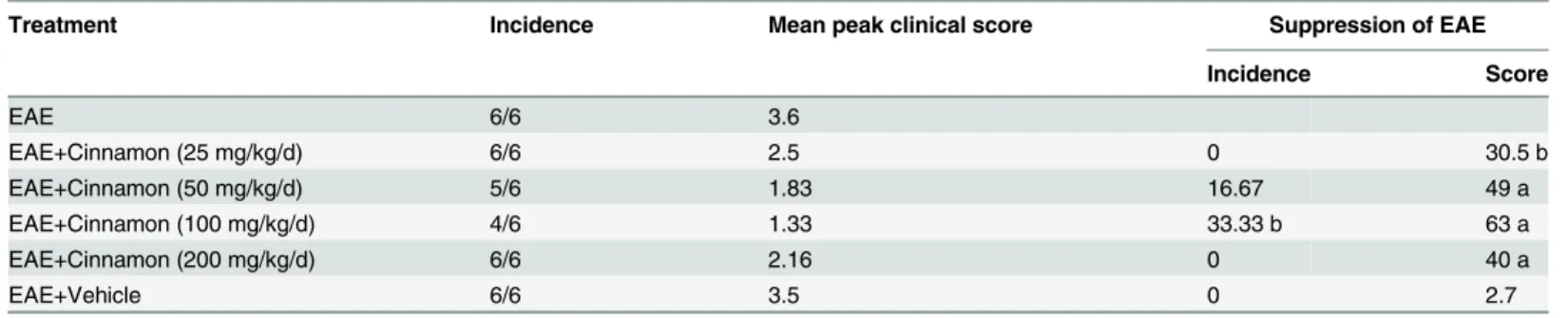
Treatment Incidence Mean peak clinical score Suppression of EAE
Incidence Score
EAE 6/6 3.6
EAE+Cinnamon (25 mg/kg/d) 6/6 2.5 0 30.5 b
EAE+Cinnamon (50 mg/kg/d) 5/6 1.83 16.67 49 a
EAE+Cinnamon (100 mg/kg/d) 4/6 1.33 33.33 b 63 a
EAE+Cinnamon (200 mg/kg/d) 6/6 2.16 0 40 a
EAE+Vehicle 6/6 3.5 0 2.7
EAE was induced in female PLP-TCR Tg mice by immunization with 10µg of PLP139–151 inM. tuberculosisin IFA. Mice also received pertussis toxin (150 ng/mouse) once on 0 day post-immunization (dpi). From 8 dpi, mice were treated with different doses of cinnamon emulsified in vehicle (0.5% methylcellulose) orally. While a clinical score of 1 was considered as the incidence of EAE in mice, a clinical score of 0 was considered to be normal.
ap<
0.001,bp<
phase (20 dpt) and was continued until 40 dpt. Here, too, cinnamon, but not vehicle, halted the disease progression (Fig. 1D). However, in contrast to the first instance, the inhibitory effect of cinnamon was manifested after 6 days of treatment (26 dpt). The EAE disease severity in the cinnamon-treated group was close to 0 (normal) in the latter part of treatment (Fig. 1D). These results clearly demonstrate that cinnamon can ameliorate the ongoing relapsing-remitting EAE when administered either early (at the onset of acute phase) or late (at the onset of relaps-ing disease).
Cinnamon inhibits chronic EAE in male C57/BL6 mice
While female SJL/J mice are used to induce RR-EAE, chronic form of EAE is modeled in male C57/BL6 mice upon immunization with MOG35–55. Next, we examined the efficacy of cinna-mon powder in suppressing the disease process of chronic EAE. Similar to its effect on RR-EAE in female PLP-TCR Tg mice and SJL/J mice, cinnamon treatment also strongly inhibited the clinical symptoms of EAE in this chronic model (Fig. 1E). Again, vehicle had no effect on chronic EAE (Fig. 1E), suggesting the specificity of the effect.
Cinnamon treatment inhibits the generation of encephalitogenic T cells
in donor mice
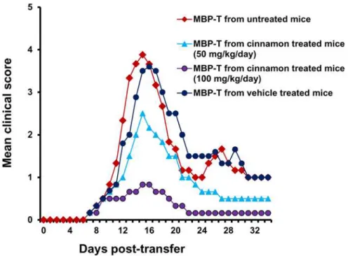
As T cells isolated from MBP-immunized donor mice are encephalitogenic, and adoptive transfer of these T cells induces EAE in recipient mice, we investigated whether treatment of donor mice with cinnamon was capable of inhibiting the production of encephalitogenic T cells. In order to test this, donor mice were treated with different doses of cinnamon orally from the day of MBP immunization. T cells isolated from cinnamon-treated and untreated MBP-immunized donor mice were then adoptively transferred to recipient mice. Our results showed that mice receiving MBP-primed T cells from cinnamon-treated donor mice exhibited significantly reduced clinical symptoms and disease severity compared to mice receiving MBP-primed T cells from either untreated donor mice or vehicle-treated donor mice (Fig. 2). These results suggest that cinnamon treatment inhibits the generation of encephalitogenic T cellsin vivoin donor mice.
Cinnamon treatment preserves the integrity of blood-brain barrier (BBB)
and blood-spinal cord barrier (BSB) in PLP-TCR transgenic mice
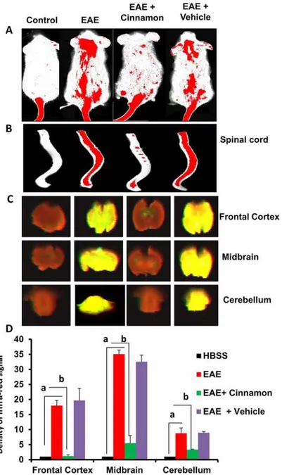
To confirm these results further, mice were sacrificed, and the spinal cord and different parts of the brain (frontal cortex, midbrain and cerebellum) were scanned for infra-red signals in Odyssey infra-red scanner. Consistent to live mice results, we did not notice much infra-red signal in the spinal cord and different parts of the brain in control HBSS-treated mice (Fig. 3B-D; lane 1) but significant amount of infra-red dye was visible in CNS tissues of EAE mice (Fig. 3B-D; lane 2). Again, treatment of EAE mice by cinnamon markedly attenuated the entry of infra-red dye into the spinal cord and different parts of the brain (Fig. 3B-D; compare lane 3 with lane 2). Taken together, these results suggest that cinnamon treatment preserves the integrity of BBB and BSB in EAE mice.
Cinnamon inhibits infiltration of mononuclear cells, inflammation and
demyelination in the spinal cord of EAE
EAE as well as MS is caused by infiltration of autoreactive T cells and associated mononuclear cells, like macrophages, into the CNS, followed by broad-spectrum inflammatory events [9,21]. We examined whether cinnamon attenuated infiltration and inflammation in RR-EAE in PLP-TCR transgenic mice. Mice receiving cinnamon from 8 dpi (onset of the acute phase) were sacrificed on 16 dpi. H & E staining showed widespread infiltration of inflammatory cells into the spinal cord (Fig. 4A) of RR-EAE mice. On the other hand, cinnamon treatment markedly inhibited the infiltration of inflammatory cells into the spinal cord of RR-EAE mice (Fig. 4A). In contrast, vehicle was unable to inhibit the infiltration of inflammatory cells (Fig. 4A). Quantitation of the relative level of inflammatory cells showed that cinnamon, but
Figure 2. Oral administration of ground cinnamon inhibits the generation of encephalitogenic T cells
in vivoin donor mice.Donor mice (4–6 week old female SJL/J) were immunized with MBP, IFA, andM. tuberculosis.From 2ndday of immunization, mice were treated with either cinnamon (50 and 100 mg/kg body
wt/d) or vehicle via gavage. On day 12 of immunization, mice were sacrificed, and total LNC were further primed with MBP (50μg/ml) for 4 days. A total of 2 × 10⁷viable MBP-primed T cells was adoptively
transferred to naïve recipient mice. Six mice were used in each group. Mice were examined for clinical symptoms every day until 34 dpt.
Figure 3. Effect of oral administration of cinnamon on the integrity of blood-brain barrier (BBB) and blood-spinal cord barrier (BSB) in EAE in PLP-TCR Tg mice.Control, EAE (14 dpi), and either cinnamon-or vehicle-treated EAE mice (14 dpi receiving cinnamon/vehicle from 8 dpi) (n= 5 in each group) received 200μl of 20μM Alexa Fluor 680-SE-NIR dye (Invitrogen) via the tail vain on 16 dpt (acute phase). After 4 h, mice were scanned in an Odyssey (ODY-0854; Licor) infrared scanner at the 700- and 800-nm channels (A). Mice were perfused with 4% paraformaldehyde. Spinal cord (B) and different parts of the brain (C) were scanned in an Odyssey infrared scanner. The red background came from an 800-nm filter, whereas the green signal was from Alexa Fluor 680 dye at the 700-nm channel. The density of the Alexa Fluor 680 signal in different parts of the brain (D) was quantified with the help of Quantity One, version 4.6.2 software, using the volume contour tool analysis module. Data are expressed as the mean±SEM of five different mice;
ap
<0.001 vs normal (HBSS);bp<0.001 vs EAE.
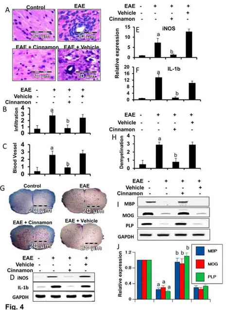
Figure 4. Oral administration of ground Cinnamon suppresses the infiltration of mononuclear cells and inhibits demyelination in the spinal cord of EAE in female PLP-TCR Tg mice.(A) Spinal cord sections of control, EAE (14 dpi) and either cinnamon- or vehicle-treated EAE mice (receiving cinnamon/ vehicle from 8 dpi) were stained with H&E. Digital images were collected under a bright-field setting using a ×40 objective. Infiltration (B) and cuffed vessel (C) were represented quantitatively by using a scale as described by us. Data are expressed as mean±SEM of five different mice.ap
<0.001 vs normal;bp<0.001
vs EAE. Spinal cord of normal, EAE, and either cinnamon- or vehicle-treated EAE mice were analyzed for iNOS and IL-1βmRNAs by semi-quantitative RT-PCR (D) and quantitative real-time PCR (E for iNOS, & F for IL-1β). Data are expressed as the mean±SEM of five different mice per group.ap<
0.001 vs normal,
bp<0.001 vs EAE. Spinal cord sections were stained with Luxol fast blue. Digital images were collected under
bright field setting using a 40X objective (G). Demyelination was represented quantitatively by using a scale as described by us (H). Data are expressed as the mean±SEM of five different mice per group;a
p<0.001 vs
normal,bp<0.001 vs EAE. Spinal cord samples were analyzed for MBP, MOG, and PLP mRNAs by
semi-quantitative RT-PCR (I) and real-time PCR (J). Data are expressed as the mean±SEM of five different mice per group;a
p<0.001 vs normal,bp<0.001 vs EAE.
not vehicle, dramatically reduced infiltration (Fig. 4B) and the appearance of cuffed vessels (Fig. 4C) in spinal cord of RR-EAE mice.
Since infiltration was inhibited, we next examined whether cinnamon was capable of inhib-iting the expression of proinflammatory molecules in the spinal cord of RR-EAE mice. Marked expression of pro-inflammatory molecules like iNOS and IL-1βwas observed in the spinal cord of untreated RR-EAE mice compared to control mice (Fig. 4D-F). However, cinnamon treatment dramatically reduced the expression of these pro-inflammatory molecules in the spi-nal cord of RR-EAE mice (Fig. 4D-F).
It is believed that infiltration of blood mononuclear cells and associated neuroinflammation plays an important role in CNS demyelination observed in MS patients and EAE animals [9,22,23]. Therefore, we examined whether cinnamon protected RR-EAE mice from demye-lination. We stained spinal cord sections by luxol fast blue (LFB) for myelin and observed wide-spread demyelination zones in the white matter (Fig. 4G-H). However, cinnamon treatment remarkably restored myelin level in the spinal cord of RR-EAE mice (Fig. 4G-H). In contrast, vehicle was unable to restore myelin level in spinal cord of RR-EAE mice (Fig. 4G-H). To con-firm this finding, we monitored the expression of three myelin genes, MBP, MOG and PLP, and observed a marked loss of mRNA expression of these genes in the spinal cord of untreated RR-EAE mice compared to control mice (Fig. 4I-J). A significant restoration of myelin gene mRNA expression was observed in RR-EAE mice that were treated with cinnamon, but not in mice treated with vehicle (Fig. 4I-J).
Next, we investigated whether cinnamon was also able to reduce inflammation and demye-lination in mice with chronic EAE (Ch-EAE). Similar to that found in RR-EAE mice, the mRNA expression of proinflammatory molecules (iNOS and IL-1β) increased (Fig. 5A-B), while the mRNA expression of myelin-specific genes (MOG, MBP and PLP) decreased in the spinal cord of mice with Ch-EAE (Fig. 5C-D). However, cinnamon treatment reduced the ex-pression of proinflammatory molecules (Fig. 5A-B) and increased the level of myelin-specific molecules (Fig. 5C-D) in the spinal cord of Ch-EAE mice. These results demonstrate that cin-namon treatment inhibits infiltration of mononuclear cells, inflammation, and demyelination in the spinal cord of EAE mice.
Enrichment of the Treg population by cinnamon treatment
Because Tregs are most important immunomodulatory subtype of T lymphocytes, in order to understand immunomodulatory effect of cinnamon, at first, we examined the effect of cinna-mon treatment on the status of Tregsin vivoin EAE mice. EAE mice receiving cinnamon or ve-hicle from 8 dpi were sacrificed on 16 dpi followed by analysis of Tregs in spleen and
To confirm these results further, we performed FACS analysis. As expected, there was a sig-nificant reduction in Foxp3+CD4+ population of T cells in EAE splenocytes as evident from FACS dot plot (Fig. 6D) and mean fluorescence intensity (MFI) (Fig. 6E). However, treatment of EAE mice with cinnamon, but not vehicle, led to the increase in Foxp3+CD4+ population in splenocytes (Fig. 6E). MFI calculation inFig. 6Ealso shows that cinnamon treatment resulted in significant increase in Foxp3.
To understand that cinnamon-induced Tregs in EAE mice were functionally active, we ex-amined the suppressive activity of these Tregs. As evident fromFig. 7A-B, IFN-γexpression was very low in normal tomato red T cells and MOG priming significantly increased the ex-pression of IFN-γin these T cells. However, cinnamon-induced MOG-primed Tregs of B6.129 mice markedly suppressed the expression of IFN-γin MOG-primed tomato red T cells (Fig. 7A-B). This result was specific as MOG-primed non-Tregs increased the expression of IFN-γin tomato red T cells (data not shown). These results demonstrate that cinnamon-induced Tregs in EAE mice are functionally active.
How does cinnamon enrich Tregs?
Recently, we have reported that NO is a critical regulator of Tregs [17]. Therefore, we were prompted to investigate whether cinnamon treatment increased the number of Tregs via de-creasing NO. At first, we examined whether cinnamon treatment would inhibit the expression of iNOS and the production of NO in the spleen of EAE mice. As expected, splenocytes isolated from EAE mice produced 5-fold more NO (measured as nitrite) as compared to that isolated from control mice (Fig. 8A). However, treatment of EAE mice with cinnamon, but not vehicle,
Figure 5. Oral administration of ground cinnamon inhibits the expression of proinflammatory molecules and upregulates the expression of myelin genes in the spinal cord of chronic EAE in C57/ BL6 mice.C57BL/6 mice were immunized with 100μg of MOG35–55, and from 8 dpi, mice were treated with either cinnamon or vehicle. On 20 dpi, spinal cord of normal, EAE, and either cinnamon- or vehicle-treated EAE mice were analyzed for mRNA expression of proinflammatory molecules ( iNOS and IL-1β) (A & B) and myelin-specific molecules (MOG, MBP and PLP) (C & D) by semi-quantitative RT-PCR (A & C) and quantitative real-time PCR (B & D). Data are expressed as the mean±SEM of five different mice per group.
ap<
0.001 vs normal;bp<0.001 vs EAE.
led to the suppression of NO production in splenocytes (Fig. 8A). To understand the mecha-nism further, we investigated the effect of cinnamon on mRNA level of iNOS. It is evident from semi-quantitative RT-PCR (Fig. 8B) and real-time PCR (Fig. 8C) analyses that treatment of EAE mice with cinnamon, but not vehicle, led to the inhibition of iNOS mRNA expression in splenocytes. These results suggest that cinnamon is capable of suppressing the expression of iNOS in EAE mice. Next, we monitored the level of Foxp3 mRNA. In contrast to iNOS, induc-tion of EAE decreased the mRNA expression of Foxp3, whereas cinnamon blocked the loss of Foxp3 in splenocytes (Fig. 8Bfor RT-PCR andFig. 8Dfor real-time PCR).
Figure 6. Oral administration of ground cinnamon enriches regulatory T cells (Tregs)in vivoin EAE in PLP-TCR Tg mice.Spleens of control, EAE (14 dpi) and either cinnamon- or vehicle-treated EAE mice (14 dpi receiving cinnamon/vehicle from 8 dpi) were analyzed for the mRNA expression of Foxp3, CD62L, CTLA4, GITR, CD25, and CD4 by semi-quantitative RT-PCR (A) and real-time PCR (Foxp3, B; and CD25, C). Data are expressed as the mean±SEM of 5 mice per group.ap<0.001 vs normal;bp<0.001 vs EAE.
LNC isolated from different groups of mice were analyzed by FACS for Foxp3 and CD4 (D). The MFI of Foxp3 in CD4+ population was calculated by using the CellQuest software (E). Data are mean±SEM of 5 mice per group.a
p<0.001 vs normal;bp<0.001 vs EAE.
Next, to directly test a role of NO in cinnamon-mediated modulation of Foxp3, we added DETA-NONOate (an NO donor) to splenocytes isolated from cinnamon-treated EAE mice. It is evident from RT-PCR (Fig. 8B), real-time PCR (Fig. 8D), FACS dot plot (Fig. 8E), and MFI analysis (Fig. 8F) that cinnamon treatment of EAE mice resulted to an increase in the level of Foxp3 in splenocytes. However, this increase and/or protection of Foxp3 mRNA and protein was completely abrogated by DETA-NONOate (Fig. 8B, D-F), indicating an important role of NO in cinnamon-mediated upregulation of Foxp3 and enrichment of Tregs.
Figure 7. Suppressive activity of Tregs isolated from cinnamon-treated EAE mice.Tomato red transgenic mice were immunized with MOG (100µg/mouse) and 12 d after immunization; splenocytes were isolated and re-primed with MOG (10µg/ml) for 2 d. Similarly, C57BL/6 mice were also immunized with MOG and from 8 dpi, mice were treated with ground cinnamon via gavage. On 16 dpi, splenocytes were isolated and re-primed with MOG for 2d followed by purification of CD4+CD25+ Tregs. Then these Tregs were added to MOG-primed splenocytes isolated from tomato red transgenic mice at a ratio of 2:1 of tomato red T cell: Tregs. After 24 h, non-adherent cells were immunostained for IFN-γ(green). Cells from tomato red transgenic mice exhibited red color (A). IFN-γexpressing red cells were counted and expressed as percent of total red cells (B). Data are mean±SEM of 20 different images.ap<
0.001 vs control tomato red T cells,bp<0.001 vs
Figure 8. Cinnamon treatment enriches Tregs in EAE in PLP-TCR Tg mice via suppressing NO production.Splenocytes isolated from control, EAE (14 dpi) and either cinnamon- or vehicle-treated EAE mice (14 dpi receiving cinnamon/vehicle from 8 dpi) were re-primed with PLP139–151 for 24 h followed by monitoring the level of nitrite in supernatants (A) and the mRNA expression of iNOS and Foxp3 by semi-quantitative RT-PCR (B) and real-time PCR (C, iNOS; D, Foxp3). Data are expressed as the mean±SEM of 3 mice per group.ap
<0.001 vs control;bp<0.001 vs EAE. Splenocytes isolated from
cinnamon-treated EAE mice (14 dpi receiving cinnamon/vehicle from 8 dpi) were re-primed with PLP139–151 for 24 h in the presence or absence of DETA-NONOate (a NO donor) followed by FACS for Foxp3 and CD4 (E). The MFI of Foxp3 in CD4+ population was calculated by using the CellQuest software (F). Data are mean±SEM of 3 mice per group.ap<0.001 vs normal;bp<0.001 vs EAE;cp<0.001 vs (EAE+cinnamon).
Suppression of the Th17 response by cinnamon treatment
After the discovery of IL-23, Th17 cells are considered to play a more active role than Th1 cells in the disease process of EAE and MS [24]. It has been found that there is an inverse relationship between Th17 cells and Tregs [25]. Because cinnamon treatment enriched Tregs, we examined whether cinnamon was capable of regulating Th17 cells in EAE mice. While induction of EAE increased the expression of IL-17 mRNA (Fig. 9A) and the level of CD4+IL-17+ T cells in sple-nocytes (Fig. 9B), cinnamon markedly suppressed EAE-induced upregulation of IL-17 mRNA (Fig. 9A) as well as the CD4+IL-17+ T cell population (Fig. 9B). On the other hand, vehicle treat-ment had no such suppressive effects on CD4+IL-17+ T cell (Fig. 9B). MFI analysis of IL-17 (Fig. 9D) within the CD4+ population also supported this finding. Th17 cells are also characterized by a transcription factor called RORγT [26]. To confirm the regulation of Th17 cells by cinnamon treatment, we also monitored RORγT. Consistent with the upregulation of IL-17 mRNA and CD4+IL-17+ T cells, induction of EAE increased the mRNA expression of RORγT (Fig. 9A) and the level of CD4+RORγT+ T cells in splenocytes (Fig. 9C) and treatment of EAE mice with cin-namon, but not vehicle, resulted in suppression of RORγT mRNA (Fig. 9A) and CD4+RORγT+ T cells (Fig. 9C). This is also corroborated by MFI analysis of RORγT within the CD4+ popula-tion (Fig. 9E). These results clearly show that cinnamon is capable of suppressing Th17 cells.
Switching of Th1 to Th2 in response to cinnamon treatment
Similar to Th17 cells, Th1 cells are also autoimmune inflammatory and switching of the Th1 to a Th2 phenotype is one of the ways to ameliorate the disease [10,24,27]. Because cinnamon in-creased Tregs, which are known to suppress Th1 cells via releasing TGF-βand IL-10, we exam-ined whether cinnamon treatment was capable of suppressing the autoimmune Th1
response in EAE mice. While T-bet-dependent IFN-γproduction is a characteristic of Th1 cells, Th2 cells display GATA3-dependent IL-10 and IL-4 release [9,28,29]. As expected, in-duction of EAE increased the mRNA expression of IFN-γand Tbet (Fig. 10A) and the level of CD4+IFN-γ+ T cells in splenocytes (Fig. 10C). Treatment of EAE mice with cinnamon, but not vehicle, led to marked suppression of EAE-induced upregulation of IFN-γand Tbet mRNAs (Fig. 10A) as well as the CD4+IFN-γ+ T cell population (Fig. 10C). MFI analysis of IFN-γ(Fig. 10E) within the CD4+ population also supported this finding. Next, we
analyzed the Th2 responses by monitoring Th2 cytokines and Th2-specific transcription factor GATA3. The induction of EAE suppressed the mRNA expression of IL-10 and GATA3 (Fig. 10B) and the level of CD4+IL-4+ T cells (Fig. 10D) in splenocytes. However,
cinnamon treatment markedly increased the mRNA expression of IL-10 and GATA3 and the level of CD4+IL-4+ T cells in splenocytes (Fig. 10B&D). MFI analysis of IL-4 (Fig. 10F) within the CD4+ population also supported this finding. This effect was specific as vehicle treatment had no effect. Together, these results suggest that cinnamon treatment was capable of suppressing the Th1 response, while augmenting the Th2 response in EAE mice.
Cinnamon suppresses EAE in mice via Tregs
Figure 9. Oral administration of ground cinnamon suppresses autoimmune Th17 cellsin vivoin EAE in PLP-TCR Tg mice.Spleens of control, EAE (14 dpi) and either cinnamon- or vehicle-treated EAE mice (14 dpi receiving cinnamon/vehicle from 8 dpi) were analyzed for the mRNA expression of Th17-specific factors (RORγt and IL-17) (A). Splenocytes isolated from different groups of mice were analyzed by FACS for RORγt & CD4 (B) and IL-17 & CD4 (C). The MFI of RORγT (D) and IL-17 (E) in CD4+ population were calculated by using the CellQuest software. Data are mean±SEM of 3 mice per group.a
p<
0.001 vs normal;bp<0.001 vs EAE.
Next, we examined whether cinnamon treatment also protected EAE via Tregs. Therefore, during cinnamon treatment, the function of Tregs was blockedin vivoin EAE mice by anti-CD25 antibody. As evident fromFig. 11B, cinnamon treatment ameliorated clinical symptoms of RR-EAE. However, functional blocking anti-CD25 antibody almost completely abrogated the cinnamon-mediated protective effect on EAE mice (Fig. 11B). This result was specific as control IgG had no such effect (Fig. 11B). Together, these results delineate an important role of Tregs in cinnamon-mediated protection of EAE.
Discussion
MS is the most common human demyelinating disease of the CNS and despite intense investi-gations, no effective therapy is available for this disease. Tysabri and different forms of interfer-on-β(IFN-β) are currently used to treat MS. However, reduced effectiveness and severe toxic
Figure 10. Oral administration of ground cinnamon switches Th1 to Th2in vivoin EAE in PLP-TCR Tg mice.Spleens of control, EAE (14 dpi) and either cinnamon- or vehicle-treated EAE mice (14 dpi receiving cinnamon/vehicle from 8 dpi) were analyzed for the mRNA expression of Th1-specific factors (Tbet and IFN-γ) (A) and Th2-specific factors (GATA3 and IL-10) (B) by semi-quantitative RT-PCR. LNC isolated from different groups of mice were analyzed by FACS for IFN-γ& CD4 (C) and IL-4 & CD4 (D). The MFI of IFN-γ (E) and IL-4 (F) in CD4+ population were calculated by using the CellQuest software. Data are mean±SEM of 3 mice per group.ap
<0.001 vs normal;bp<0.001 vs EAE.
effects over chronic use, as well as treatment costs, often limit these available therapies. For ex-ample, IFN-βhas a number of side effects including flu-like symptoms, menstrual disorders in women, decrease in neutrophil and white blood cell count, increase in AST and ALT levels, and development of neutralizing antibodies to IFN-β[9,30,31]. Similarly, treatment with
Figure 11. Oral administration of cinnamon protects EAE in PLP-TCR Tg mice via Tregs.Female PLP-TCR Tg mice were immunized with 10μg of PLP139–151, and from 8 dpi, mice were treated with cinnamon (100mg/kg of body weight/d) via gavage. After 8 d of treatment, splenocytes were isolated followed by purification of CD4+CD25+ Tregs. In a parallel experiment, EAE was induced in female PLP-TCR Tg mice and from 4 dpi, EAE mice were treated (i.p.) once with cinnamon-induced Tregs (1×10⁶Tregs/mouse). Purified CD4+CD25- non-Tregs were also used for comparison. Six recipient mice (n = 6) were included in each group. Mice were examined for clinical symptoms daily until 30 dpi (A). EAE was induced in female PLP-TCR Tg mice and from 2 dpi, mice were treated with cinnamon (100mg/kg of body weight/d) via gavage followed by one i.p. injection of anti-CD25 antibody (50µg/mouse) on the same day. One group of mice also received same amount of control IgG. Mice (n = 6) were examined for clinical symptoms daily.
Tysabri can cause lung infection, breathing problems, chest pain, wheezing, urinary tract infec-tion, vaginitis, nausea, vomiting, and liver damage. Tysabri also increases the chance of getting a severe brain infection, leading to progressive multifocal encephalopathy, which may cause disability and death. Therefore, it is necessary to identify safe, effective and economical thera-peutic option for MS.
Cinnamon, the brown bark of cinnamon tree, is a commonly used spice and flavoring mate-rial for deserts, candies, chocolate, etc. It has a long history as a medicine as well. Medieval phy-sicians used cinnamon in medicines to treat a variety of disorders including arthritis, coughing, hoarseness, sore throats, etc. In addition to containing manganese, dietary fiber, iron, and calci-um, cinnamon contains a major compound, cinnamaldehyde, which is converted into cin-namic acid by oxidation. In the liver, this cincin-namic acid isβ-oxidized to benzoate [32] that exists as sodium salt (NaB) or benzoyl-CoA. It has been reported that minor amount of NaB is also excreted in the urine of human [33,34]. NaB is of medical importance as it is a component of Ucephan, a FDA-approved drug used in the treatment for hepatic metabolic defects associat-ed with hyperammonemia such as urea cycle disorder in children [35,36]. It is also widely used as a preservative in broad range of foods and cosmetic products [37]. Earlier we have demon-strated that NaB modifies T cells at multiple steps and protects experimental allergic encepha-lomyelitis, an animal model of MS [13]. Here we provide the first evidence that oral feeding of cinnamon powder is capable of suppressing the disease process of EAE in mice. Our conclusion is based on the following observations. PLP immunization remained unable to induce clinical symptoms of EAE in female PLP-TCR transgenic mice receiving cinnamon orally. In contrast, PLP priming induced EAE in PLP-TCR mice receiving methyl cellulose (vehicle). From a ther-apeutic point of view, it is important to test whether a drug candidate is efficacious when ad-ministered after the onset of disease symptoms. Oral cinnamon powder fulfilled this
requirement and inhibited the progression of RR-EAE when administered either early or at a late stage of the disease progression. Therapeutic treatment of EAE animals with cinnamon was also capable of inhibiting the invasion of mononuclear cells into the spinal cord, as well as the expression of inflammatory molecules (iNOS and IL-1β), and restored myelination and the expression of myelin genes within the CNS. We did not notice any side effect (e.g. hair loss, weight loss, untoward infection etc.) in any of the mice used during cinnamon treatment. These results suggest that cinnamon may be considered to mitigate the disease process in MS patients.
red T cells by cinnamon-induced Tregs suggests that cinnamon-induced Tregs are functionally active.
How does cinnamon induce Tregs? Recently, we have demonstrated that NO negatively reg-ulates Foxp3 and Tregs [17]. While blocking NO either by inhibiting iNOS or direct scavenging of NO or by pharmacological drugs restores the expression of Foxp3 in MBP-primed T cells, NO donors decrease Foxp3 [17]. Therefore, we hypothesized that cinnamon treatment could enrich Tregs via regulating NO production. Suppression of iNOS expression and NO produc-tion in splenocytes of cinnamon-treated EAE mice as compared to vehicle treatment and abro-gation of cinnamon-mediated protection of Foxp3 by NO donor suggest that cinnamon treatment protects Tregs via suppression of NO production. Recently we have shown that oral administration of cinnamon (Cinnamonum verum) produces sodium benzoate (NaB) in serum and brain of mice [15]. We have also reported that NaB attenuates the production of NO [39] and that NaB restores Foxp3 in MBP-primed splenocytes via suppression of NO [17]. There-fore, it is likely that after oral administration, cinnamon is metabolized to sodium benzoate, which then suppresses the production of NO in spleen to protect Tregs in EAE mice. This sug-gestion is supported by the fact that NaB also enriches Tregs and protect mice from EAE [13].
Being the master regulator, Tregs are known to maintain immune homeostasis. While in one hand, Tregs suppress the proliferation of autoimmune Th1 cells by secreting TGF-βand IL-10, on the other, Tregs release IL-35 to control the proliferation of autoimmune Th17 cells [1,3,38,40,41]. Accordingly, oral cinnamon treatment suppressed both Th1 and Th17 im-mune responses in PLP-TCR mice. Theoretically, Tregs should also suppress Th2 via releasing TGF-βand IL-35 [3,41,42]. However, in our study, cinnamon treatment suppressed autoim-mune Th1 and Th17 responses, while stimulating the anti-autoimautoim-mune Th2 response in EAE. It is also possible as Tregs may contribute to Th2 polarization. For example, McKee and Pearce [43] have demonstrated that Tregs contribute to Th2 polarization during Helminth infection by suppressing the development of Th1 response. Similarly, Kohm et al [44] have shown that supplementation of Tregs by adoptive transfer before EAE induction significantly reduces the severity of clinical disease, potentially by promoting anti-autoimmune Th2 response. Here, we also must remember that Tregs produce substantial amount of IL-10, a cytokine that is also produced by Th2 cells. Therefore, whether the stimulation of Th2 response by cinnamon treat-ment is a direct effect of cinnamon metabolites, an indirect effect via enrichtreat-ment of Tregs, or both, needs further study.
Because Tregs have been implicated in the pathogenesis of autoimmune diseases, we exam-ined the effect of cinnamon-induced Tregs in EAE mice and, in a different experiment, used an anti-CD25 antibody in cinnamon-treated EAE mice. Suppression of EAE by cinnamon-in-duced Tregs and abrogation of cinnamon-mediated protection of EAE by anti-CD25 antibody clearly suggest that cinnamon-induced Tregs are capable of ameliorating EAE and that the ef-fect of cinnamon treatment is Treg-dependent.
In summary, we have demonstrated that oral administration of cinnamon powder upregu-lates anti-autoimmune Treg/Th2 cells, down-reguupregu-lates autoimmune Th17/Th1 cells and blocks the disease process of EAE when administered either prophylactially or therapeutcially. These results highlight a novel immunomodulatory role of cinnamon and suggest that this widely-used spice may be explored for therapeutic intervention in MS.
Acknowledgments
Author Contributions
Conceived and designed the experiments: SM KP. Performed the experiments: SM. Analyzed the data: SM KP. Wrote the paper: SM KP.
References
1. Coffer PJ, Burgering BM (2004) Forkhead-box transcription factors and their role in the immune system. Nat Rev Immunol 4: 889–899. doi:10.1038/nri1488PMID:15516968
2. Hori S, Nomura T, Sakaguchi S (2003) Control of regulatory T cell development by the transcription fac-tor Foxp3. Science 299: 1057–1061. doi:10.1126/science.1079490PMID:12522256
3. Sakaguchi S (2005) Naturally arising Foxp3-expressing CD25+CD4+ regulatory T cells in immunologi-cal tolerance to self and non-self. Nat Immunol 6: 345–352. doi:10.1038/ni1178PMID:15785760
4. Huan J, Culbertson N, Spencer L, Bartholomew R, Burrows GG, et al. (2005) Decreased FOXP3 levels in multiple sclerosis patients. J Neurosci Res 81: 45–52. doi:10.1002/jnr.20522PMID:15952173
5. Viglietta V, Baecher-Allan C, Weiner HL, Hafler DA (2004) Loss of functional suppression by CD4 +CD25+ regulatory T cells in patients with multiple sclerosis. J Exp Med 199: 971–979. doi:10.1084/ jem.20031579PMID:15067033
6. Paust S, Cantor H (2005) Regulatory T cells and autoimmune disease. Immunol Rev 204: 195–207. doi:10.1111/j.0105-2896.2005.00247.xPMID:15790360
7. McGeachy MJ, Stephens LA, Anderton SM (2005) Natural recovery and protection from autoimmune encephalomyelitis: contribution of CD4+CD25+ regulatory cells within the central nervous system. J Immunol 175: 3025–3032. doi:10.4049/jimmunol.175.5.3025PMID:16116190
8. Venken K, Hellings N, Thewissen M, Somers V, Hensen K, et al. (2008) Compromised CD4+ CD25 (high) regulatory T-cell function in patients with relapsing-remitting multiple sclerosis is correlated with a reduced frequency of FOXP3-positive cells and reduced FOXP3 expression at the single-cell level. Im-munology 123: 79–89. doi:10.1111/j.1365-2567.2007.02690.xPMID:17897326
9. Pahan K (2010) Neuroimmune pharmacological control of EAE. J Neuroimmune Pharmacol 5: 165–167. doi:10.1007/s11481-010-9219-6PMID:20414732
10. Pahan K (2011) Immunomodulation of experimental allergic encephalomyelitis by cinnamon metabolite sodium benzoate. Immunopharmacol Immunotoxicol 33: 586–593. doi:10.3109/08923973.2011. 561861PMID:21425926
11. Dasgupta S, Jana M, Zhou Y, Fung YK, Ghosh S, et al. (2004) Antineuroinflammatory effect of NF-kappaB essential modifier-binding domain peptides in the adoptive transfer model of experimental aller-gic encephalomyelitis. J Immunol 173: 1344–1354. doi:10.4049/jimmunol.173.2.1344PMID:
15240729
12. Dasgupta S, Zhou Y, Jana M, Banik NL, Pahan K (2003) Sodium phenylacetate inhibits adoptive trans-fer of experimental allergic encephalomyelitis in SJL/J mice at multiple steps. J Immunol 170: 3874–
3882. doi:10.4049/jimmunol.170.7.3874PMID:12646656
13. Brahmachari S, Pahan K (2007) Sodium benzoate, a food additive and a metabolite of cinnamon, modi-fies T cells at multiple steps and inhibits adoptive transfer of experimental allergic encephalomyelitis. J Immunol 179: 275–283. doi:10.4049/jimmunol.179.1.275PMID:17579047
14. Mondal S, Roy A, Pahan K (2009) Functional blocking monoclonal antibodies against IL-12p40 homo-dimer inhibit adoptive transfer of experimental allergic encephalomyelitis. J Immunol 182: 5013–5023. doi:10.4049/jimmunol.0801734PMID:19342681
15. Jana A, Modi KK, Roy A, Anderson JA, van Breemen RB, et al. (2013) Up-regulation of neurotrophic factors by cinnamon and its metabolite sodium benzoate: therapeutic implications for neurodegenera-tive disorders. J Neuroimmune Pharmacol 8: 739–755. doi:10.1007/s11481-013-9447-7PMID:
23475543
16. Khasnavis S, Pahan K (2014) Cinnamon treatment upregulates neuroprotective proteins Parkin and DJ-1 and protects dopaminergic neurons in a mouse model of Parkinson’s disease. J Neuroimmune Pharmacol 9: 569–581. doi:10.1007/s11481-014-9552-2PMID:24946862
17. Brahmachari S, Pahan K (2010) Myelin basic protein priming reduces the expression of Foxp3 in T cells via nitric oxide. J Immunol 184: 1799–1809. doi:10.4049/jimmunol.0804394PMID:20083653
18. Brahmachari S, Pahan K (2009) Suppression of regulatory T cells by IL-12p40 homodimer via nitric oxide. J Immunol 183: 2045–2058. doi:10.4049/jimmunol.0800276PMID:19587012
20. Waldner H, Whitters MJ, Sobel RA, Collins M, Kuchroo VK (2000) Fulminant spontaneous autoimmuni-ty of the central nervous system in mice transgenic for the myelin proteolipid protein-specific T cell re-ceptor. Proc Natl Acad Sci U S A 97: 3412–3417. doi:10.1073/pnas.97.7.3412PMID:10737797
21. Kuerten S, Lehmann PV (2011) The immune pathogenesis of experimental autoimmune encephalomy-elitis: lessons learned for multiple sclerosis? J Interferon Cytokine Res 31: 907–916. doi:10.1089/jir. 2011.0072PMID:21936633
22. Martin R, McFarland HF, McFarlin DE (1992) Immunological aspects of demyelinating diseases. Annu Rev Immunol 10: 153–187. doi:10.1146/annurev.iy.10.040192.001101PMID:1375472
23. Chun J, Hartung (2010) HP Mechanism of action of oral fingolimod (FTY720) in multiple sclerosis. Clin Neuropharmacol 33: 91–101. doi:10.1097/WNF.0b013e3181cbf825PMID:20061941
24. El-behi M, Rostami A, Ciric B (2010) Current views on the roles of Th1 and Th17 cells in experimental autoimmune encephalomyelitis. J Neuroimmune Pharmacol 5: 189–197. doi: 10.1007/s11481-009-9188-9PMID:20107924
25. Chaudhry A, Rudra D, Treuting P, Samstein RM, Liang Y, et al. (2009) CD4+ regulatory T cells control TH17 responses in a Stat3-dependent manner. Science 326: 986–991. doi:10.1126/science.1172702
PMID:19797626
26. Shi G, Cox CA, Vistica BP, Tan C, Wawrousek EF, et al. (2008) Phenotype switching by inflammation-inducing polarized Th17 cells, but not by Th1 cells. J Immunol 181: 7205–7213. doi:10.4049/jimmunol. 181.10.7205PMID:18981142
27. Schrempf W, Ziemssen T (2007) Glatiramer acetate: mechanisms of action in multiple sclerosis. Auto-immun Rev 6: 469–475. doi:10.1016/j.autrev.2007.02.003PMID:17643935
28. Zhu J, Paul WE (2010) CD4+ T cell plasticity-Th2 cells join the crowd. Immunity 32: 11–13. doi:
10.1016/j.immuni.2010.01.001PMID:20152167
29. Dasgupta S, Roy A, Jana M, Hartley DM, Pahan K (2007) Gemfibrozil ameliorates relapsing-remitting experimental autoimmune encephalomyelitis independent of peroxisome proliferator-activated recep-tor-alpha. Mol Pharmacol 72: 934–946. doi:10.1124/mol.106.033787PMID:17625103
30. Miller A (1997) Current and investigational therapies used to alter the course of disease in multiple scle-rosis. South Med J 90: 367–375. doi:10.1097/00007611-199704000-00001PMID:9114824
31. Cohen JA, Reingold SC, Polman CH, Wolinsky JS (2012) Disability outcome measures in multiple scle-rosis clinical trials: current status and future prospects. Lancet Neurol 11: 467–476. doi:10.1016/ S1474-4422(12)70059-5PMID:22516081
32. Abd El-Mawla AM, Schmidt W, Beerhues L (2001) Cinnamic acid is a precursor of benzoic acids in cell cultures of Hypericum androsaemum L. but not in cell cultures of Centaurium erythraea RAFN. Planta 212: 288–293.
33. Bridges JW, French MR, Smith RL, Williams RT (1970) The fate of benzoic acid in various species. Bio-chem J 118: 47–51. PMID:4990586
34. Kubota K, Ishizaki T (1991) Dose-dependent pharmacokinetics of benzoic acid following oral adminis-tration of sodium benzoate to humans. Eur J Clin Pharmacol 41: 363–368. doi:10.1007/BF00314969
PMID:1804654
35. Leonard JV, Morris AA (2002) Urea cycle disorders. Semin Neonatol 7: 27–35. doi:10.1053/siny.2001. 0085PMID:12069536
36. Scaglia F, Carter S, O’Brien WE, Lee B (2004) Effect of alternative pathway therapy on branched chain amino acid metabolism in urea cycle disorder patients. Mol Genet Metab 81 Suppl 1: S79–85. doi:
10.1016/j.ymgme.2003.11.017PMID:15050979
37. Nair B (2001) Final report on the safety assessment of Benzyl Alcohol, Benzoic Acid, and Sodium Ben-zoate. Int J Toxicol 20 Suppl 3: 23–50. PMID:11766131
38. Ziegler SF (2006) FOXP3: of mice and men. Annu Rev Immunol 24: 209–226. doi:10.1146/annurev. immunol.24.021605.090547PMID:16551248
39. Brahmachari S, Jana A, Pahan K (2009) Sodium benzoate, a metabolite of cinnamon and a food addi-tive, reduces microglial and astroglial inflammatory responses. J Immunol 183: 5917–5927. doi:
10.4049/jimmunol.0803336PMID:19812204
40. Niedbala W, Wei XQ, Cai B, Hueber AJ, Leung BP, et al. (2007) IL-35 is a novel cytokine with therapeu-tic effects against collagen-induced arthritis through the expansion of regulatory T cells and suppres-sion of Th17 cells. Eur J Immunol 37: 3021–3029. doi:10.1002/eji.200737810PMID:17874423
42. Reddy J, Waldner H, Zhang X, Illes Z, Wucherpfennig KW, et al. (2005) Cutting edge: CD4+CD25+ regulatory T cells contribute to gender differences in susceptibility to experimental autoimmune encephalomyelitis. J Immunol 175: 5591–5595. doi:10.4049/jimmunol.175.9.5591PMID:16237044
43. McKee AS, Pearce EJ (2004) CD25+CD4+ cells contribute to Th2 polarization during helminth infection by suppressing Th1 response development. J Immunol 173: 1224–1231. doi:10.4049/jimmunol.173.2. 1224PMID:15240714