Serologic and molecular diagnostic and bioassay in mice for
detection of
Toxoplasma
gondii
in free ranges chickens from
Pantanal of Mato Grosso do Sul
1Luciane Holsback2*, Hilda F. de J. Pena3, Alessandra Ragozo3, Estela G. Lopes3, Solange M.Gennari3 and Rodrigo M. Soares3
ABSTRACT.- Holsback L., Pena H.F.J., Ragozo A., Lopes E.G., Gennari S.M. & Soares R.M.
2012. Serologic and molecular diagnostic and bioassay in mice for detection of
Toxo-plasmagondii in free range chickens from Pantanal of Mato Grosso do Sul.Pesquisa Veterinária Brasileira 32(8):721-726. Setor de Veterinária e Produção Animal, Universidade Estadual do Norte do Paraná, Campus Luiz Meneghel, Rodovia BR 369 Km 54, Bandeiran-tes, PR 86360-000, Brazil. E-mail: [email protected]
The aim of this study was to investigate the occurrence of Toxoplasma gondii and
com-pare the results obtained in the Modiied Agglutination Test (MAT), Polimerase Chain Reac-tion (PCR) and bioassay in mice. In order to accomplish this, 40 free-range chickens from eight farms in neighboring areas to the Pantanal in Nhecolândia, Mato Grosso do Sul, were
euthanized and blood samples, brain and heart were collected. The occurrence of anti-T.
gondii antibodies found in chickens was 67.5% (27 samples), considering as a cutoff point the dilution 1:5. Among the samples analyzed, 7 (25.9%) were positive in the dilution 1:5, 3 (11.1%) in 1:10, 2 (7.4%) in 1:20, 3 (11.1%) in 1:320, 1 ( 3.7%) in 1:640, 3 (11.1%) in 1:1280, 2 (7.4%) in 1:2560, 4 (14.8%) in 1:5120 and 2 (7.4%) in 1:10.240. From the mixture of tissue samples (brain and heart) from the chickens analyzed, 16 (40%) presented electro-phoretic bands compatible with T. gondii by PCR (gene B1). In the comparison of techniques, 59.26% positivity in PCR was revealed among animals that were seropositive in MAT (cutoff 1:5). From 141 inoculated mice, six (4.44%) died of acute toxoplasmosis between 15 and 23 days after inoculation. Surviving mice were sacriiced at 74 days after inoculation, and a total of 28 cysts were found in the brains of 10 distinct groups. From the seropositive hens, 27 bioassays were performed and 11 (40.7%) isolates were obtained. A greater number of isolations happened in mice that were inoculated with tissues from chickens that had high titers for anti-T. gondii antibodies. Chronic infection in mice was observed in nine groups (33.3%) from ive different properties. Among the surviving mice, 25.6% were positive for
T. gondii in MAT (1:25). From mice positive in PCR, 87.5% were also positive in MAT. Among the PCR-negative mice, 5.2% were positive for T. gondii in MAT. It can be concluded through this study that the occurrence of infecton by T. gondii in the rural properties studied was high, that PCR directed to gene B1 does not conirm the viability of the parasite, but it can be used as a screening method for the selection of chickens infected by T. gondii, that the ani-mals with titer greater than 10 must be prioritized for the selection of aniani-mals for bioassay, since for them, the chances of isolating the parasite are greater and that seroconversion in experimentally infected mice is not a good indicator for isolating the agent.
INDEX TERMS: Toxoplasmosis, bioassay in mice, free-range chicken, parasite disease, Toxoplasma gondii, comparison of diagnosis techniques.
1 Received on September 27, 2011.
Accepted for publication on April 7, 2012.
2 Setor de Veterinária e Produção Animal, Centro de Ciências Agrárias,
Uni-versidade Estadual do Norte do Paraná, Rodovia BR 369 Km 54, Bandeiran-tes, PR 86360-000, Brazil. *Corresponding author: [email protected]
3 Departamento de Medicina Veterinária Preventiva e Saúde Animal,
RESUMO.- [Diagnóstico sorológico e molecular e bio-ensaio em camundongo para detecção de Toxoplasma gondii em galinhas de criação livre do Pantanal do Mato Grosso do Sul.] Os objetivos deste estudo foram investigar
a ocorrência de Toxoplasma gondii e comparar os
resul-tados obtidos no Teste de Aglutinação Modiicada (MAT), Reação em Cadeia pela Polimerase (PCR) e o bioensaio em camundongos. Para tanto, 40 galinhas de criação livre de oito fazendas em áreas limítrofes ao Pantanal da Nhecolân-dia, Mato Grosso do Sul, foram eutanasiadas e amostras de sangue, o cérebro e o coração foram coletados. A frequência de anticorpos anti-T. gondii encontrada nas galinhas foi de 67,5% (27 amostras), considerando como ponto de corte a diluição 1:5. Entre as amostras analisadas, 7 (25,9%) foram positivas na diluição 1:5, 3 (11,1%) em 1:10, 2 (7,4%) em 1:20, 3 (11,1%) em 1:320, 1 ( 3,7%) em 1:640, 3 (11,1%) em 1:1.280, 2 (7,4%) em 1:2.560, 4 (14,8%) em 1:5.120 e 2 (7,4%) em 1:10.240. A partir da mistura de amostras de tecidos (cérebro e coração) das galinhas analisadas, 16 (40%) apresentaram bandas eletroforéticas compatíveis com T. gondii por PCR (gene B1). Na comparação das téc-nicas, revelou-se 59,26% de positividade na PCR entre os animais soropositivos no MAT (ponto de corte 1:5). Dos 141 camundongos inoculados, seis (4,44%) morreram de toxoplasmose aguda entre 15 e 23 dias após a inoculação. Os camundongos que sobreviveram foram sacriicados 74 dias após a inoculação, sendo encontrados 28 cistos nos cérebros de 10 grupos distintos. Das galinhas soropositi-vas, foram realizados 27 bioensaios e obtidos 11 (40,7%) isolados. Um maior número de isolamentos ocorreu em camundongos que foram inoculados com tecidos de gali-nhas que tinham altos títulos de anticorpos anti-T. gondii. Infecção crônica em camundongos foi observada em nove grupos (33,3%) de cinco propriedades diferentes. Entre os camundongos que sobreviveram, 25,6% foram positivos
para T. gondii no MAT (1:25). Dos camundongos positivos
na PCR, 87,5% também foram positivos no MAT. Já entre os camundongos PCR negativos 5,2% foram positivos para
T. gondii no MAT. Concluiu-se através deste estudo que a ocorrência de infecção pelo T. gondii nas propriedades ru-rais estudadas foi alta, que a PCR direcionada ao gene B1, não conirma a viabilidade do parasita, porém pode ser uti-lizada como método de triagem para a seleção de galinhas infectadas por T. gondii, que os animais com título maior que 10 devem ser priorizados para a seleção de animais para bioensaio, pois para eles, as chances de isolamento do parasita são maiores e que a soroconversão em camundon-gos infectados experimentalmente não é um bom indicador de isolamento do agente.
TERMOS DE INDEXAÇÃO:Toxoplasmose, bioensaio em camun-dongo, galinhas de criação livre, doença parasitária, Toxoplasma gondii, comparação de técnicas diagnósticas.
INTRODUCTION
Potentially capable of infecting many mammals and birds
(Smith & Reduck 2000), Toxoplasma gondii is known to
cause congenital disease and abortion in humans and do-mestic animals (Dubey & Beattie 1988, Remington & Des-monts 1990).
In Brazil, Vergara et al. (1985) showed that 70% of the human population has been exposure at some stage of life. Among several serological surveys already carried out have been identiied in various regions Brazilian prevalence as 73.9, 74.7, 51.6, 78.7, 50.3 and 69% in region the Amazon, city of Recife, region the Xingu from state of Mato Grosso, Rio de Janeiro, Minas Gerais and São Paulo states, respec-tively (Araújo 1970, Baruzzi 1970, Coutinho et al. 1981, Jamra & Guimarães 1981, Ferraroni & Lacaz 1982, Azevedo et al. 1983, Guimarães et al. 1993).
Chickens, turkeys, ducks and canaries can be infected by
Toxoplasma gondii, which conirms the eurixenic character of this parasite that may infect animals belonging to diffe-rent groups. Resistance to infection might be related to age of animals because younger animals are the most affected (Amaro Neto et al. 1995). The role of breeding chicken in industrial scale in toxoplasmatic transmission to humans is of low importance, since their breeding is fast and contact with felines is not allowed. However, it contrasts with the domestic breeding in low scale, where the birds live for ye-ars in the same ecosystem as felines (Araujo et al. 1989, Li-terak & Hejlicek 1993). In this case, free-range chicken are susceptible to infection by T. gondii through feed or water contaminated with oocysts.
For years, papers are published about seroprevalence of toxoplasmosis in chickens using various methods of im-munodiagnosis. The prevalence of 2.8% indirect hemagglu-tination test in chickens for slaughter from Rio Grande do Sul, was showed by Araujo et al. (1989) and Barbosa (2007)
detected antibodies anti- T. gondii by modiied
agglutina-tion test (MAT) in one (0.33%) of 300 sera of broilers from Belem, Pará state, from samples from poultry slaughtered illegally .
The infection and the disease have been detected with greater sensitivity by molecular methods of diagnosis such as Polymerase Chain Reaction (PCR). However, the limita-tion of the technique is to not distinguish whether the am-pliied DNA is derived from viable parasites or fragments of the parasite (Holliman 1994). Because of this, the bioassay in susceptible animals reproduce the infection when the-re is viable parasites in animal tissues, the-relecting the active presence of the same. The objectives of this study were to determine the occurrence antibodies anti-T. gondii in adult free ranges chickens from farms in the Pantanal region of Nhecolândia, Mato Grosso do Sul, and compare the perfor-mance of PCR with the perforperfor-mance of MAT for diagnosing
Toxoplasma infection in chickens naturally infected and in
experimentally infected mice by bioassay.
MATERIALS AND METHODS
The state of Mato Grosso do Sul is located in mid-western Brazil and occupies an area of 358,158 Km2. The climate is tropical
A total of 40 free-range chickens from eight properties located in the Pantanal sub-region of Nhecolândia, in the state of Mato Grosso do Sul were used in this study. The approximate age of the chickens was 2 to 6 years, with males being the older animals. Regarding sex, 23 chickens were euthanized and 17 roosters in accordance with the choice of owners.
The chickens were killed by cervical dislocation and blood col-lected directly by incision of the jugular vein. Samples of blood, heart and brain from each chicken were individually identiied and kept on ice and taken to the laboratory of Parasitic Diseases of the VPS-FMVZ-USP, São Paulo, at most three days after eutha-nasia. The carcasses were left with the owners for incineration.
Serum samples were examined by modiied agglutination test (MAT) (Dubey & Desmonts 1987). The antigen was kindly provi-ded by Dr. J.P. Dubey Laboratory of Parasitic Diseases of Animals, Department of Agriculture of the United States.
For screening of animals, dilutions of sera were performed in series of 1:5 to 1:40. Chickens with titers greater than or equal to 5 were considered positive (Dubey et al. 2003a) and titers greater than or equal to 40 were diluted and tested again until you reach the title maximum of the reaction. It was used positive and nega-tive controls previously known.
For the bioassay were used Balb-c female mice about 2 mon-ths old. Each group (27 in total, G1 to G27) was formed by three to six mice kept in the same box. After inoculation, the mice were observed once a day. All mice were euthanized 74 days after ino-culation by cervical dislocation with prior sedation for blood col-lection and detection of Toxoplasma gondii. The serum was exami-ned by the modiied agglutination test (MAT) (Dubey & Desmonts
1987). All mice used in the bioassay were examined for T. gondii
in the tissues, as previously described (Dubey & Beattie 1988). The mice whose tissues were found some stage of the parasite, detected by direct examination of brain fragment or molecular diagnosis, were considered infected from T. gondii. Isolation was considered positive when there was ampliication of parasite DNA using PCR, regardless of the visualization of biological forms (ta-chyzoites or bradyzoites) in tissues of mice bioassay.
DNA extraction was based on protocols described by Ausubel et al. (1999). For to increase the sensitivity of the technique, DNA extraction was performed in triplicate. Samples of brain and heart of all chickens was analyzed by PCR using the primer pair for gene B1 (F-5 ‘GGA ACT GCA TCC GTT CAT GAG3’ and R 5’-TCT TTA AAG CGT TCG TGG TC-3’). The same was done in the tissues of inocu-lated mice. In each PCR run, a positive control (RH sample), at le-ast two negative controls (pure autoclaved water) and extraction control (TE – Tris-EDTA buffer) were inserted.
The comparison of proportions and the statistical concordan-ce indexes (kappa coeficient calculation) were obtained through the program EPITABLE (EPI-INFO 6.0). The Chi-square test was performed with the aid of the program STATCALC (EPIINFO 6.0).
RESULTS AND DISCUSSION
The frequency of anti-Toxoplasma gondii found in chickens was 67.5% (27 samples) considering a cutoff dilution 1:5. Of positive samples, seven (25.9%) were positive at 1:5 dilution, three (11.1%) diluted 1:10, two (7.4%) diluted 1:20, three (11.1%) at 1:320 dilution, one (3.7%) diluted
1:640, three (11.1%) diluted 1:1,280, two (7.4%) diluted 1:2,560, four (14.8%) diluted 1:5,120 and two (7.4%) di-luted 1:10,240. The relative frequency of animals positive for T. gondii of all farms valued between 40 and 100%. The results of this study showed a high frequency of animals positive for T. gondii by MAT, which agrees with other in-dings in surveys conducted in Brazil (Dubey et al. 2006a, 2007a, De Oliveira et al. 2009).
The highest frequency of seropositive chickens free ran-ge by the modiied agglutination test (cutoff 1:5) was found in Illinois (100%), followed by Nicaragua (85.7%) (Dubey et al. 2007b,c). In Brazil, the frequency of seropositive chi-ckens T. gondii by MAT (1:5) ranged from 39% (São Paulo) to 100% (Alagoas) (Dubey et al. 2002, De Oliveira et al. 2009). Probably due to dificult access to the regions prone to looding, such as the sub-region where the animals were evaluated in this study, the publications are rare, leading to impairments in comparison of data obtained in this stu-dy to others alreastu-dy made. These reports include studies on the prevalence of toxoplasmosis in pregnant women in the state (Figueiró-Filho et al. 2005), whose result was only 0.42%, seroprevalence studies in animals such as horses by immunoluorescence (Larangeira et al. 1985) with 32.8%
of positive animals and pampas deer (Ozotocerus
bezoarti-cus) in the region near the Nhecolândia, and found 12%
se-ropositive animals through various serological techniques (Ferreira et al. 1997). In the last decade, Silva (2005) eva-luated 15 horses from seven rural property subregions of the Pantanal Mato Grosso do Sul (including the sub-region Nhecolândia) and showed only 1.33% of animals positive by hemagglutination test, however, no animals were positi-ve in the sub-region Nhecolândia.
In the present study 59.26% of seropositive chickens were positive in PCR (cutoff dilution 1:5) (Table 1). The fair agreement (kappa = 0.486) between tests (MAT and PCR) in chickens may be explained by several factors such as
cross-reactions in the MAT, DNA ampliication of T. gondii
nonviable in tissue of chickens. Other explaination is loca-lization of the parasite in skeletal muscles of chickens or other organs that were not used in the bioassay. Besides the possibility of amplifying the DNA of a microorganism dead (Holliman 1994), another limitation of PCR is the possibility of false-negative results when the parasite is not present in the material used. For this reason DNA extrac-tion was performed in triplicate to increase the sensitivity of the technique. Of the 16 positive samples from chickens in the PCR, 25% were obtained from third sample extrac-ted, demonstrating that this test failure can be repaired by repeating the extraction.
When considering the PCR as “gold standard” and analyzing the diagnostic sensitivity and speciicity of MAT, it was concluded that the sensitivity of diagnosis by MAT (cutoff 1:5) was 100% and speciicity of only 54.2%. Ho-wever, when considering an upper cutoff point (1:40), the speciicity rises to 100%. This could be due to the presen-ce of elementary pulses nonspeciic (for example,
cross--reaction with Hammondia). This hypothesis is supported
by analyzing the statistical correlation between values of positive animals in the MAT (cutoff 1:40) and the animals positive on PCR, showing an excellent agreement between these diagnoses (Kappa = 0.947, p<0.0001).
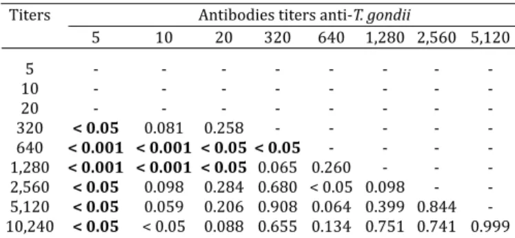
Six mice (4.44%) died from acute toxoplasmosis be-tween 15 and 23 days after inoculation conirmed by ob-servation of tachyzoites in peritoneal luid, lung and liver through optical microscopy and PCR. The antibodies title of anti-T. gondii in both chickens whose tissues were ino-culated in these mice was of 5,120. From 129 surviving mice, T. gondii cysts were found in the brain of 28 mice in 10 different groups. Higher frequency of cysts (P<0.0001, X2=52.87) was observed in mice inoculated with tissues from chickens with high anti-T. gondii antibody titers, but not necessarily from those with highest titers. Correlating the frequencies of cysts in mice and the titers in chicken, a moderate positive correlation (r=0.302) could be observed between these two parameters. This means that, although a greater proportion of mice with cysts were found among the ones inoculated with chicken tissues whose tittering for anti-T. gondii antibodies were medium or high, this re-lation was not directly proportional (Table 2).
Table 1. Number of chickens positive and negative in the modiϐied agglutination test (MAT, cutoff
1:5) and Polymerase Chain Reaction (PCR)
MAT PCR Total
Positive Negative
Positive 16 11 27
Negative 0 13 13
Total 16 24 40
X2 = 12.84; P<0.001. Kappa = 0.486; regular agreement.
Table 2. P values resulting from comparison of the ratios of the number of cysts in the brains of mice inoculated with
tissues from chickens of different titles of antibodies to
Toxoplasma gondii
Titers Antibodies titers anti-T. gondii
5 10 20 320 640 1,280 2,560 5,120
5 10 20 320 < 0.05 0.081 0.258 640 < 0.001 < 0.001 < 0.05 < 0.05 1,280 < 0.001 < 0.001 < 0.05 0.065 0.260 - - 2,560 < 0.05 0.098 0.284 0.680 < 0.05 0.098 - 5,120 < 0.05 0.059 0.206 0.908 0.064 0.399 0.844 10,240 < 0.05 < 0.05 0.088 0.655 0.134 0.751 0.741 0.999 P<0.05 (statistical difference).
Correlation between these parameters associated was cited by Ragozo (2007), obtaining 69.2% isolation (cysts found in brains) in mice inoculated with tissues from sheep with 3200 titer. However, Yai (2007) did not ind differen-ces between the titers from antibodies in capybaras and the percentage of isolates obtained in bioassay mice. Pena (2004) also found greater frequency of isolation in groups of mice inoculated with tissues from cats with high titers for anti-T. gondii antibodies.
Thirty-three mice (25.6%) were positive for T. gondii in the MAT (1:25). Of these, 28 were PCR positive and 5 negative. The number of mice positive and negative MAT and PCR are shown in Table 3.
The four mice positive PCR and negative MAT, were from four different groups (G3, G5, G6 and G14). The titles of the chickens inoculated in these mice, ranged from 320 to 10,240. Besides the samples were positive by PCR were found two cysts in the brains of mice in G3 and G14. Five mice MAT positive and negative by PCR were also from ive distinct groups (G2, G3, G6, G14 and G21) and the titles of chickens between 5 and 5120.
Isolates of T. gondii from MAT-negative mice may be due to absence of seroconversion or low diagnostic sensitivity (87.5%). The lack of seropositivity in infected mice may also have occurred by a prozone effect on MAT. The absence of seroconversion in mice with positive isolation of T. gon-dii was not cited in any previous work done like this. Howe-ver, Pena (2004) by diluting the serum of mice positive in the MAT, to verify the maximum degree of anti-T. gondii an-tibody titers, found in up to 409,600. Although this author has conducted a study with cats, this suggests that mice produce large quantities of antibodies which could explain the occurrence of the prozone phenomenon in MAT. Pena (2004), Yai et al. (2009) and Ragozzo et al. (2008) found no cysts in the brains of mice were seronegative, moreover, these authors did not perform molecular analysis of tissue samples from these mice.
Seven of 28 mice with cysts in the brain were negative by PCR and also had four who did not react serologically with antigens of T. gondii in the modiied agglutination test (MAT). These four animals were inoculated with tissues from
chi-ckens with high titers of anti-T. gondii (over 320). Increased frequency of mice infected in the MAT was found between the groups of chickens inoculated with organs whose evi-dence of anti-T. gondii were 640 (80%), 10,240 (77.8%) and 1,280 (73.3%) (Q2 = 64.24, p<0.0001) as shown in Table 4.
Despite high levels of antibodies to be associated with higher levels of the parasite in tissues, mainly in the brain (Aigner et al., 2010), probably was not obtained isolating in these mice because there were few bradyzoites in the ali-quot used what may have led to not capture these cells in DNA extraction and subsequent ampliication in PCR. Even if the parasite DNA was extracted from this suspension, the same may not have been captured in the sample pipetted. Another reason could be that the parasite found at a loca-tion other than the brain, since no analysis was performed from pool of skeletal muscles of these animals (chickens).
CONCLUSIONS
The occurrence of infection by Toxoplasma gondii in
the farms studied was high; the PCR targeting the B1 gene, does not conirm of the viability of the parasite, but can be used as a screening method for the selection of chickens infected for T. gondii.
Chickens with titers greater than 10 should be prioriti-zed for screening of animals for bioassay, because the chan-ces of isolation of the parasite from them are larger.
The results of this study show that seroconversion in experimentally infected mice is not a good indicator for isolation of the agent.
Acknowledgements.- To the Military Police of Rio Negro municipality, and to the employees and owners of the farms participating in this expe-riment. To FAPESP for inancing part of this study. Beneiciary of inancial assistance CAPES - Brazil.
REFERENCES
Aigner C.P., Silva A.V., Sandrini F., Osório P.S., Pioares L. & Largura A. 2010. Real-time PCR-based quantiication of Toxoplasma gondii in tissue sam-ples of serologically positive outdoor chickens. Mem. Inst. Oswaldo Cruz 105(7):935-937.
Amato Neto V., Medeiros E.A.S., Levi G.C. & Duarte M.I.S. 1995. Toxoplas-mose. 4th. ed. Editora Sarvier, São Paulo, p.154.
Araújo F.G. 1970. Anticorpos anti-Toxoplasma gondii em doadores de san-gue. Revta Inst. Med. Trop., São Paulo, 12(2):105-111.
Araújo F.A.P., Silva N.R.S., Chaplin E.L. & Bigatti L.E. 1989. Prevalência de anticorpos toxoplásmicos em frangos abatidos para consumo humano em Porto Alegre, Rio Grande do Sul. Arqs Fac. Vet. UFRGS 17:23-28. Azevedo D.S., Jamra L.M.F. & Ribeiro M.F. 1983. Isolamento de oocistos de
Toxoplasma gondii em dois bairros do Recife (PE). Revta Inst. Med. Trop. São Paulo 25(1):31-36.
Ausubel F.M., Brent R., Kingston R.E., Moore D.D., Seidman J.G., Smith J.A. & Struhl K. 1999. Short Protocols in Molecular Biology. 4th ed. Sect. 2-3.
Wiley, New York.
Barbosa S.A.A. 2007. Survey of anti-Toxoplasmagondii antibodies by mo-diied agglutination test in chicken slaughtered for consumption in Be-lém city, Northern Brazil. Revta Inst. Adolf Lutz, São Paulo, 66(2):228. Baruzzi R.G. 1970. Contribution to the study of the toxoplasmosis
epide-miology: serologic survey among the Indians of the Upper Xingu River, Central Brazil. Revta Inst. Med. Trop. São Paulo 12(2):93-104.
Coutinho S.G., Souza W.J.S., Camillo-Coura L., Marzochi M.C.A. & Amendo-eira M.R.R. 1981. Levantamento dos resultados das reações de imuno-Table 3. Number of mice seropositive and
seronegative at MAT and positive and negative in the PCR
MAT PCR Total
Positive Negative
Positive 28 05 33
Negative 04 92 96
Total 32 97 129
X2 = 85.7; P<0.001. Kappa = 0.815; excellent agreement.
Table 4. Frequencies of isolation and seropositivity for Toxoplasmagondii by MAT between total mice bioassays distributed between the various titles of
the chickens
Antibodies titers anti- Amount of mouse Isolation in Positive mice
T. gondii (chickens) bioassays mice (%) (MAT) (%) 1:25
5 39 0 a 2.6 a
10 17 0 a 0 a
20 9 0 a,b,e 0 a
320 15 26.7 b,d 26.7 a,b
640 5 100c,f 80 b
1,280 15 60 d,f 73.3 b
2,560 11 27.3 b,d 9.1 c
5,120 9 33.3 b,d 55.6 b,c
10,240 9 44.4 e,d 77.8 b
luorescência indireta para toxoplasmose em 6079 pacientes de am-bulatório ou gestantes do Rio de Janeiro realizadas durante os anos de 1971 a 1977. Revta Inst. Med. Trop., São Paulo, 23(2):48-56.
De Oliveira L.N., Costa L.M., Melo C.B., Silva J.C.R., Bevilaqua C.M.L., Aze-vedo S.S., Muradian V., Araújo D.A.F.V., Dubey J.P. & Gennari S.M. 2009.
Toxoplasma gondii isolates from free-range chickens from the northeast region of Brazil. J. Parasitol. 95(1):235-237.
Dubey J.P. & Desmonts G. 1987. Serological responses of equids fed Toxo-plasma gondii oocysts. Equine Vet. Med. J. 19:337-339.
Dubey J.P. & Beattie C.P. 1988. Toxoplasmosis of Animals and Man. CRC Press, Boca Raton, Florida. 220p.
Dubey J.P., Graham D.H., Blackston C.R., Lehmann T., Gennari S.M., Ragozzo A.M.A., Nishi S.M., Shen S.K., Kwok O.C.H., Hill D.E. & Thulliez P. 2002. Biological and genetic characterization of Toxoplasma gondii isolates from chickens (Gallus domesticus) from São Paulo, Brazil: Unexpected indings. Int. J. Parasitol. 32:99-105.
Dubey J.P., Navarro I.T., Graham D.H., Dahl E., Freire R.L., Prudencio L.B., Sreekumar C., Vianna M.C. & Lehmann T. 2003a. Characterization of
Toxoplasma gondii isolates from free range chickens from Paraná. Vet. Parasitol. 117:229-234.
Dubey J.P., Gennari S.M., Labruna M.B., Camargo L.M.A., Vianna M.C.B., Marcet P.L. & Lehmann T. 2006a. Characterization of Toxoplasma gondii
isolates in free-range chickens from Amazon, Brazil. J. Parasitol. 92:36-40.
Dubey J.P., Sundar N., Pineda N., Kyvsgaard N.C., Luna L.A., Rimbaud E., Oliveira J.B., Kwok O.C.H., Qi Y. & Su C. 2006b. Biologic and genetic cha-racteristics of Toxoplasma gondii isolates in free-range chickens from Nicaragua, Central America. Vet. Parasitol. 42:47-53.
Dubey J.P., Sundar N., Gennari S.M., Minervino H.A., Farias R., Ruas J.L., Dos Santos T.R.B., Cavalcante G.T., Kwok O.C.H. & Su C. 2007a. Biologic and genetic comparison of Toxoplasma gondii isolates in free-range chickens from the northern Pará state and the southern state Rio Grande do Sul, Brazil revealed highly diverse and distinct parasite populations. Vet. Pa-rasitol. 143:182-188.
Dubey J.P., Webb D.M., Sundar N., Velmurugan G.V., Bandini L.A., Kwok O.C.H. & SU C. 2007b. Endemic avian toxoplasmosis on a farm in Illinois: Clinical disease, diagnosis, biologic and genetic characteristics of Toxo-plasma gondii isolates from chickens (Gallus domesticus) and a goose (Anser anser). Vet. Parasitol. 148:207-212.
Ferraroni J.J. & Lacaz C.S. 1982. Prevalência de anticorpos contra agentes causadores da hepatite, malária, síilis e toxoplasmose em cinco popula-ções humanas distintas da Amazônia Brasileira. Revta Inst. Med. Trop., São Paulo, 24(3):155-161.
Ferreira R.A., Mineo J.R., Duarte J.M., Silva D.A.O. & Patarroyo J.H. 1997.
Toxoplasmosis in naturally infected deer from Brazil. J. Wildl. Dis. 33(4):896-899.
Figueiró-Filho E.A., Lopes A.H.A., Senefonte F.R.A., Souza Júnior V.G., Bote-lho C.A., Figueiredo M.A. & Duarte G. 2005. Toxoplasmose aguda: estu-do da freqüência, taxa de transmissão vertical e relação entre os testes diagnósticos materno-fetais em gestantes em estado da Região Centro--Oeste do Brasil. Revta Bras. Ginecol. Obstetr. 27(8):442-449.
Guimaraes A.C., Kawarabayashi M., Borges M.M., Tolezano J.E. & Andra-de H.F.J. 1993. Regional variation in toxoplasmosis seronegativity in the São Paulo metropolitan region. Revta Inst. Med. Trop., São Paulo, 35:479-483.
Holliman R.E., Raymond R., Renton N. & Johnson J.D. 1994. The diagnosis of toxoplasmosis using IgG avidity. Epidemiol. Infect. Dis. 112:399-408. Jamra L.M.F. & Guimarães E.C. 1981. Conversão sorológica para Toxoplas-mose em crianças de um centro de saúde de São Paulo. Revta Inst. Med. Trop., São Paulo, 23(3):133-137.
Larangeira N.L., Ishizuka M.M. & Hyakutake S. 1995. Prevalência da toxo-plasmose eqüina avaliada pelo Teste de Imunoluorescência Indireta, Mato Grosso do Sul, Brasil. Boletín de la Oicina Sanitaria Panamericana 99(2).
Literák I. & Hejlícek K. 1993. Incidence of Toxoplasma gondii in population of domestic birds in the Czech Republic. Avian Pathol. 22:275-281. Pena H.F.J., Gennari S.M., Dubey J.P. & Su C. 2008. Population structure and
mouse-virulence of Toxoplasmagondii in Brazil. Int. J. Parasitol. 38:561-569.
Ragozzo A.M.A., Yai L.E.O., Oliveira L.N., Dias R.A., Dubey J.P. & Gennari S.M. 2008. Seroprevalence and isolation of Toxoplasma gondii from sheep from São Paulo state, Brazil. J. Parasitol. 94(6):1259-1263.
Remington J.S. & Desmonts G. 1990. Congenital toxoplasmosis: Variability in the IgM-luorecent antibody response and some pitfalls in diagnosis. J. Pediatric 83(1):27-30.
Silva R.A.M.S. 2005. Antibodies to Toxoplasmagondii in horses from Panta-nal, Brazil. Vet. Zootec., Botucatu, 12(1/2):20-24.
Smith J.E. & Reduck N.R. 2000. Toxoplasma gondii strain variations and pathogenicity, p.405-431. In: Cary J.W., Linz J.E. & Bhatnagar B. (Eds), Microbial Food-Borne Diseases: Mechanisms of pathogenesis and toxin syntesis. Technnomic Publishing, Mancaster, Philadelphia.
Vergara T.R.C. 1985. Epidemia de toxoplasmose do sistema nervoso cen-tral em enfermos com AIDS na cidade do Rio de Janeiro. Arq. Bras. Med. 59(6):397-406.