UNIVERSIDADE FEDERAL DE SÃO CARLOS CENTRO DE CIÊNCIAS BIOLÓGICAS E DA SAÚDE PROGRAMA DE PÓS-GRADUAÇÃO EM FISIOTERAPIA
ADAPTAÇÕES SENSÓRIO-MOTORAS EM CURTO PRAZO APÓS UMA ÚNICA SESSÃO DE TERAPIA ROBÓTICA ASSOCIADA AO VIDEOGAME
MARCELA DE ABREU SILVA COUTO
ADAPTAÇÕES SENSÓRIO-MOTORAS EM CURTO PRAZO APÓS UMA ÚNICA SESSÃO DE TERAPIA ROBÓTICA ASSOCIADA AO VIDEOGAME
MARCELA DE ABREU SILVA COUTO
Tese de doutorado apresentada ao Programa de Pós-graduação em Fisioterapia da Universidade Federal de São Carlos, como parte dos requisitos par aa obtenção do título de Doutora em Fisioterapia.
Orientador: Prof. Dr. Thiago Luiz de Russo Co-orientador: Prof. Dr. Adriano A. G. Siqueira
Apoio Financeiro: Bolsista de Doutorado pela Coordenação de Aperfeiçoamento de Pessoal de Nível Superior (Capes).
Dedico este trabalho ao meu pai que sempre foi meu suporte, meu incentivo e sempre foi vibrante com todas as minhas conquistas. Obrigada por me permitir voar e por ter sido minha motivação nas horas difíceis. Somos o resultado da sua vida, a continuação de algo que Deus começou em você. Estaremos juntos na eternidade. Serei sempre grata.
é bom; o seu amor dura para sempre”. (Salmos 118) Obrigada por me sustentar, me manter firme, me colocar em segurança e trabalhar por mim quando já não tenho mais condições de alcançar por mim mesma. A Ti o mérito de todas as minhas conquistas. Sem sua presença nos meus dias, nada teria o mesmo significado.
Minha gratidão ao meu parceiro nesta “empreitada”, sempre disposto a encarar os desafios juntos, sempre me faz encontrar um novo sentido, um novo olhar. Obrigada por sempre me mostrar as circunstâncias de um novo ângulo, por me fazer mais otimista, e principalmente por me mostrar como chegar mais perto de Deus. Como é bom chegar até aqui com a nossa família completa! Obrigada por dividir tantos desafios comigo.
Agradeço a minha mãe e a meus irmãos por me apoiarem e me incentivarem em todas as horas. Obrigada pela nossa união. Por se esforçarem em minimizar a distância e não deixarem faltar amor que nos move, nos alimenta! Um agradecimento especial ao Marcus Paulo que não hesitou em estar ao meu lado nos momentos que eu mais precisei. Você é fera mano! Amo vocês.
Agradeço aos orientadores Thiago Luiz de Russo e Adriano A.G. Siqueira. Muito obrigada por me permitirem crescer, pela referência de excelência e maestria, ou poderia até dizer acurácia e destreza. Obrigada por todo o suporte, pelas soluções, pelas longas tardes de trabalho e pela dedicação em multiplicar conhecimento. Agradeço pela oportunidade, pelo precioso tempo, interesse, e principalmente, por não economizarem investimento na minha formação.
Agradeço a querida orientadora Dr. Carolynn Patten que foi muito atenciosa e me recebeu com muito carinho em seu laboratório. Obrigada por ter proporcionado tantas oportunidades de aprendizagem e de crescimento. Muito obrigada pelo suporte, paciência e compreensão nos momentos inesperáveis que vivi durante este período de trabalho. Foi sem dúvida a experiência mais intensa da minha vida!
muito agradável, trouxeram muita coisa boa para a minha formação e para a minha vida. Obrigada pelo apoio, pela ajuda, e pelo companheirismo. Serei sempre grata!
Aos pacientes e voluntários, muito obrigada! Sem vocês não existiria trabalho. Obrigada pela confiança. Que vocês sejam motivados a se superarem sempre. Desejo que vocês sejam inundados pelo renovo de Deus!
AVC- acidente vascular cerebral/encefálico FAC- Functional Ambulation Categories EMG- electromyography
HG- hemiparesis group CG- control group
MVIC- maximum voluntary isometric contraction SD- standard deviation
CV- coefficient of variation RMSE- root mean square error DST- Dynamical Systems Theory
MIT- Massachusetts Institute of Technology
DP- dorsi/plantarflexion IE- inversion/eversion
FMA- Fugl-Meyer Assessment MAS-Modified Ashworth Scale ROM- range of motion
Nm/rad: newton meters per radians i- initial
f- final; s- second
Tabela 1. Characteristics of subjects
Tabela 2. Steadiness
Tabela Material suplementar 1. Game score
MANUSCRITO 2
Tabela 1. Characteristics of Subjects
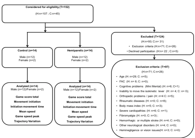
Figura 1. Flowchart of enrolled participants. T: total of recruited individuals, H: hemiparesis group and C: control group. FAC: Functional Ambulation Categories, EMG: electromyography. Corrupted files were the reason for exclusions during analyses except for the EMG and Concentric post-sessions due to two individuals who presented fatigue symptoms (H:2).
Figura 2. Image of patient positioned in the anklebot during the game task.
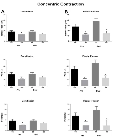
Figura 3. Concentric isokinetic outcomes (torque peak, work and power) ankle dorsiflexors and plantiflexors muscles. CG: control group and HG: hemiparetic group. The asterisk symbol (*) represents the paretic group difference when compared to the control group in pre or post-robotic therapy (p<0.01).
Figura 4. Maintenance of submaximal torques during ankle dorsiflexion or plantarflexor. Representation of generate torque data (black line) and target torque (pink line). A) control group, (B) hemiparesis group during sensorimotor control assessment. Representative data of one individual from each group.
Figura Material complementar 1. Assessment design: MVIC: maximal voluntary isometric contraction. Individuals attended the laboratory over three days. On the second day, they carried out the familiarization protocol. After one week, the evaluation protocol was carried out. Above the line is an indication of rest time between procedures.
Figura Material complementar 2. Maximun EMG peak during torque peak of concentric contraction. CG: control group; HG: hemiparesis Group; DF: dorsiflexion; PF: plantarflexion; *Significant difference between paretic group when compared to the control group in pre or post-robotic therapy (p≥0.05). Significant differences concerning
interaction between pre and post-robotic assisted sessions for both movements.
Figura Material complementar 3. Minimum EMG peak during torque peak of concentric contraction. CG: control group; HG: hemiparesis group; DF: dorsiflexion; PF: plantarflexion; * Significant differences were identified between the paretic group when compared to the control group on pre or post-robotic therapy (p≥0.05). Significant
differences were identified between groups during the dorsiflexion on the tibialis anterior muscle in the pre and post-robotic assisted session. Significant differences between the groups were found only in the post-sessions for the lateral gastrocnemius and soleus muscles during dorsiflexion. The lateral gastrocnemius and soleus muscles presented significant differences between groups just in post-session during dorsiflexion. The hemiparesis group increased when compared to the control group for all situations except for the tibialis anterior muscle in the pre robotic-session, when the hemiparesis group decreased during dorsiflexion. There were no significant differences between the groups in the plantarflexion. There was no interaction between the pre and post-robotic assisted session for both movements.
MANUSCRITO 2
Figura 1. Flowchart of enrolled participants. T: total of recruited individuals, H: hemiparesis group and C: control group. FAC: Functional Ambulation Categories.
Figura 2. Videogame Interface.
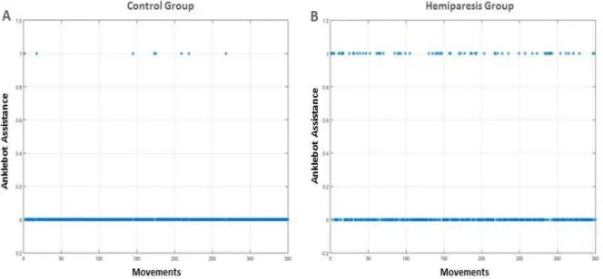
Figura 3. Initiation without robotic assistance. Line 0: no robotic assistance to initiate the movement; Line 1: robot assistance to initiate the movement.
APRESENTAÇÃO 1
CONTEXTUALIZAÇÃO 2
REVISÃO DA LITERATURA 4
MANUSCRITO I 11
MANUSCRITO II 43
CONCLUSÃO GERAL 70
PRODUÇÃO NO PERÍODO 71
BIBLIOGRAFIA DA REVISÃO DA LITERATURA 78
aplicados durante tarefas específicas e orientadas por metas com significados a fim de evocar movimentos coordenados em pacientes com hemiparesia. No entanto, não existem diretrizes suficientes para prescrever a terapia para a reabilitação dos membros inferiores. O primeiro objetivo deste trabalho foi verificar se uma única sessão de terapia robótica promove adaptação motora, influenciando a coordenação dos movimentos do tornozelo (manutenção do torque submáximo do tornozelo - Steadiness), as variáveis de força máxima concêntrica, como torque, potência e trabalho e o desempenho funcional de indivíduos hemiparéticos crônicos; e o segundo objetivo foi descrever as análises dos dados métricos relacionados à precisão, velocidade, suavidade e movimentos iniciados sem assistência, bem como a variação da trajetória registrada durante o protocolo de terapia do tornozelo robótico com base nos critérios de ajuste de aumento e diminuição da assistência robótica. O desenho experimental de ambos os estudos foi double-arm pilot, sendo uma amostra de conveniência de participantes com AVC crônico (n = 14) que apresentaram déficits hemiparéticos residuais e um número igual de sujeitos controle saudáveis pareados por idade e sexo. O equilíbrio, a mobilidade e a função foram avaliados. No primeiro manuscrito, os testes concêntrico isocinético e steadiness foram avaliados utilizando a dinamometria. Os picos máximos e mínimos de ativação muscular foram registados por eletromiografia simultaneamente com os testes concêntricos. No segundo manuscrito, a pontuação do jogo, o número de movimentos iniciados sem assistência, o tempo de iniciação, a velocidade média, o número de picos de velocidade e a variação da trajetória foram variáveis registradas em sete blocos de repetições com níveis variáveis de assistência. Um questionário de motivação foi dado ao grupo hemiparético. Os principais achados foram: adaptações motoras foram identificadas preferencialmente durante a dorsiflexão. O grupo hemiparético apresentou ganhos imediatos na manutenção do torque submáximo, maior destreza, melhor reação inicial e suavidade durante a dorsiflexão após a terapia robótica. O protocolo de terapia robótica promoveu a preservação e manutenção do desempenho neuromuscular quando comparado ao grupo controle saudável. Além disso, a análise dos dados métricos obtidos durante a sessão de terapia robótica corroborou para identificar adaptação motora relacionada à velocidade, precisão e erros na posição da trajetória. Além disso, a estratégia de mudança de impedância foi eficaz para promover um ambiente terapêutico desafiador e adaptações do controle motor.
specific and oriented tasks. However, there are insufficient guidelines to prescribe rehabilitation therapy of the lower limbs. The first aim of study was to verify whether a single session of robotic therapy promotes short-term ankle motor adaptations, influencing the coordinate movements (sub-maximum torque maintenance of ankle – steadiness), maximal strength outcomes, such as torque, power and work and functional performance in chronic post-stroke individuals; and the second objective was to describe the analysis the metric data related to accuracy, speed, smoothness and movement initiated without assistance, as well as trajectory variation recorded during robotic ankle therapy protocol based on the criteria of increasing and decreasing impedance adjustments. Both studies had double-arm pilot design, on a convenience sample of participants with chronic stroke (n = 14) who had residual hemiparetic deficits and an equal number of age- and sex-matched non-disabled control subjects. In the first manuscript, balance, mobility and function were measured. Concentric isokinetic and steadiness tests were assessed using dynamometry. The maximum and minimum muscle activation peaks were recorded by electromyography simultaneously with concentric tests. For submaximal sensorimotor control analysis (Steadiness), the standard deviation, coefficient of variation and root mean standard error (RMSE) were recorded. In the second manuscript, game score, movement initiated without assistance, initiation time, mean speed, number of speed peaks and trajectory variation were variables recorded over seven blocks with variable assistance levels. A motivation questionnaire was given to the hemiparesis group. The main results were motor adaptations in sub maximum maintenance, which were identified preferentially during the dorsiflexion. The hemiparesis group showed greater dexterity, initial reaction and smoothness during dorsiflexion post-robotic assistance therapy. People with chronic hemiparesis presented short-term performance gains in submaximal torque maintenance, especially during dorsiflexion after a single robotic therapy session. The actual robotic therapy protocol promoted preservation and maintenance of neuromuscular performance when compared to the healthy control group. In addition, analyzing the metric data obtained during the robotic therapy session corroborated to identify motor adaptation related to the speed, accuracy and errors in the trajectory position. Moreover, impedance change strategy was effective to promote challenge and motor control adaptation.
APRESENTAÇÃO
Esta tese está organizada conforme as recomendações do Programa de
Pós-graduação em Fisioterapia da Universidade Federal de São Carlos. Inicialmente
são apresentadas uma breve contextualização e uma revisão do problema a ser
abordado. A seguir, os manuscritos intitulados “Short-term adaptations of ankle
sensorimotor control due to a single session of robot therapy in chronic
post-stroke subjects” e “Ankle adaptive sensorimotor strategies in chronic post-stroke
subjects due to variable assistance of robotic therapy and videogame”. Estes
trabalhos foram submetidos a revistas internacionais de alto impacto da área.
Finalmente, uma conclusão geral do trabalho, bem como as atividades
CONTEXTUALIZAÇÃO
O presente estudo é o resultado de uma parceria estabelecida pelo
Laboratório de Pesquisa em Fisioterapia Neurológica do Departamento de
Fisioterapia da UFSCar (LaFiN) e do Laboratório de Reabilitação Robótica do
Departamento de Engenharia Mecânica da Escola de Engenharia de São
Carlos, USP. Verificou-se o efeito de uma sessão de terapia robótica
(Anklebot), associada a um jogo computacional, sobre o controle
sensório-motor de indivíduos com acidente vascular cerebral (AVC), na fase crônica.
Um aspecto inovador vinculado a este estudo relaciona-se ao sistema de
assistência ao movimento utilizado pelo robô, baseado na impedância. Uma
estratégia inédita de acréscimo e retirada gradual de assistência robótica foi
utilizada para se estudar as modificações neuromusculares relacionadas à
força muscular e coordenação do tornozelo (manuscrito 1), bem como os
mecanismos de controle motor frente ao protocolo de treinamento (manuscrito
2).
Esta tese contribuiu para a criação de uma linha de pesquisa no
laboratório e serviu como base para futuras intervenções terapêuticas em
pacientes neurológicos. Além disso, auxilia no desenho de futuros protocolos
de terapia robótica, trazendo informações sobre as estratégias adaptativas
sensório-motoras frente ao tipo de estímulo utilizado e reflexões sobre o uso
deste recurso na prática clínica.
Por fim, este estudo emprega alta tecnologia no tratamento de indivíduos
hemiparéticos crônicos, que estão submetidos a um processo de reabilitação
árduo e prolongado. Muitas vezes este processo é visto pelo paciente como
facilitação de movimentos, com a inclusão do indivíduo em um ambiente
terapêutico inovador e motivacional são necessárias. Além disso, a
propagação do conhecimento relacionado a tecnologias empregadas na
reabilitação corrobora para o aumento da usualidade e acessibilidade de
tecnologias existentes, como também, promove o desenvolvimento de
tecnologias nacionais de custo reduzido que possibilitem ampliar o acesso
REVISÃO DA LITERATURA
O Acidente Vascular Cerebral (AVC) é um grave problema de saúde
pública em todo o mundo. Atualmente, é considerada a segunda principal
causa de morte e a terceira causa de incapacidade. Afeta principalmente os
indivíduos no auge de sua vida produtiva, exercendo um enorme impacto sobre
o desenvolvimento socioeconômico dos países (JOHNSON et al ., 2016). Há
uma previsão realizada pela American Heart Association que considera que se
as medidas apresentadas pelos governos continuarem sendo implantadas com
rigor, haverá uma redução de 20% das ocorrências de AVC em 2020 (GO et al
., 2013).
No Brasil, o AVC ainda está entre as principais causas de morte e
internação. A estimativa é de aproximadamente dois milhões de pessoas com
sequelas de AVC, dentre elas, quinhentos mil apresentam incapacidade grave.
A prevalência de 1,6% ocorre em homens e 1,4% em mulheres, e a de
incapacidade 29,5% em homens e de 21,5% em mulheres (BONSENHOR et
al ., 2015).
Os pacientes que apresentam incapacidades passam por um longo e
complexo programa de reabilitação. Esses indivíduos tornam-se limitados na
realização das atividades de vida diária, como caminhar, subir e descer
degraus, realizar cuidados pessoais e trabalhar (GILES e ROTHWELL, 2008;
MAKI et al ., 2006). Em relação ao caminhar, a fraqueza nos músculos
dorsiflexores que elevam o pé durante a caminhada, comumente referida como
marcha, mesmo em pacientes hemiparéticos deambuladores comunitários que
apresentam um nível satisfatório de recuperação funcional. As duas principais
complicações do pé caído são a falta do apoio do calcanhar e o arrastar dos
dedos do pé durante a fase de balanço, além de ser um fator desencadeante
de quedas e ser um dos principais fatores para a instabilidade durante a
marcha (KNARR et al ., 2013).
O tornozelo desempenha um papel fundamental na locomoção de várias
maneiras. A estabilidade desta articulação contribui para a manutenção da
postura ortostática estável, absorção do impacto durante a locomoção
atenuando a força de impacto com o solo (HANSEN et al ., 2004). Além disto,
também é responsável pela manutenção da velocidade da marcha e promove
uma maior flexão do joelho durante a fase de balanço (KNARR, 2013).Ainda,
atua como um dos principais contribuintes para a estabilidade da marcha.
Dentre os músculos envolvidos no caminhar destacam-se o sóleo,
responsável pela propulsão, o gastrocnêmio atua na estabilidade postural e o
tibial anterior que é fundamental para o final do apoio na retirada do pé (toe-off)
durante a marcha (KNARR et al . 2013). Em relação à ativação muscular de
hemiparéticos, um estudo prévio do nosso grupo de pesquisa demostrou
diminuição a ativação dos músculos agonistas do joelho. Clark et al .
mostraram que a alteração da ativação agonista é a principal causa da
fraqueza de torque-velocidade em sujeitos hemiparéticos crônicos. A
diminuição da ativação agonista e aumento da co-ativação muscular estão
presentes nos membros paréticos durante atividades funcionais. Em tarefas
reduções nas taxas de disparo das unidades motoras do tronozelo
hemiparético.
Exoesqueleto – Anklebot
Roy e colaboradores (2009) desenvolveram um exoesqueleto para
reabilitação do tornozelo, denominado Anklebot (Interactive Motion
Technologies, Watertown, MA), que segue a mesma linha dos robôs para
membros superiores desenvolvidos pelo mesmo grupo de pesquisa.
Desenvolvido no Massachusetts Institute of Technology (MIT), ele possui baixo
atrito e utiliza o controle de impedância para garantir que o sistema seja
backdrivable, ou seja, o robô se deslocará à medida que o paciente exercer
força sobre ele. Os movimentos executados foram plantiflexão e dorsiflexão no
plano sagital.
O controle de impedância, proposto inicialmente por Neville Hogan, em
1985, atua diretamente na especificação da interface de contato entre o robô e
o ambiente. Além disso, também modula a forma como o sistema robótico
reage às perturbações geradas pelo paciente e garante um comportamento
complacente. Uma baixa impedância significa que o robô se deslocará à
medida que o paciente exercer força sobre ele (backdrivability). Portanto, um
sistema robótico com controle de impedância pode ser programado para
permitir que o paciente pós-AVC realize movimento no todo ou em parte,
mesmo quando a tentativa for fraca ou descoordenada. Neste caso, o robô
auxilia o paciente a completar a tarefa.
O Anklebot tem se mostrado efetivo para recuperação da função do
possibilita o tratamento de muitos pacientes com capacidade de deambulação
limitada. Também minimiza riscos de quedas e focaliza o treinamento para
articulações específicas. Assim, o uso da terapia robótica, pode promover
ganhos na função, considerando diferentes períodos e níveis de recuperação
de acidente vascular cerebral através da melhora da coordenação do tornozelo
(FORRESTER et al., 2014; ROY et al ., 2009).
Um melhor desempenho observado na precisão de alvos, na velocidade
e na suavidade durante movimentos de plantiflexão e dorsiflexão sem
assistência foi mostrado em estudos que utilizaram o Anklebot em indivíduos
hemiparéticos crônicos por seis semanas (1-3 dias por semana) (FORRESTER
et al ., 2016; FORRESTER et al ., 2014; ROY et al ., 2011). Além das
melhorias relacionadas ao treino é possível observar a tranferência para tarefas
funcionais. Estudos que avaliam a cinemática da marcha apontam mudanças
na simetria intermembros, ganhos na velocidade e cadência durante a marcha,
mesmo após o treino de repetição na posição sentada (FORRESTER et al .,
2016; ROY et al ., 2011).
Em indivíduos saudáveis, Perez et al . (2004) demonstraram que o
tornozelo obteve o aumento da habilidade motora em curto prazo e o aumento
da excitabilidade cortical referente ao músculo tibial anterior após uma sessão
de repetições passivas. Este aumento da excitabilidade foi associado com a
redução de erros em uma tarefa de desempenho motor do tornozelo, indicando
melhora do controle motor do tornozelo. Assim, se um mecanismo semelhante
é estimulado por um protótipo de tornozelo em sujeitos hemiparéticos, é
possível inferir que novas estratégias motoras poderiam contribuir para
Terapia robótica
O uso de dispositivos robóticos para complementar a terapia
convencional ganhou atenção considerável, pois oferece recursos para o
alcance de muitos objetivos relacionados à aprendizagem motora (HUANG et
al ., 2009; MILLER et al ., 2010).A prática repetitiva em alta intensidade, que
orienta o membro acometido por meio de biofeedback e metas que oferecem
significados a tarefa, tem sido um grande foco de investigação para
restabelecer a função motora em indivíduos com hemiparesia (MEHRHOLZ,
2013; MIRELMAN et al ., 2009).
Os efeitos da terapia robótica foram ampla e previamente estudados em
membros superiores e este conhecimento tem sido utilizado para justificar os
estudos em membros inferiores (MURATORI, et al . 2013). Em relação à
terapia robótica aplicada em membros inferiores, ainda há uma abordagem
estreita considerando que poucas práticas de reabilitação realmente
concentram-se nas alterações do tornozelo comprometido. Ainda, poucos
estudos descrevem os efeitos agudos do protocolo de terapia robótica. A
descrição dos efeitos agudos da terapia robótica corrobora para futuras
propostas do uso da terapia robótica em longo prazo e fornece orientação para
o desenvolvimento de um protocolo desafiador, promovendo assim motivação
para indivíduos hemiparéticos crônicos.
Além disso, dispositivos robóticos podem ser associados a interfaces de
videogames, possibilitando o registro de dados durante a terapia. Esses dados
facilitam o processo de progressão da terapia robótica, bem como,
complementam informações ao processo de avaliação clínica.
Em relação aos efeitos de aprendizagem após uma única sessão, a
retenção e a transferência de habilidades foram demonstradas em um estudo
prévio sobre o Anklebot (ROY A, FORRESTER e MACKO, 2011). Os pacientes
melhoraram a exatidão e a precisão dos movimentos dirigidos por metas. Estas
alterações observadas na análise das variáveis métricas obtidas durnate a
sessão robótica foram associadas ao melhor desempenho observado na
análise da cinemática da marcha. Os ganhos foram mantidos após 48h. Porém
este estudo não incluiu variáveis relacionadas com o desempenho
neuromuscular e a coordenação motora. Estas variáveis foram observadas no
presente estudo que apresenta um protocolo de terapia robótica similar, porém
com estratégias de assistência robótica e videogame diferentes.
Videogame
Videogames têm sido amplamente utilizados em intervenções
somatossensoriais para idosos, pacientes neurológicos e outras populações.
Profissionais que trabalham com a reabilitação de pacientes têm desenvolvido
diferentes abordagens para utilização deste recurso, deixando de ser
brinquedos para diversão para se tornar ferramentas terapêuticas para
reabilitação e promoção da saúde. Green e Bavelier (2003) afirmaram que
jogar videogames efetivamente promove o aumento de habilidades visuais e
atenção dos jogadores, além da contribuição na promoção da memória de
curto prazo, atenção seletiva e motivação para adultos e idosos (GAMBERINI
terapêutico pela utilização de biofeedback visual, sonoro e tátil. O biofeedback
é um estímulo para reforço ou adequação de alguma função ou comportamento
fisiológico, que ajuda jogadores a obter controle consciente sobre específicas
funções ou tarefas (BYUN et al ., 2016). O uso de biofeedback em ambiente
terapêutico também está baseado na literatura que investiga os princípios do
controle motor (LOHSE et al ., 2014)
A utilização desses recursos técnológicos mostra-se promissora em
relação à reabilitação neurológica, porém ainda há muitas lacunas. Por
exemplo, ainda não está claro como a terapia robótica pode influenciar o
controle sensório-motor durante tarefas específicas que exigem força e controle
motor de membros inferiores de indívíduos hemiparéticos crônicos. A
quantidade de repetições ou assistência poderia afetar o desempenho
neuromuscular? Quais seriam as melhores estratégias de biofeedback?
Considerando estas questões, a proposta inicial deste estudo foi verificar se o
protocolo de terapia robótica utilizado promove alterações sobre níveis de força
durante contrações concêntricas e a coordenação motora durante o torque
submáximo. Além disso, analisar a acurácia, a velocidade, a suavidade e o erro
MANUSCRITO 1 – SUBMETIDO À PHYSICAL THERAPY
Title: Short-term adaptations of ankle sensorimotor control due to a single session of robot therapy in chronic post-stroke subjects
Short title: Ankle paresis and robotic therapy
Authors: Silva-Couto MA1, MS, Siqueira AGS, PhD, Santos GL, MS, Russo TL1, PhD
1Department of Physical Therapy, Federal University of São Carlos (UFSCar),
São Carlos, SP, Brazil.
São Carlos School of Engineering2 (EESC), Department of Mechanical
Engineering, University of São Paulo (USP), SP, Brazil.
Correspondence to: Marcela de Abreu Silva-Couto and Thiago Luiz de Russo. Laboratory of Neurological Physical Therapy Research (LaFiN). Department of Physical Therapy. Federal University of São Carlos – UFSCar. Rodovia Washington Luís, km 235, Monjolinho, São Carlos, São Paulo, Brasil, CEP 13565-905. Telephone: +551633519578 / Fax: +551633612081;
E-mail: marcela.deabreu5@gmail.com;thiagoluizrusso@gmail.com
Itemized list of tables and figures: Tables 1 and 2; Figures 1, 2, 3 and 4, Supplementary material: Table 1, Figures 1, 2 and 3.
Number of words: 4.400
Abstract
strength outcomes, such as torque, power and work and functional performance in chronic post-stroke individuals. Methods: This was a double-arm pilot study on a convenience sample of participants with chronic stroke (n = 14) who had residual hemiparesis deficits and an equal number of age- and sex-matched non-disabled control subjects. Balance, mobility and function were measured. Concentric isokinetic and steadiness tests were assessed using dynamometry. The maximum and minimum muscle activation peaks were recorded simultaneously with concentric tests. For submaximal sensorimotor control analysis (Steadiness), the standard deviation, coefficient of variation and root mean standard error (RMSE) were recorded. Results:
A significant difference was identified only when comparing the groups (p=0.001) during the 10-meter walking test, however no significant differences were identified in pre and post-session analyses. Among the pre-session concentric isokinetic variables, the control group presented higher strength levels when compared to the hemiparesis group during movements of
dorsiflexion (p≤0.03) and plantarflexion (p=0.01). Among the post-session concentric isokinetic
variables, the control presented higher strength levels when compared to the hemiparesis group
only for the plantarflexion movement (p≤0.01). There was a decrease in the maximum peak and
an increase in the minimum activation peak observed mainly during dorsiflexion of the paretic group. Regarding the pre and post-robotic assistance sessions, no significant difference was observed for any comparison (p>0.05) except for the steadiness test. Considering the steadiness test initial values, the hemiparesis group had a low performance during dorsiflexion
and plantarflexion (p≥0.03) when compared to the control group. However, the hemiparesis
group showed greater dexterity during dorsiflexion post-robotic assistance therapy (p≥0.02).
Conclusion: People with chronic hemiparesis presented short-term performance gains in submaximal torque maintenance, especially during dorsiflexion after a single robotic therapy session. The actual robotic therapy protocol promoted preservation and maintenance of neuromuscular performance when compared to the healthy control group.
Keywords: Neurological Rehabilitation, videogame therapy, Chronic stroke, Anklebot, Ankle rehabilitation, Assistive Technology.
Introduction
Using robotic systems to complement conventional post-stroke rehabilitation
programs is a promising resource. Robotic devices can provide high-intensity,
repetitive, specific and oriented task practice. They allow for varied and
videogames and biofeedback interactions can promote additional motor control
adjustments and improve motivation, optimizing task performance in individuals
with hemiparesis.2,3 Using robots to treat post-stroke patients has come to the
forefront over the last decade, however there is little information in the literature
on how this type of therapy can affect sensorimotor performance and
relearning.4 In addition, to the best of our knowledge, there are no guidelines
that describe how to design projects, prescribe and use lower limb devices in
clinical practice.5,6
Most studies that described the effects of robotic therapy have focused on the
upper limbs.4,6,7 Moreover, there is little evidence for its effects on the recovery
of the lower limbs and especially the ankle joints.5 Many robotic devices are
used for gait training, seeking to bring stability and correction to knee and hip
movements.8 However, foot drop and the ankle weakness during stance phase
of gait are very common clinical problems. Whereas strength and coordination
of the ankle are critical for the performance of functional tasks, such as
walking.9,10,11
In order to improve the sensorimotor control of the ankle joint, Anklebot, which
is a lower-extremity robotic module, developed at the Massachusetts Institute of
Technology (MIT) has been shown to be effective.1,2,9,12 It isolates the ankle in a
seated position during post-stroke therapy, consequently, helping to treat many
patients with limited ambulatory capacity, consequently, it may improves
function in different stroke recovery periods.2,13 The increase in targeting
accuracy, speed and smoothness in dorsiflexion and plantar flexion variations
over six weeks (1-3 days per week).2,9,12 Furthermore, the observed shifts
toward greater interlimb symmetry, gains in gait velocity and cadence suggest
improvements in the ability to coordinate and control the limbs while walking.9,13
Ankle training in a seated position in response to visual stimuli is related to
training gait function.2,13
The retention of performance and transfer of abilities after a single session
robotic therapy was demonstrated in a previous Anklebot study.12 Patients
improved the accuracy and precision of goal-directed movements. However, it
is still unclear how robotic therapy could influence sensorimotor control during
specific tasks that require strength and motor control. Could the amount of
repetitions or assistance affect neuromuscular performance? In order to
aggregate information on sensorimotor adaptations after robotic therapy, it can
be stated that the innovation of this study was to investigate the influence of the
robotic therapy protocol in activities that require specific ankle control to
maintain the submaximal force. This control is observed by Steadiness test,
which is a sensorimotor control analysis that verifies torque fluctuations during
submaximal contractions around a target torque.
Accordingly, the aim of the study was to verify whether a single session of
robotic therapy promotes short-term ankle adaptations, influencing the
coordination of movements (sub-maximum torque maintenance of ankle –
steadiness), maximal strength outcomes, such as torque, power and work and
functional performance in chronic post-stroke individuals. The hypothesis was
motor control, including force generation and maintenance and these changes
can influence walking test performance.
Material and methods
Ethical Guidelines
The study was conducted according to the guidelines and standards for human
research (Resolution 196/1996, the National Health Council) and it was
approved by the local ethics committee (report no.: 527.556/2014). This was a
double-arm pilot study.
Experimental Design
The hemiparesis subjects were recruited from the Hospital Santa Casa de
Misericordia of São Carlos and the waiting list from the local medical center.
The control subjects were recruited of local community. The individuals with
hemiparesis were not linked to any rehabilitation programs and this experience
was the first contact with robotic therapy. The following inclusion criteria were
considered: 6 or more months post-stroke; men or women aged between 50
and 75 years; low spasticity (less than level 3 on the modified Ashworth scale 14
so that the individual would be able to perform the isokinetic test); and
independent overground walking (levels 2 and 5 according to the Functional
Ambulation Categories). 15 The control group participants had to score greater
than 8 on the Physical Activity Questionnaire Basal, which indicates they were
not sedentary.16,17 This is important because sedentary people have deleterious
control group performed physical activity (mainly aerobic activities) at least 3
times a week. No further criteria regarding the physicalactivity level were used.
The exclusion criteria were: clinical signs of severe heart failure or chronic
metabolic disease; severe cognitive or communication impairments; minimum
score on the Mini-Mental State Examinationaccording to the education level;19
a history of lower limb injuries, deformity or contractures of the ankle joint; a
smaller range of motion than 10o for dorsiflexion and 20o for plantarflexion;
sensory deficits and neglect absence defined by clinical exams. Bell’s 20 and
Clock drawing 21 tests were used to identify the neglect. Six or more errors
indicate visuospatial hemi-neglect in the Bell´s test. Concerning the Clock
drawing test,neglect patients are often found to omit numbers or to place all of
the numbers on the right hand side of the clock face. The statistical power was
calculated at the end of the study using the standard deviation variable of
submaximal sensorimotor control during dorsiflexion (power = 0.96, effect size =
0.7, critical F = 4.2). These values were found using GPower® software.
Participants
The flowchart for participant exclusion and inclusion is presented in
Figure 1. Thus, 28 individuals took part in the study. The hemiparesis group
(n=14) comprised 12 men and 2 women with ischemic and unilateral stroke.
The mean time after stroke was 4.2±6 years. The control group (n=14) also
consisted of 12 men and 2 women who were matched to the hemiparesis group
by age (± 3 years), gender and body mass index (± 4 kg/m2). For ethical
reasons, after protocol, all the hemiparesis participants were invited to attend an
physiotherapists. The control group received information about the importance
of doing regular physical activity.
Procedure and Measuring Instruments
The evaluations were performed as follows: on the first day, screening was
performed to apply the inclusion and exclusion criteria (clinical assessment), as
well as functional tests. The second day was for becoming familiarized with the
evaluation protocol of the robotic session (the protocol of robotic assisted
therapy associated to videogame was defined as a session). After one week,
the evaluation protocol was carried out (Suppl Mat Fig 1).
Clinical assessment
For a detailed description of the hemiparesis group, their balance and mobility
“Timed Up & Go” Test23, and the 10-Meter walking test24. Furthermore, motor
performance and activities of daily living were assessed using the Fugl-Meyer
Assessment for lower limbs25 and Barthel Index 26, respectively.
Assessment protocol and robotic assisted therapy familiarization
The familiarization procedures were conducted as follows: 10-meter walking
test, concentric contraction, maximum voluntary contraction, steadiness and
robotic assisted therapy. The 10-meter walking test was used to verify
functional task (gait performance), concentric contraction to verify force levels,
maximum voluntary contraction to calculate the sub-maximum torque (target
torque), steadiness to verify the ankle sensorimotor control during maintenance
of the sub-maximum torque (target torque). One week after familiarization
procedures, the same assessment protocol was applied in pre and post robotic
assisted therapy session times. In addition, the concentric, steadiness and
10-meter walking tests were redone after the robotic therapy. The sub-maximum
torque value obtained during pre-therapy was used in the post-therapy
steadiness test for comparison purposes.
10-meter test
Participants were instructed to perform a preferred walking speed or
comfortable walk with their usual walking device. The participant time was
recorded for the middle 10-m of the 12-m walkway, and the average gait
velocity was calculated. Three trials were carried out and the average was
recorded for data analysis. A brief rest was given in between trials. 24
Five maximal concentric contractions, during ankle dorsiflexion and
plantarflexion, at a speed of 30°/s were performed using the Biodex System 3
dynamometer (Biodex Multi-joint System 3, Biodex Medical System Inc., New
York). This speed reflects the function of the ankle during walking. 27, 28 The
range of motion testing was set between 10° (dorsiflexion) and 20°
(plantarflexion). Subjects performed three repetitions of dorsiflexion and
plantarflexion movements with submaximal strength in order to familiarize
themselves with the movement.29 During contractions, the subjects were
encouraged by visual feedback to achieve maximum strength. These tests were
carried out with the paretic limb of the hemiparesis group and the dominant limb
of the control group. 29,30 The variables measured were peak torque, work and
power. To complement the interpretation about torque, the electromyographic
methodology and data can be found in the supplementary material (Figures 2
and 3).
Steadiness test
The higher peak torque of three CIVM repetitions was used to calculate the
target strength (20% of CIVM).31 The submaximal sensorimotor control was
evaluated during five repetitions and conducted by visual feedback, displayed
as a horizontal line for 20 seconds, with a 60 second rest between sets. Only
three repetitions were used in the analysis. The first and last sets were
excluded. Considering the initial and final motor adjustments, the three first
seconds and the last second were excluded. The low-pass second-order,
Butterworth filter, with a cut-off frequency of 7Hz was used. The following
(CV), and root mean square error (RMSE). Standard deviation (SD) is the
absolute measurement of the amplitude fluctuation torque. Coefficient of
variation (CV) is a measure of torque fluctuations expressed as a percentage of
average torque (CV=SD/ average torque x 100). SD and CV values for torque
fluctuation were used to probe the subject’s ability to sustain a steady
submaximal contraction at a constant torque level. The root mean square error
(RMSE) is the square of the vertical distance between the target torque and
generated torque.32
Ankle Robotic therapy (Anklebot) and videogame
Anklebot device: The Anklebot robotic system (Interactive Motion Technologies,
Inc., Watertown, MA, USA) is a wearable and backdrivable system that can be
used in a seated position or during walking. It allows actuation and sensing
within the normal range of motion in dorsiflexion/ plantarflexion (DP) and
inversion/ eversion (IE) movements of the ankle joint.1 Anklebot uses an
impedance controller with adjustable stiffness and damping.
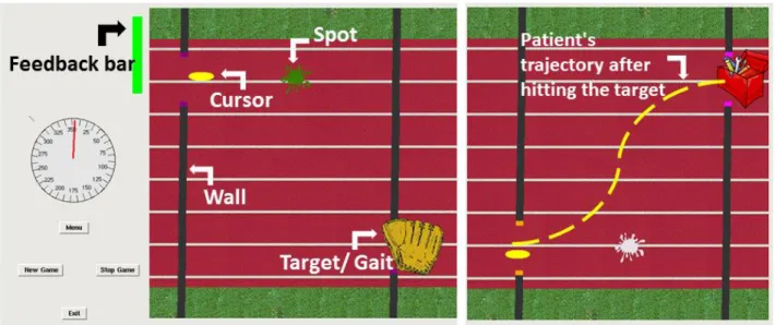
Anklebot setups: The patient was positioned according to the description of Roy
et al (2009).1 The paretic lower limb was positioned at ~45° on a cushioned
knee support, isolating the foot to move freely about the ankle (Fig 2). Subjects
were then introduced to the videogame “race” that was subsequently used to
assess the paretic ankle motor control. The chair was positioned at 1.5 meters
Figure 2: Image of patient positioned in the anklebot during the game task.
Videogame: The movements were visually promotes (movement initiated by
visual feedback) and the objectives were presented in a videogame that
reproduces an obstacle course. The ankle was free to move in the frontal plane,
the patient controlled the ankle during movements of dorsiflexion and plantar
flexion to achieve the targets. This interactive approach used an impedance
controller. The Anklebot control functioned as a cursor showing "up" or "down".
The individual was directed, along a trajectory, by target in two vertical levels
corresponding to 10° dorsiflexion and 20° plantarflexion degrees. Assistance at
the beginning of the movement was provided when necessary, allowing
individuals to achieve the target, i.e., if the subject was not able to start moving
after two seconds, the Anklebot provided torque to initiate the movement
towards the target1,13.
Repetition protocol and varying impedances: 350 ankle movements were
distributed in seven blocks and there was a 3.5-second interval between each
target. The protocol comprised switched robotic-assistance levels that were
modified at each block of 50 movements/targets. The first and the last block
was a gradual impedance increase of 50Nm/rad. The fifth and sixth blocks
presented gradual impedance decrease. Thus, the following impedance
sequence was applied: 0Nm/rad ► 50Nm/rad ► 100Nm/rad ► 150Nm/rad ►
100Nm/rad ► 50Nm/rad ►0Nm/rad.
Game score: The total score recorded in each block was 1500 points. The
player´s aim was to move through the gate passage and reach the target object
(30 points). Bumping the cursor on game obstacles on the wall (-20 points) and
on the spots on the race track (-10 points) were the error possibilities. At the
end of the session, the total number of errors and correct answers were shown.
During the game, according to the performance of the player, a bar remained
green or red as a visual feedback. The Electromyography information and the
game score values are presented in the Supplementary Material (Figure 2 and
3;Table 1; respectively). There was no difference between the groups and
between the blocks (p≥0.05). A video about the game is presented in the
material supplementary (Suppl Mat; Video 1).
Data Analysis
Data were tested for normality and homogeneity (Shapiro-Wilk and Levene
tests, respectively). The demographic and functional data (parametric data),
were applied Independent t-test (group comparison). Regarding 10 meters
walking test, concentric contraction and steadiness variables (parametric data),
ANOVA two-way (group x evaluation) with repeated measures (evaluation: pre
and post-robotic assisted session) were used to verify the effect of the ankle
performed using MatLab software (v.7.0.1) and SPSS for statistics analysis
(v.17). The GraphPrism (v.7) software was used to develop the graphs.
Results
Demographic and Functional data
Groups were similar according to the demographic variables. Both groups
presented mental integrity and independence during activities of daily living
according to the Mini-Mental Exam and Barthel Index, respectively. The
hemiparesis group presented good mobility and no risk of falls according to the
Berg Balance and Timed Up and Go tests. Moreover, the hemiparesis group
presented a reduced level of spasticity and slight motor impairment according to
the Modified Ashworth Scale and Fugl-Meyer Assessment, respectively. Eight
patients presented lesions in the dominant hemisphere. The descriptive data
(e.g., demographic and functional data) are presented in Table 1.
Table 1. Subjects´Characteristics
Control Group
(n= 14) Hemiparesis Group (n=14)
Gender (Male/Female) 11/ 2 11/ 2
Age (years) 64 (74-53) 65 (74-54)
Height (meters) 1.66 (1.83-1.50 1.65 (1.75-1.52)
Body Weight (Kg) 70 (94-55) 71(93-58)
Body Mass Index (Kg/m2) 25.4 (30.3-20.8) 25.8 (35.6-20.7)
FAC (0/1/2/3/4/5) NA (0/0/0/2/12)
Mini Mental 28 (29-24) 26 (29-24)
Barthel Index 20 (20-19) 19.5 (20-17)
MAS (0/1/1+/2/3/4)
Hip NA 10/4/0/0/0/0
Knee NA 5/6/2/1/0/0
Ankle NA 2/8/4/0/0/0
FMA NA 32 (24-42)
TUG test (sec) 8.1(11.1-6.3) 12 (31.1-7.8)*
Measurements are reported as median (maximum and minimum values). FMA: Fugl-Meyer Assessment; FAC: Functional Ambulation Categories: 0=nonfunctional ambulation, 1=ambulatory, dependent for physical assistance, level 2, 2=ambulatory, dependent for physical assistance, level 1, 3=ambulatory, dependent for supervision, 4=ambulatory, independent, level surfaces only, 5=ambulatory, independent; MAS=Modified Ashworth Scale: 0=no increase in muscle tone, 1=slight increase in muscle tone, manifested by a catch and release or by minimal resistance at the end of the range of motion [ROM], 1=slight increase in muscle tone, manifested by a catch, followed by minimal resistance throughout the remainder of the ROM, 2=more marked increase in muscle tone through most of the ROM, but affected parts easily moved, 3=considerable increase in muscle tone, passive movement difficult, 4=affected parts rigid in flexion or extension; TUG test= Timed Up & Go test. *Significantly different compared to control group (p=0.001).
10-meter Walking Test
Differences were identified only in the comparison between groups (F=29.1;
p=0.001). The control group presented an average gait velocity of 2.01±0.4m/s
in the pre-robotic session and 1.9±0.3m/s in the post-robotic session. The
Hemiparesis group presented 1.07± 0.4m/s in the pre-robotic session and
1.04±0.3 m/s in the post-robotic session.
Concentric isokinetic test
Regarding pre-session assessments, the torque peak, work and power
variables presented significant differences between the control and hemiparesis
groups during movements of dorsiflexion (F=4.9; p=0.03; F=7.06, p=0.01;
F=6.05, p=0.01) and plantarflexion (F=7.7, p=0.01; F=10.07, p=0.01; F=10.5,
p=0.01), respectively. Regarding torque peak, work and power in post-session
variables, significant differences between groups were observed only for
plantarflexion movements (F=8.90, p=0.01; F=10.42, p=0.01; F=12.6, p=0.01),
respectively. Regarding to the comparisons between pre and post-robotic
Figure 3: Concentric isokinetic outcomes (torque peak, work and power) ankle dorsiflexors and plantiflexors muscles. CG: control group and HG: hemiparetic group. The asterisk symbol (*) represents the paretic group difference when compared to the control group in pre or post-robotic therapy (p<0.01).
Steadiness
Significant differences were observed between the hemiparesis and the control
group for SD (F=6.6; p=0.018), CV (F=12.90; p=0.002) and RMSE (F=5.00;
p=0.03) during dorsiflexion, and SD (F=8.40; p=0.008), CV (F=27.80; p=0.001)
and RMSE (F=20.10; p=0.001) during plantarflexion. Regarding to the
comparison between pre and post post-robotic assisted session analyses, only
the dorsiflexion movement presented significant differences for SD (F=7.10;
Table 2. Steadiness variables during dorsiflexion and plantar flexion for both groups after and before robotic session.
PRE SESSION POST SESSION
SD CV RMSE SD CV RMSE
DF HG 2.10 (±0.93)* 23.73 (±11.90) * 3.12 (±0.80) * 1.57 (±0.73)
†* 17.35 (±7.16) †* 3.20 (±0.44) *
HG 1.27 (±0.60) 11.87 (±6.10) 2.85 (±0.32) 1.00 (±0.77)† 9.14 (±0.77)† 2.77 (±0.34)
PF HG 1.59 (±1.11) * 17. 65 (±8.75) * 3.71 (±0.45) * 1.33 (±0.81) * 14.68 (±7.94) * 3.53 (±0.62) *
CG 0.56 (±0.45) 4.65 (±4.36) 2.89 (±0.47) 0.80 (±0.58) 5.28 (±4.24) 2.80 (±0.34)
Discussion
This study presents innovations that help prescribe robotic therapy to people with chronic
hemiparesis. The robotic therapy protocol associated to the videogame proved to be safe
and efficient to promote adjustments of ankle sensorimotor control during dorsiflexion,
ensuring the maintenance of neuromuscular performance. According to the aim of the
study, this study showed short-term effects of a single robotic therapy session by observing
the change in sensorimotor control during the submaximal torque maintenance and force
generated capacity during concentric contraction for individuals with chronic hemiparesis.
Thus, the variables observed in this study initiated a discussion about the interaction
between neuromuscular system and the environment due to robotic therapy.
Variable impedance strategy used here showed an improvement in sensorimotor control
during dorsiflexion after robotic therapy, observed in steadiness analysis. A lower torque
variability or movement fluctuation is usually associated with a greater dexterity.31 There
was a decrease in the standard deviation and coefficient of variation variables when the
hemiparesis was compared to the control group in pre and post the robotic therapy
assessments. In addition, it can be observed in Figure 4 that the sensorimotor control of
the hemiparesis ankle presents less variability after the robotic therapy session. This
strategy of improving coordination is related to short-term sensorimotor control
Previous studies that used the Anklebot device associated the changes in motor control to
the gait optimization of people with chronic hemiparesis.1,33 The only variable that we have
in our study related to gait is mean velocity during the 10-meter test. The gait velocity after
the robotic therapy session was the same when compared to the initial value. Therefore,
the functional capacity was preserved, not increased. However, a study conducted by
Forrester et al 13 verified that patients significantly increased their walking speed in the
10-meter test (from 19.1±3.0 to 37.2±4.9 m/s) after a shorter protocol of 200 repetitions
associated with computer games during one day, which was not the case in our study with
chronic patients. It suggests that the earlier the robotic therapy, the more responsive
people with hemiparesis are to the treatment. The 10 meter test only tells us about time
and not about number/length of steps or angular joint measures, thus it is also important to
recommend using kinematic analysis for future studies.
It should be mentioned that we stumbled across an important finding that cannot be
ignored. We observed two patients in this study, who finished the robotic therapy, however,
they showed fatigue, which made it difficult to perform the tests after the robotic session.In
the functional profile analysis of the participants, no characteristic was found to justify this
behavior. Therefore, when proposing a robotic therapy protocol, it is important to establish
criteria to determine specific details concerning progression protocol. In addition, the
difficulty of the task corresponding to the skill level while performing is an important
criterion to prevent frustration, boredom and fatigue when engaging the learner in robotic
Regarding strength level after robotic therapy, subjects maintained their initial torque, work
and power levels, which indicates that the capacity to generate force was not affected by
the intensity, repetitions, impedance change strategy and time of therapy. This is important
because the results do not indicate changes compatible to fatigue (except for two isolated
cases). In fact, an increase in strength after a single robotic therapy session was not
expected, however if the recruitment pattern of muscle fibers was modified, this would be
important information for the discussion on the effects of long-term robotic therapy. A
previous study carried out by Forrester et al .33 demonstrated that strength levels of chronic
hemiparetic individuals were not altered by robotic therapy. After 6 weeks (18 sessions),
individuals with chronic hemiparesis presented a possible change in force exclusively
during dorsiflexion movements (from 74.7 ± 15.9 N to 101.6 ± 11.0 N), but failed to reach
statistical significance, similar to the present study.
Regarding the EMG analysis, alterations in the minimum and maximum muscle activation
of the paretic group were observed when compared to the control group, mainly during the
dorsiflexion movement. Moreover, the graphs showed no significant difference between the
pre and post-robotic assisted session for both groups and all movements (Figures 2 and 3
of Supplementary Material). These results suggest that the sensoriomotor control promoted
by the single robotic-assisted session applied was not enough to change the pattern of
activation of the ankle muscles during maximal contractions. One positive point is that
these findings confirm that muscles do not show signs of fatigue because no decrease in
Regarding the videogame and impedance change criterion, it is worth mentioning that it
represented an equivalent level of challenge for both groups. Based on the presented
results, we can infer that the strategy of impedance change between blocks of 50
movements was effective to promote sensorimotor adaptation. Both groups were
challenged to complete the game, in which the difficulty level was equivalent for both
groups (see Table 1; suppl material game score). The game protocol was similar to the
protocol of Roy et al .38, however, the author used the decreasing criterion for assistance
level changes. The patient started the game with a lot of assistance and ended the game
without assistance. The present protocol presented two situations, one criterion of
increasing assistance and the second criterion of decreasing assistance; however, the
differences between the different assistance levels in the game score analyzed were not
identified. Roy et al .38 were concerned with patient motivation and used the 80% accuracy
criterion to increase the level of difficulty between sessions. Our patients presented levels
greater than 80% accuracy during the game. The association of videogames to robotic
therapy allows for suitable control of important task variables (accuracy, difficult level,
smoothness) and individuals (motivation, enjoyment, adherence) during
rehabilitation.39,40,41 Resources provide meaningful feedback that encourage motor learning
based on motor control principles of movement organization.39,40
Some limitations should be reported, such as the fact that lack of resting periods after
evaluations could affect neuromuscular performance. In addition, no information about
retention can be presented considering the study design. Finally, the evaluations order
Conclusion
Individuals with chronic hemiparesis presented short-term gains in submaximal torque
maintenance performance (sensorimotor control) during dorsiflexion after a single robotic
therapy session. Subjects with chronic hemiparesis presented greater dexterity in the
dorsiflexion movement after a single robotic assistance therapy. Thus, the actual robotic
therapy protocol proved to be useful for patients who presented preservation and
maintenance of neuromuscular performance when compared to the health control,
considering concentric torque, muscle activation and function.
Considerations for clinical practice: It is necessary to comply with the functional
capacity of the patient by screening, indicating whether or not he/she is eligible for this
therapy. This study presented references for protocol design that can be used for a
long-term robotic therapy program. In addition, the association with other exercises is possible,
considering that the robotic treatment protocol is accomplished in 30 minutes and a
conventional session in approximately 1 hour.
Suppl Mat Figure 1. Assessment design: MVIC: maximal voluntary isometric contraction. Individuals attended the laboratory over three days. On the second day, they carried out the familiarization protocol. After one week, the evaluation protocol was carried out. Above the line is an indication of rest time between procedures.
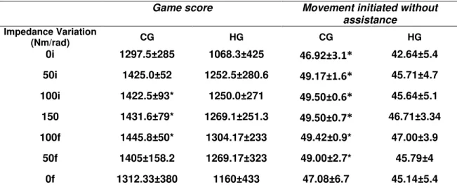
Suppl. Mat Table 1. Game score.
1◦block
(0Nm/rad) 2
◦ block
(50Nm/rad)
3◦ block
(100Nm/rad
)
4◦ block
(150Nm/rad
)
5◦ block
(100Nm/rad
) 6
◦ block
(50Nm/rad) 7
◦ block
(0Nm/rad)
CG 1297.5±285 1425.0±52 1422.5±93 1431.6±79 1445.8±50 1405±158.2 1312.33±380
HG 1068.3±425 1252.5±280.6 1250.0±271 1269.1±251.3 1304.17±233 1269.17±323 1160±433
CG: control group; HG: hemiparesis group. Impedance or exoskeleton assistance K= Newton meters per radians (Nm/rad). The comparison between game scores during several blocks did not present significant
differences (p≥0.005).
Assessment of muscle activity
Electromyography activity was simultaneously assessed during concentric isokinetic tests.
Tibialis anterior (TA), peroneus longus (PL), medial gastrocnemius (GM) and lateral
gastrocnemius (LG) and soleus (SL) activations were investigated. Maximum and minimum
activation for each muscle were measured in millivolts (mV) [30]. The EMG signals were
acquired using an 8-channel recording system. A portable system (Myomonitor IV, Delsys,
Boston, USA) at a sampling rate of 2000 Hz using rectangular-shaped (19.8 mm wide and
35 mm long) bipolar surface electrodes with 1 x 10 mm 99.9% Ag conductors and an inter
-conductor distance of 10 mm were also used. The system has an input impedance of
>1015Ω // 0.2pF, a common mode rejection ratio of >80 dB, signal-to-noise ratio<1.2 μV
parallel bars (1mm2 x 1 cm, 99.9%) double-phase (Delsys) was used. The signals were
corrected for offset and filtered at 20 to 400 Hz using a fourth-order, zero-lag Butterworth
band-pass filter. The root mean square amplitude was quantified with a window duration of
20 milliseconds and a temporal overlap of 50%. The maximum and minimum muscle
activation was calculated (in millivolts) for each repetition.
Statistical Analysis
For the maximum and minimum muscle activation (nonparametric data), a Mann- Whitney
U test (hemiparetic x control group) and a Wilcoxon test (pre and post-robotic assisted
session) were used. Delsys-EMG data processing was performed using MatLab software
(v.7.0.1) and SPSS v.17 for statistics analysis.
Muscle activity - EMG
Maximum activation: The anterior tibialis muscle was lower during dorsiflexion in the
hemiparesis group when compared to the control group at pre (p=0.03) and post-session
(p=0.01) assisted robotic therapy assessments. Peroneus longus and gastrocnemius
lateral muscles were lower during dorsiflexion in the hemiparetic group when compared to
the control group at post-session (p=0.02). The soleus muscle was lower during
dorsiflexion in the hemiparetic group when compared to the control group at pre-session
(p=0.01). Regarding the plantarflexion, only the medial gastrocnemius muscle was lower
(p=0.01) There was no significant difference between the pre and post-robotic assisted
session analyses for both movements and groups (Suppl Mat Fig 2).
Minimum activation: The anterior tibialis muscle was higher during dorsiflexion in the
paretic group when compared to the control group at pre (p=0.03) and post-session
(p=0.04) assisted robotic therapy assessments. The lateral gastrocnemius (p=0.01) and
Soleus (p=0.02) muscles were higher during dorsiflexion in the hemiparetic when
compared to the control group at the post-session. There were no significant differences
between the groups during plantarflexion. There was no significant difference between pre