UNIVERSIDADE DA BEIRA INTERIOR
Ciências da Saúde
Bisphenol A effects in transthyretin and barrier
integrity markers in the choroid plexus:
implications in amyloid beta catabolism
Ana Catarina Abreu Duarte
Dissertação para obtenção do Grau de Mestre em
Ciências Biomédicas
(2º ciclo de estudos)
Orientador: Prof. Doutora Cecília Santos
Coorientador: Prof. Doutora Helena Marcelino
Acknowledgments
This work would not be possible without the contribution of diverse people, to which I have to thank.
First, I would like to thank PhD professor Cecília Santos for receive me in the neuroscience group, for the opportunity to develop a work in a field of my interest, for all scientific knowledge, and for the guide over this year.
To PhD professor Helena Marcelino, for the scientific knowledge transmitted, for all support in laboratory activities and for the important advices that lead me over this journey, and allowed me to realize the present work.
To pass for the diverse situations that had emerged many other persons had contributed to my success, namely to MS Joana Tomás for its availability and advices, to Ana Raquel Costa for the friendship and for be always available to help, to Nádia Morete and Flávio Neves, two great lab colleagues and friends, with whom was always possible to discuss the experiments and rely. Also would like to thank to my other group colleagues of the neuroscience group for contribute to this great experience in my life.
For last, I thank to all my friends and my family for all comprehension and support, and in specially to my boyfriend André, for listen and support me, and principally, for always encourage me to keep following until the end.
Abstract
The choroid plexus is a multifunctional tissue responsible for a wide range of homeostatic functions crucial to the central nervous system, including secretion of cerebrospinal fluid, synthesis and secretion of important peptides and regulation of the chemical substances exchange between the blood and the cerebrospinal fluid, through the blood-cerebrospinal fluid barrier. Transthyretin, a protein highly expressed and secreted by choroid plexus to the cerebrospinal fluid, is the major amyloid-beta scavenger protein, contributing to its clearance. Sex hormones, as estrogens, upregulate transthyretin expression in choroid plexus, and as a consequence its regulation may be disrupted by substances that interfere with various cellular pathways regulated by endogenous hormones, known as endocrine disruptors chemicals. The human population is exposed to many chemicals with such properties, such as bisphenol A. Therefore, the present study analysed the effects of the endocrine disruptor bisphenol A on transthyretin expression in newborn rats by Whole-Mount fluorescent staining and Western blot, and at the mRNA level by Real time RT-PCR. Moreover, the effects of beta-amyloid on transthyretin expression were also investigated using the same techniques in choroid explants of newborn and young rats. Blood-cerebrospinal fluid barrier plays an important role in the regulation of molecules movement between choroid plexus and cerebrospinal fluid, and it disruption can happen when choroid plexus functions are impaired. Thus, one purpose of this work was determine the effects of both compounds, beta-amyloid and bisphenol A, in blood-cerebrospinal fluid barrier integrity through the evaluation of some membrane protein levels present in this barrier, namely, occludin, E-cadherin, claudin-1 and zonula occludens-1. Beta-amyloid treatment in rat choroid plexus seems to trigger transthyretin upregulation, in a dose-response manner. Transthyretin mRNA levels in newborn rat choroid plexus explants increased much more than in young explants. Increased transthyretin expression levels were not correlated with secretion levels. Additionally, beta-amyloid at 1ug/mL increased reactive oxygen species production in choroid plexus. Low doses of bisphenol A affected transthyretin expression in rat choroid plexus in a non-monotonic dose response way, accordingly to data previously reported in other studies with bisphenol A. The same response profile was observed in transthyretin protein and mRNA levels measured, with higher transthyretin levels verified at 50nM of bisphenol A. As reported before with beta-amyloid treatment, also bisphenol A lead to an increase of transthyretin expression in choroid plexus cells, which was not altered with significance the secretion levels of this protein.
Beta-amyloid and bisphenol A clearly influence transthyretin expression in rat choroid plexus, in a dose-response manner, and in a non-monotonic dose response, respectively. In accordance to previous reports, increasing beta-amyloid levels induced transthyretin upregulation.
Increased transthyretin production by choroid plexus seems to be a protective mechanisms to avoid beta-amyloid fibrillization and consequent toxicity. Bisphenol A interfered with transthyretin expression, in both positive and negative ways. Therefore, bisphenol A levels might lead to up or down of transthyretin regulation, and consequently, leading to impairment of beta-amyloid levels in brain. Blood-cerebrospinal fluid barrier integrity might be compromised by beta-amyloid and bisphenol A injuries, which explains alteration in secretion rates of controls for treated choroid plexus explants. However, further investigation is required to analyse evolution of transthyretin expression by choroid plexus throughout life, and would be also important evaluate bisphenol A effects in blood-cerebrospinal fluid barrier protein levels, to better understand bisphenol A consequences in beta-amyloid clearance.
Keywords
Choroid Plexus, Transthyretin, Amyloid-Beta, Bisphenol A, Blood-cerebrospinal fluid barrier, Tight junctions
Resumo alargado
No sistema ventricular cerebral encontram-se 4 plexos coróides, um em cada ventrículo, os quais desempenham importantes funções, diretamente envolvidas na homeostasia do sistema nervoso central. Destas funções destacam-se a secreção do líquido cefalorraquidiano, a síntese e secreção de inúmeras substâncias bioactivas (proteínas, citocinas, vitaminas), bem como a regulação da passagem de moléculas entre a corrente sanguínea e o líquido cefalorraquidiano, através da barreira sangue-líquido cefalorraquidiano. Esta barreira formada pelas células epiteliais do plexo coróide é composta por inúmeras proteínas membranares denominadas tight
junctions e adherens junctions, que são fundamentais para manter a sua integridade, e
assegurar a função secretora do plexo coróide. O plexo coróide é ainda responsável pela síntese e secreção da transtirretina, a principal proteína envolvida no catabolismo do péptido beta amilóide, cuja deposição no cérebro é uma das principais marcas etimológicas da doença de Alzheimer. Quando secretada para o líquido cefalorraquidiano, a transtirretina forma complexos estáveis com o beta-amilóide, evitando a sua agregação e fibrilação, e consequentemente, os efeitos tóxicos inerentes à acumulação destes agregados. A expressão da transtirretina é regulada positivamente pelas hormonas sexuais, nas quais se incluem os estrogénios. Estas hormonas podem ver as suas ações mimetizadas por certos compostos denominados por disruptores endócrinos, cuja crescente difusão no meio ambiente e entre as populações tem levantado várias questões sobre a sua implicação na saúde pública. Um dos disruptores endócrinos mais estudado é o bisfenol A, um xenoestrogénio, altamente difundido no meio ambiente, e ao qual a exposição humana foi comprovada em inúmeros estudos. Posto isto, este trabalho tem como principal objetivo avaliar o efeito do bisfenol A na expressão da transtirretina, em explantes de plexo coroide de rato, e a sua consequente relação no catabolismo do beta-amilóide. Além disto, também se averiguou se a expressão da transtirretina no plexo coróide sofre modificações, em ratos de diferentes idades, e se essa expressão tem alguma relação com o aumento dos níveis de beta-amilóide. Adicionalmente, a expressão de algumas proteínas descritas na barreira sangue-fluido cefalorraquidiano (occludina, e-caderina, claudina-1 e zonula occludens-1) foi avaliada nos explantes de plexos coróides após o tratamento com o beta-amilóide e o bisfenol A, de modo a perceber se estes compostos interferem na integridade desta barreira.
A expressão da transtirretina foi analisada ex vivo, em explantes de plexos coróides de ratos recém-nascidos e de ratos jovens tratados com beta-amilóide e, em explantes de recém nascidos tratados com bisfenol A. Para tal, recorreu-se a várias técnicas: Whole mount (imunohistoquímica por fluorescência), Real-time PCR e Western Blot. A localização e expressão das proteínas membranares foi estudada através de Whole mount e Western blot.
Nos explantes de plexo coróide tratados com beta-amilóide houve um aumento na expressão da transtirretina, correlacionado com o aumento da concentração de beta-amilóide, que se verificou em ambas as idades estudadas, de forma semelhante. A maior diferença ocorreu relativamente à expressão de mRNA da transtirretina nos explantes dos animais recém-nascidos, onde os níveis de transtirretina são bastante elevados, mais do que nos explantes dos jovens. Por outro lado e, contrariando o esperado, não houve um aumento na excreção desta proteína, mas sim um ligeiro decréscimo. Foi ainda avaliada a produção de espécies reativas de oxigénio nos explantes de plexo coróide de ratos jovens onde houve um aumento significativo relativamente aos controlos. Nos plexos coróides tratados com bisfenol A, verificaram-se modificações na expressão da transtirretina mesmo com baixas doses do composto, as quais estão de acordo com os níveis referidos em estudos epidemiológicos. Além disto, os níveis de expressão da transtirretina nestes explantes mostraram seguir uma curva com resposta não-monotónica, tal como observado em vários outros estudos, e característico da exposição aos disruptores endócrinos. A expressão da transtirretina nos plexos coroides foi maior para a concentração de 50nM de BPA comparativamente às outras concentrações testadas, tanto para os níveis da proteína como para os de mRNA. Contudo, a secreção da transtirretina não acompanhou o aumento da sua expressão.
Tanto o beta-amilóide como o bisfenol A mostraram-se capazes de interferir na expressão da transtirretina no plexo coróide de rato, de forma dose-dependente e não-monotónica, respetivamente. O aumento da produção de transtirretina pelo plexo coróide quando os níveis de beta amilóide estão aumentados parece tratar-se de um mecanismo de proteção para evitar a agregação do péptido e consequente toxicidade. Relativamente ao bisfenol A, este consegue modular a expressão da transtirretina tanto positivamente como negativamente, o que poderá ter consequências nos níveis de transtirretina produzidos e libertados para o líquido cefalorraquidiano, e assim, interferir nos níveis de beta amilóide no cérebro. Tendo em conta as discrepâncias observadas entre os níveis de expressão da transtirretina e da sua secreção, para ambos os compostos estudados, a integridade da barreira sangue-líquido cefalorraquidiano poderá estar comprometida, e dessa forma, contribuir para o desequilíbrio do beta-amilóide. Assim, tanto o beta-amilóide como o bisfenol A interferem na produção de transtirretina no plexo coróide de rato. Contudo, é necessário investigar a relação entre a transtirretina e o beta-amilóide ao longo do envelhecimento, para perceber se é a diminuição da transtirretina a responsável pela acumulação do beta-amilóide, nomeadamente na doença de Alzheimer, ou se é o péptido que contribuiu para a disfunção do plexo coróide levando à diminuição da transtirretina. Será ainda importante investigar os níveis das proteínas da barreira sangue-líquido cefalorraquidiano, para melhor entender de que forma o bisfenol A pode levar a um aumento de beta amilóide, ao interferir não só na expressão da transtirretina como também nas funções do plexo coróide.
Palavras-chave
Plexo coróide, transtirretina, beta-amiloide, bisfenol A, barreira sangue-líquido cefalorraquidiano, tight junctions
Index
I. Introduction ... 1
1. Choroid Plexus ... 2
1.1. Functions ... 3
1.1.1. Production and secretion of cerebrospinal fluid ... 3
1.1.2. The Blood-Cerebrospinal Fluid Barrier ... 4
1.1.2.1. The Epithelial Junctional Complex of the Blood-cerebrospinal fluid barrier 5
1.1.2.2. Principal adherens and tight junctions proteins of the blood-cerebrospinal fluid barrier ... 7
1.1.3. Protein Synthesis ... 8
1.2. Transthyretin ... 8
1.2.1. Transthyretin role in Amyloid Beta clearance ... 9
1.2.2. Transthyretin regulation by Sex Hormones ... 10
2. Implications of Choroid Plexus Senescence ... 11
2.1. Amyloid Beta Metabolism ... 11
2.2. Choroid plexus dysfunction and Amyloid Beta accumulation in brain ... 12
3. Endocrine Disruptors ... 13
3.1. Bisphenol A ... 14
3.1.2. Bisphenol A metabolism and molecular mechanism of action ... 14
3.1.3. Bisphenol A levels in human tissues and fluid ... 15
3.1.1. Metabolic effective dose ... 16
II. Aim ... 17
III. Materials and Methods ... 19
1. Animals and Z310 cell line ... 20
1.1. Cell Culture ... 21
1.1.1. Cells Passage ... 21
1.1.2. Cell Counting ... 21
1.1.3. Cells Freezing and Thawing ... 22
2. Whole Mount fluorescent staining ... 22
2.2. Confocal microscopy images analysis ... 23
3. Western Blot ... 23
3.1. Protein Extraction ... 23
3.1.1. CP explants ... 23
3.1.2. Z310 cells ... 24
3.2. Western Blot – TTR, Occludin, E-cadherin and Cld-1 ... 24
4. Extraction of total RNA ... 24
4.1. Extraction ... 24
4.2. Determination of total RNA integrity ... 25
4.3. Quantification of total RNA ... 25
5. cDNA synthesis ... 25
6. PCR ... 26
7. Real-Time RT-PCR ... 27
8. Reactive Oxygen Species Assay ... 27
9. Statistical analysis ... 28
IV. Results ... 29
1. Protocols optimization and establishment ... 30
1.1. Expression and localization of TTR in rat CP ... 30
1.1.1. Whole Mount fluorescent staining ... 30
1.1.2. Western Blot ... 30
1.1.3. RT-PCR ... 32
1.2. Localization and expression of tight and adherens junctions proteins at the blood-cerebrospinal fluid barrier of rat CP ... 32
1.2.1. Whole-Mount fluorescent staining ... 32
1.2.2. Western Blot ... 34
2. TTR expression in rat Choroid Plexus in response to A-Beta insult ... 35
2.1. Evaluation of TTR levels after treatment with A-Beta42 for 24 hours ... 35
2.1.1. Whole-Mount fluorescent staining ... 35
2.1.2. Real Time RT-PCR ... 37
2.1.3. Western Blot ... 38
2.2. Blood-Cerebrospinal fluid barrier membrane proteins expression in response to A-Beta insult ... 39
2.2.1. Whole-Mount fluorescent staining ... 39
2.3. Amyloid Beta oxidative stress in choroid plexus: production of reactive oxygen species ... 44
3. BPA effects in TTR expression in rat Choroid Plexus ... 46
3.1. TTR expression after 24 hours of BPA treatment ... 46
3.1.1. Whole-Mount fluorescent staining ... 46
3.2. TTR expression after 6 hours of BPA treatment ... 48
3.2.1. Whole-Mount fluorescent staining ... 49
3.2.2. Real-Time RT-PCR ... 50
3.2.3. Western Blot ... 51
V. Discussion ... 53
VI. Conclusion & Future Perspectives ... 57
Figures list
Figure 1 - Ventricular system of brain and CSF circulation. ... 2
Figure 2 - Barrier interfaces in adult and developing brain. ... 4
Figure 3 - Scavenger function of TTR. ... 9
Figure 4 - Correlation between CP dysfunction and accumulation of A-Beta in brain... 13
Figure 5 - Scheme of experimental studies about BPA and A-Beta effects in TTR and TJs BCSFB proteins levels, in rat choroid plexus explants. ... 20
Figure 6 - Expression and localization of TTR in rat CP by confocal microscopy. ... 30
Figure 7 - Western blot optimization protocol of TTR protein in Z310 and CP. ... 31
Figure 8 - TTR Western blot performed with culture medium samples after incubation for 6h with and without CP. ... 31
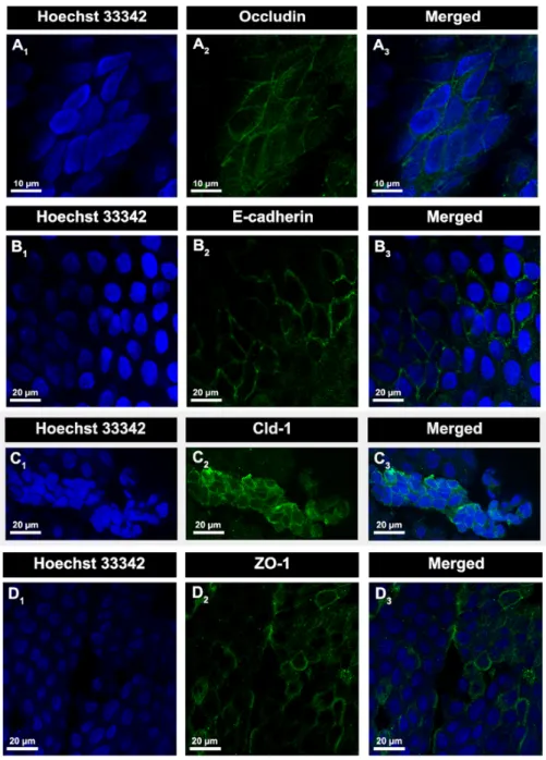
Figure 9 - Electrophoresis in 1.5% agarose gel stained with GreenSafe of cDNA PCR products of TTR gene. ... 32
Figure 10 - Localization and expression of some TJs and AJs proteins of BCSFB, in rat CP explants. ... 33
Figure 11 - Western blot optimization and establishment to membrane protein of BCSFB .... 34
Figure 12 - A-Beta42 effect in TTR expression in rat CP explants at different ages. ... 37
Figure 13 - Comparison of TTR mRNA levels by Real time RT-PCR in newborn and young rat CP explants treated with A-Beta42 for 24h. ... 38
Figure 14 - Evaluation of A-Beta42 effect in TTR secretion by Western blot. ... 39
Figure 15 - A-Beta42 effect in occludin protein expression in rat CP explants ... 40
Figure 16 - A-Beta42 effect in E-cadherin protein expression in rat CP explants ... 41
Figure 17 - A-Beta42 effect in Cld-1 protein expression in rat CP explants. ... 42
Figure 19 - Ex vivo effects of A-Beta42 on ROS production in young rat CP explants ... 45
Figure 20 - TTR expression in newborn CP explants after 24h of BPA treatment ... 48
Figure 21 - TTR expression in rat CP explants after 6h of BPA treatment ... 50
Figure 22 - Comparison of TTR mRNA levels by Real time RT-PCR in CP explants treated for 6h with BPA.. ... 51
Abbreviations and Acronymes List
A-Beta Amyloid-Beta AD Alzheimer’s Disease AJ Adherens Junction Apo Apolipoprotein APP Amyloid Precursor Protein BBB Blood-Brain Barrier
BCSFB Blood-Cerebrospinal Fluid Barrier BPA Bisphenol A
BSA Bovine Serum Albumin
cDNA Complementary Deoxyribonucleic Acid Cld Claudin
CNS Central Nervous System CP Choroid Plexus CPEC Choroid Plexus Epithelial Cells CSF Cerebrospinal Fluid
DCFHA-DA 2,7-Dichlorodihydrofluorescein Diacetate DEPC Diethylpyrocarbonate
DMEM Dulbecco’s Modified Eagle Medium DMSO Dimethyl Sulfoxide ED Endocrine Disruptor EDC Endocrine Disruptor Chemical ER Estrogen Receptor
JAM Junctional Adhesion Molecule PBS Phosphate Buffered Saline PCR Polymerase Chain Reaction PFA Paraformaldehyde PMSF Phenylmethylsulfonyl Fluoride PVDF Polyvinylidene Difluoride RBP Retinol Binding Protein RNA Ribonucleic Acid ROS Reactive Oxygen Species
RT-PCR Reverse Transcription Polymerase Chain Reaction SDS Sodium Dodecyl Sulfate
TJ Tight Junction TTR Transtyretin ZO Zonula Occludens
1. Choroid Plexus
Choroid plexuses (CPs) are highly vascularized tissues, with a simple structure, confined to the ventricular system of the brain (Figure 1). This system is divided in four ventricles: two laterals, one third and a fourth, and each one detains the localization of one choroid plexus (CP) (Johansson et al., 2008, Wolburg and Paulus, 2010, Damkier et al., 2013). In the lateral ventricles of the mammalian brain, CPs form a sheet-like structure, whereas in the third and fourth ventricles, branched villus-like structures are observed instead (Damkier et al., 2010). CP is formed by a single layer of cuboidal epithelial cells that reside on a basement membrane. This epithelial basement membrane is characterized as a network of fenestrated capillaries surrounded by connective tissue composed by fibroblasts and immune cells (e.g., mast cells, macrophages, granulocytes), and a rich extracellular matrix (Miyoshi and Takai, 2005, Wolburg and Paulus, 2010, Terry et al., 2010, Damkier et al., 2013). The choroid plexus epithelial cells (CPEC) are connected by tight junctions (TJs), adherens junctions (AJs) and desmosomes (Damkier et al., 2013). CPEC contain numerous mitochondria, Golgi apparatus, smooth endoplasmic reticulum and lysosome-like vesicles which demonstrate their intense synthetic capacity (Marques et al., 2013). Ependymal cells have numerous microvilli from the ventricle facing (apical side), and extensive infolding at blood facing (basolateral side), thus providing a large surface for contact between epithelium and the cerebrospinal fluid (CSF) the apical side and epithelium and the CP interstitial fluid on the basolateral side (Skipor and Thiery, 2008).
Superior sagittal sinus Choroid Plexus Aquaductus ceribi Foramen of Magendie Foramen of Monro 3rd ventricle 4th ventricle Foramina of Luschka Arachnoid granulations Lateral ventricles
Figure 1 - Ventricular system of brain and CSF circulation. CSF flow forms in lateral CPs, drains via the
foramen of Monro to the 3rd ventricle and of this to 4rd ventricle through aquaductus ceribi/Sylvius. CSF
leaves ventricular brain thorugh foramina of Luschka and foramen of Magendie. Adapted from (Damkier et al., 2013).
1.1. Functions
CP is a multifunctional tissue responsible for a wide range of homeostatic functions crucial for the central nervous system (CNS), among which stand out: secretion of the CSF, regulation of the exchange of chemical substances from the blood to the CSF through the blood-cerebrospinal fluid barrier (BCSFB), and synthesis and secretion of important biologically active substances as vitamins, growth factors, peptides and hormones (Serot et al., 2000, Johansson et al., 2008, Skipor and Thiery, 2008, Johanson et al., 2011b). These functions will be clarified in the next sections, with more emphasis on the characteristics of BCSFB and synthesis of transthyretin (TTR) due to their role in the present work.
1.1.1. Production and secretion of cerebrospinal fluid
CP is responsible for the constant formation and drainage of CSF, producing about 70-80% of this fluid (Skipor and Thiery, 2008, Johanson, 2008). This allows the formation of a unique circulatory system capable to perform diverse metabolic and signalling functions, which directly affect brain homeostasis (Johanson, 2008). CPs from the lateral ventricles form the flow of the CSF, which drains via the foramen of Munro into the third ventricle, and then by the Aqueduct of Sylvius into the fourth ventricle. At this point, CSF leaves the ventricular system and passes through paired foramens of Luschka and foramen of Magendie into the subarachnoid spaces where it fills the basal cisterns of the brain and the spinal cord (Figure 1). The return of the CSF occurs either directly, via arachnoid villi in the venous sinuses of the brain, or via lymphatic drainage pathways (Segal, 2001, Smith et al., 2004, Skipor and Thiery, 2008, Lehtinen et al., 2013). CSF functions in the CNS include physical protection, intracranial pressure regulation, waste removal and provision of a supportive environment (Serot et al., 2003, Johanson et al., 2008, Erickson and Banks, 2013). The CP is an important source of molecules that circulate in the CSF, contributing with compounds like vitamins, hormones and peptides (Skipor and Thiery, 2008, Spector and Johanson, 2013). Whether CSF plays an important role in controlling and maintaining a proper environment to the CNS, CSF homeostasis depends mainly of CP. Composition of CSF is strictly regulated by the transport systems expressed in CPEC, located at the basolateral and apical membranes, which are essential for the bidirectional movement of various substances across the BCSFB (Redzic et al., 2005, Coisne and Engelhardt, 2011, Erickson and Banks, 2013). As the CSF rapidly and widely disseminates the substances circulating on it that have crossed the BCSFB, throughout the CNS, stability and integrity of this barrier is essential to avoid the access of deleterious substances to brain (Johanson et al., 2011a).
1.1.2. The Blood-Cerebrospinal Fluid Barrier
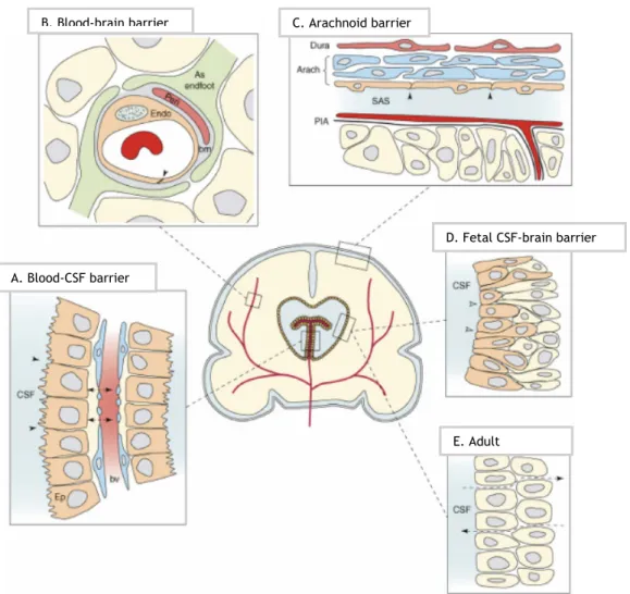
Three barrier layers limit and regulate molecular exchange at the interfaces between the blood and the neural tissue or its fluid spaces: the blood-brain barrier (BBB), the BCSFB and the arachnoid barrier (Figure 2) (Abbott et al., 2006, Ueno, 2007, Abbott et al., 2010). CNS barriers have an essential role in CNS homeostasis maintenance, establishing a stable and adequate environment that allows normal brain function. Movement of molecules between blood stream and CSF is tightly controlled, to avoid oscillations in ionic composition, by preventing the entrance of macromolecules, neurotransmitters and neurotoxins, by elimination of metabolites, and provision of nutrient support (Skipor and Thiery, 2008, Abbott et al., 2010, Marques et al., 2013).
C. Arachnoid barrier
E. Adult
D. Fetal CSF-brain barrier A. Blood-CSF barrier
B. Blood-brain barrier
Figure 2 - Barrier interfaces in adult and developing brain (Saunders et al., 2008). A. BCSFB is a
barrier between CP blood vessels and the CSF. CPEC have apical tight junctions that restricts intercellular passage of molecules; B. BBB constitutes a barrier between the lumen of cerebral blood vessels and brain parenchyma; C. The arachnoid barrier. The blood vessels of the dura are fenestrated providing a weak barrier function; D. The fetal CSF-brain barrier has it localization between the CSF and brain parenchyma. It shows properties of a functional barrier at the early developing of brain; E. The adult ventricular parenchyma shows fatten cells and loss of strap junctions which are present in the fetal CSF-brain barrier. Consequently, the passage of large molecules as proteins is no longer prevented.
The arachnoid barrier is provided by the avascular arachnoid epithelium, underlying the dura, and completely enclosing the CNS. Due to its avascular nature and relatively small surface area, this barrier layer do not show a conditional role in exchange of molecules between the blood and the CNS (Abbott et al., 2006). BBB, the interface with largest surface area (Neuwelt et
al., 2011), is a selective barrier that separates the nervous system from circulating blood, and
is formed by the cerebrovascular endothelial cells between blood and brain interstitial fluid (Abbott et al., 2006, Goncalves et al., 2013). BBB and BCSFB constitutes the two main barriers that divide the CNS from the periphery, participating together in CNS homeostasis. The BCSFB is formed by CPEC facing the CSF at their apical membrane (Abbott et al., 2010).
The BCSFB has three general barrier functions. First, the TJs between epithelial cells lining the CP form a physical barrier to facilitate the diffusion of molecules, especially the large and hydrophilic ones, between blood and CSF, and therefore paracellular diffusion does not occur to any great extent. Second, CPEC form an enzymatic barrier involved in uptake and degradation of many substances originated either in the brain or in the blood (e.g., serotonin, noradrenalin and their metabolites). CPEC express a lot of enzymes that degrade peptides including carboxy-, amino-, and endopeptidases. And third, CPEC contain a variety of specific and non-specific transport systems, that both mediate the entry of essential nutrients (e.g., glucose, amino acids) and regulatory substances into the brain, and also facilitate the elimination of xenobiotics and endogenous waste products from the CSF to the circulating blood. The BCSFB restricts and regulates large molecular traffic, which justifies the low amount of protein found in CSF compared with plasma (Skipor and Thiery, 2008). The barrier and secretory functions of CPEC are maintained by the polarized expression of a number of specific transmembrane transport systems that allow for the directed transport of nutrients into the CSF and removal of toxic compounds out of the CSF (Coisne and Engelhardt, 2011). The presence of intercellular TJ between cells that form the interface periphery-CNS is essential for BCSFB carry out all these functions (Saunders et al., 2013).
1.1.2.1.
The Epithelial Junctional Complex of the
Blood-cerebrospinal fluid barrier
TJs are the most apical component of the epithelial junctional complex, which includes also AJ and desmosomes (Figure 3A) (Tsukita et al., 2001, Balda and Matter, 2008). AJs play an important role, being responsible for generating the polarized distribution of plasma membrane proteins and for establishing other cell-cell junctional complexes, as TJs and desmosomes (Szmydynger-Chodobska et al., 2007). The principal constituents of AJs are the transmembrane proteins of the cadherin family, to which E-cadherin belongs (Miyoshi and Takai, 2005). TJs
assembly is promoted by homotypic interactions between two proteins on the surface of adjacent cells: E-cadherin and nectin (Ca2+ independent adhesion molecule and member of
Immunoglobulin G superfamily) (Schneeberger and Lynch, 2004). This interaction is established indirectly, Afadin binds to the C-terminal of nectin and also to β-catenin, which in turn binds to C-terminal of E-cadherin (Schneeberger and Lynch, 2004). TJs act as dynamic barriers, regulating the diffusion of water, ions, and other small molecules through the paracellular space between neighbouring cells. They also play a barrier function, maintaining cell polarity, through the restriction of the diffusion of apical and basolateral membrane components. TJs signalling is bi-directional, therefore signals that are transmitted from the cell to TJs regulate its assembly and function, and TJs coordinately receive and transmit information back to the cell, regulating gene expression, which consequently produces cellular responses such as proliferation and differentiation (Terry et al., 2010).
Generally, epithelial TJs composition consists in transmembrane and cytoplasmic proteins (Goncalves et al., 2013). The major transmembrane proteins are the tetraspan proteins occludin, claudins and the single-span proteins JAMs (junctional adhesion molecule) (Balda and Matter, 2008). The JAMs (~40 kDa) belongs to the immunoglobulin subfamily, and therefore are associated with the immune system (Hwang et al., 2013). It is possible to found JAMs both at TJs and AJs (Miyoshi and Takai, 2005). These proteins are involved in cell-cell adhesion/junctional assembly of epithelial cells (Tsukita et al., 2001). Transmembrane proteins are intrinsically related with permeability and paracellular transport at TJs (Terry et al., 2010, Hwang et al., 2013). The cytoplasmic plaque proteins of TJs, the zonula occludens (ZO) proteins (ZO-1, 2, 3), are responsible for connecting transmembrane proteins to actin cytoskeleton, and
B
A
Figure 3 – Epithelial junctional complex of BCSFB. A. Typical composition of epithelial junctional
complex with tight and adherens junctions, and desmosomes; B. Some of proteins found at tight junctions (occuldin, claudins, JAM and ZO proteins), and at adherens junctions (E-cadherin, catenins, nectin, afadin). Adapted from (Terry et al., 2010) and (Miyoshi and Takai, 2005, Corbett et al., 2012).
also contribute to the recruitment of cytosolic molecules which are involved in cell signalling (Schneeberger and Lynch, 2004).
1.1.2.2. Principal adherens and tight junctions proteins of the
blood-cerebrospinal fluid barrier
The expression of AJs and TJs proteins in CP was analysed in various studies (Lippoldt et al., 2000a, Lippoldt et al., 2000b, Wolburg et al., 2001), among them are E-cadherin, occludin, claudin-1 (Cld-1) and ZO-1.
E-cadherin is a single-pass transmembrane glycoprotein which exhibits Ca2+ dependent
homophilic interactions with opposing molecules on neighbouring cells (Szmydynger-Chodobska
et al., 2007, Canel et al., 2013), and is important for tissue morphogenesis and polarity
(Tunggal et al., 2005). This protein has a major role in the assembly of AJs (Miyoshi and Takai, 2005), by establishing links to the actin cytoskeleton through catenins, and then forming adhesive contacts between cells (Ueno, 2007). The intracellular domain of E-cadherin binds to β-catenin and p120-catenin, which binds to α-catenin, and this in turn interacts with actin. This connection between the catenin-E-cadherin complex and the actin cytoskeleton promotes strong cell-cell adhesion (Tunggal et al., 2005, Szmydynger-Chodobska et al., 2007, Canel et
al., 2013). Occludin (60-65 kDa) was the first transmembrane protein of TJs identified (Furuse et al., 1993) and is expressed in several tissues with similar patterns, including the brain (Hwang et al., 2013). This protein seals neighbouring cells (Miyoshi and Takai, 2005) and, together with
claudins (Clds), participates in TJs formation (Overgaard et al., 2011). Claudins, more exactly Cld-1 and -2, were found to belong to TJs, some years after occludin (Furuse et al., 1998). These proteins belong to a family of small proteins (20-34 kDa) (Van Itallie et al., 2004) with 27 members known in mice and humans (Goncalves et al., 2013). Of all transmembrane proteins in TJs, Clds are those that demonstrate a major important role in specific paracellular barrier properties that characterize the epithelial barriers as BCSFB (Overgaard et al., 2011). Claudins, and thus Cld-1, are pointed as the basis of the selective size, charge, and conductance properties of the paracellular pathway (Van Itallie and Anderson, 2004, Tunggal et al., 2005). Another protein expressed in CP, ZO-1 (220 kDa) is a protein member of the membrane-associated guanylate kinase family of proteins (Miyoshi and Takai, 2005, Szmydynger-Chodobska
et al., 2007, Balda and Matter, 2008). ZO-1 provides structural support to the epithelial cells,
connecting actin skeleton to several proteins of TJs (occludin, clds, ZO-2 and -3) and indirectly to E-cadherin, contributing to the control of TJ assembly (Miyoshi and Takai, 2005, Ueno, 2007, Balda and Matter, 2008).
As mentioned, the expression of AJs and TJs proteins in BCSFB has extensivelly been studied. Both AJs proteins, cadherins and catenins, were identified in the epithelial cells of this barrier
(Wolburg et al., 2001), and the expression of diverse Clds (Cld-1, 2, 3) was reported in CP (Kratzer et al., 2012) and CPEC (Coisne and Engelhardt, 2011). Kratzer et al., 2012 analysed the expression of 14 members of Clds family in CP. Of all tested, Cld-1, -2, and -3 showed the highest levels of expression, and of those Cld1 stands out as the most expressed. Cld6, 9, -10, -12, -19 and -22 expression was also observed in CP. Additionally, the expression of those TJs proteins during the various phases of development was analysed. During development, the expression of Cld-2, -9 and -22 increased whereas Cld-3 and -6 decreased. Therefore expression of TJs proteins in CP starts early development, indicating an active barrier function. Moreover, occludin and ZO-1 are also expressed in CP. The immortalized epithelial cell line derived from rat CP (Z310 cell line) also expresses occludin, ZO-1, E-cadherin and β-catenin making it a suitable model for barrier studies, since alterations in their expression contributes to barrier breakdown (Vargas et al., 2010, Goncalves et al., 2013).
1.1.3. Protein Synthesis
CP has the ability to regulate chemicals on brain by limiting the access of substances from the blood stream to the CNS, and also by serving as unique source of essential molecules to the cerebral compartment. Several studies demonstrated the presence of protein and/or mRNA of cytokines, growth factors and hormones in the CP. Example of those are interleukin-1β, interleukin-6, tumour necrosis factor-α, Insulin-like growth factor 2, nerve growth factor, transforming growth factor-β, vascular endothelial growth factor, transferrin, TTR, vasopressin, retinol acid and leptin (Redzic et al., 2005, Skipor and Thiery, 2008). CP also transport folate, vitamin B6, vitamin B12, vitamin C, and probably vitamin E. TTR is the most abundant protein, representing 25% of the newly proteins synthesized by CP and 50% of CP secreted proteins to CSF (Serot et al., 1997, Segal, 2001, Abate-Shen and Shen, 2002, Serot et
al., 2003)).
1.2. Transthyretin
Transthyretin, originally named “prealbumin”, is a protein with 55 kDa found in human serum and CSF, mainly produced in liver and CP (Soprano et al., 1985). This protein is composed by four identical subunits (≈ 14 kDa each) (Du and Murphy, 2010). Its gene is expressed in liver, pancreas, kidney, CP, retinol epithelium and leptomeningeal epithelium (Richardson, 2009, Buxbaum and Reixach, 2009, Li and Buxbaum, 2011). In brain, TTR production is restricted to CP and meninges, with the first being the major site of its production (Sousa et al., 2007). Prealbumin was renamed to TTR, due to its function in transport of thyroid hormones and
hormone required for cell cycle regulation in the CNS, and is the major plasma retinol transporter through binding to retinol binding protein (RBP) (Blay et al., 1993, Southwell et al., 1993, Richardson, 2009, Li and Buxbaum, 2011, Du et al., 2012). Binding of TTR to RBP prevents filtration of retinol by kidneys, and it is also indirectly involved in the delivery of retinol to target cells. To date, apolipoprotein A-I, neuropeptide Y and amyloid-beta (A-beta) peptide have been identified as natural TTR substrates (Liz et al., 2012). TTR acts as an endogenous detoxifier of protein oligomers with potential neuroprotective effects, through the inhibition of amyloid fibril formation (Cascella et al., 2013). For this reason, much attention has been given to this protein and its role in reducing A-beta oligomers, and consequently, by protecting against Alzheimer’s disease (AD) (Buxbaum and Reixach, 2009).
1.2.1. Transthyretin role in Amyloid Beta clearance
TTR was the third CSF protein found to interact with A-Beta, after apolipoprotein E (ApoE) and Apolipoprotein J (Apo J) (or clusterin), and is the major A-Beta scavenger protein in human CSF (Schwarzman et al., 1994). The ability of TTR to form stable complexes with A-Beta through its monomers, avoiding amyloid formation (Schwarzman et al., 1994), and the decreased TTR levels in CSF of AD patients (Riisoen, 1988, Serot et al., 1997, Velayudhan et al., 2012), point to an important role of this protein in AD. Additionally, lower TTR levels in plasma were correlated with severe cognitive decline, making this protein a serious candidate as an AD biomarker (Velayudhan et al., 2012).
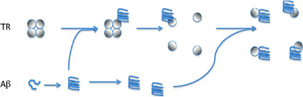
TTR can bind, in both intra and extracellular environment, due a chaperone-like manner (Buxbaum et al., 2008), to all A-Beta forms (monomers, oligomers and fibrils). However it binds with more affinity to soluble fibrillar A-Beta oligomers, which are believed to be the most toxic forms of A-Beta (Yang et al., 2013, Cascella et al., 2013). Usually, TTR tetramers are very stable with this conformation being essential to the transport function of this protein. However,
Figure 3 - Scavenger function of TTR. TTR in A-Beta presence change its tetrameric conformation to
TTR monomers bind more to A-Beta than tetramers (Du and Murphy, 2010), indicating that conformational molecular changes are important to the binding (Du et al., 2012) and TTR monomers are required to sequester A-Beta (Yang et al., 2013). It was, recently, reported that the presence of A-Beta triggers the destabilization of TTR tetramers, allowing TTR monomers to bind A-Beta, preventing its aggregation, deposition and interaction with cells (Figure 5) (Yang
et al., 2013). These results together with the upregulation of TTR in transgenic mice
overexpressing mutant amyloid precursor protein (APP), resulting in slow disease progression and lack of neurodegeneration (Stein and Johnson, 2002), and accelerated A-Beta deposition in the absence of TTR (Choi et al., 2007), supports the natural protective role assigned to TTR in AD (22).
Moreover, TTR inhibits the influx of Ca2+ caused by the oligomers, further preventing other
consequences of A-Beta, such as oxidative stress, membrane leakage and apoptosis. Overall, the molecular mechanism by which TTR protects cells against the deleterious effects of protein aggregation seems to involve two different levels of intervention: (1) inhibition of protein aggregation and fibril formation, as previously demonstrated; and (2) neutralization of protein oligomer toxicity once the oligomers are formed (Cascella et al., 2013). Proteolysis has been proposed as a possible mechanism by which TTR process A-Beta, cleaving this in smaller and less amyloidogenic peptides, which can be eliminated by cells (Costa et al., 2008a).
1.2.2. Transthyretin regulation by Sex Hormones
Several epidemiological and neuropathological studies have provided evidence for gender related differences in AD, with women being more susceptible to this disease than men (Ruitenberg et al., 2001, Oliveira et al., 2011). A possible explanation relates the loss of estrogens and progesterone that occur during menopause, which is much more abrupt than testosterone loss by men during aging (Yue et al., 2005, Oliveira et al., 2011). TTR probably regulates brain A-Beta levels in a gender-associated manner, under the control of testosterone and estrogen levels (Oliveira et al., 2011).
Both 17B-estradiol and dehydrotestosterone (DHT) up-regulated TTR expression in liver (Goncalves et al., 2008) and CP (Quintela et al., 2008, Quintela et al., 2009) of mice and rat. Furthermore, progesterone also enhanced TTR expression (Quintela et al., 2009, Quintela et
al., 2011). Estrogens are indicated by the majority of studies as neuroprotective, reducing
A-Beta formation, and protecting against neuronal toxicity, apoptosis and oxidative stress (Wilson
et al., 2000, Greenfield et al., 2002, Amtul et al., 2010). Moreover, 17B-estradiol, by increasing
TTR levels in brain, leads to inhibition of A-Beta aggregation (Amtul et al., 2010), which highlight the importance of this sex hormone in AD, and also supports the relevant role of TTR in AD onset and progression.
2. Implications of Choroid Plexus Senescence
Generally, aging has an impact in all systems of human body, and CP is not an exception. CP morphology alterations comprise epithelial atrophy, basement membrane thickening, fibrosis and calcifications of stroma, and increased number of cells presenting Biondi bodies (Serot et
al., 2000, Serot et al., 2012), that intensifies with aging. BCSFB permeability increases while
CP protein synthesis and CSF turnover are reduced (Johanson et al., 2011b). The CSF volume increases, following augmented resistance to CSF drainage, probably as a result of a combination of calcification of the arachnoid villi, thickening of the arachnoid membrane, and central vascular hypertension (Redzic et al., 2005, Erickson and Banks, 2013). The decrease of CSF antioxidant properties might be explained by reduction in vitamin E and ascorbic levels which are two principal scavengers of the free radicals of CSF. A decrease in turnover leads to an increasing contact-time between proteins and glucose which promotes the glycation of proteins, including TTR, and also increases oxidative stress (Shuvaev et al., 2001, Vicente Miranda and Outeiro, 2010, Serot et al., 2012). Further, A-Beta retention time in brain increases, contributing to its accumulation in CP, and other processes as apoptosis, oxidative stress, BCSFB disruption, cell death and probably further decreases CSF turnover (Erickson and Banks, 2013).
CP is involved in the most basic aspects of neural function, which means that even modest changes in CPs may trigger some important effects (Vargas et al., 2010). Moreover, the age-related alterations in CP, described above, also take place in AD, but are much more accentuated (Serot et al., 2000, Serot et al., 2012). Therefore, this ventricular structure has received particular attention in the last years due to its importance and role in the CNS pathologies, particularly in AD (Krzyzanowska and Carro, 2012).
2.1. Amyloid Beta Metabolism
A-Beta, a 4 kDa amino acid metalloprotein, is a consequence of APP metabolic processing, that occurs when APP is cleaved by the enzymes β-secretase (BACE-I) and Y-secretase (Figure 5) (Shirwany et al., 2007, Rowe and Villemagne, 2013). APP point of cleavage mainly result in three forms of A-Beta with 38, 40 and 42 amino acid residues. This is particularly important because A-Beta42, the longer form, is far more prone to oligomerize and form fibrils than
another A-Beta form (Walsh and Selkoe, 2007). It is likely that A-Beta form aggregates, forming toxic species as dimers, oligomers and fibrils. It is still unknown which of these species presents more toxicity (Ittner and Gotz, 2011), although some studies points to be smaller species of A-Beta (oligomers) (Walsh et al., 2002, Gong et al., 2003, Li et al., 2011). The secreted A-A-Beta may have several fates, which include reabsorption in capillaries, degradation by proteases and drainage into the CSF (Shirwany et al., 2007, Serot et al., 2012).
Currently, A-Beta is seen as the best marker of AD pathology (Rembach et al., 2013). Typically, 90% of healthy individuals present the A-Beta40 form predominantly, and only 10% present
A-Beta42. In contrast, in AD, this ratio changes dramatically, with A-Beta42, the principal
compound of amyloid plaques, increasing to approximately 50% (Shirwany et al., 2007).
2.2. Choroid plexus dysfunction and Amyloid Beta accumulation
in brain
CP dysfunction normally means that its functions, as secretion, synthesis and transport are compromised (Krzyzanowska and Carro, 2012). Consequently occurs a decrease in CSF turnover, TTR levels, and increases sequestering of proteins synthesized by CP, and oligomerization of A-Beta42. TTR as a scavengerr of A-Beta, plays an important role in its clearance, whereby changes
in TTR gene expression in the CP and decreased TTR levels in CSF, as consequence of CP age-related modifications, might relate to AD (Serot et al., 2000, Sousa et al., 2007). Concordantly, deposition of A-Beta isoforms, including A-Beta42, in AD patients, is mainly due to failures in its
clearance rather than to overproduction (Mawuenyega et al., 2010, Pascale et al., 2011), and levels of TTR in the CSF of AD patients are inversely correlated with A-Beta plaque burden (Merched et al., 1998).
Thus, it is expected that A-Beta accumulates in CP, as already observed in the CP of AD patients (Vargas et al., 2010, Krzyzanowska and Carro, 2012). Moreover, impairment of CP functions as consequence of A-Beta deposition, mainly of A-Beta42, was shown in AD patients CP, by
increasing nitric oxide production and interfering with BBB integrity, due to disrupted expression of TJs proteins as ZO-1 (Vargas et al., 2010). In other words, CP dysfunction with aging initiates a cascade that leads to accumulation of A-Beta in brain, which consequently enhances modifications in CP performance (Figure 4). As already mentioned, estrogens have an important role in the regulation of TTR expression, and decreased levels of this hormone are verified with aging. Consequently, this reduction in estrogen production might block the natural protective response, where TTR expression increases, and so contributing to A-Beta accumulation.
3. Endocrine Disruptors
There is an increasing concern about the effects of human and wildlife exposition to compounds that have the ability to interfere with endogenous hormones, the so called endocrine disruptors (EDs). These are exogenous compounds that possess the potential to interfere with hormonal regulation and the normal endocrine system, through several mechanisms including blocking, mimicking and displacing endogenous hormones. Consequently, production, release, metabolism, and elimination of natural hormones can be modified (Wetherill et al., 2007, Casals-Casas and Desvergne, 2011, Weiss, 2012). Although there are many sources of endocrine disruptors, the major concern is attributed to synthetic chemicals with endocrine-disrupting properties (EDCs), due to its large and continuous production in western societies (Casals-Casas and Desvergne, 2011).
Figure 4 - Correlation between CP dysfunction and accumulation of A-Beta in brain. Aging contributes
to impaired CP functions, which in turn leads to decreased TTR levels. Additionally, estrogens levels decrease with aging, contributing to TTR decrease. Crucial to A-Beta clearance, compromised TTR expression might lead to A-Beta deposition in brain.
A major mechanism of EDC-mediated metabolic disruption is through EDC interaction with nuclear receptors, including sex steroid hormone receptors, receptors acting as xenobiotic sensors, and receptors specialized in metabolic regulations (Casals-Casas and Desvergne, 2011). EDCs include biocides, industrial compounds, surfactants, and plasticizers. Bisphenol A (BPA), probably the most studied EDC (Flint et al., 2012, Rogers et al., 2013), belongs to this last category.
3.1. Bisphenol A
BPA is a nonsteroidal xenoestrogen that exhibits approximately 10-4 the activity of estradiol
(Flint et al., 2012). Heat resistance and elasticity are two of BPA characteristics that lead to its increased use and crescent production worldwide, with more than 10 million of tons produced every year (Fenichel et al., 2013). This chemical is mainly used as intermediate to produce resins and polymers. It is possible to found BPA in a wide range of materials and products: bottles, coatings, pipes, dental sealants, printing store receipts, canned food, nail polishes and flame-retardant materials (Weiss, 2012, Boas et al., 2012, Asimakopoulos et al., 2012). Human exposure to BPA is extensive, as many of these products are easily handled by humans, and moreover, BPA can leach from those (high temperatures, acidic or basic environments) (Weiss, 2012, Boas et al., 2012, Asimakopoulos et al., 2012, Fenichel et al., 2013). Furthermore, BPA has been detected in samples of water, sewage leachates, indoor and outdoor air and dust (Vandenberg et al., 2010). Although, the oral via is the most likely route of human exposure to BPA, transdermal and inhalation should also be considered, as these can avoid most of the first-pass hepatic metabolism, contrarily to oral exposure. The dermal contact may exist with air, dust and water contaminated with BPA (Vandenberg et al., 2007).
3.1.2. Bisphenol A metabolism and molecular mechanism of
action
After oral absorption, BPA is rapidly metabolized in the liver into bisphenol A-glucuronide, a highly soluble metabolite, without hormonal activity, that is then eliminated through urine (Mileva et al., 2014). The half-life of BPA estimated is about 6h (Volkel et al., 2002, Calafat et
al., 2008). Although, BPA is mostly metabolized in liver and intestines, BPA can originate others
metabolites, which may present more risk to physiological functions than BPA itsef (Mileva et
al., 2014). Unconjugated BPA has been frequently detected in urine and blood samples in many
different studies. This indicates that an internal exposure to the parent compound exist, which is estrogenically active (Vandenberg et al., 2010).
Estrogenic properties of BPA were described for the first time in 1936, and since then various experiments were performed that confirmed these properties (Casals-Casas and Desvergne,
2011). BPA binds differently to both estrogen receptors (ERs) - ERα and ERβ - displaying a 10-fold higher affinity for ERβ (Gould et al., 1998, Kuiper et al., 1998). Initially, BPA was thought to have a very weak estrogenic function due to be much less potent than estradiol. More recently, it was observed that BPA promotes cellular responses through many different pathways, and at very low doses, with changes in cell function from picomolar doses, below the levels expected for classical nuclear ERs binding (Wozniak et al., 2005, Welshons et al., 2006). BPA has also been shown to bind a membrane-associated ER and produce non-genomic steroid actions with the same efficacy and potential than estradiol. BPA can cause effects in animal models at doses in the range of human exposures, indicating that it can act at lower doses than predicted from some in vitro and in vivo assays (Vandenberg et al., 2010). In addition to affect estradiol hormone function, BPA can also antagonize thyroid hormone and androgen actions (Welshons et al., 2006, Flint et al., 2012). Yang et al., 2009, reported a possible relation between BPA exposure and increased oxidative stress and inflammation, and that postmenopausal women present more predisposition related with estrogen levels and receptor occupancy. BPA exerts cellular and tissue-type specific effects and non-monotonic dose-responses at cellular and intracellular levels at low physiologically relevant concentrations (Wetherill et al., 2007). In the same tissue, different cell types present unique estrogen-stimulated gene expression, and BPA activity may alternate between agonist and antagonist (Welshons et al., 2006).
BPA may elicit cellular responses through several mechanisms. Briefly, BPA may disturb the proper estrogen nuclear hormone receptors activity, and consequently, interfere with endogenous estrogens activity; also can affect the androgen system, disrupt the thyroid hormone function, and affect development, differentiation and function of the CNS and of the immune system (Moriyama et al., 2002, Wetherill et al., 2007).
3.1.3. Bisphenol A levels in human tissues and fluid
With the aim of evaluate if BPA represents a threat to the human population, several studies have been performed for this purpose recently. Unconjugated BPA, the form that has estrogenic activity, has been measured repeatedly in various types of human samples including urine, plasma, saliva, breast milk, among others, using various analytical techniques (Welshons et al., 2006, Vandenberg et al., 2007, Calafat et al., 2008, Gentilcore et al., 2013).
For reference, in a study where urine samples were collected and analysed, 92.6% of people accused the presence of BPA (Calafat et al., 2008). Vandenberg et al. (2007) reviewed the published data of more than 80 studies about human exposure to BPA, and reported that the average urine BPA levels had a range of 1-3 ng/mL, and moreover, the concentration of BPA in serum were between 0.3-4.4 ng/ml, corresponding to 1-19.4 nM. Furthermore, the exposure of human population to BPA is widespread, with BPA levels detected in most children and adults
(Vandenberg et al., 2007). This data suggested a continuous exposure to BPA (Calafat et al., 2008) and this amount of BPA to which human are exposed may cause adverse effects in health (Vandenberg et al., 2010).
All endogenous estrogens, androgens, and thyroid hormones decline with aging, and play a role in brain plasticity. Moreover, all three endocrine systems are vulnerable to EDCs (Weiss, 2012). In addition, BPA showed to be able to cross BBB (Sun et al., 2002), which demonstrates that the brain is exposed to this compound.
3.1.1. Metabolic effective dose
BPA has become the chemical model for examples of non-monotonic dose-response functions, due to its dose-response curve called U-shaped or inverted U-shaped. Effects are observed at very low doses and high doses, but no effects are observed at intermediary doses. This contradicts the conventional idea that the lack of an adverse effect at certain dose implies that to lower doses the absence of an effect will maintain. Thus, to non-monotonic dose-response curves, the effects of low doses cannot be predicted by the effects observed at high doses. These U-shaped curves suggest the existence of two independent mechanisms for low and high doses (De Coster and van Larebeke, 2012), probably ER-dependent and ER-independent mechanisms, although far from being completely understood. Thus, BPA might not act as estradiol, which increases the importance of its study and how it affects the various human systems, including the brain.
Aim
The important role of TTR, the principal protein synthesized in choroid plexus, in A-Beta clearance has been supported by many studies, and here its interaction will be assessed in animals with different ages. For other side, it is well documented the importance of sex hormones in TTR regulation and production. For this reason, this work intends to investigate if an endocrine disruptor, Bisphenol A, well known for the widespread exposure to human population, is able to interfere with TTR expression in rat CPs, and consequently if it may affects A-Beta clearance. Additionally, the effect of Bisphenol A, and also A-Beta, in the blood-cerebrospinal barrier integrity will also be subject of analysis, through the measurement of expression of certain membrane proteins found at this barrier.
III.
Materials and Methods
1. Animals and Z310 cell line
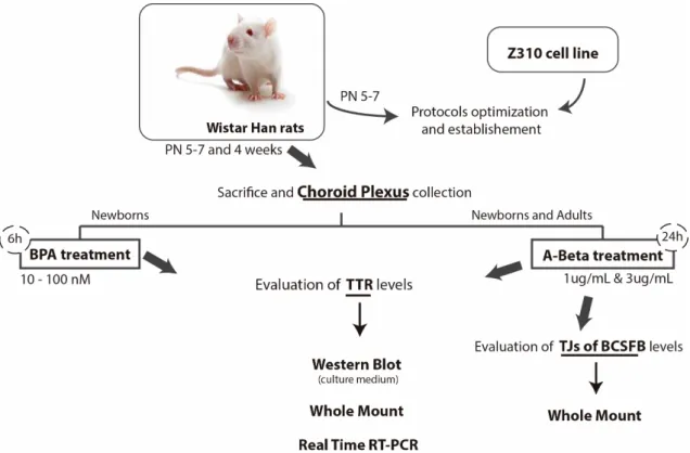
In this work, several assays with CP explants collected from Wistar Han rats (newborn and young) were carried out. All animals were handled in compliance with the National Institute of Health guidelines and the National and European Union rules for the care and handling of laboratory animals (Directive 2010/63/EV). The explants were treated with BPA and A-Beta42
and the effects of these two compounds were evaluated using diverse techniques. In brief, the experiments scheme carried out with these explants are represented in figure 5. Additionally to CP explants, an immortalized rat choroidal epithelium cell line, called Z310, with potential for application in BCSFB research was also used. The major role of Z310 cells in this work relates with optimization processes, specifically Western blot, allowing to avoid a considerer number of animals that would be required to the different optimization steps of this technique, as amount of total protein loaded, transfer time, antibodies dilutions and incubation times.
Figure 5 - Scheme of experimental studies about BPA and A-Beta effects in TTR and TJs BCSFB proteins levels, in rat choroid plexus explants. Evaluation of BPA effects was carried out in CPs from newborn
Wistar Han rats, while A-Beta effects were evaluated in newborn and young rats. CPs were subjected to various techniques, such as Western blot, Whole mount immunohistochemistry and Real-Time PCR. PN-post natal; BPA-Bisphenol A; A-Beta- Beta amyloid; TTR- transthyretin; TJs- tight junctions; BCSFB- Blood cerebrospinal fluid barrier; CP- Choroid plexus.
1.1. Cell Culture
Cell line Z310 was supplemented with Dulbecco´s Modified Eagle Medium (DMEM, Sigma-Aldrich), 10% fetal bovine serum (FBS, Biochrom AG, Berlin) and 1% penicillin/streptomycin (Sigma, Saint Louis) in a t-flask, and was kept in AutoFlow DHD CO2 Air-Jacketed Incubator (NuAire, USA) at 37ºC, in a 5% CO2 atmosphere.
1.1.1. Cells Passage
Always that cellular confluence achieved about 90% it was proceeded to cellular passage, decreasing cellular density to secure the continuous expansion of Z310 cell line. These cells are adherents. Culture medium was removed and cells were washed with phosphate buffered saline (PBS) 1x. PBS was removed and replaced by trypsin-EDTA 0.25% in a volume that ensure cells totally cover, and were incubated for 3 to 5 minutes at 37ºC. When most of the cells have detached, culture medium at an equivalent volume of used to trypsin was added. Resuspended cells were collected to a falcon and were centrifuged for 5 minutes, 1300 rpm. Supernatant resulted from centrifuging was discarded and pellet containing cells was resuspended in PBS 1x, and again centrifuged for 5 minutes, 1300 rpm. Supernatant was removed and cells were resuspended in 1 mL of medium culture and placed in the incubator (37ºC, 5% CO2).
1.1.2. Cell Counting
For cell counting, 20 uL were taken from cellular suspension, and equal volume of trypan bue was added, followed by homogenization. Of these preparation, 20 uL were transferred to a Neubauer chamber, for viable cells counting. After counting the cells at the different quadrants, the cells number/mL and the total number of cells in t-flask were estimated, following these formulas:
1.1.3. Cells Freezing and Thawing
This process was performed to ensure the perpetuation of the cell line. With this purpose cells were trypsinized, as described before. Cells were diluted in DMEM with 10% of DMSO (Dimethyl sulfoxide), to prevent the formation of crystals that could cause cell lysis. Cells were stored, at first, at -20ºC, after at -80ºC, and finally, in liquid nitrogen to permanent storage. To thawing cells, cryopreserved cells were resuspended in DMEM with 10% of FBS and 1% of penicillin/streptomycin, after previous thawing at 37ºC. Then, resuspension was centrifuged for 5 minutes, 1300 rpm, supernatant rejected and cells washed again with culture medium. After a final centrifuge it was proceeded to cellular culture as previously described.
2. Whole Mount fluorescent staining
Whole mount fluorescent staining is a technique that allows to determinate the cellular localization and distribution of certain proteins in whole tissue, using specific antibodies. In this work, location and expression of TTR (cytoplasmic protein), and occludin, E-cadherin, Cld-1 and ZO-Cld-1 (membrane proteins) were assessed by this technique. Further, tissues were subjected to treatment with A-Beta and BPA at nanomolar (nM) concentrations, with the aim of analysing its effect in CP and BCSFB integrity.
2.1. Whole Mount – TTR, Occludin, E-cadherin, Cld-1 and ZO-1
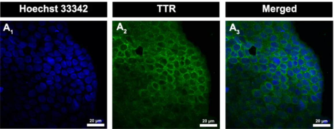
Briefly, lateral CPs were collected from Wistar Han rats with 5-7 days and placed into a 48 wells microplate (one CP per well) with paraformaldehyde (PFA) 4% for 45 min at room temperature. CPs were then incubated with a solution of sucrose 30% until deposit in the bottom, and next with a blocking solution 2,5% bovine serum albumin (BSA) 0,2% Triton X-100, overnight at 4ºC. In CPs intended to incubate with membrane proteins antibodies, Triton X-100 wasn’t used in blocking solution, to avoid compromising cellular membranes. CPs were washed once with PBS-Tween 0.01% (PBS-T) and incubated overnight at 4ºC, with respective antibodies diluted in block solutions: rabbit anti-mouse TTR antibody (1:200, Dako), rabbit anti-occludin and anti-E-cadherin (1:50, Santa Cruz Biotechnology) and goat anti-Cld-1 and anti-ZO-1 (1:50, Santa Cruz Biotechnology). After this, CPs were washed several times with PBS-T and then incubated with secondary antibodies, diluted in blocking solutions, Alexa 488 goat anti-rabbit (1:2000, Molecular Probes) and donkey anti-goat 488 (1:1000, Santa Cruz Biotechnology) for 3 hours on dark at room temperature. From the incubation with secondary antibody all next procedures were performed in dark. CPs were again washed with PBS-T before incubation with Hoechst 33342 dye (Molecular Probes, EUA) diluted 1:1000. Finally, CPs were mounted with Entellan and visualized on a confocal microscope LSM 710 (Zeiss, Germany).
The protocol described above was followed to evaluate A-Beta and BPA effects in CP proteins studied in this work. Thus, CPs collected from Wistar Han rats with 5-7 days were placed into a microplate (one CP per well) with complete DMEM, and incubated for 6 or 24h at 37ºC, in a 5% CO2 atmosphere, with or without BPA treatment (control). BPA (Sigma Aldrich, Sant Louis)
was dissolved in DMSO and diluted in this culture medium at 10, 25, 50, 75, 100 nM and 10 µM. The final concentration of DMSO in each well was less than 0,01%. incubated with or without BPA. Also, CPs collected from Wistar Han rats with 5-7 days and 30 days were incubated in complete DMEM for 24h in a 5% CO2 atmosphere, with or without A-Beta42 (AnaSpec), at 1µg/mL
or 3µL/mL. After respective incubation times, the culture medium was removed and CP tissues were washed once with PBS 1x. Then, normal protocol was followed and CPs were incubated with PFA 4%.
2.2. Confocal microscopy images analysis
Confocal microscope allowed the acquisition of various Z-stacks of each CP, at different localizations, after the whole fluorescent staining procedure. These Z-series collects a sequence of several different images/sections of the same region of the tissue. Then, using Blue Edition from Zen 2011 software (Zeiss), mean fluorescence intensity of each stack corresponding to proteins staining was obtained. This staining represents proteins expression in CPs, and then was used to quantify and evaluate the expression levels of proteins in CP explants after treatments.
3. Western Blot
Western blot was carried on with total protein extracted from CPs and Z310 cell line, and with culture medium collected after CP explants incubation with A-Beta42 and BPA.
3.1. Protein Extraction
3.1.1. CP explants
To extract CPs protein, CPs were digested with buffer lysis (5mM HEPES (pH 7,5), 250 mM Sucrose, 10mM NaNO3, 0.2 mM PMSF, 25 µg/mL leupeptin, 1mM orthovanadate, 10 mM sodium phosphate (pH 7,4), 150 mM NaCl, 2% Triton X-100, 2% deoxycholate, 0.2% SDS and 0.2 mM PMSF) by manual homogenization with a pestle, followed by centrifugation at 10,000 g, for 10 min. After centrifugation, the supernatant, containing protein, was collected. Total protein content in samples was measured using the Bio-Rad Protein Assay according with manufacturer’s protocol (Bio-Rad, Hercules, USA).
3.1.2. Z310 cells
Z310 cells resuspension obtained after trypsinization, and twice centrifuged for 5 minutes, 1300 rpm. Then, cells were washed with PBS 1x, centrifuged for 7 minutes, 11000 rpm, 4ºC, and supernatant rejected. This process was repeated twice, and at final, cells were resuspended in lysis buffer (same used to CP protein extraction). Total protein content of samples was measured as described before.
3.2. Western Blot – TTR, Occludin, E-cadherin and Cld-1
Total protein 50 µg was separated by SDS-PAGE using 8% and 12,5% gels, after boiled at 95ºC for 10 min, and electrophoretically transferred to polyvinylidene difluoride (PVDF) membranes (Amersham Biosciences). Membranes were blocked with 5% non-fat dry milk in Tris-buffered saline (TBS). After 1h30 in blocking solution, membranes were washed 10 min with washing solution, TBS containing 0,1% Tween (TBS-T), and then incubated respectively, overnight, at 4ºC with anti-TTR (1:250, Dako), anti-Occludin, anti-E-cadherin, anti-Cld-1 and anti-ZO-1 (1:200, Santa Cruz Biotecnhology) antibodies diluted in TBS-T. Membranes were washed for 45 minutes in TBS-T, at room temperature, with replacement of washing solution each 15 minutes. Then incubation with HRP-conjugated anti-rabbit and anti-goat secondary antibodies (1:20000) was done. The washing process was repeated as described before, and antibody binding was detected using the ECF substrate (ECF Western Blotting Reagent Packs, GE Healthcare) according to the manufacturer’s instructions. Images of blots were captured with the Molecular Imager FX Pro Plus MultiImager system, and densitometry of bands was carried out using the software Quantity OneTM (Bio-Rad).
4. Extraction of total RNA
TTR expression was analyzed by Real Time Polymerase Chain Reaction (Real Time PCR) to evaluate the A-Beta and BPA effect on its expression. Before proceeding to Real Time PCR, total RNA was extracted from rat CPs, previously collected, frozen in liquid nitrogen and stored at -80ºC. RNAt integrity was checked through visualization in 1% agarose gel. Total RNA was quantified in a nanophotometer.
4.1. Extraction
For CP RNA extraction 300 µL of TRIzol Reagent were added to each tube containing a CP followed by manual homogenization with a pestle in order to allow dissolution of cellular