Preparation and Characterization of Chitosan/mPEG-PCL Blended Membranes for Wound
Dressing and Controlled Gentamicin Release
Samira Faleiros Silva Brianezia, Karine Cappuccio Castroa,c, Rodolfo Debone Piazzab, Maria do
Socorro Fernandes Meloa, Rafael Matsumoto Pereirac, Rodrigo Fernando Costa Marquesb, Maria
Gabriela Nogueira Camposa,c*
Received: October 21, 2017; Accepted: August 04, 2018
In this paper, a novel wound dressing membrane for controlled release of gentamicin (GE), while covering and protecting the wound was investigated. Chitosan (CHI) was associated with methoxy polyethylene glycol - polycaprolactone copolymer (mPEG-PCL) to prepare the blended wound dressing membranes. The use of copolymer mPEG-PCL was necessary to improve the compatibility between CHI and PCL. The association of CHI and PCL was required to control the water retention and release rate of encapsulated GE. In vitro release studies were performed with the mPEG-PCL/
CHI-GE membranes in order to evaluate the effect of copolymer concentration on the kinetics of GE release and water uptake. Reduced burst release rates and swelling ratios were observed for the 1/2
and 1/4 mPEG-PCL/CHI-GE membranes. In addition, all gentamicin-loaded membranes inhibited S. aureus and E. coli growth, and demonstrated color, moisture and thermal stability. Therefore, mPEG-PCL/CHI-GE membranes showed important features for potential wound dressing and drug delivery applications.
Keywords: Copolymer, blend, methoxy polyethylene glycol, polycaprolactone, antibiotic, drug delivery.
*e-mail: [email protected]
1. Introduction
Skin is the largest organ of the body and protect it
against infections, injuries, traumas and sunrays1. Skin also
is important in the control of body temperature2. Skin lesions affect a large number of people in Brazil and in the world
and can be caused by burns, chronic infections, vascular
insufficiency, diabetes and hypertension, which can lead to death due to the loss of large areas of the skin1. When skin is
lost or lesioned, the wound should be covered and treated to avoid contamination of microorgamism2. Gentamicin (GE) is
one the most commonly used antibiotics due to its low cost, wide antibacterial action, low rate of pathogenic resistance and allergy, good stability and water solubility3. Based on
the wound type, suitable dressing material must be used. Many researches can be found in the Literature regarding to the development of wound dressings, drug delivery
systems and skin substitutes3,4,5. Collagen chitosan, alginate,
cotton, hyaluronic acid, gelatin, poly lactic acid, cellulose, polycaprolactone and other polymers can be used to prepare to
prepared hydrogels, films and membranes for wound dressing
application6. The ideal dressing should provide or maintain
moist environment; enhance epidermal migration; promote angiogenesis and connective tissue synthesis; allow gas
exchange between wounded tissue and environment; maintain
appropriate tissue temperature to improve the blood flow to
the wound bed and enhances epidermal migration; provide protection against bacterial infection and be non-adherent to
the wound and easy to remove after healing. Besides, it must
provide debridement action to enhance leucocytes migration
and support the accumulation of enzyme and must be sterile,
non-toxic and non-allergic7. It is not possible for a unique
material to meet all the requirements for an ideal wound dressing. Thus, materials association can be an alternative
to improve the performance of skin substitutes and to meet
most of the desired requirements.
Chitosan (CHI) is a hydrophilic and cationic polymer obtained by chemical deacetylation of chitin, a natural polysaccharide found in shrimp and crab shells8. It has been
extensively studied in biomedical area due to its interesting biological properties such as biodegradability, biocompatibility, bioactivity, antibacterial, antitumor, antifungal and hemostatic activities8. Disadvantage property of chitosan membrane that
is unappropriated for controlled drug delivery application, is
its high water uptake/swelling because of its large number of
hydrogen bonds8. This can compromise the sustained release
of hydrophilic/water-soluble drugs, such as gentamicin3.
Therefore, an appropriate association to a hydrophobic
aInstituto de Ciência e Tecnologia, Universidade Federal de Alfenas, Poços de Caldas, MG Brasil bInstituto de Química, Universidade Estadual Paulista “Júlio de Mesquita Filho”, Araraquara, SP
Brasil
polymer could achieve the controlled retention and release of a drug from chitosan.
Polycaprolactone (PCL) is a synthetic polymer, derived
from petroleum and classified as semi-crystalline polyester.
Due to its composition/structure, PCL has hydrophobic character and can be slowly degraded by the body. It has found several biomedical applications, such as controlled
release of drugs, sutures and scaffolds for tissue engineering,
due to its compatible properties and biodegradability9. Thus,
combination of CHI and PCL can be used to prepare a blended membrane with unique properties for wound dressing and gentamicin controlled release.
One way to blend hydrophilic and hydrophobic molecules is to add chemicals that have both hydrophilic and hydrophobic moieties, such as surfactants10. Another alternative is to chemically modify the polymeric backbone to
attach hydrophilic/hydrophobic molecules in order improve
the compatibility. Copolymerization is also a strategy for polymeric materials. Copolymerization of PCL with methoxy polyethylene glycol (mPEG) forms block copolymers that
are biocompatible and biodegradable polymers for potential use as drug delivery systems11. In addition, the miscibility
between hydrophilic CHI and hydrophobic PCL may be increased by the use of the mPEG-PCL copolymer12.
Therefore, the aim of this paper was to prepare and
characterize chitosan-mPEG-PCL membranes loaded with
gentamicin to be later used as wound dressing for sustained
antibiotic release. The blended membranes were characterized
for chemical composition, morphology, color, thermal
stability, moisture content and water uptake. Moreover,
the in vitro gentamicin controlled release was studied over
an extended period of 1 week and the effect of copolymer concentration on the kinetics of gentamicin release was also
evaluated. The antibacterial activity of the CHI/mPEG-PCL membranes was tested in vitro against E. coli and S. aureus,
most common bacteria found in skin wounds.
2. Materials and Methods
2.1 Materials
High molecular weight chitosan (CAS# 419419),
ε-caprolactone monomer (molecular weight 114.14 g/mol),
methoxy polyethylene glycol (molecular weight 550 g/mol) and ninhidrin (CAS# N4876) were purchased from Sigma/ Aldrich - USA. Gentamicin sulfate injectable solution (280
mg/2mL) was purchased from Neo Química - Brazil. Glacial
acetic acid (60.05 g/mol and density 1.05 g/cm3) and acetone were purchased from Proquímios - Brazil.
2.2 Copolymerization of PCL and mPEG
The poly (ethyleneglycol) methyl-ether-poly(ε-caprolactone) (mPEG-PCL) copolymer was synthetized by
Dr. Marques’ research group, using the aforesaid monomers, as previously described9.
2.3 Preparation of CHI/mPEG-PCL membranes
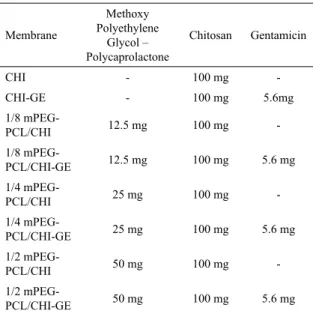
Eight membranes were prepared by varying the concentration of mPEG-PCL and GE in the formulation, according to the Table 1. For all compositions, 9mL of 1M acetic acid was used to dissolve CHI, and 5mL of acetone was used to dissolve mPEG-PCL. Acetone was also added to the formulations free of mPEG-PCL. For GE loaded membranes, 1mL of 5.6 mg/mL gentamicin solution was added to the polymeric solution. The antibiotic solution was replaced by Milli-Q water for GE free formulations.
Therefore, the final solution volume for all formulations
was 15mL. The membranes were obtained by casting 1.5 mL of each solution in a circular mold (diameter 2.5 cm), followed by the evaporation of solvent at room temperature.
3. Characterization of the Membranes
3.1 Thickness
A bench micrometer (Outside Micrometer, Vonder) was
used to measure the thickness in 10 different points of the membrane. The average thickness was calculated by the
arithmetic mean of the 10 values.
3.2 Color and opacity
The color and opacity of the membranes were analyzed
in a Colorimeter Minolta CR-300. The luminance parameters
L* were determined, ranging from 0 (black) to 100 (white);
a*, green (-60) to red (+60); and b*, from blue (-60) to
Table 1. Composition of membranes (w)
Membrane
Methoxy Polyethylene
Glycol – Polycaprolactone
Chitosan Gentamicin
CHI - 100 mg
-CHI-GE - 100 mg 5.6mg
1/8
mPEG-PCL/CHI 12.5 mg 100 mg
-1/8
mPEG-PCL/CHI-GE 12.5 mg 100 mg 5.6 mg
1/4
mPEG-PCL/CHI 25 mg 100 mg
-1/4
mPEG-PCL/CHI-GE 25 mg 100 mg 5.6 mg
1/2
mPEG-PCL/CHI 50 mg 100 mg
-1/2
yellow (+60). The total color difference (∆E*) was calculated
according to Equation 1.
(1)
3.3 Morphological analysis
A Scanning Electron Microscopy (Leo 440i, England)
was used to morphological characterization of the membranes
before and after gentamicin in vitro release. Samples were
previously coated with a 16 nm thick of gold film in a Sputter Coater (model SCD 050, Bal-Tec) for 60 seconds
at an operating pressure of 2x10⁻² Pa at 24 ºC.
3.4 Infrared analysis (FTIR)
Samples were characterized by FTIR (ATR) in a
Spectrometer (Cary 630, Agilents Technologies) from 650 to 4000 cm-1. The Microlab software was used to treat the data.
3.5 Moisture content
Moisture content of the membranes was determined by the gravimetric method. The percentage of moisture was calculated according Equation 2.
(2)
3.6 Swelling behavior
Membranes were dried in a desiccator for 24 hours and weighed (Wd). After such procedure they were immersed
in PBS, pH 7.4 and were kept at 37ºC (to simulate body
temperature) for 24 hours, when they were weighed again (Ws). The percentage of swelling, S(%), was obtained through Equation 3.
(3)
4. Thermal Analyses
4.1 Thermogravimetric analysis (TG) and derived
thermogravimetry (DTG)
TG analysis was performed in a TG-DTA / DSC Apparatus
(STA 449 F3 Jupiter, Netzsch). The analyzes were performed
under a nitrogen atmosphere in the temperature range from 30ºC to 900ºC, at a heating rate of 10ºC / min.
4.2 Differential scanning calorimetry (DSC)
Thermal analysis were performed in a Calorimeter (TA-Q100) by using the following conditions: temperatures range from - 130ºC to 200ºC, at a heating rate of 2ºC / min, under atmosphere of nitrogen.5. In vitro Gentamicin Release and
Antibacterial Activity
5.1 Standard curve for gentamicin
In order to obtain the standard curve relating the
gentamicin concentration to the absorbance, different samples of known concentrations of gentamicin were prepared from
the dilution of the injectable gentamicin solution (280 mg/2mL). A colorimetric procedure, previously established
was used for gentamicin quantification3. The method is based
on ninhydrin reaction with primary and secondary amines present in gentamicin. This reaction produces a purple color and the absorption of the gentamicin-ninhydrin mixtures at 400 nm has a linear relationship with the gentamicin concentration. The samples were previously reacted with ninhydrin for 15 min at 95ºC and cooled in an ice bath, followed by determination of visible absorbance at 400 nm.
5.2 In vitro release assay
Experiments on in vitro release of gentamicin from the
membranes were performed at 37ºC in 0.2 M PBS, pH 7.4.
Two independent experiments were performed in order to evaluate the initial release (one hour) and long-term release
(one week). In the first experiment, the release medium was collected and replaced with fresh buffer after each 15 minutes
until complete one hour (15, 30, 45 and 60 minutes). The second experiment consisted in collecting samples from the release medium after each 24 h and replaced with fresh
buffer each time. This procedure was repeated for 7 days
and the experiment was carried out in triplicate. For both experiments, the collected medium was previously reacted with ninhydrin for 15 min at 95ºC, followed by determination of visible absorbance at 400 nm. Gentamicin concentration
was taken from the standard curve.
5.3 Antibacterial activity
Both bacteria, Staphylococcus aureus and Escherichia coli, were independently grown in nutrient broth for 24 hours at 37ºC. The antibacterial activity study was carried out by
agar diffusion technique: 30 ml of PCA (Plate Count Agar)
were added to each Petri dish and then inoculated with 0.1 ml of bacterial solution (E. coli and S. aureus). The membranes were then placed in contact with the agar (1 membrane of 2.5 cm diameter/quadrant) and the plate was incubated for 24
* * * * * * *
E L Lp a ap b bp .
2 2 2 0 5
D ="
Q
-V
+Q
-V
+Q
-V
%(%)
*
%
;
;
M
Mi
Mf
Mi
M
percentage of moisture
Mi
initial sample mass g
Mf
final sample mass g
100
=
-=
=
=
Q
R
R
R
V
W
W
W
"
%
%
*
;
S
Ws
Wd
Ws
Ws
mass of the swollen sample g
Wd
mass of the dry sample g
hours at 37ºC. After this, bacterial growth’s inhibition zones
were measured around the membranes. This antibacterial activity study was carried out in triplicates.
6. Results and Discussion
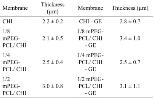
6.1 Thickness
Thickness of membranes was measured in order to verify
the distribution of the solution in the mold. Uniformity of
thickness plays an important role in the barrier property of the membranes. The average thickness of the membranes is shown in Table 2. As expected, o significant difference was observed comparing the average thickness of the membranes.
6.2 Color and opacity
Color and opacity are properties of great relevance for membranes used as wound dressings, since it is of great
importance that the wound can be visualized, to evaluate
the presence of infection and the healing process evolution7.
As expected, all prepared membranes are translucent.
Table 3 summarizes the results of the colorimetric analysis.
In relation to the luminescence parameter (that varies from
0 for black to 100 for white), the values of L* was around
70 for all samples, but CHI-GE showed the highest L*. Regarding to the a* parameter, the chromatic coordinate that varies from -60 (green) to +60 (red), all membranes showed
results near to zero, indicating the absence of these colors.
Similar behavior was observed for the b* parameter, which varies from -60 (blue) to +60 (yellow). Positive values were observed for all samples, suggesting a discrete yellowish of the samples with mPEG-PCL and GE.
The total color difference (∆E*) was calculated by the Equation 1 and no significant difference was observed
comparing the membranes, except for CHI-GE, which showed
higher total color difference. Mei et al. (2013) investigated the properties of blended films incorporated with perilla oil13. The authors also reported an increase in total color difference (ΔE*) after the oil incorporation13. Thus, incorporation of
yellow components, such as gentamicin sulfate solution and
perilla oil, may cause an increase in total color difference.
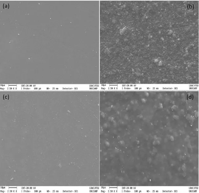
6.3 Morphological analysis
Although all membranes were visually homogeneous, microscopic analysis was performed to evaluate the interaction between CHI and mPEG-PCL and the microstructural characteristics of the membranes. Figure 1 shows the images obtained by SEM for the CHI, CHI-GE, 1/2 mPEG-PCL/CHI and 1/2 mPEG-PCL/CHI-GE membranes. Figure 1a and 1c
shows a dense and homogeneous chitosan film. By Figure
1, it was possible to observe the uniformly incorporation of the mPEG-PCL copolymer in the chitosan matrix (Figure 1b and 1d).
Cooper et al. (2013) prepared chitosan-polycaprolactone based membranes by Electrospinning technique14. SEM images
of the electrospun membranes showed better incorporation of PCL in the CHI matrix for 1/3 PCL/CHI composition, however, more than one phase was observed for all formulations14. Sarasam and Madihaly (2005) reported the characterization
of chitosan-polycaprolactone blends prepared by solvent evaporation in an oven at various temperatures followed by solvent annealing with chloroform vapors15. According
to the authors, separation of phases was not observed only for membranes drying at 55ºC15. Therefore, the copolymer
mPEG-PCL used in our study promoted a better miscibility between of PCL-CHI, as observed in Figure 1b and 1d.
The incorporation of gentamicin in the 1/2 mPEG-PCL/ CHI-GE membrane can be suggested by the presence of gentamicin sulfate crystals on the surface of the membranes in the Figure 1c and 1d. Nevertheless, antibiotic incorporation
in the membranes was confirmed by FTIR analysis.
6.4 Infrared Spectroscopy analysis
This analysis was performed for the raw materials: chitosan powder, mPEG-PCL powder and gentamicin solution; and for the obtained membranes in order to identify the Table 2. Average thickness (µm) of membranes unloaded and
loaded with gentamicin.
Membrane Thickness (µm) Membrane Thickness (µm)
CHI 2.2 ± 0.2 CHI - GE 2.8 ± 0.7
1/8 mPEG-PCL/ CHI
2.1 ± 0.5
1/8 mPEG-PCL/ CHI
- GE
3.4 ± 1.0
1/4 mPEG-PCL/ CHI
2.5 ± 0.4
1/4 mPEG-PCL/ CHI
- GE
2.5 ± 0.7
1/2 mPEG-PCL/ CHI
3.0 ± 0.8
1/2 mPEG-PCL/ CHI
- GE
3.1 ± 1.1
Table 3. Luminescence parameter, L*; chromatic coordinates a*
and b* and total color difference ∆E* of membranes unloaded and
loaded with gentamicin.
Membrane ∆E* L* a* b*
CHI 71.72 71.71 - 0.40 0.77
CHI - GE 74.35 74.34 - 0.61 1.22
1/8 mPEG - PCL/
CHI 72.21 72.21 - 0.30 0.51
1/4 mPEG - PCL/
CHI 71.25 71.23 - 0.46 1.75
1/2 mPEG - PCL/
CHI 71.62 71.60 - 0.36 1.69
1/8 mPEG - PCL/
CHI - GE 71.20 71.17 - 0.59 1.96
1/4 mPEG - PCL/
CHI - GE 71.36 71.34 - 0.46 1.69
1/2 mPEG - PCL/
Figure 1. Scanning electron micrographs of (a) CHI; (b) 1/2 mPEG-PCL/ CHI; (c) CHI - GE; (d) 1/2 mPEG-PCL/ CHI - GE.
Figure 2. Infrared spectra of raw materials: (black) mPEG-PCL,
(red) chitosan and (green) gentamicin solution.
characteristic groups of the raw materials in the membranes. The infrared spectra of raw materials are shown in Figure 2.
According to Yao et al. (2013), the main characteristic peak
of chitosan is found at 1655 cm-1 as it can be observed for this
sample16. Ketones, which are functional groups characteristic
of PCL (C=O) show as intense band between 1870 cm-1
and 1540 cm-1, more specifically absorb at 1715 cm-1 16,17. It is also verified that the band corresponding to the amine
group of gentamicin is between 1650 cm-1 and 1500 cm-1, coinciding with the chitosan peak, but being of a slightly
higher intensity. For the gentamicin solution there is an intense band corresponding to the OH group between 3500 cm-1 and 3000 cm-1. Moreover, there is a low intensity band
at 3442 cm-1 that can be attributed to water and the hydroxyl group of PEG in the copolymer. Bands between 2946 cm-1
mPEG- PCL. The ester group (-OC=O) characteristic peak
is also observed at 1725 cm-1 in the copolymer spectrum17.
Figure 3 shows the FTIR spectra of the membranes unloaded with gentamicin. As expected, the main groups of CHI and mPEG-PCL was found in all spectra. The band corresponding to chitosan (N-H) is of low intensity, while the band corresponding to PCL (C = O) appears intensely.
Figure 4 shows the FTIR spectra of gentamicin-loaded membranes and gentamicin solution. As expected, an increase on the intensity of the hydroxyl and amine groups
peaks was observed for the gentamicin-loaded membranes, confirming the antibiotic incorporation in these membranes.
Aquino et al. (2013) reported absorption bands between 1645 and 1550 cm−1 in gentamicin loaded microparticles, which were attributed to the bending of N-H bond of the primary and secondary amines18. Bajpai et al. (2017) also reported a characteristic peak of -NH2 functionality at the
vibrational wavenumbers 3802-3462 cm−1 in the spectrum of
gentamicin sulfate loaded alginate films, as well as observed
in the Figure 419.
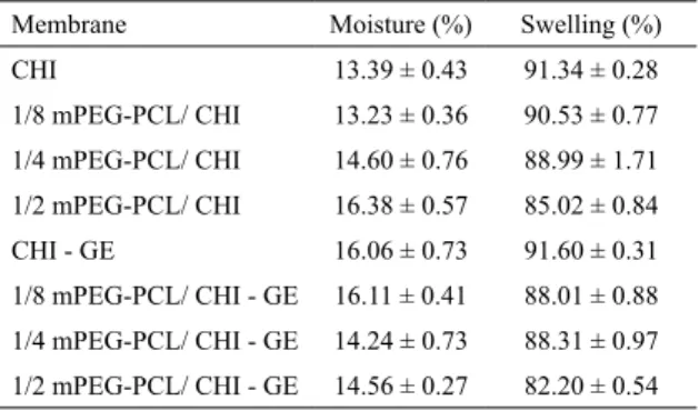
6.5 Moisture content
The percentage of moisture of the membranes, calculated
by the Equation 2, is summarized in Table 4. It was not
possible to establish a trend of moisture as a function of the concentration of mPEG-PCL in the membranes, as well as of the presence of gentamicin. However, all membranes presented values similar to those reported for chitosan membranes in the Literature, suggesting that the presence of gentamicin
and mPEG-PCL did not influence the water content3.
6.6 Swelling behavior
Water uptake by the membranes is an important feature
for application as wound dressing, since in the healing process
the wound site must be kept moist and the membrane should be able to absorb the exudate fluids6. On the other hand, for
Figure 3. Infrared spectra of membranes: (black) CHI, (red) 1/8
mPEG-PCL/CHI, (green) 1/4 mPEG-PCL/CHI, and (blue) 1/2 mPEG-PCL/CHI.
Figure 4. Infrared spectra of membranes: (black) CHI-GE, (red)
1/8 mPEG-PCL/CHI-GE, (green) 1/4 mPEG-PCL/CHI-GE, (blue) 1/2 mPEG-PCL/CHI-GE and (light blue) Gentamicin solution.
Table 4. Moisture content and percentage of swelling of membranes unloaded and loaded with gentamicin.
Membrane Moisture (%) Swelling (%)
CHI 13.39 ± 0.43 91.34 ± 0.28
1/8 mPEG-PCL/ CHI 13.23 ± 0.36 90.53 ± 0.77
1/4 mPEG-PCL/ CHI 14.60 ± 0.76 88.99 ± 1.71
1/2 mPEG-PCL/ CHI 16.38 ± 0.57 85.02 ± 0.84
CHI - GE 16.06 ± 0.73 91.60 ± 0.31
1/8 mPEG-PCL/ CHI - GE 16.11 ± 0.41 88.01 ± 0.88
1/4 mPEG-PCL/ CHI - GE 14.24 ± 0.73 88.31 ± 0.97
1/2 mPEG-PCL/ CHI - GE 14.56 ± 0.27 82.20 ± 0.54
medicated wound dressings, the swelling percentage can
affect the drug release8. The percentages of swelling of the
membranes were calculated by the Equation 3 and are shown
in Table 4. As expected, a reduction of water uptake was
observe for the membranes prepared with the mPEG-PCL
copolymer. However, no significant difference was observed
comparing CHI and 1/8 mPEG-PCL/CHI membranes. This behavior can be attributed to the hydrophobic character of
PCL. Moreover, the effect of the copolymer, and hence of
the swelling rate on the gentamicin release was evaluated by in vitro test.
6.7 Thermal analyses
Thermal characterization of the membranes is important to evaluate their stability at different temperatures and the
main thermal transitions that occur.
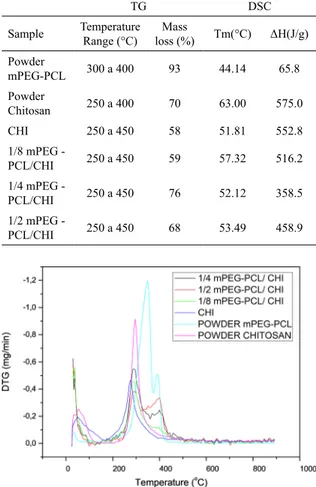
6.7.1 Thermogravimetric (TG) and thermogravimetric analysis (DTG)
The mass loss was determined by the thermogravimetric method. The percentage of mass loss of the raw materials and obtained membranes are shown in Table 5. Cooper et
al. (2011) found a peak of chitosan degradation around
peaks of mass loss was observed in the derivative curves
of CHI and mPEG-PCL (Figure 5). According to Niamsa et al., (2008) the peak mass loss for mPEG was found at 418ºC and the degradation of PCL was observed at 360ºC, coinciding with the temperature range of mass loss observed for the membranes containing mPEG-PCL10.
6.7.2 Calorimetric analysis (DSC)
The melting temperatures (Tm) and enthalpy for the raw materials and CHI/mPEG-PCL membranes are shown in Table 5. As expected, Tm values of the membranes are well above the body temperature (37ºC), suggesting good thermal stability for wound dressing application. Moreover, membranes showed intermediary values of Tm between the CHI and mPEG-PCL melting points. Cardoso et al. (2014) also investigated thermal properties of hybrid membrane of
chitosan/poly (ε-caprolactone) for tissue engineering21. The
authors also reported an increase on the melting temperature of the PCL-CHI hybrid membrane in comparison to PCL21.
On the other hand, the fusion enthalpy of 1/4 mPEG - PCL/CHI and 1/2 mPEG - PCL/CHI blended membranes was considerably lower than that found for CHI. Reduced enthalpy of fusion suggests lower crystallinity degree that can be explained by the presence of mPEG-PCL in the membranes. This copolymer has lower fusion enthalpy and may reduce the crystallinity of the chitosan-based membranes22. Crystallinity degree may also influence other properties, such as the water uptake by the membranes.
7. In vitro Gentamicin Release and
Antibacterial Activity
7.1 Standard curve for gentamicin
The gentamicin standard curve was obtained using the colorimetric method, which is based on the reaction of ninhydrin with the primary and secondary amines of gentamicin. The linear model between absorbance and concentration is given by the equation y = 0.00207x + 0.19376 and R2= 0.983, where
x is the gentamicin concentration and absorbance. The R2 parameter is greater than 0.95, indicating a satisfactory fit
of the equation to the data obtained. Thus, this curve was used to calculate the concentrations of gentamicin released in the in vitro release assays, which were performed in two experiments.
7.2 Initial Burst Release
Firstly, the release of gentamycin was measured for 15 minutes, 30 minutes, 45 minutes and 1 hour (Figure 6) in order
to simulate the initial burst release. In the first 15 minutes,
greater release of gentamicin from the membranes CHI-GE and 1/8 mPEG-PCL/CHI-GE was observed due to the higher percentage of swelling of these samples. The 1/4 and 1/2 mPEG-PCL/CHI-GE membranes released less gentamicin, indicating that the blend of CHI and mPEG-PCL copolymer
can be effective in delaying the burst release process. Table 5. Results of thermal analyses of raw materials and membranes
unloaded with gentamicin: thermogravimetric and calorimetric parameters.
TG DSC
Sample Temperature Range (°C)
Mass
loss (%) Tm(°C) ΔH(J/g)
Powder
mPEG-PCL 300 a 400 93 44.14 65.8
Powder
Chitosan 250 a 400 70 63.00 575.0
CHI 250 a 450 58 51.81 552.8
1/8 mPEG -
PCL/CHI 250 a 450 59 57.32 516.2
1/4 mPEG -
PCL/CHI 250 a 450 76 52.12 358.5
1/2 mPEG -
PCL/CHI 250 a 450 68 53.49 458.9
Figure 5. Derived derivative thermogravimetric curves of polymeric
raw materials and membranes unloaded with gentamicin. (pink)
chitosan, (light blue) mPEG-PCL, (blue) CHI, (green) 1/8 mPEG-PCL/
CHI, (black) 1/4 mPEG-PCL/CHI and (red) 1/2 mPEG-PCL/CHI.
Figure 6. In vitro Gentamicin burst release from (black) CHI-GE,
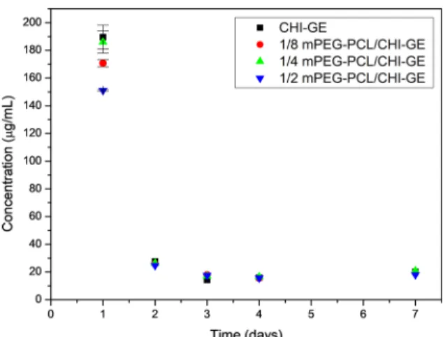
7.3 Long-term Release
The long-term release results are shown in the Figure 7.
It was only possible to observe significant decrease on the
gentamicin released from the membrane 1/2 mPEG-PCL/ CH-GE and 1/8 mPEG-PCL/GE when compared to CHI-GE. Again, the membrane with the highest concentration of mPEG-PCL released the lowest concentration of gentamicin
in the first day. Nevertheless, from the 2nd to the 7th day, no significant difference was observed for the gentamicin released
from all tested membranes. In addition, the minimal inhibitory concentration (MIC) of gentamicin against Staphylococcus aureus (MIC50 0.2 µg/mL) was delivered during the analyzed period, suggesting that membranes are capable to protect the wound from S. aureus infection for one week23.
7.4 Antibacterial activity
Agar diffusion technique was used to evaluate the
antibacterial activity of the CHI, mPEG-PCL/CHI and gentamicin-loaded membranes. The main bacteria found in infected wounds, E. coli and S. aureus, were tested and
the inhibition zone diameter was measured around the samples. It was not possible to observe zone of inhibition
of the bacterial growth around the membranes without the incorporation of the antibiotic in the plates inoculated with E. coli. According to Dutta et al. (2009), chitosan has high antimicrobial activity against pathogenic organisms including fungi and Gram-positive and Gram-negative bacteria24. However, this property could not be verified by this test,
probably due to the low concentration of chitosan used to prepare the membranes.
On the other hand, after 24 h, all gentamicin-loaded membranes inhibited E. coli growth. The inhibition zone diameters ranged between 1.0 and 1.5cm (Table 6), indicating the release of the antibiotic from the membranes to the culture
Figure 7. Long-term in vitro gentamicin release from (black)
CHI-GE, (red) 1/8 mPEG-PCL/CHI, (green) 1/4 mPEG-PCL/CHI
and (blue) 1/2 mPEG-PCL/CHI at every 24 hours for one week.
Table 6. Diameter of the zone of inhibition (cm) around gentamicin
loaded membranes against E. coli and S. aureus.
Menbrane E. coli S. aureus
Diameter (cm) Diameter (cm)
CHI-GE 1.4 ± 0.1 1.2 ± 0.1
1/8 mPEG-PCL/ CHI-GE 1.1 ± 0.1 1.0 ± 0.1
1/4 mPEG-PCL/ CHI-GE 1.5 ± 0.0 1.2 ± 0.1
1/2 mPEG-PCL/ CHI-GE 1.2 ± 0.1 1.1 ± 0.1
medium. Bajpai et al. (2016) investigated the antibacterial activity of alginate films loaded with gentamicin19. The authors reported a zone of inhibition of 3cm in the Plate inoculated
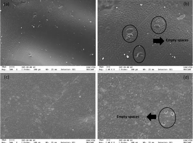
with E. coli, suggesting the film has fair antibacterial activity19. In order to confirm gentamicin release from the membranes,
scanning electron micrographs of the CHI-GE and 1/2
mPEG-PCL/CHI were taken before and after the Agar Diffusion
test. Figure 8 illustrates the release of gentamicin from the 1/2 mPEG-PCL/CHI membrane, indicated by the presence of empty spaces in place of the gentamicin sulfate crystals.
Regarding to the antibacterial activity of the membranes against S. aureus (Figure 9), similar behavior observed for E. coli was found, i.e. only gentamicin-loaded membranes inhibited the S. aureus growth. Similar diameters of inhibition were measured for both bacteria.
Sorensen and Sorensen (1993) investigated the antibacterial activity of gentamicin against four strain of S. aureus using
Mueller-Hinton (MH) broth and surgical wound fluid (WF)
as test media25. According to the authors, during the first hour of incubation, there was a marked concentration-dependent kill rate25. Thus, it is important to have a high
concentration of gentamicin released to the media in the
first hour, as we demonstrated in the Figure 6, in order to
eliminate all bacterial contamination. These authors also
reported that after the first hour of incubation, the kill rate
was independent of the gentamicin concentration in both media (MH and WF), suggesting that the gentamicin-loaded membranes can control the bacterial growth for at least one
week (Figure 7)25.
8. Conclusions
We prepared homogeneously blended CHI and mPEG-PCL membranes loaded with gentamicin for potential medicated wound dressing application. The obtained membranes are
translucent, which would facilitate the visualization of the wound, and capable of absorbing fluids that would help keeping
the wound moisture. The membranes are thermally stable at physiological temperature and able to release incorporated gentamicin at physiological pH and temperature conditions. Released antibiotic from the gentamicin-loaded membranes inhibited E. coli and S. aureus growth, suggesting their use as medicated wound dressings. Nevertheless, in vivo studies must be performed in order to evaluate the biocompatibility
Figure 8. Scanning electron micrographs of (a) CHI-GE before antibacterial test (500x); (b) CHI-GE (1000x) after antibacterial test; (c) 1/2 mPEG-PCL/CHI-GE before antibacterial test (500x) and (d) 1/2 mPEG-PCL/CHI-GE after antibacterial test (1000x).
9. Acknowledgments
This research was supported by CNPq - Conselho
Nacional de Desenvolvimento Científico e Tecnológico,
CAPES - Coordenação de Aperfeiçoamento de Pessoal de Nível Superior, FAPEMIG - Fundação de Amparo à Pesquisa do Estado de Minas Gerais and FAPESP - Fundação de Amparo à Pesquisa do Estado de São Paulo.
10. References
1. Singer AJ, Clark RA. Cutaneous wound healing. The New England Journal of Medicine. 1999;341(10):738-746.
2. Alves H, Machado MT, Noronha ANW. Análise Quantitativa
do Processo de Reparo em Cicatriz Cirúrgica de Ratos Tratados
com Extrato de Musa Sapientum, Aloe Vera, Colagenase e Placebo. Revista Ciências em Saúde. 2011;1(2):1-11.
3. Campos MGN, Rawls HR, Innocentini-Mei LH, Satsangi N. In vitro gentamicin sustained and controlled release from chitosan
cross-linked films. Journal of Materials Science: Materials in Medicine. 2009;20(2):537-542.
4. Campos MGN, Satsangi N, Rawls HR, Mei LHI. Chitosan
Cross-Linked Films for Drug Delivery Application. Macromolecular Symposia. 2009;279(1):169-174.
5. Campos MGN, Mei LHI, Santos AR Jr. Sorbitol-Plasticized and Neutralized Chitosan Membranes as Skin Substitutes.
Materials Research. 2015;18(4):781-790.
6. Viji CS, Amritha TS, Rajalekshmi G, Pandimadevi M. Potential
wound healing materials from the natural polymers - A review. International Journal of Pharma and Bio Sciences.
2015;6(3):B1365-B1389.
7. Dhivya S, Padma VV, Santhin E. Wound dressings - a review.
BioMedicine (Taipei). 2015;5(4):22.
8. Hoemann CD, Hurtig M, Rossomacha E, Sun J, Chevrier A,
Shive MS, et al. Chitosan-Glycerol Phosphate/Blood Implants
Improve Hyaline Cartilage Repair in Ovine Microfracture Defects.
The Journal of Bone & Joint Surgery. 2005;87(12):2671-2686.
9. Souza MA, Santos HT, Pretti TS, Nascimento ACGG, da Costa TP, Santos FJ, et al. Magnetic Nanoparticles Surface Modified with Biodegradable Polymers for Controled Methotrexate
Delivery in Cancer Therapy. Journal of Nanopharmaceutics and Drug Delivery. 2015;3:1-8.
10. Niamsa N, Puntumchai A, Sutthikhum V, Srisuwan Y, Baimark Y. Preparation and characterization of biodegradable chitosan
and methoxy poly(ethylene glycol)-b-poly (e-caprolactone)
blend homogeneous films. Journal of Applied Polymer Science. 2008;109(1):418-423.
11. Sahoo S, Sasmal A, Sahoo D, Nayak P. Synthesis and characterization of chitosan- polycaprolactone blended with
organoclay for control release of doxycycline. Journal of Applied Polymer Science. 2010;118(6):3167-3175.
12. El-Hefian EA, Nassef MM. Chitosan-Based Polymer Blends:
Current Status and Applications. Journal of the Chemical Society of Pakistan. 2014;36(1):11-27.
13. Mei J, Yuan Y, Wu Y, Li Y. Characterization of edible starch-chitosan film and its application in the storage of Mongolian
cheese. International Journal of Biological Macromolecules. 2013;57:17-21.
14. Cooper A, Oldinski R, Ma H, Bryers JD, Zhang M. Chitosan-based nanofibrous membranes for antibacterial filter applications.
Carbohydrate Polymers. 2013;92(1):254-259.
15. Sarasam A, Madihally SV. Characterization of
chitosan-polycaprolactone blends for tissue engineering applications.
Biomaterials. 2005;26(27):5500-5508.
16. Yao Q, Nooeaid P, Roether JA, Dong Y, Zhang Q, Boccaccini AR. Bioglass(r)-based scaffolds incorporating polycaprolactone
and chitosan coatings for controlled vancomycin delivery.
Ceramics International. 2013;39(7):7517-7522.
17. Silverstein RM, Webster FX, Kiemle DJ. Identificação Espectrométrica de Compostos Orgânicos. 7ª ed. Rio de Janeiro: LTC; 2007.
18. Aquino RP, Auriemma G, Mencherini T, Russo P, Porta A, Adami R, et al. Design and production of gentamicin/dextrans microparticles by supercritical assisted atomisation for the treatment of wound bacterial infections. International Journal of Pharmaceutics. 2013;440(2):188-194.
19. Bajpai SK, Shah FF, Bajpai M. Dynamic release of gentamicin sulfate (GS) from alginate dialdehyde (AD)-crosslinked casein (CAS) films for antimicrobial applications. Designed Monomers and Polymers. 2017;20(1):18-32.
20. Cooper A, Bhattarai N, Zhang M. Fabrication and cellular compatibility of aligned chitosan-PCL fibers for nerve tissue
regeneration. Carbohydrate Polymers. 2011;85(1):149-156.
21. Cardoso GB, Machado-Silva AB, Sabino M, Santos AR Jr, Zavaglia CA. Novel hybrid membrane of chitosan/poly
(e-caprolactone) for tissue engineering. Biomatter. 2014;4. pii: e29508.
22. Ahmed S, Sheraz MA, Rehman IU. Studies on tolfenamic acid-chitosan intermolecular interactions: effect of pH, polymer
concentration and molecular weight. AAPS PharmSciTech. 2013;14(2):870-879.
23. Yoshimura H, Ishimaru M, Kojima A. Minimum Inhibitory concentrations of 20 antimicrobial agents against Staphylococcus aureus isolated from bovine intramammary infections in Japan. Journal of Veterinary Medicine. B, Infectious Diseases and Veterinary Public Health. 2002;49(9):457-460.
24. Dutta PK, Tripathi S, Mehrotra GK, Dutta J. Perspectives for
chitosan based antimicrobial films in food applications. Food Chemistry. 2009;114(4):1173-1182.
25. Sørensen TS, Sørensen AI. Bactericidal activity of gentamicin