CATARINA FORTUNA
DOS REMÉDIOS
EFEITO CITOTÓXICO DO SULFORAFANO EM
CÉLULAS DE OSTEOSSARCOMA HUMANO
CYTOTOXIC EFFECT OF SULFORAPHANE IN
HUMAN OSTEOSARCOMA CELLS
CATARINA FORTUNA
DOS REMÉDIOS
EFEITO CITOTÓXICO DO SULFORAFANO EM
CÉLULAS DE OSTEOSSARCOMA HUMANO
CYTOTOXIC EFFECT OF SULFORAPHANE IN
HUMAN OSTEOSARCOMA CELLS
Dissertação apresentada à Universidade de Aveiro para cumprimento dos requisitos necessários à obtenção do grau de Mestre em Biologia Aplicada, Ramo de Biologia Molecular e Celular, realizada sob a orientação científica da Doutora Helena Cristina Correia de Oliveira, Investigadora Pós-Doutoramento do Departamento de Biologia da Universidade de Aveiro, e co-orientação da Professora Doutora Maria da Conceição Lopes Vieira dos Santos, Professora Associada com Agregação do Departamento de Biologia da Universidade de Aveiro, e do Doutor José Miguel Pimenta Ferreira de Oliveira, Investigador Pós-Doutoramento do Departamento de Biologia da Universidade de Aveiro.
Aos meus pais, irmã e F. que todos os dias encurtam os quilómetros que nos separam. À Bia, por todo o amor, por todo o apoio, por toda uma vida, por tudo.
o júri
Presidente Prof. Doutor João António de Almeida Serôdio
Professor Auxiliar do Departamento de Biologia da Universidade de Aveiro
Arguente Principal Prof. Doutor Francisco Luís Maia Mamede Pimentel
Professor Associado Convidado com Agregação da Secção Autónoma de Ciências da Saúde da Universidade de Aveiro
Orientadora Doutora Helena Cristina Correia de Oliveira
Investigadora Pós-Doutoramento do Departamento de Biologia da Universidade de Aveiro
Co-Orientadora Prof. Doutora Maria da Conceição Lopes Vieira dos Santos
Professora Associada com Agregação do Departamento de Biologia da Universidade de Aveiro
Co-Orientador Doutor José Miguel Pimenta Ferreira de Oliveira
agradecimentos Gostaria de agradecer a todos que, directa ou indirectamente, me acompanharam durante as várias etapas de realização desta dissertação e cujo apoio, incentivo e amizade serei eternamente grata.
Começo por agradecer aos meus pais e irmã pelo apoio e amor incondicionais, por compreenderem a minha ausência e indisponibilidade em tantas ocasiões, pelos sacrifícios que permitem esta minha caminhada e por, independentemente da distância que nos separa, permanecerem diariamente presentes na minha vida. Aos meus pais agradeço particularmente tudo o que fazem, cada um à sua maneira, para assegurar a minha estabilidade a todos os níveis e o entusiástico incentivo ao longo destes últimos cinco anos. À minha irmã, obrigada pelo seu humor inteligente e peculiar que tantos momentos magníficos e irrepetíveis proporciona, pela cumplicidade muitas vezes demonstrada sem recurso a palavras e pelos sábios conselhos, apesar da sua juventude.
A realização deste trabalho não teria sido possível sem o apoio dos meus três orientadores. A eles, o meu profundo agradecimento por toda a paciência que demonstraram, pela sua disponibilidade (muitas vezes fora do horário de trabalho) e pelo conhecimento que generosamente partilharam comigo. A cada um deles quero deixar algumas palavras. À Dra. Helena Oliveira, obrigada por ter acolhido esta jovem e inexperiente estudante há alguns anos atrás e por todos os ensinamentos que moldaram o meu crescimento enquanto bióloga. Ao Dr. José Miguel Oliveira agradeço o muito que aprendi numa área que há muito me fascinava e todo o tempo e trabalho dispendidos no desenho dos primers e no tratamento de dados da análise da expressão génica, cruciais para a finalização deste trabalho. À Prof. Maria da Conceição Santos, obrigada por me ter aberto as portas do laboratório que dirige e ter permitido a aprendizagem e a vivência de inúmeras experiências com a sua equipa, pelas ideias que partilhou comigo enquanto Professora e orientadora e pelos desafios lançados que tanto contribuíram para a minha evolução e que me abriram novos caminhos.
Aos meus colegas de laboratório Fernanda Rosário, Susana Barros, Pedro Pinto, Tiago Pedrosa, Francisco Pinho, Cristina Monteiro, Sónia Pinho, Bruno Ladeiro, Verónica Bastos, Andreia Ascenso, Marta Pinheiro e Hugo Santos não sei como expressar a minha enorme gratidão. O cunho de todos eles está presente neste trabalho, quer seja através de toda a experiência que partilharam comigo, do auxílio prestado no trabalho experimental, do companheirismo e amizade que nunca esquecerei ou de um último jantar que tanto me tocou e surpreendeu. Em especial gostaria de agradecer à Jenny, minha parceira de trabalho, companheira de aventuras e desventuras em Portugal e além fronteiras. A sua amizade incondicional foi o meu pilar em diversas ocasiões e a sua generosidade foi essencial na entrega desta dissertação durante a minha ausência.
À Cláudia Deus, colega de mestrado e amiga, um grande obrigada pela paciência que demonstrou ao longo destes dois anos e pela sua alegria inata. Foi um enorme prazer conhecê-la e partilhar tantos pensamentos, trabalhos e risos.
Por fim, à Teele Karafin, que conheci no início de uma nova etapa da minha vida, e que, apesar dos poucos meses de convívio, já se revelou uma grande amiga, ouvinte e conselheira. Obrigada pelas trocas de ideias, pelos telefonemas diários em alturas de crise e por tudo o que ainda há-de vir. A todos o meu sincero obrigado e desejos de felicidades e sucesso!
“I find hope in the darkest of days, and focus in the brightest. I do not judge the universe.”
palavras-chave Osteossarcoma, MG-63, sulforafano, citotoxicidade, terapia do cancro, ciclo celular, apoptose, expressão génica
resumo O osteossarcoma é o tipo mais comum de tumor maligno do osso. Esta doença apresenta uma elevada incidência em crianças e adolescentes. Adicionalmente, metástases, ocorrendo principalmente nos pulmões, afectam uma percentagem considerável dos pacientes com osteossarcoma. Os tratamentos actuais incluem ressecção cirúrgica, quimioterapia e radioterapia, mas os pacientes com osteossarcoma apresentam uma baixa capacidade de resposta a estas opções de tratamento. Assim, é urgente desenvolver novas abordagens de tratamento. O sulforafano é um fitocomposto que exibe actividade quimiopreventiva e quimioterápica. Numerosos estudos têm demonstrado que o sulforafano modula muitos eventos associados ao cancro, como susceptibilidade a agentes carcinogénicos, ciclo celular, apoptose, angiogénese e metastização. Apesar de bem caracterizados em diversos tumores, os efeitos do sulforafano são ainda insuficientemente conhecidos no osteossarcoma, assim como o mecanismo de acção subjacente. O objectivo desta dissertação foi avaliar os potenciais efeitos citostático e apoptótico do sulforafano e revelar o mecanismo de acção por trás destes efeitos. Para isso, a linha celular de osteossarcoma humano MG-63 foi exposta a sulforafano (no intervalo de 0 a 20 µM) durante 24 e 48 h. Parâmetros como confluência, morfologia celular, viabilidade celular, ciclo celular, clastogenicidade, apoptose, actividade específica de caspases e expressão de genes associados ao ciclo celular foram analisados após o tratamento com sulforafano. As culturas expostas ao sulforafano apresentaram uma diminuição da confluência, acompanhada de alterações na morfologia celular. O sulforafano também induziu uma diminuição da viabilidade celular dependente da dose e do tempo de exposição que pode ser explicada pelo aumento de células apoptóticas e por citostaticidade com bloqueio na fase G2/M. Este bloqueio do ciclo celular foi mediado por Chk2, pela sub-expressão de CDC25C e, provavelmente, pela sub-expressão de CCNB1, assim como de CDK2. Um efeito clastogénico foi também detectado após 48 h de tratamento com sulforafano. Estes dados sugerem que o sulforafano deve ser considerado um candidato a agente quimioterápico no tratamento de osteossarcoma, embora sejam necessários mais estudos para lançar luz sobre vias de sinalização moduladas pelo sulforafano, assim como sobre a possível citotoxicidade deste fitocomposto em osteoblastos normais.
keywords Osteosarcoma, MG-63, sulforaphane, cytotoxicity, cancer therapy, cell cycle, apoptosis, gene expression
abstract Osteosarcoma is the most common type of malignant bone tumor. This disease shows high incidence in children and adolescents. Additionally, metastases occurring mainly at the lungs affect a considerable percentage of osteosarcoma patients. Current treatments include surgical resection, chemotherapy, and radiotherapy, but osteosarcoma patients show poor responsiveness to these treatment options. Thus, it is urgent to develop novel treatment approaches. Sulforaphane is a phytochemical that displays chemopreventive and chemotherapeutic activity. Many studies have been shown that sulforaphane modulates many cancer-related events, as susceptibility to carcinogenic agents, cell cycle, apoptosis, angiogenesis, and metastasis. Although well characterized in several tumors, the effects of sulforaphane are still poorly understood in osteosarcoma, as well as the underlying mechanism of action. The aim of this dissertation was to evaluate the potential cytostatic and apoptotic effects of sulforaphane and to unveil the mechanism of action behind these effects. In order to do that, the human osteosarcoma cell line MG-63 was exposed to sulforaphane (in the 0 to 20 µM range) for 24 and 48 h. Parameters as confluence, cell morphology, cell viability, cell cycle, clastogenicity, apoptosis, caspase specific activity, and expression of cell cycle-related genes were analyzed after sulforaphane treatment. Cultures exposed to sulforaphane showed a decrease in confluence, accompanied by alterations in cell morphology. Sulforaphane also induced a dose- and time-dependent impairment of cell viability, which can be explained by an increase of apoptotic cells and cytostaticity with G2/M phase arrest. This cell cycle blockage was mediated by Chk2 and CDC25C downregulation, and probably CCNB1 as well as CDK2 downregulation. A clastogenic effect was also detected after 48 h of sulforaphane treatment. These data suggest that sulforaphane should be considered as a chemotherapeutic agent candidate in osteosarcoma treatment, although more studies are needed to shed insight on signaling pathways modulated by sulforaphane, as well as the possible cytotoxicity of this phytochemical on normal osteoblasts.
Pinto, P., Pinho, F., Pedrosa, T., Pinho, S., Rosário, F., Ferreira de Oliveira, J.M.P., Oliveira, H. and Santos, C. (2012) Sulforaphane-induced DNA Damage in Osteosarcoma Cells: A Correlation with Oxidative Stress, Cell Cycle Arrest and Apoptosis.
Table of Contents
List of Abbreviations ... xi
List of Figures ... xvi
List of Tables... xvii
I. INTRODUCTION... 1
1.1. Cancer... 1
1.1.1 General considerations about cancer ... 1
1.1.2. Target mechanisms in cancer therapy ... 6
1.1.2.1. Cell cycle ... 6
1.1.2.2. Apoptosis ... 8
1.2. Osteosarcoma ... 11
1.2.1. Bone physiology ... 11
1.2.2. Osteosarcoma: epidemiology, clinical aspects, diagnostic methods, histologic features, and etiology... 12
1.2.3. Pathogenesis of osteosarcoma ... 14
1.2.4. Conventional treatment approaches in osteosarcoma... 16
1.2.5. MG-63: an in vitro model for osteosarcoma ... 17
1.3. Sulforaphane... 19
1.3.1. Cruciferous vegetables, glucosinolates, and isothiocyanates ... 19
1.3.2. Sulforaphane and cancer... 20
1.3.3. Chemopreventive and chemotherapeutic pathways targeted by sulforaphane.. 21
1.3.3.1. Modulation of Phase I and Phase II enzymes ... 21
1.3.3.2. Induction of cell cycle arrest ... 22
1.3.3.3. Induction of apoptosis ... 22
1.3.3.4. Inhibition of angiogenesis and metastasis... 23
II. OBJECTIVES... 25
III. MATERIALS AND METHODS ... 26
3.2. Cell culture ... 26
3.3. Sulforaphane preparation and cell exposure... 27
3.4. Characterization of confluence and cell morphology... 27
3.5. Cell viability ... 28
3.6. Cell cycle and clastogenicity analysis ... 28
3.7. Apoptosis assessment ... 30
3.8. Protein quantification ... 31
3.9. Caspase activity assessment ... 31
3.9.1. Caspase 3 activity... 32
3.9.2. Caspase 8 and caspase 9 activity ... 32
3.9.3. Determination of caspase specific activity ... 33
3.10. Gene expression... 34
3.10.1. Primers ... 34
3.10.2. RNA extraction ... 34
3.10.3. cDNA synthesis ... 36
3.10.4. qRT-PCR... 37
3.11. Data and statistical analysis ... 38
IV. RESULTS ... 39
4.1. Characterization of confluence and cell morphology... 39
4.2. Cell viability ... 39
4.3. Cell cycle and clastogenicity analysis ... 39
4.4. Apoptosis assessment ... 41
4.5. Caspase activity assessment ... 45
4.6. Gene expression... 45
V. DISCUSSION... 48
VI. CONCLUSION AND FUTURE PERSPECTIVES ... 57
List of Abbreviations
Akt RAC-alpha serine/threonine-protein kinase
Apaf-1 Apoptotic protease-activating factor 1
ARE Antioxidant response element
ATCC American Type Culture Collection
ATM Ataxia telangiectasia mutated
ATP Adenosine triphosphate
ATR ATM- and Rad3-related protein
Bak B-cell lymphoma protein 2-antagonist/killer 1
Bax B-cell lymphoma protein 2-associated X protein
Bcl-2 B-cell lymphoma protein 2
Bcl-XL B-cell lymphoma protein 2-related protein, long isoform
Bid BH3-interacting domain death agonist
BLM Bloom syndrome protein coding gene
BMP Bone morphogenic protein
BSA Bovine serum albumin
CCNB1 Cyclin B1 coding gene
Cdc25 Cell division cycle 25
CDC25C M-phase inducer phosphatase 3 coding gene
Cdc25C M-phase inducer phosphatase 3
CDK Cyclin-dependent kinase
CDK1 Cyclin-dependent kinase 1 coding gene
Cdk1 Cyclin-dependent kinase 1
CDK2 Cyclin-dependent kinase 2 coding gene
Cdk2 Cyclin-dependent kinase 2
Cdk4 Cyclin-dependent kinase 4
CDKN2A Cyclin-dependent kinase inhibitor 2A coding gene
CDS Coding DNA sequence
CHEK1 Checkpoint kinase 1 coding gene
CHEK2 Checkpoint kinase 2 coding gene
Chk1 Checkpoint kinase 1
Chk2 Checkpoint kinase 2
CIN Chromosomal instability
Cip Cyclin-dependent kinase interacting protein
CKI Cyclin-dependent kinase inhibitor
c-Myc Myc proto-oncogene protein
Ct Cycle threshold
CYP Cytochrome P450 enzyme
Cyt c Cytochrome c
DEPC Diethylpyrocarbonate
DISC Death-inducing signaling complex
DMSO Dimethyl sulfoxide
DNA Deoxyribonucleic acid
dNTP Deoxyribonucleotide triphosphate
DTT Dithiothreitol
EDTA Ethylenediaminetetraacetic acid
FADD Fas-associated death domain
Fas Tumor necrosis factor receptor superfamily member 6
FasL Fas ligand
FBS Fetal bovine serum
FITC Fluorescein isothiocyanate
FPCV Full peak coefficient of variation
GAPDH Glyceraldehyde-3-phosphate dehydrogenase coding gene
GLS Glucosinolate
HDAC Histone deacetylase
Hh Hedgehog
HIF-1 Hypoxia-inducible factor 1
HIF-1α Hypoxia-inducible factor 1-alpha
ICAD Inhibitor of caspase-activated DNase
Ink4 Inhibitor of cyclin-dependent kinase 4
ITC Isothiocyanate
KDR/flk-1 Kinase insert domain receptor/ Fetal liver kinase 1
Keap1 Kelch-like ECH-associated protein 1
Kip Kinase inhibitor protein
Mdm2 Human homologue of murine double minute 2
αααα-MEM α-Minimum Essential Medium
MIRA-1 Mutant p53 reactivation and induction of rapid apoptosis
MMP Matrix metalloproteinase
MMP-2 Matrix metalloproteinase 2
MMP-9 Matrix metalloproteinase 9
MPF Maturation-promoting factor
mRNA Messenger ribonucleic acid
mTOR Mammalian target of rapamycin
MTT 3-(4,5-dimethyl-thiazolyl-2)-2,5-diphenyl tetrazolium bromide
Myt1 Membrane-associated tyrosine- and threonine-specific cdc2-inhibitory kinase
pNA p-nitroanilide
Noxa Phorbol-12-myristate-13-acetate-induced protein 1
Nrf2 Nuclear factor erythroid 2-related factor 2
OS Osteosarcoma
p15Ink4b Cyclin-dependent kinase inhibitor 2B
p16Ink4a Cyclin-dependent kinase inhibitor 2A
p18Ink4c Cyclin-dependent kinase inhibitor 2C
p21Cip1 Cyclin-dependent kinase inhibitor 1A
p27Kip1 Cyclin-dependent kinase inhibitor 1B
p53 Tumor protein p53
p57Kip2 Cyclin-dependent kinase inhibitor 1C
PARP Poly (ADP-ribose) polymerase
PBS Phosphate-buffered saline
PCR Polymerase chain reaction
PDGF Platelet-derived growth factor
PI Propidium iodide
PI3K Phosphatidylinositol 3-kinase
PRIMA-1 p53 reactivation and induction of massive apoptosis
PS Phosphatidylserine
Puma B-cell lymphoma protein 2-binding component 3
qRT-PCR Quantitative reverse transcription polymerase chain reaction
Rb Retinoblastoma-associated protein
RecQ4 ATP-dependent DNA helicase Q4
RECQL4 ATP-dependent DNA helicase Q4 coding gene
RFU Raw fluorescence unit
RNA Ribonucleic acid
RNase Ribonuclease
ROS Reactive oxygen species
RUNX2 Runt-related transcription factor 2 coding gene
Runx2 Runt-related transcription factor 2
SD Standard deviation
SFN Sulforaphane
Smac/DIABLO Second mitochondria-derived activator of caspase/Direct IAP-binding
protein with low pI
TGF-β Transforming growth factor β
TP53 p53 coding gene
TRADD Tumor necrosis factor receptor-associated death domain
TRAF2 Tumor necrosis factor receptor-associated factor 2
TRAIL Tumor necrosis factor-related apoptosis-inducing ligand
TRAIL-R1 Tumor necrosis factor-related apoptosis-inducing ligand receptor 1
VEGF Vascular endothelial growth factor
Wee1 Wee1-like protein kinase
Wnt Wingless-type
List of Figures
Figure 1. Cancer hallmarks established over the past decade. ... 5
Figure 2. General scheme of the main cell cycle phases of mammalian cells and its key regulators. ... 7
Figure 3. The intrinsic and extrinsic pathways to caspase activation in apoptosis. ... 9
Figure 4. Bioconversion pathway of glucosinolates... 20
Figure 5. Enzymatic conversion of glucoraphanin to sulforaphane and the respective chemical structures. ... 21
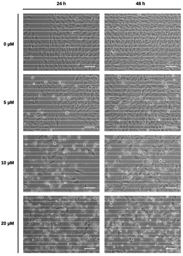
Figure 6. Microscopic visualization of control and sulforaphane-treated MG-63 cells. ... 40
Figure 7. Cytotoxic effect of sulforaphane on MG-63 cells... 41
Figure 8. Cytostatic effect of sulforaphane on MG-63 cells. ... 42
Figure 9. Sulforaphane-induced clastogenicity. ... 43
Figure 10. Activation of apoptosis in MG-63 cells exposed to sulforaphane. ... 44
Figure 11. Specific activity of two initiator caspases and one executioner caspase after sulforaphane treatment. . ... 46
Figure 12. Relative expression of cell cycle-related genes after exposure to 10 µM sulforaphane for 48 h. ... 47
Figure 13. Sulforaphane-induced changes in gene expression of MG-63 that may contribute to activation of G2/M DNA damage checkpoint. ... 56
List of Tables
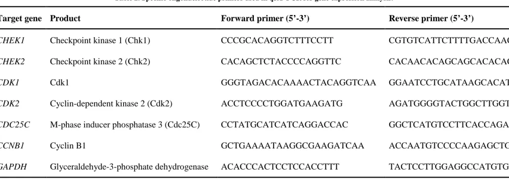
Table 1. Mechanisms of action of conventional chemotherapeutic agents used in osteosarcoma treatment. ... 17 Table 2. Specific oligonucleotide primers used in qRT-PCR for gene expression analysis.
I. INTRODUCTION
1.1. Cancer
1.1.1 General considerations about cancer
Cancer is a major burden worldwide. Economically developed countries present overall cancer incidence rates twice as high as the ones found in developing countries (Jemal et
al., 2011), mainly due to the Western lifestyle. According to the American Cancer Society,
it is estimated that 1,638,910 men and women will be diagnosed with cancer and 577,190 men and women will die of cancer in the United States in 2012, being these rates slightly higher for men than for women and with lung, colorectal, breast, and prostate cancers accounting for the largest numbers of new cases and deaths (Siegel et al., 2012). In Europe, cancer was listed as the second most common cause of death after cardiovascular diseases (OECD, 2012) with approximately 3.2 million estimated new cases and 1.7 million estimated deaths in 2008 (Ferlay et al., 2010). Recent data project 1,283,101 cancer deaths to occur in Europe during the current year, with lung (262,250), colorectal (163,106), breast (88,101), and prostate (69,960) cancers as the main causes (Malvezzi et
al., 2012), similarly to the United States.
From a simplistic point of view, cancer can be defined as a group of diseases in which abnormal cells proliferate in an uncontrolled manner and spread to other tissues. However, during the past few decades, an immense body of knowledge evidenced the inherent complexities of cancer, but fortunately also unveiled its characteristics and the underlying molecular mechanisms.
The etiological factors of cancer vary considerably from case to case. Chemical agents, lifestyle (e.g., diet, obesity, tobacco smoke, alcohol consumption), infections, radiation, genetic problems, and hormones are some of the factors already proved to correlate with carcinogenesis (Langeberg et al., 2007; Khan and Shrivastava, 2010; Khan et al., 2010; Vahakangas, 2011). However, the general established conception is that cancer results from cumulative genomic alterations that lead to atypical RNA and protein profiles, which
in the end originate the abnormal cancer phenotype. Such alterations are often found in genes that in the cancer scenario behave as either tumor suppressors or oncogenes (Hahn and Weinberg, 2002). The range of cancer-inducing genomic changes is vast, yielding from intragenic mutations to aneuploidy events. Loss of function of tumor suppressors is usually directly or indirectly related with intragenic mutations (Malkin et al., 1990) and deletions or allelic loss (Wei et al., 1999; Yoshimoto et al., 2007) of the corresponding genes. In the case of oncogenes, mutations (Akagi et al., 2007), chromosomal translocations (MacKenzie et al., 1993), and genomic amplifications (Birkeland et al., 2012) are the three major events explaining gain of function. In recent years, epigenetic alterations, heritable alterations in gene expression without changes in the primary DNA sequence (Sadikovic et al., 2008a), were also found to be main contributors for cancer development. Variations in the DNA methylation profile of genes or gene promoters are common drivers of carcinogenesis (Futscher et al., 2004), but histone modifications (Kondo et al., 2007) and changes in nucleosome positioning (Medina and Sanchez-Cespedes, 2008) are important players in that process, as well.
Based on these and other findings of the past decades and on the assumption that the molecular machinery owned by mammalian cells is equable, Douglas Hanahan and Robert A. Weinberg suggested that most – if not all – human cancers share the same functional capabilities that allow cancer cells to survive, proliferate, and disseminate (Hanahan and Weinberg, 2000). The six hallmarks of cancer proposed by these authors in 2000 include:
1) Self-sufficiency in growth signals. In order to grow and proliferate, normal cells require the binding of growth signals to transmembrane receptors and the subsequent activation of intracellular signal transduction cascades that culminate in a pro-growth cellular response. Tumor cells can reduce the dependency on exogenous signals by different means, such as producing their own growth signals (e.g., platelet-derived growth factor (PDGF) (Furuhashi et al., 2004)) and overexpressing growth signal receptors (e.g., human epidermal growth factor receptor 2 (HER2) (Loi et al., 2011)), thus breaching homeostasis and achieving abnormal growth rates.
2) Insensitivity to antigrowth signals. Besides growth signals, a normal cell is also exposed to antigrowth signals from the environment. As the name suggests, these molecules act as antiproliferative signals, inducing either a quiescent state or cell differentiation. Deregulation of proteins involved in signaling pathways transducing
antigrowth signals is often found in cancers and usually such proteins are intimately related with cell cycle progression. One example is retinoblastoma-associated protein (Rb), a major transducer of antigrowth signals that inhibits transcription factors, preventing cell cycle progression (Jonsson et al., 2012).
3) Evasion of apoptosis. Together with proliferation rates, the life span of a tumor cell is an important determinant of the overall tumor progression. Apoptosis is the cell death pathway commonly activated to maintain tissue homeostasis, but cancer cells seem to be resistant to this mechanism. Alterations in apoptotic mediators are extremely abundant in cancers. Amongst such mediators, p53, a tumor suppressor also known as “guardian of the genome” due to its function when DNA damage occurs, is frequently found to suffer different deregulations (Malkin et al., 1990; Pogribny and James, 2002), preventing activation of the apoptotic cascade.
4) Limitless replicative potential. Within a normal tissue, cells undergo a finite number of replications, after which they become senescent. This event can be explained by the existence of telomeres, thousands of short nucleotide sequence repeats protecting the ends of chromosomes. During each replication cycle, chromosomes lose telomeric DNA, resulting in a progressive telomere shortening throughout cell life that eventually leads to death (Londono-Vallejo and Wellinger, 2012). However, tumor cells appear to undergo unlimited replications, thanks to the expression of functional telomerase, the DNA polymerase that adds DNA sequence repeats to the 3’ ends of chromosomal DNA for telomere maintenance, which is almost absent in normal cells (Chen and Chen, 2011).
5) Sustained angiogenesis. A critical step in tumor progression is the maintenance of the expansion rate. As the tumor becomes larger, the inner cells of the tumor mass experience unfavorable conditions and often die due to the lack of blood supply that enables delivery of nutrients and oxygen, as well as removal of waste products and carbon dioxide. In order to surpass short blood supply and to continue to grow, tumors must activate an “angiogenic switch” and induce angiogenesis, the process of blood vessel formation from quiescent blood vessels (Hanahan and Folkman, 1996). Under hypoxic conditions, hypoxia-inducible factor 1 (HIF-1), an heterodimer possessing an oxygen sensor subunit (HIF-1α), is responsible for prompting angiogenesis in several human cancers via transcription factor activity that results in overexpression of vascular endothelial growth factor (VEGF); VEGF is a major growth signal that binds to tyrosine
kinase receptors of endothelial cells, stimulating proliferation and generation of new blood vessels (Semenza, 2000). Both oxygen deprivation (Shweiki et al., 1992) and genetic alterations causing increased activity of HIF-1 (Maxwell et al., 1999; Ravi et al., 2000) seem to be two reasonable mechanisms behind angiogenesis in tumors.
6) Tissue invasion and metastasis. This pivotal step in tumor development comprises the escape of tumor cells from the initial tumor mass, the invasion of other tissues (adjacent or distant), and the settlement of new colonies: metastatic or secondary tumors. The fifth hallmark is intrinsically connected with the metastatic process providing the main route for primary tumor cells evasion and displacement (Zetter, 1998). Cancer cells that acquired this capability often present loss of molecules mediating cell-cell adhesion (e.g., cadherins (Yoshida et al., 2001)). Other feature that enhances the invasive potential of tumor cells is the upregulation of matrix metalloproteinases (MMPs) (Lafleur et al., 2005; Kryczka et al., 2012), zinc-dependent extracellular matrix-degrading proteases.
These functional capabilities are attained through a multistep tumorigenic process driven by the accumulation of genomic alterations, but one particular lesion may give rise to several capabilities at the same time (Hanahan and Weinberg, 2000).
Four years later, Dunn, Old, and Schreiber proposed a seventh hallmark of cancer: avoidance of immunosurveillance (Dunn et al., 2004). In truth, highly immunogenic cancer cells can be detected and destroyed by the immune system, but the remaining ones are spared due to their weak immunogenic phenotype (immunoediting) and ability to dismantle components of both arms of the immune system (Dunn et al., 2004).
In 2008, Guido Kroemer and Jacques Pouyssegur discussed the complex interactions between these seven hallmarks and tumor cell metabolism (Kroemer and Pouyssegur, 2008). Many tumor cells undergo a “metabolic switch” in which their energy production is highly limited to glycolysis instead of mitochondrial oxidative phosphorylation even in normoxic conditions, a concept brought up by Otto Warburg many decades ago (Warburg, 1956). Underlying this metabolic reprogramming is, among others, the upregulation of glucose transporters and enzymes of the glycolytic pathway (Jones and Thompson, 2009). Last year, Hanahan and Weinberg (2011) revisited their former list of hallmarks, highlighted the importance of tumor microenvironment in the carcinogenic process, included the mechanisms that have been discussed in the meantime, and considered new
hallmarks: genome instability and mutation, and tumor-promoting inflammation. The first refers to the acceleration in the accumulation of mutations by the cancer cell through a variety of mechanisms, such as the collapse of DNA damage sensors or DNA damage repairing machinery (Jackson and Bartek, 2009). The second, explores the ability of tumor cells to induce an inflammatory response in the tumor microenvironment, that promotes several steps of the carcinogenic process; innate immune cells play here an important role by releasing reactive oxygen species (ROS) and growth factors in the vicinity of the tumor (Qian and Pollard, 2010).
Figure 1 illustrates the establishment of cancer hallmarks throughout the years as result of the incessant research on this subject.
Figure 1. Cancer hallmarks established over the past decade. The 2011 up-to-date Hanahan and Weinberg’s scheme enclosing: the six acquired hallmarks of cancer cells (sustaining proliferative signaling, evading growth suppressors, resisting cell death, enabling replicative immortality, inducing angiogenesis, and activating invasion and metastasis) initially proposed by these authors in 2000; a seventh hallmark (avoiding immune destruction) recommended by Dunn and colleagues in 2004; the deregulated cellular energetics integrated by Kroemer and Pouyssegur in 2008; and two enabling characteristics (genome instability and mutation, and tumor-promoting inflammation) added by Hanahan and Weinberg in 2011. Adapted from Hanahan and Weinberg (2011).
1.1.2. Target mechanisms in cancer therapy
The compelling body of research and knowledge accomplished in the past decades lead to a better understanding of carcinogenesis and the molecular mechanisms behind it, and concomitantly to the progress in identifying novel targets for mechanism-based targeted therapies in human cancers. These therapies target specific key molecules that are involved in aberrant signaling pathways, thus modulating proliferation, differentiation, and death of cancer cells (Cattley and Radinsky, 2004). Such specificity can bring advantages for cancer treatment, as it is expected to show more effective results in cancer cells than current treatment approaches and less harmful effects on normal cells. As is already apparent, the mechanisms underlying cancer hallmarks offer attractive therapeutic targets and, in the next sections, two of these mechanisms – cell cycle and apoptosis – will be described in more detail and some targeted therapies exploiting them will be enumerated.
1.1.2.1. Cell cycle
Mammalian cell proliferation consists of repeated cell cycles that are divided in two main stages: mitosis (M), the process of nuclear division, and interphase, the period from the end of one mitosis to the beginning of the next one. Mitosis is subdivided in prophase, metaphase, anaphase, and telophase, whereas interphase comprises G1 (growth phase), S (DNA synthesis phase), and G2 (gap phase) phases (Norbury and Nurse, 1992). Non-proliferating cells, which accounts for the majority of cells in the human body, enter a quiescent state at G1 that prevents further divisions – the G0 phase. As many other processes, cell cycle is strongly regulated by a wide number of proteins.
Progression through the cell cycle is controlled by active protein kinase complexes, composed of a catalytic cyclin-dependent kinase (CDK, a family of serine/threonine protein kinases) and a regulatory cyclin. Throughout the cell cycle, CDK protein levels remain steady, but cyclin protein levels oscillate due to cyclic synthesis and degradation, leading to a periodically activation of CDKs (Pines, 1995). Consequently, CDKs are sequentially activated by specific cyclins during different phases, inducing cell cycle progression by phosphorylation of downstream proteins (Figure 2).
CDK activity can also be counteracted by CDK inhibitors (CKIs) from two distinct families: Cip/Kip (for CDK interacting protein/kinase inhibitor protein), which includes p21Cip1, p27Kip1, p57Kip2; and Ink4 (for inhibitor of cyclin-dependent kinase 4 (Cdk4)), that
Figure 2. General scheme of the main cell cycle phases of mammalian cells and its key regulators. The specific CDK/cyclin associations required in each cell cycle phase and the CKIs that negatively regulate the main drivers of cell cycle progression. Adapted from Fuster et al. (2010).
comprises p16Ink4a, p15Ink4b, p18Ink4c, p19Ink4d. The Cip/Kip proteins interact with and inhibit various CDK/cyclin complexes (Harper et al., 1995), whereas Ink4 proteins specifically bind to G1 CDKs, preventing their association with cyclin D (Carnero and Hannon, 1998) (Figure 2). In turn, CKIs are regulated by different internal and external signals. For instance, p21Cip1 expression is transcriptionally activated by the tumor suppressor p53 when DNA damage is detected (el-Deiry et al., 1993) and the expression and activation of p15Ink4b and p27Kip1, respectively, are influenced by transforming growth factor β (TGF-β) (Reynisdottir et al., 1995).
In addition, CDK activity is regulated by phosphorylation/dephosphorylation on conserved threonine and tyrosine residues. For example, Wee1-like protein kinase (Wee1) and membrane-associated tyrosine- and threonine-specific cdc2-inhibitory kinase (Myt1) inactivate cyclin-dependent kinase 1 (Cdk1) by tyrosine-15 and/or threonine-14 phosphorylation, and dephosphorylation of these sites is required for re-establishment of Cdk1 activity, which is mediated by phosphatases of the cell division cycle 25 (Cdc25) family (Medema and Macurek, 2012). Besides these molecules, others involved in
different signaling pathways (e.g., PI3K/Akt/mTOR pathway) can interact and modulate the cell cycle machinery.
Investigation of cell cycle deregulations in the cancer context resulted in the development of some agents that target components of these pathways. Alvocidib is a small molecule that inhibits CDKs, inducing cell cycle arrest (Lee and Sicinski, 2006). NVP-BEZ235, an imidazo [4,5-c] quinoline derivative, induces G1 arrest by targeting the ATP-binding cleft of PI3K (for phosphatidylinositol 3-kinase) and mTOR (for mammalian target of rapamycin), whose activity is frequently altered in human cancer cells (Maira et al., 2008).
1.1.2.2. Apoptosis
Apoptosis, also known as programmed cell death, is a controlled type of cell death that plays an important role in tissue homeostasis. This process occurs in physiological conditions (e.g., embryonic development, normal cell turnover), but also as a response to pathological conditions (e.g., immune reactions) and cell injuries (e.g., radiation, hypoxia, harmful chemical agents) (Norbury and Hickson, 2001). Apoptosis is generally characterized by distinct morphological and biochemical characteristics. These hallmarks of apoptosis include expression of phosphatidylserine (PS) in the outer layer of the cell membrane, cell shrinkage, retraction of pseudopods, rounding up of the cell, membrane blebbing, ultrastructural modification of cytoplasmic organelles, chromatin condensation, DNA fragmentation, and loss of membrane integrity (Kroemer et al., 2005). At the end of the apoptotic process, the cellular content is enclosed into apoptotic bodies (“budding”), which are subsequently engulfed and degraded by phagocytic cells. Due to the restriction of the cellular constituents in apoptotic bodies, the rapid phagocytosis of apoptotic remains by surrounding cells and the absence of anti-inflammatory cytokines production by the latter cells, apoptosis is in essence not associated with inflammatory reactions (Kurosaka et
al., 2003). Another feature of apoptosis is the activation of a group of enzymes belonging
to the caspase (cysteine-aspartic acid protease) family that function as initiators and executioners of the apoptotic process (Lavrik et al., 2005). The activation of caspases can occur through two major pathways (Figure 3).
The extrinsic or death receptor pathway is triggered when external signals bind to surface cell death receptors (e.g., tumor necrosis factor (TNF), Fas ligand (FasL), and TNF-related apoptosis-inducing ligand (TRAIL) bind to TNF receptors, TNF receptor superfamily member 6 (Fas), and TRAIL receptors, respectively) (Hengartner, 2001). Then, the
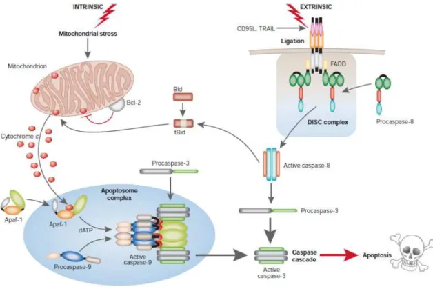
Figure 3. The intrinsic and extrinsic pathways to caspase activation in apoptosis. Activation of caspase 9 by apoptosome complex and caspase 8 by DISC complex as a result of the triggering of mitochondrial and death receptor apoptotic pathways, respectively. The initiator caspases 8 and 9 cleave procaspase 3, leading to the activation of an executioner caspase cascade that ends in apoptosis. Members of the Bcl-2 family modulate apoptosis through the control of mitochondrial Cyt c release. Extracted from MacFarlane and Williams (2004).
intracellular domain of death receptors recruits adapter proteins, such as TNF receptor-associated death domain (TRADD) or Fas-receptor-associated death domain (FADD), as well as procaspase 8, forming a ligand-receptor-adapter protein complex, named death-inducing signaling complex (DISC), that activates the initiator caspase 8 (Schneider and Tschopp, 2000).
On the other hand, the intrinsic or mitochondrial pathway integrates various intracellular signals (e.g., irreparable DNA damage, hypoxia, severe oxidative stress) that cause mitochondrial permeability and release of pro-apoptotic molecules, such as cytochrome c (Cyt c) into the cytoplasm. Once released, Cyt c binds to apoptotic protease-activating factor 1 (Apaf-1), which in turn recruits procaspase 9, resulting in the formation of the apoptosome complex and activation of the initiator caspase 9 (Kroemer et al., 2007). The release of Cyt c from the mitochondria is partly regulated by B-cell lymphoma protein 2
(Bcl-2) family members, with anti-apoptotic (e.g., Bcl-2, Bcl-2-related protein, long isoform (Bcl-XL)) and pro-apoptotic (e.g., associated X protein (Bax), Bcl-2-antagonist/killer 1 (Bak), BH3-interacting domain death agonist (Bid)) activity (Reed, 1997).
Each pathway culminates in the activation of a proteolytic cascade involving the sequential breakdown of procaspases 3, 6, and 7, and hence their activation, by the corresponding initiator caspases. This execution phase leads to the cleavage of downstream cellular substrates that in part explain the biochemical and morphological changes during apoptosis.
Alternatively, a third less well-known apoptotic pathway can occur, in which it is believed that caspase 12 is activated by the adapter protein TNF receptor-associated factor 2 (TRAF2) as a result of an injured endoplasmic reticulum (O'Brien and Kirby, 2008). In addition, DNA damage can lead to apoptotic death through a p53-dependent pathway. The pro-apoptotic proteins Bax, phorbol-12-myristate-13-acetate-induced protein 1 (Noxa), and Bcl-2-binding component 3 (Puma) were described as transcriptional targets of p53. Alternatively, p53 can also activate death receptors and repress anti-apoptotic proteins (Amaral et al., 2010).
Some of the described constituents of the apoptotic cascades have been pivotal for the design of new therapies. Anti-apoptotic members of these cascades are promising targets for new therapeutic agents, as ABT-737, a small molecule capable of inhibiting the activity of Bcl-XL through binding to its BH3 groove (Stauffer, 2007), and Oblimersen, an antisense oligonucleotide that inhibits the expression of Bcl-2 by blocking the translation of its mRNA (Moreira et al., 2006). Mapatumumab, a fully human agonistic monoclonal antibody, is another therapeutic alternative that binds and activates TRAIL-R1 (for TRAIL receptor 1) to induce apoptosis (Carlo-Stella et al., 2007). The induction of p53-dependent apoptosis can also be achieved with PRIMA-1 (for p53 reactivation and induction of massive apoptosis) and MIRA-1 (for mutant p53 reactivation and induction of rapid apoptosis), two compounds that reactivate the function of mutant p53 (Selivanova and Wiman, 2007), and with Nutlin-3 that binds to Mdm2 (human homologue of murine double minute 2, a negative regulator of p53) and inhibits the binding and ubiquitination of p53 (Vassilev, 2007).
1.2. Osteosarcoma 1.2.1. Bone physiology
Throughout life, bone tissue undergoes different processes. Longitudinal and radial growth of bones occurs mainly during childhood and adolescence, a rapid growth phase in an individual’s life. The process of bone modeling (change of the general bone shape influenced by physiological or mechanical forces) and bone remodeling (bone renewal to maintain mineral homeostasis and bone strength) are also important for safeguarding the health and performance of skeleton (Clarke, 2008). In one way or another, these critical processes are dependent on the action of two cellular types: osteoclasts and osteoblasts. Osteoclasts are cells capable of resorbing bone, which is essential for the removal of old, microdamaged bone tissue. These cells bind to bone matrix and secrete H+ ions and acidified vesicles containing MMPs, cathepsin K, and other enzymes (Teitelbaum et al., 1995). The resorption area is limited by osteoclasts through a rearrangement of its cytoskeleton that forms a sealing zone wherein the degradation of bone tissue occurs (Vaananen et al., 2000).
In contrast, osteoblasts are responsible for formation of new bone matrix on appropriate surfaces. Osteoblasts are generated from osteoprogenitor cells that descend from mesenchymal stem cells, and amongst osteoblasts’ functions one can count: synthesis of bone matrix components, such as collagen-related proteins; regulation of the mineralization process of bone matrix via release of vesicles that concentrate calcium and phosphate; and destruction of mineralization inhibitors, such as pyrophosphate or proteoglycans (Anderson, 2003). After bone formation, 50-70% of osteoblasts undergo apoptosis (Clarke, 2008).
Bone remodeling is particularly highly dependent on the coupled action of osteoclasts and osteoblasts that sequentially remove old bone and replace it with newly synthesized and mineralized bone matrix. Thus, any imbalance in the osteoblast physiology may cause severe damages in the bone structure.
1.2.2. Osteosarcoma: epidemiology, clinical aspects, diagnostic methods, histologic features, and etiology
Osteosarcoma (OS) or osteogenic sarcoma is a primary malignant bone tumor characterized by the formation of immature bone or osteoid (the unmineralized, organic component of bone matrix) by tumor cells.
This tumor is the most frequent primary bone sarcoma, representing around 20% of all bone tumors, and the most common primary malignant bone tumor in children and adolescents (Marina et al., 2004; Tang et al., 2008). Nevertheless, OS is considered a rare disease, accounting for only 0.2% of all malignant tumors (Greenlee et al., 2001). OS has a bimodal age distribution, with a first and larger peak incidence during the second decade of life (the adolescent growth spurt) and a minor second peak in elderly adults (Marina et al., 2004). Children and adolescents represent approximately 75% of OS patients (Picci, 2007) and boys are more frequently affected than girls (Marina et al., 2004). A slightly higher incidence of OS in African-American children than in Caucasian children is also reported (Marina et al., 2004). In 80-90% of cases, OS occurs in long tubular bones and is usually originated in the metaphysis. Children, adolescents, and young adults affected with this malignant disease normally present high-grade tumors in long bones of the extremities (rapid bone growth areas), including femur, tibia, and humerus (Marina et al., 2004; Tang
et al., 2008; Gorlick and Khanna, 2010). In the second OS peak incidence, corresponding
to individuals over the age 60, low-grade tumors are typically found in axial sites (Gorlick and Khanna, 2010). These differences may suggest different underlying mechanisms for the development of OS in younger and older patients.
The most common and early symptom of OS (2-4 months before diagnosis) is pain, caused either by the stretching of the periosteum or bone deterioration due to stress fractures, and generally arises after vigorous physical exercise or trauma (Marina et al., 2004; Ta et al., 2009). Some patients complain about swelling, related to soft tissue extension, and rare systemic symptoms as weight loss, pallor, fever, and anorexia can also be reported (Picci, 2007). In 80% of cases, conventional OS is localized in one bone site, presenting metastases in about 20% of patients (Ta et al., 2009). Still, a great percentage of patients with localized OS (80%) develop metastatic disease after surgical resection, which means that almost all OS patients are affected by metastatic disease (Marina et al., 2004). The most frequent metastatic site is the lung, with secondary tumors occurring in this organ in
approximately 90% of patients with metastatic OS (Longhi et al., 2003; Gorlick and Khanna, 2010). Besides lung, bone metastases are also common, generally appearing after pulmonary metastases are already established (Ta et al., 2009). However, the occurrence of multiple bone metastases may actually reflect multifocal primary tumors (Marina et al., 2004). “Skip metastases” (outside the reactive zone, but within the same bone or across the neighboring joint) and lymph node metastases are rare (Ta et al., 2009) and metastatic disease at OS recurrence can affect the central nervous system (Marina et al., 1993), as well. Death in OS patients is usually caused by progressive pulmonary metastasis that leads to respiratory failure (Marina et al., 2004).
The initial imaging diagnostic modality for OS is commonly radiography, showing sclerotic, lytic or mixed lesions in the affected bone with periosteal elevation and often production of soft tissue swelling (Picci, 2007). Other important diagnostic methods include computed tomography and magnetic resonance imaging, which are useful to predict the tumor extension and determine surgical resection (Panicek et al., 1997). Elevated levels of alkaline phosphatase and lactate dehydrogenase have been reported to have prognostic significance (Marina et al., 2004; Picci, 2007). Biopsy of the affected area for confirmation is a mandatory step in diagnosis.
Conventional OS consists of primary intramedullary high-grade sarcoma and presents a wide spectrum of histologic appearances that share common characteristics: proliferation of malignant mesenchymal stem cells and production of bone and/or osteoid (Marina et al., 2004). Histologically, the World Health Organization classifies conventional OS in three major subtypes - osteoblastic, chondroblastic, and fibroblastic - that reflect the predominant type of matrix in the tumor, as well as the mesenchymal origin of the malignant cells and their ability to differentiate into various cell types (Raymond et al., 2002). Osteoblastic OS is found in 70% of conventional OS cases (Tang et al., 2008) and is characterized by the production of osteoid or bone as the main type of matrix and the presence of malignant plasmacytoid to epithelioid osteoblasts (Marina et al., 2004). Chondroblastic and fibroblastic OS tumors account for 10% of conventional OS, each (Tang et al., 2008). The first subtype shows mostly chondroid matrix with malignant cells within the lacunae, while the second subtype is composed of malignant spindle cells with scarce osteoid (Marina et al., 2004). In addition to the conventional OS subtypes, anaplastic, telangiectatic, giant cell-rich, and small cell OS are recognized as rare OS
subtypes (Tang et al., 2008). Despite the variety found between patient samples and even within a single tumor, the clinical behavior of OS doesn’t seem to be affected by this divergence.
The current understanding of OS etiology is still limited. Because the major peak incidence coincides with the adolescent growth spurt, it is believed that the occurrence of OS is correlated with rapid bone growth. This is supported by the fact that girls have an OS peak incidence a little earlier than boys, corresponding to the earlier age of growth spurt (Rytting et al., 2000). Exposure to radiation is the only well-established environmental risk factor already associated with OS (Weatherby et al., 1981; Mark et al., 1994), but the long period between radiation exposure and tumor appearance (10 to 20 years) suggests that this is not relevant for the development of most conventional OS tumors. Yet, radiation can be responsible for the appearance of rare OS cases in adults and the development of secondary OS in individuals that received radiation therapy for treatment of certain primary tumors (Tang et al., 2008). Orthopedic implant-related OS has also been reported in a few patients (Keel et al., 2001). Furthermore, several human genetic disorders and familial cancer syndromes are linked with higher incidence of OS: Li-Fraumeni syndrome, an autosomal dominant disorder characterized by a germline mutation of TP53, which encodes p53; Rothmund-Thomson, RAPADILINO, and Bloom syndromes, autosomal recessive disorders associated with mutations in RECQL4 (in the case of the first two syndromes) and BLM (in the case of Bloom syndrome), genes encoding RecQ DNA helicases; hereditary retinoblastoma, caused by a mutation in the Rb tumor suppressor gene; among others (Calvert et al., 2012). OS tumors observed in older age individuals can also relate to Paget's disease (Picci, 2007).
1.2.3. Pathogenesis of osteosarcoma
The underlying molecular mechanisms of OS pathogenesis are characterized by a vastly heterogeneous array of genomic abnormalities, which hampers the identification of consensus changes in the development of this malignant tumor.
High levels of genomic instability, particularly chromosomal instability (CIN, elevated rate of gain or loss of entire chromosomes or sections of chromosomes), are often found in OS tumors (Selvarajah et al., 2006). Numerical CIN (caused by mitotic errors, segmental amplifications/deletions) usually leads to gene copy number alterations, whereas structural CIN (resultant from failure in DNA damage response mechanisms or replication errors)
promotes additional genomic rearrangements and chromosomal breakages. Such variety of outcomes in CIN results in complex and diverse aberrations among OS patients (Martin et
al., 2012). Among the most commonly observed numerical abnormalities in OS, one can
count gain of chromosome 1, loss of chromosomes 9, 10, 13, and/or 17, and partial/complete loss of the long arm of chromosome 6, whilst structural abnormalities include rearrangements of chromosomes 11, 19, and 20 (Marina et al., 2004). CIN-dependent or -inCIN-dependent genetic alterations found in conventional OS consist of amplifications, deletions, mutations, or loss of heterozygosity affecting diverse tumor suppressors (e.g., RecQ4, p16Ink4a, p15Ink4b) and oncogenes (e.g., Cdk4, Mdm2) (Martin et
al., 2012). Variation in the DNA methylation profile of genes encoding these same proteins
is described in OS tumors, as well (Sadikovic et al., 2008a).
Nevertheless, within this lack of consistency reported in genetic alterations of different OS tumors, the importance of the tumor suppressors p53 and Rb in OS pathogenesis was underlined by several studies. Alterations of TP53 by point mutations are present in 30% of OS tumors (Papachristou and Papavassiliou, 2007), while deletion or loss of heterozygosity has a frequency of approximately 40% (Martin et al., 2012). Functional inactivation of p53 at the post-translational level cannot be forgotten, since the oncoprotein Mdm2, a p53 inhibitor that promotes its degradation and downregulation of its transcription, is frequently amplified in OS metastases and at OS recurrences (Miller et al., 1996). RB1, the gene encoding Rb, presents mutations and loss of heterozygosity associated with inactivation of Rb in 50% of OS tumors (Wadayama et al., 1994). Together with the amplification of Cdk4 gene found in 10% of OS cases (Martin et al., 2012), Rb inactivation leads to an uncontrolled cell cycle through G1/S progression. Additionally, inactivation of p53 and Rb causes CIN in vivo (van Harn et al., 2010). The association of p53 and Rb with OS is further supported by the high risk of OS in individuals carrying a germline mutation of TP53 or RB1 (Calvert et al., 2012).
As for the potential cancer cell responsible for OS development, our understanding is rather restricted. OS arises from mesenchymal cells that have or acquire the capacity to produce osteoid (Gorlick et al., 2003). OS tumors exhibit osteoblast-like features, but the presence of distinct histologic forms of OS and the broad range of differentiation status of OS cells (highly differentiated, poorly differentiated, undifferentiated) suggest that the cell of origin is preosteoblast and retains the potential for pluripotent differentiation (Gorlick et
al., 2003; Haydon et al., 2007; Gorlick and Khanna, 2010). Actually, emerging data
suggest that OS should be regarded as a differentiation disease caused by genetic and epigenetic alterations that disrupt differentiation of mesenchymal stem cells (Tang et al., 2008). Several signaling pathways, such as wingless-type (Wnt), bone morphogenic protein (BMP), and Hedgehog (Hh), play an important role in regulating osteogenic differentiation (Yuasa et al., 2002; Zhou et al., 2008). Transcription factors are also key regulators of this process. Among them, runt-related transcription factor 2 (Runx2) is a central player in osteogenic lineage commitment and terminal differentiation, and its transcription regulation and activity are complexly affected by numerous signaling pathways (including the ones described above), interaction with Rb, and epigenetic modifiers (Lian et al., 2004; Tang et al., 2008; Martin et al., 2011). The disruption of Runx2 signaling by these interactors may interrupt osteoblast differentiation and RUNX2 overexpression resulting from gain and amplification of chromosome 6p12-p21, which encompasses the RUNX2 gene, is also probably involved in this pathogenesis (Won et al., 2009; Martin et al., 2011).
1.2.4. Conventional treatment approaches in osteosarcoma
Surgical resection of localized OS tumors is crucial for the treatment of this disease. Currently, the surgical strategies to deal with OS include amputation, limb salvage (removal of tumor without amputation, with subsequent replacement of bones and joints with allografts or prosthetic devices), and rotationplasty, being limb salvage the choice for 80-90% of patients (Picci, 2007). However, surgical treatment alone frequently fails in the eradication of OS because high-grade OS patients usually present micro-metastases (Marina et al., 2004). When surgery is not practicable, radiotherapy can be used, but presents limited effects (Picci, 2007). During the last three decades, the development of chemotherapeutic agents, as well as the efficient combination of chemotherapy regimens, improved the outcome for patients with OS. In fact, before the use of effective chemotherapy, surgical resection and/or radiotherapy only allowed 2-year overall survival rates of 15-20% (Gorlick et al., 2003; Marina et al., 2004). Nowadays, multimodal treatment programs are frequent, in which chemotherapy is not only applied after surgery, but also as a pre-surgery tool (neoadjuvant chemotherapy) (Gorlick et al., 2003). The most common chemotherapeutic agents used in OS treatment are Doxorubicin, Cisplatin, Ifosfamide, and Methotrexate, generally used in combination, and the mechanisms of
action of these drugs are described in Table 1. Other drugs, such as taxanes, may be used to sensitize cancer cells to conventional chemotherapeutic agents.
When metastatic OS occurring in the lung is diagnosed, the nodules should be resected and pre- and post-operative chemotherapeutic treatment should be the same as that for patients with localized disease (Picci, 2007). In the case of metastatic OS located in other sites, the probability of tumor eradication is less than 5% (Bacci et al., 2003). Unfortunately, treatment failure is rather common in metastatic patients and recurrence of OS located in the lung occurs in 90% of them (Gorlick et al., 2003; Picci, 2007).
Although the integration of chemotherapeutic agents in OS treatment programs has contributed to the achievement of better overall survival rates, recurrence of OS is still a major problem (Chou and Gorlick, 2006). The failure of current treatments suggests acquired resistance to chemotherapy and/or ability of metastatic cells to develop a protective microenvironment in the lung (Gorlick et al., 2003).
Table 1. Mechanisms of action of conventional chemotherapeutic agents used in osteosarcoma treatment. Adapted from Ta et al. (2009).
Agent Mechanism of action
Doxorubicin Intercalates at point of local uncoiling of the DNA double helix and inhibits the synthesis of DNA and RNA
Cisplatin Binds directly to tumor DNA and inhibits the synthesis of DNA through the formation of DNA cross-links
Ifosfamide Causes crosslinking of DNA strands, inhibiting the synthesis of DNA and protein
Methotrexate Inhibits the synthesis of purine and thymidylic acid by binding dihydrofolate reductase
1.2.5. MG-63: an in vitro model for osteosarcoma
The past few decades have been prolific in the development of cell isolation and culturing techniques, conducting to the emergence of multiple cell culture models. The use of cell lines allows the study of general cell biology, biocompatibility testing, drug discovery,
among others. In particular, cancer cell lines have been a useful instrument towards a better comprehension of this malignancy, and the subsequent identification of promising therapeutic agents and novel targets for specific therapies (Masters 2002; Sharma et al., 2010). In general, OS cell lines derived from patient samples retain many markers of the osteoblast phenotype (Pautke et al., 2004), but their altered molecular background and their high proliferative rates explain the selection of these cell lines as in vitro OS models. The human OS MG-63 cell line is derived from a 14 years old Caucasian male and is deposited in the American Type Culture Collection (ATCC) center. In culture, these cells develop as an adherent monolayer, present a fibroblast-like morphology, and require 48 h for one population doubling (Mohseny et al., 2011). MG-63 cells size is approximately 1/6 of the size of normal osteoblasts in culture and, contrarily to normal osteoblasts, MG-63 cells do not vary their cell size dependent on cell density and do not show branching cell processes (Pautke et al., 2004).
A molecular characterization with immunohistochemical and genetic data from MG-63 demonstrated that these cells present disturbances that affect important signaling pathways. A homozygous deletion of CDKN2A, the gene coding for cyclin-dependent kinase inhibitor 2A (also known as p16Ink4a), was found in these cells (Ottaviano et al., 2010). The complete deletion of CDKN2A may have serious effects on carcinogenesis, since p16Ink4a is no longer available to perform G1 cell cycle control through the inhibition of Cdk4. Moreover, MG-63 cells presented a wild-type TP53 and a normal chromosomal band 17p13.1 (harboring the TP53 gene); however, levels of TP53 transcripts were found to be extremely low and p53 protein was not detectable by immunohistochemical staining (Ottaviano et al., 2010). The downregulation of p53 can be explained by post-transcriptional regulation mechanisms of this protein (Vogelstein et al., 2000), but rearrangements of TP53 can also lead to the absence of TP53 transcripts in the TP53 wild-type cell line MG-63 (Chandar et al., 1992). The lack of p53 may lead to uncontrolled proliferation of these OS cells, due to its crucial role in cell cycle and apoptosis, as well as in other relevant pathways. In addition, the important role of Runx2 is not limited to osteoblast lineage determination and differentiation, but a cell cycle-dependent expression of RUNX2 is required for the regulation of cell cycle progression at G1 in osteoblasts (Galindo et al., 2005). In MG-63 cells, Runx2 protein levels are constitutively expressed throughout the cell cycle (Lucero et al., 2012), suggesting the involvement of this protein
in the disrupted proliferative mechanism of this OS model. Furthermore, Sadikovic and colleagues reported that MG-63 present genomic imbalance, different DNA methylation profiles, and changes in gene expression when compared to normal osteoblasts, particularly showing hypomethylation and gain of OS-related genes accompanied by overexpression of these genes (Sadikovic et al., 2008b).
Overall, the genetic scenario commonly found in OS patients and shared by MG-63 cells makes this cell line a representative in vitro model of OS.
1.3. Sulforaphane
1.3.1. Cruciferous vegetables, glucosinolates, and isothiocyanates
Research on the action of bioactive constituents of plants has shown, during the last decades, their anti-carcinogenic properties and epidemiologic studies confirm that consumption of fruits and vegetables, particularly cruciferous vegetables, may decrease the frequency of certain cancer types (Beliveau and Gingras, 2007; Hayes et al., 2008).
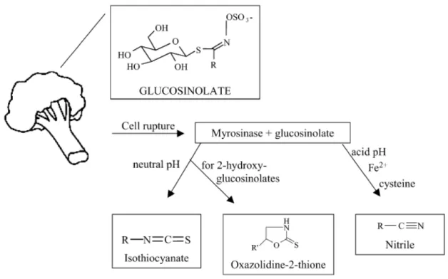
Cruciferous vegetables belong to the Brassicaceae family (formerly known as Cruciferae), and include economically important plants, as broccoli, cabbage, cauliflower, kale, bokchoy, Brussels sprouts, radish, and mustards. Among the phytochemicals that can be found at high concentrations in cruciferous vegetables is the group of glucosinolates (GLSs). This well-characterized group is responsible for the bitter or hot taste intensively present in mustard, radish, and horseradish (Hayes et al., 2008). GLSs are β-thioglucoside-N-hydroxysulfates that when enzymatically hydrolyzed by the action of myrosinase (β-thioglucoside glucohydrolase) originate different compounds (Fimognari and Hrelia, 2007), as illustrated in Figure 4. One of the possible products of GLSs hydrolysis is a group of bioactive sulphur-containing compounds, named isothiocyanates (ITCs). GLSs and myrosinase are stored in separate cell compartments and their release is triggered by cell damages, such as microbial attack, insect predation, and food processing (e.g., chewing, cutting, boiling) (Fimognari and Hrelia, 2007). However, recent data show that GLSs hydrolysis can also be mediated by the microflora of the mammalian gastrointestinal tract (Lai et al., 2010). This means that besides the GLSs conversion occurring before the digestive process, further formation of ITCs may take place by myrosinase of the gastrointestinal tract microflora.
Figure 4. Bioconversion pathway of glucosinolates. The hydrolysis of GLSs catalyzed by myrosinase results in glucose release and formation of an instable intermediate compound (R-C(-SH)=N-O-SO3-), which, dependent on the reaction conditions, can originate ITCs, nitriles, or oxazolidine-2-thiones, among others. Extracted from Fimognari and Hrelia (2007).
1.3.2. Sulforaphane and cancer
Considerable research interest has been given in the past years to one particular ITC compound – sulforaphane (SFN, 1-isothiocyanato-4-(methylsulfinyl)-butane) – due to the variety of exerted anti-cancer effects (Clarke et al., 2008). SFN is the hydrolysis product of the GLS precursor glucoraphanin (4-(methylsulfinyl)butyl glucosinolate) (Figure 5) and is found at particular high levels in broccoli and broccoli sprouts (Fahey et al., 1997).
The great interest in SFN is related to its ability to simultaneously modulate multiple cellular targets involved in different stages of cancer development that include: DNA protection by modulation of carcinogen-metabolizing enzymes; inhibition of cell proliferation and induction of apoptosis; and inhibition of tumor angiogenesis and metastases formation. Therefore, SFN is able to prevent cancer initiation, as well as act on cancer cells as a therapeutic agent (Fimognari and Hrelia, 2007).
The basis for anticancer SFN effects rely on the highly electrophilic central carbon in the -N=C=S group. This structural feature enables SFN to bind to proteins and modify their