Dipyridamole increases the cytotoxicity
of cisplatin in human larynx cancer
cells
in vitro
1Departamento de Química e Física Molecular, Instituto de Química de São Carlos,
Universidade de São Paulo, São Carlos, SP, Brasil 2Cena, Universidade de São Paulo, Piracicaba, SP, Brasil M. Rodrigues1,
F. Barbosa Junior2 and J.R. Perussi1
Abstract
This paper describes the effect of dipyridamole (DIP) on the cytotox-icity of cisplatin in HEp-2 human larynx cancer cells in vitro and the nature of the interaction between cisplatin and dipyridamole. Cyto-toxic assays were performed to obtain the IC50 for cisplatin. The cells
were treated with 0, 20, 40, 80, 120 or 200 µM cisplatin, with or without a single concentration of DIP and incubated for 60 min at 37ºC and 5% CO2 for 3 days and then counted with a hemocytometer. The
accumulation of cisplatin in the cells was measured by atomic absorp-tion and fluorescence was used to determine the membrane binding constant of DIP. In the presence of 10, 20 and 30 µM DIP, the IC50 of
cisplatin was reduced by 25, 60 and 82% in HEp-2 cells. Combination index analysis revealed that cisplatin and DIP interact synergistically. In larynx cancer cells, the accumulation of cisplatin increased by 13, 27 and 65% as the DIP concentration was increased from 10 to 20 and 30 µM, respectively. The binding constant of DIP to the cell mem-brane was estimated to be (0.36 ± 0.12 mg/ml)-1 (N = 2) by
fluores-cence and cisplatin did not suppress DIP fluoresfluores-cence. These results suggest that DIP significantly enhances cisplatin cytotoxicity in HEp-2 cells by increasing cisplatin accumulation, probably by altering the cell membrane as suggested by its binding constant. The results obtained reinforce the importance of combination therapy to reduce the doses of chemotherapeutic drugs and therefore the side effects of chemotherapy.
Correspondence
J.R. Perussi
Departamento de Química e Física Molecular
Instituto de Química de São Carlos USP
Caixa Postal 780 13560-970 São Carlos, SP Brasil
Fax: +55-16-273-9985 E-mail: janice@iqsc.sc.usp.br
This work is dedicated to Valeria C.M. Carvalho (1968-2003).
Research supported by FAPESP. M. Rodrigues was the recipient of a CNPq fellowship.
Received June 9, 2003 Accepted January 27, 2004
Key words
•Cisplatin
•Dipyridamole
•Synergism
•Tumor cells
•Cytotoxicity
•IC50
Introduction
Cisplatin has been used to treat different kinds of cancer although toxic side effects are known (1). The antitumor activity of cisplatin involves induction of intra- and interstrand cross-links that severely distort the DNA helix and block replication (1-3). Since cisplatin accumulation is a major
de-terminant of its antitumor activity, modula-tors of cisplatin accumulation have received much attention.
in-cluding 5-fluorouracil (7), methotrexate (8,9), adriamycin (10), etoposide (11), doxorubi-cin and vinblastine (12), and cisplatin (13). Jekunen et al. (13) showed that DIP syner-gistically enhanced the cytotoxicity of cis-platin in ciscis-platin-sensitive 2008 human ovar-ian carcinoma cells by a factor of 4.7, and in the cisplatin-resistant 2008/C13*5.25 sub-line by a factor of 5.8. In a nude mouse model with human bladder cancer (14), tu-mor size decreased by 20% when cisplatin was combined with DIP. Using human tes-ticular carcinoma in the same model, com-plete tumor regression was achieved (14). Barberi-Heyob et al. (15) found that DIP synergistically increased the growth-inhibi-tory activity of cisplatin in MCF-7 human breast cancer cells. Perussi et al. (16) have studied the potentiation of cisplatin cytotox-icity by DIP in two human breast cancer cells, one of them cisplatin sensitive (MDA/ S) and the other cisplatin resistant (MDA/
R). In the presence of 30 µM DIP, the IC50 of
cisplatin was reduced by 39% for both cell lines. In the MDA/S cells, the cellular accu-mulation of cisplatin increased by 57 ± 8% in the presence of 30 µM DIP, which did not affect the accumulation of cisplatin in MDA/ R. The cited investigators suggested that the enhancement of cisplatin cytotoxicity by DIP in MDA/S cells may be related to a DIP-induced increase in cisplatin accumulation, but the enhanced cytotoxicity in MDA/R cells employs a mechanism that does not involve an increase in the cellular accumula-tion of cisplatin.
In the present study we report the en-hancement of cisplatin cytotoxicity by DIP in human larynx cancer cells (HEp-2) that is probably related to a DIP-induced increase in cisplatin accumulation.
Material and Methods
Chemicals
Cisplatin, cis-diaminedichloroplatinum
(II), and DIP, 2,6-bis (diethanolamine)-4,8-dipiperidinopyrimido [5,4-d] pyrimidine were purchased from Sigma (St. Louis, MO, USA). Cisplatin stock solutions were made fresh 24 h before each experiment in unsup-plemented medium to minimize the hydroly-sis of cisplatin. Dipyridamole stock solu-tions were prepared in DMSO and kept in the dark at 4ºC. The maximum DMSO con-centration used was 0.1%. Drug solutions were sterilized via syringe filtration just be-fore use. All chemicals were of analytical quality and were used as purchased.
Cell culture
HEp-2, a human larynx cancer cell line obtained from Adolfo Lutz Institute, São Paulo, SP, Brazil, was grown as a monolayer
in 25-cm2 flasks with Dulbecco’s minimal
essential medium supplemented with 10% fetal bovine serum, streptomycin and ampi-cillin at 37ºC in a humidified atmosphere of
5% CO2 in 95% air. Confluent cell cultures
were harvested with Versene (a solution con-stituted mainly of 0.526 mM EDTA) and removed from the flask with phosphate-buff-ered saline (PBS) solution. Cells were cen-trifuged, resuspended in medium and counted with a hemocytometer. Cell viability was assessed by the Trypan blue exclusion method.
Cytotoxic assays
HEp-2 cells were seeded on Petri dishes
at 4.104 cells/dish and treated with 0, 20, 40,
80, 120 or 200 µM cisplatin in
unsupple-mented medium, with or without a fixed
concentration of DIP ranging from 0 to 35 µM. It should be mentioned here that DIP is insoluble in aqueous solutions at concentra-tions above 40 µM. The cells were incubated with the drug(s) for 60 min at 37ºC and 5%
CO2. After drug removal ordinary
harvested with Trypsin, fixed in 37% for-maldehyde and counted using a hemocytom-eter. All experiments were carried out in duplicate. The number of cells for 0.0 µM cisplatin and 0.0 µM DIP was taken as the control, corresponding to 100% cell sur-vival. An index of survival was calculated at each cisplatin concentration, i.e., the num-ber of live cells with the drug divided by the number of control live cells without the drug. Median effect analysis was used to
deter-mine the mean inhibitory concentration (IC50)
for cisplatin and for DIP (17). Data were visualized graphically by plotting the index of cell survival against the cisplatin concen-tration. The degree of synergism between cisplatin and DIP was determined by using combination index analysis at a non-constant ratio as described above. According to Chou and Talalay (18), combination index <1 stands for synergism while combination in-dex = 1 indicates summation and combina-tion index >1 suggests antagonism (18).
Data are reported as means ± SD. The
Student unpaired t-test was used to
deter-mine differences between pairs of means, with the level of significance set at P < 0.05.
Determination of cisplatin accumulation in the presence of dipyridamole
The method used to calculate the accu-mulation of cisplatin has been described (19). Briefly, cells were seeded into 60-mm plas-tic culture dishes and grown to confluence as monolayers in ordinary supplemented
medi-um at 37ºC and 5% CO2. The cells were
incubated with 200 µM cisplatin in the pres-ence of 0, 5, 10, 20 or 30 µM DIP in unsup-plemented medium for 60 min at 37ºC, then washed four times with ice-cold PBS. Next, 1.0 ml of 0.1% Triton X-100 in 0.1 N HCl was added to each dish and the cells were scraped from the bottom surface. The de-tached cells were frozen in cryogenic vials for at least 24 h. Upon thawing, the cells were sonicated at 7 watts for 15 s.
Twenty-microliter aliquots of the lysate were used for platinum analysis with a Perkin-Elmer Zeeman Atomic Absorption Spectrometer Model 4110ZL (Wellesley, MA, USA). Cis-platin accumulation is reported as pmol of platinum/mg protein. The protein content of each sample was measured by the method of Lowry et al. (20). One-way analysis of vari-ance (ANOVA) was used to analyze the atomic absorption data.
Determination of the association constant of dipyridamole with the cell membrane
The determination of the association con-stant of DIP with the cell membrane was performed by titration. The cells in a high-density cell suspension were lysed by soni-cation or by adding a hypotonic solution and centrifuged. The protein concentration of the supernatant and membrane fraction was determined by the method of Lowry et al. (20). DIP solution (1 µM) was added to variable concentrations of membrane sus-pensions (0 to 1.30 mg of protein/ml), the samples were mixed in a Vortex mixer and
incubated for 60 min at 37ºC and 5% CO2.
The samples were then centrifuged at 4000 g
for 10 min and the fluorescence spectra of the supernatant were obtained. The pellet obtained by centrifugation was washed twice with 1% phosphate buffer and solubilized in 1 ml of 2% Triton X-100 and 10% NaCl. The supernatant contains the cytoplasmic con-stituents and the pellet contains the mito-chondria as well as nuclear and cell mem-branes. The fluorescence of the solubilized pellet and the supernatant was detected with a Hitachi F-4500 fluorometer (Ibaraki, Japan) in the region of 400-600 nm with excitation at 415 nm since DIP absorbs at this wavelength. A 1-µM DIP solution was used as a standard for fluorescence intensity. Data from the titra-tion were analyzed by directly fitting to the law of mass action (21,22) or as double recipro-cal plots of fluorescence intensity changes
Considering the equilibrium:
D + M ⇔ D - M (Eq. 1)
where D represents the drug and M the cell membrane, the total fluorescence observed
is due to the free and bound species of D. ∆F
represents the difference between the initial
fluorescence intensity (F0) in the absence of
the cell membrane and the fluorescence in-tensity of the drug in the presence of cell
membrane (F). This difference (∆F) is
re-lated to the quantity of the drug associated with the membrane. In order to quantitate the association constant for the binding of DIP
to the membrane (Kb) and ∆Fmax, the
fluores-cence data for the supernatant obtained by titration of the drug with membrane suspen-sions were treated by the double reciprocal method (23,24). This treatment is based on the following equation:
1 1 1 1 1
= + x x (Eq. 2)
∆F ∆Fmax ∆Fmax Kb [M]
where ∆Fmax is the variation of fluorescence
intensity between the fluorescence intensity in the absence of the membrane and the
fluorescence emission intensity of the drug at a saturation membrane concentration (based on the membrane protein concentra-tion). This value can be obtained from the fit of the experimental data using Equation 2.
Investigation of complex formation between dipyridamole and cisplatin
The possibility of a direct interaction between DIP and cisplatin was investigated by determining the suppression of the fluo-rescence of 5 µM DIP after each addition of 20 µl of a 2 mM cisplatin solution prepared in phosphate buffer, pH 7.2. The fluores-cence spectrum of each sample was obtained from 425 to 600 nm with a Hitachi F-4500 fluorometer, with excitation at 415 nm. Sample absorbance was monitored after each addition of the titrating compound and the spectra from 250 to 600 nm were recorded with a Shimadzu UV-1601 PC spectropho-tometer.
Results and Discussion
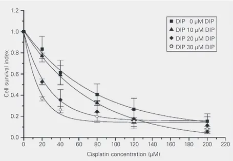
Figure 1 shows the indices of cell sur-vival for HEp-2 cells as a function of cis-platin concentration, in the absence and pres-ence of 10, 20 and 30 µM DIP. At each cisplatin concentration, the indices of sur-vival of the cells were reduced in the
pres-ence of DIP. In this cell line, the survival
curve for cisplatin alone was significantly different from that for cisplatin plus DIP (P < 0.001). These results suggest that the cyto-toxicity of cisplatin is increased in the
pres-ence of DIP. Table 1 presents the IC50 values
obtained from plots shown in Figure 1. In
HEp-2 cells, the IC50 of cisplatin was
re-duced by 25, 60 and 82% in the presence of 10, 20 and 30 µM DIP, respectively. The degree of enhancement of cisplatin cytotox-icity increased with DIP concentration.
Perussi et al. (16) observed that the IC50
value for cisplatin in human breast cancer cells sensitive to cisplatin (MDA/S)
de-Cell survival index
1.2
1.0
0.8
0.6
0.4
0.2
0.0
0 20 40 60 80 100 120 140 160 180 200 220
Cisplatin concentration (µM)
DIP 0 µM DIP
DIP 10 µM DIP
DIP 20 µM DIP DIP 30 µM DIP
creased by 39% in the presence of 30 µM DIP, suggesting that HEp-2 cells are more sensitive to the combination of the drugs.
Figure 2 shows the sensitivity of the cells to DIP alone. DIP alone decreased the sur-vival of HEp-2 cells by 10, 17 and 29% at concentrations of 10, 20 and 30 µM, respec-tively. This reduction was statistically sig-nificant (P < 0.01) for 20 and 30 µM DIP.
The IC50 of DIP was estimated to be 94 ± 10
µM since the solubility of DIP is very low in aqueous solutions and just a short range of DIP concentrations could be used in the assay (0 to 35 µM DIP). The cytotoxic effect of cisplatin was 1.6 times more potent than
that of DIP alone in these cells (IC50 was 94
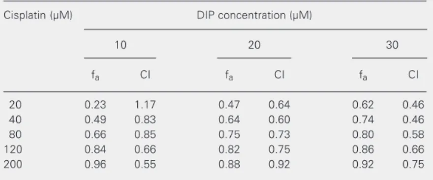
µM for DIP and 60 µM for cisplatin). Table 2 presents the affected fractions and the combination index obtained for the cytotoxic experiments performed at a non-constant ratio with cisplatin in combination with DIP in HEp-2 cells. Combination index analysis indicated that this parameter was lower than one in 93% of the combinations used, suggesting that the interaction between cisplatin and DIP is synergistic (18). Similar results of synergistic interaction were re-ported by Jekunen et al. (13) for an ovarian carcinoma cell line treated with the combi-nation of these two drugs.
The data in Figure 1 suggest that the fraction affected by 80 µM cisplatin with 10 µM DIP was similar to the fraction affected by 40 µM cisplatin with 20 µM DIP and 20 µM cisplatin with 30 µM DIP. However, the indices obtained for these drug combina-tions were different (Table 2), suggesting that the interaction of cisplatin with DIP was synergistic when low cisplatin concentra-tions were combined with moderate and high DIP concentrations, and moderately syner-gistic when low cisplatin concentrations were combined with low DIP concentrations. Fur-thermore, Figure 1 indicates that the fraction affected by 120 µM cisplatin with 10 µM DIP may be similar to the fractions affected by 120 µM cisplatin with 20 µM DIP and by
Table 1. Effect of dipyridamole on the cytotoxicity of cisplatin in the human larynx HEp-2 cell line.
Dipyridamole IC50 (µM) Decrease in
(µM) the IC50 (%)
0 60 ± 9
-10 45 ± 2 25
20 24 ± 2 60
30 11 ± 3 82
The cells were exposed to several cisplatin con-centrations (0, 20, 40, 80, 120 and 200 µM) with and without dipyridamole for 1 h at 37ºC (N = 5-13). Data are reported as means ± SD. The IC50 values for the association of the drugs were sta-tistically different (P < 0.05; ANOVA).
Cell survival index
1.0
0.8
0.6
0.4
0.2
0.0
*
*
0 10 20 30
Dipyridamole concentration (µM)
Figure 2. Effect of dipyridamole (DIP) on the survival index of HEp-2 cells. Cells were treated with several DIP concentra-tions (0, 5, 10, 15, 20 and 30 µM) for 1 h at 37ºC in the pres-ence of 5% CO2. Each column represents the mean ± SD for two experiments each per-formed in duplicate. *P < 0.005 compared to no DIP (ANOVA). 120 µM cisplatin with 30 µM DIP. The
indices obtained for these drug combina-tions (Table 2) were about the same, sug-gesting that the interaction of cisplatin with DIP was also synergistic at high cisplatin concentrations.
Table 2. Combination indexes for cisplatin and dipyridamole (DIP) in the HEp-2 cell line obtained by the median effect analysis.
Cisplatin (µM) DIP concentration (µM)
10 20 30
fa CI fa CI fa CI
20 0.23 1.17 0.47 0.64 0.62 0.46
40 0.49 0.83 0.64 0.60 0.74 0.46
80 0.66 0.85 0.75 0.73 0.80 0.58
120 0.84 0.66 0.82 0.75 0.86 0.66
200 0.96 0.55 0.88 0.92 0.92 0.75
the increase in cisplatin accumulation by DIP increased the cytotoxicity of cisplatin in HEp-2 cells.
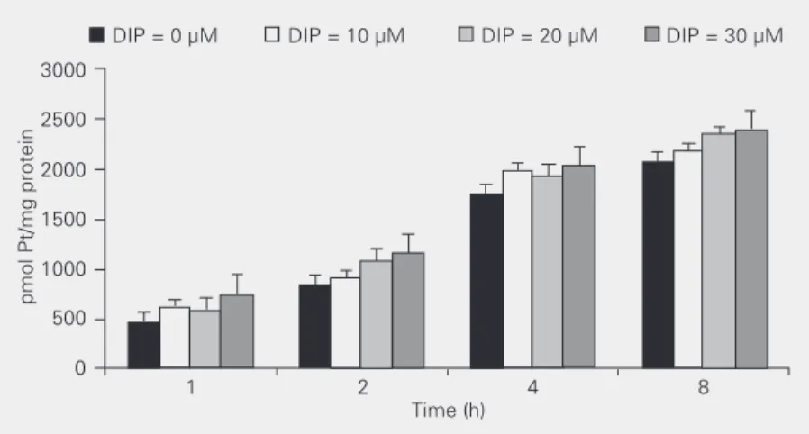
Experiments were performed to investi-gate the effect of incubation time on the intracellular accumulation of cisplatin in HEp-2 cells. When cisplatin was combined with DIP (0, 10, 20 and 30 µM) (Figure 3) the increase in incubation time of the drugs was found to lead to an increase in intracel-lular cisplatin concentration. For each incu-bation time used there was a significant in-crease (P < 0.005, ANOVA) in platinum accumulation. So, we may conclude from these results that the amount of intracellular cisplatin depends on DIP concentration as well as on the duration of incubation with the drugs.
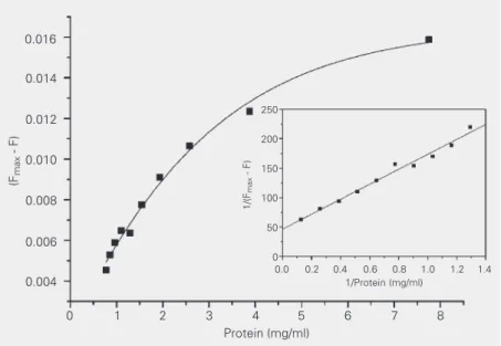
Titrations of membrane suspensions have indicated that the association depends on the increase of the membrane concentration in solution. The results were closely similar to those obtained for the interaction of DIP with mitochondrial and erythrocyte mem-branes (21,22). As the membrane concentra-tion increased, the fluorescence intensity in the supernatant decreased, while it increased in the pellet. These alterations in the emis-sion spectra were used to estimate the asso-ciation constant for DIP with the cell mem-brane.
A plot of 1/∆F (data for the supernatant
fluorescence which was proportional to free DIP concentration) as a function of 1/[M]
may be used to obtain Kb, the binding
con-stant. The results obtained for both treat-ments, the mass-action law and the method of double reciprocal plot, are shown in
Fig-ure 4. The Kb value obtained by the double
reciprocal plot was 0.36 ± 0.12 (mg protein/
ml)-1. This value was obtained as the ratio of
the intercept to the slope in the linear fit
shown in the insert in Figure 4 and ∆Fmax was
obtained from the reciprocal of the intercept value. This value for the binding constant was quite similar to those obtained for the association constant of DIP with the erythro-Figure 3. Effect of dipyridamole (DIP) and incubation time on intracellular accumulation of
cisplatin in the human larynx tumor cell line HEp-2 at 37ºC and 5% CO2. Data are reported as means ± SD for 5 replicates. Platinum (Pt) content for each incubation time was statistically different (P < 0.005) from that of 1 h of incubation.
pmol Pt/mg protein
3000
DIP = 0 µM DIP = 10 µM DIP = 20 µM DIP = 30 µM
2500
2000
1500
1000
500
0
1 2 4 8
Time (h)
In order to assess the mechanism involved in the synergism between cisplatin and DIP, cellular accumulation of cisplatin was deter-mined in the presence of DIP. Table 3 shows that at 0.0 µM DIP the accumulation of cisplatin in HEp-2 cells was 830 ± 400 pmol Pt/mg membrane protein and this value in-creased with increasing DIP concentrations from 0 to 30 µM. In the presence of 30 µM DIP, the cisplatin accumulation increased significantly by 65% to 1373 ± 400 pmol Pt/
mg protein (P < 0.005), while the IC50 value
for cisplatin combined with 30 µM DIP de-creased by 82%. These results suggest that
Table 3. Effect of dipyridamole (DIP) on the accu-mulation of cisplatin in human larynx HEp-2 tumor cells.
Dipyridamole pmol Pt/mg Increase in
(µM) protein Pt (%)
0 830 ± 400 0
10 934 ± 470 13.0
20 1053 ± 500 27.0
30 1373 ± 401 65.0
cyte ghost membranes of 0.40 ± 0.02
(mg protein/ml)-1 (22) and for the
associa-tion constant of DIP with the mitochondrial
membrane of 0.8 ± 0.1 (mg protein/ml)-1
(21). On the basis of the dependence of the DIP fraction in the pellet on membrane con-centration, we found that 36% of the drug was present in the pellet. This means that with 1 µM DIP concentration and an excess of membrane, about 36% of the drug was bound. This value is quite close to the maxi-mum DIP saturation of 47% reported for the mitochondrial membrane (21).
Borges et al. (25) studied the interaction of DIP with bovine serum albumin (BSA) and membrane model systems (micelles). It was shown that DIP binds strongly to BSA, in agreement with the high level of DIP binding to human plasma albumin. This study (25) also showed that DIP binds more strongly to neutral micelles and the results of fluores-cence suppression suggest that DIP is lo-cated in the interface of the micelle, close to the beginning of the hydrophobic region. In fact, DIP incorporation seemed to occur in a region close to the border of the hydrophobic and polar parts of a phospholipid monolayer (26). It has been shown that the protective effect of DIP against the lipid peroxidation caused by cumene hydroperoxide in mito-chondrial membrane was strongly depend-ent on the duration of incubation with the drug prior to the addition of the oxidant (27). A similar feature was observed for the pro-tective effect of DIP on red blood cell lysis. The protection was quite sensitive to the time of incubation with the drug and to its concentration (22).
The experiments of fluorescence sup-pression were performed in order to deter-mine the complex formation between DIP and cisplatin. The fluorescence at 480 nm of a 10 µM DIP solution in 0.02 M phosphate buffer, pH 7.2, was monitored after each addition of cisplatin (0 to 0.4 mM final cis-platin concentration). The Stern-Volmer plot (data not shown) resulted in a straight line
parallel to the x-axis in the entire range of the suppressor concentration used. Thus, the
Stern-Volmer constant (KSV) could not be
obtained in this case because there was no fluorescence suppression, indicating that no complexation occurred between DIP and cis-platin at the concentrations used.
The present study has demonstrated that the combination of cisplatin and DIP leads to an increased cytotoxicity in HEp-2 cells. A
reduction of up to 82% in the IC50 of
cis-platin was obtained when ciscis-platin was com-bined with 30 µM DIP. This reduction was much greater than that observed in other cell lines, such as in cisplatin-sensitive and cis-platin-resistant breast tumor cell lines previ-ously studied by us (16). In that
investiga-tion, in the presence of 30 µM DIP, the IC50
of cisplatin was reduced just by 39% for both cell lines. This result suggests that the com-bination of these drugs can also improve the cytotoxicity of cisplatin in resistant cell lines. The results of that study as well as others in the literature (13) permit us to conclude that the co-administration of cisplatin and DIP is feasible and may permit the treatment of
(Fmax
- F)
0.016
0.014
0.012
0.010
0.008
0.006
0.004
0 1 2 3 4 5 6 7 8
Protein (mg/ml) 250
200
150
100
50
0
1/(F
max
- F)
0.0 0.2 0.4 0.6 0.8 1.0 1.2 1.4
1/Protein (mg/ml)
cancer patients with cisplatin concentrations that are effective in killing cisplatin-resistant cancer cells by reducing or eliminating the severe side effects of high drug concentra-tions. In this way, resistance to cisplatin may be overcome.
The atomic absorption experiments showed that DIP increases the uptake of cisplatin by cells in a concentration-depend-ent manner. These results agree with those reported by Perussi et al. (16) and by Jekunen et al. (13) who observed an increased accu-mulation of cisplatin due to DIP, but without increasing Trypan blue or propidium iodide uptake or changing cell size. The cited inves-tigators concluded that the DIP-induced in-crease in cisplatin accumulation was not associated with a nonspecific increase in membrane permeability. In the present study we showed that the cellular accumulation of cisplatin is concentration and time depend-ent. It has been shown that DIP incorpora-tion into model membranes is time depend-ent (27,22).
The analysis of the median effect showed that the interaction between these two drugs was synergistic and our fluorescence
sup-pression experiments showed no complex-ation between DIP and cisplatin. Fluores-cence experiments also allowed to deter-mine the binding constant of DIP to the cell
membrane as 0.36 ± 0.12 (mg protein/ml)-1,
a value similar to those obtained for the binding of this drug to mitochondrial and red blood cell membranes.
Our results suggest that the enhancement of cisplatin cytotoxicity by DIP in HEp-2 cells may be related to a DIP-induced in-crease in cisplatin accumulation. The results obtained support the importance of com-bined therapy to reduce the doses of chemo-therapeutic drugs and therefore the side ef-fects of chemotherapy.
Acknowledgments
The authors would like to thank Dr. Marcel Tabak (Instituto de Química de São Carlos, USP, São Carlos, SP, Brazil) for valuable suggestions and to Dr. Robert G. Canada (Howard University College of Medi-cine, Washington DC, USA) for a critical reading of the manuscript.
References
1. Zenker A, Galanski M, Bereuter L, Bernhard KK & Kinder W (1999). Capillary electrophoretic study of cisplatin interaction with nucleo-side monophosphates, di- and trinucleotides. Journal of Chromatog-raphy. A, 852: 337-346.
2. Zamble DB & Lippard SJ (1995). Cisplatin and DNA repair in cancer chemotherapy. Trends in Biochemical Sciences,20: 435-439.
3. Pillaire MJ, Hoffmann JS, Defais M & Villani G (1995). Replication of DNA containing lesions and its mutagenic consequences. Biochi-mie,77:803-807.
4. Fitzgerald GA (1987). Dipyridamole. New England Journal of Medi-cine, 316: 1247-1257.
5. Iuliano L, Pedersen JZ, Ferro D & Violi F (1995). A potent chain-breaking antioxidant activity of the cardiovascular drug dipyridamole.
Free Radical Biology and Medicine, 18: 239-247.
6. Plagemann PGW, Wohlhueter RM & Woffendin C (1988). Nucleo-side and nucleoNucleo-side transport in animal cells. Biochimica et Biophy-sica Acta, 947: 405-443.
7. Grem JL & Fisher PH (1989). Enhancement of 5-fluorouracil antican-cer activity by dipyridamole. Pharmacology and Therapeutics, 40: 349-371.
8. Cabral S, Leis S, Bover L, Nembrot M & Mordoh J (1984). Dipy-ridamole inhibits reversion by thymidine of methotrexate effect and increases drug uptake in sarcoma 180 cells. Proceedings of the National Academy of Sciences, USA, 81: 3200-3203.
9. Fisher PH, Pamukcu R, Bittner G & Willson JKV (1984). Enhance-ment of the sensitivity of human colon cancer cells to growth inhibition by acivicin achieved through inhibition of nucleic acid precursor salvage by dipyridamole. Cancer Research, 44: 3355-3359.
10. Kusumoto F, Maehara Y, Anai H, Kusumoto T & Sugima-Chi K (1988). Potentiation of adriamycin cytotoxicity by dipyridamole against HeLa cells in vitro and sarcoma 180 cells in vivo. Cancer Research, 48: 1208-1212.
11. Turner RN & Curtin NJ (1996). Dipyridamole increases inhibition, accumulation and retention in parental and multidrug-resistant CHO cells. British Journal of Cancer, 73: 856-860.
13. Jekunen A, Vick J, Sanga R, Chan CK & Howell SB (1992). Syner-gism between dipyridamole and cisplatin in human ovarian carcino-ma cells in vitro. Cancer Research, 52: 3566-3571.
14. Keane TE, Rosner G, Donaldson JT, Norwood DL, Poulton SH & Walther PJ (1990). Dipyridamole-cisplatin potentiation: enhanced in vivo cytotoxic in xenograft models of human testicular and bladder cancers. Journal of Urology, 144: 1004-1009.
15. Barberi-Heyob M, Griffon G, Merlin JL & Weber B (1993). Se-quence-dependent growth-inhibitory effects of the in vitro combina-tion of fluorouracil cisplatin and dipyridamole. Cancer Chemotherapy and Pharmacology, 33: 163-170.
16. Perussi JR, Paltoo DN, Toppin VAL & Canada RG (2003). Synergism between dipyridamole and cisplatin in human breast cancer cells in vitro. Química Nova, 26: 340-343.
17. Chou TC & Hayball M (1996). CalcuSyn: Windows Software for Dose Effect Analysis. Biosoft, Cambridge, UK.
18. Chou TC & Talalay P (1984). Quantitative analysis of dose-effect relationships: the combined effects of multiple drugs or enzyme inhibitors. Advances in Enzyme Regulation, 22: 27-55.
19. Mack KM, Canada RG & Andrews PA (1997). The effects of terbium on the cellular accumulation of cisplatin in MDA-MB-231 human breast tumor cells. Cancer Chemotherapy and Pharmacology, 39: 217-222.
20. Lowry OH, Rosebrough NJ, Farr AL & Randall RJ (1951). Protein measurement with the Folin phenol reagent. Journal of Biological Chemistry, 193: 265-275.
21. Nepomuceno MF, Mamede MEO, Macedo DV, Alves AA,
Pereira-da-Silva L & Tabak M (1999). Antioxidant effect of dipyridamole and its derivative RA-25 in mitochondria: correlation of activity and loca-tion in the membrane. Biochimica et Biophysica Acta,1418: 285-294.
22. Ruggiero AC, Nepomuceno MF, Jacob RF, Dorta DJ & Tabak M (2000). Antioxidant effect of dipyridamole (DIP) and its derivative RA 25 upon lipid peroxidation and hemolysis in red blood cells. Physi-ological Chemistry and Physics and Medical NMR, 32: 35-48. 23. Borissevitch IE, Borges CPF, Borissevitch GP, Yushmanov VE, Louro
SRW & Tabak M (1996). Binding and location of dipyridamole de-rivatives in micelles: the role of drug molecular structure and charge.
Zeitschrift für Naturforschung, 51: 578-590.
24. Tabak M & Borissevitch IE (1992). Interaction of dipyridamole with micelles of lysophosphatidylcholine and with bovine serum albu-min: fluorescence studies. Biochimica et Biophysica Acta, 1116: 241-249.
25. Borges CPF, Honda S, Berlinck RGS, Imasato H, Berci Filho P & Tabak M (1995). Synthesis, characterization and interaction with ionic micelles of tetraacetylated dipyridamole. Spectrochimica Acta
(Part A), 51: 2575-2584.
26. Borissevitch GP, Tabak M & Oliveira ON (1996). Interaction of dipyridamole with lipids in mixed Langmuir monolayers. Biochimica et Biophysica Acta, 1278: 12-18.