2013
Maria Madalena
Ribeiro Cabral
Caspase 3: potencial marcador para o sucesso da
fertilização in vitro
Caspase 3: a potential marker for in vitro fertilization
outcome
2013
Departamento de Biologia
Maria Madalena
Ribeiro Cabral
Caspase 3: potencial marcador para o sucesso da
fertilização in vitro
Caspase 3: a potential marker for in vitro fertilization
outcome
Dissertação apresentada à Universidade de Aveiro para cumprimento dos requisitos necessários à obtenção do grau de Mestre em Biologia Molecular e Celular, realizada sob a orientação científica da Doutora Margarida Sâncio da Cruz Fardilha, Professora auxiliar convidada da Secção Autónoma de Ciências da Saúde da Universidade de Aveiro e co-orientação da Dra. Helena Maria Sá Figueiredo, Responsável do Laboratório da Unidade de Medicina da Reprodução Dra. Ingeborg Chaves – Centro Hospitalar de Vila Nova de Gaia/ Espinho, EPE
o júri
Presidente Prof. Doutora Maria Lourdes Pereira
Professora Associada com Agregação do Departamento de Biologia da Universidade de Aveiro
Doutora Rita da Costa Ramalho Ferreira
Responsável pelo Laboratório da Unidade de Medicina da Reprodução do Centro Hospitalar de Alto Ave, EPE
Prof. Doutora Margarida Sâncio da Cruz Fardilha
Professora auxiliar convidada da Secção Autónoma de Ciências da Saúde da Universidade de Aveiro
Dra. Helena Maria Sá Figueiredo
agradecimentos À Professora Margarida Fardilha por todo o apoio e conhecimentos que me passou no decurso do mestrado. Por me mostrar que é possível num laboratório de investigação viver um ambiente de entre ajuda e companheirismo, quando os valores corretos de conhecimento e humildade são passados aos alunos.
À Joana pois sem ela este trabalho não seria possível. Pela total disponibilidade, enorme ajuda e eterna boa-disposição. Por tudo o que me ensinou no tempo que partilhamos. E por tornar tão gratificante a elaboração deste trabalho.
Ao Miguel pela incansável ajuda na análise estatística.
A todos os colegas do Laboratório de Transdução de Sinais da Universidade de Aveiro: Maria João, Juliana, Korrodi, Emanuel, Mega. Por toda ajuda, disponibilidade e boa disposição.
À Dra. Helena Figueiredo porque é sempre um prazer aprender com alguém com os seus conhecimentos e simpatia. Por ser um exemplo de honestidade, profissionalismo e comportamento ético.
À Dra. Ilda Pires pelos imensos conhecimentos passados, pelo companheirismo e amizade. Por ser um exemplo de humanidade e por me contagiar com a sua interminável vontade de saber sempre mais. Por ser um exemplo que, por mais difícil que seja, é possível ser profissional, mulher e mãe, sem que nenhuma das valências seja prejudicada.
E como não poderia deixar de ser à minha família e amigos pois no final do dia são eles que me animam, consolam e dão energia para prosseguir.
palavras-chave Apoptose, caspase-3, células do cumulus, fertilização in vitro, gravidez
resumo Mundialmente é estimado que aproximadamente 70 milhões de casais tenham
problemas de infertilidade, o que corresponde a um em cada sete casais em idade reprodutiva. O rápido aumento de problemas reprodutivos nas últimas décadas sugere uma maior probabilidade deste aumento ser devido a factores do estilo de vida e/ou ambientais, do que resultado de uma variação genética. Em Portugal 2,2% dos bébes nascidos resultam de técnicas de reprodução medicamente assistida. A transferência de vários embriões realizada, por vezes, no decurso destas técnicas pode resultar em gravidezes múltiplas, o que advém em complicações bem conhecidas para as mães e os bébes. São necessárias novas ferramentas de diagnóstico para que se possa transferir menos embriões com resultados similares ou melhores.
Neste estudo foi investigado o impacto de vários factores do estilo de vida no potencial reprodutivo de 47 casais que recorreram a técnicas de reprodução medicamente assistida.
Para além disso, foi realizada também, a correlação entre os níveis de expressão de um marcador da apoptose (caspase-3 clivada) nas células do cumulus do ovócito e a obtenção de uma gravidez (n=30). Uma concentração significativamente (p<0.01)) maior de caspase-3 clivada foi observada nas células do cumulus dos casais que não obtiveram uma gravidez.
Dada a dificuldade de obter respostas reais nos questionários dos voluntários e a pequena dimensão da amostra para avaliar parâmetros com tanta variação inter-individual, o estudo não conseguiu obter resultados estatísticamente significativos na correlação do impacto de factores do estilo de vida no potencial reprodutivo.
O estudo permitiu concluir que o nível de caspase-3 clivada nas células do cumulus parece ser um bom marcador da qualidade ovocitária e um bom predictor do resultado de gravidez.
keywords Apoptosis, caspase-3, cumulus cells, in vitro fertilization, pregnancy
Abstract Worldwide it is estimated that approximately 70 million couples suffer from infertility, which corresponds to one in each seven couples at fertility age. The rapid increase in reproductive problems in recent decades suggests that they are more likely to be caused by lifestyle and/or environmental factors than as a result of genetic variations. In Portugal 2.2% of the babies born result from an assisted reproduction technology (ART) technique. The multiple embryo transfer performed sometimes in ART techniques may result in multiple pregnancies, which have well known complications for mothers and babies. New diagnostic tools are needed to improve embryo selection, in order to transfer less embryos to achieve similar or better results.
In this study the impact of lifestyle factors on the reproductive potential of 47 couples who resort to ART was investigated. Also, the correlation between the expression levels of an apoptotic marker (cleaved caspase-3) in oocytes cumulus cells and the achievement of pregnancy was performed (n=30). Significant (p<0.01) higher concentration of cleaved caspase-3 was observed in cumulus cells of couples who did not achieve pregnancy.
Given the difficulty in obtain reliable answers from the volunteers in the questionnaires and the small sample size to evaluate parameters with such a wide inter-subject variability it failed to give conclusive statistical significant data in the lifestyle impact into reproductive potential.
The present study allowed concluding that the level of cleaved caspase 3 in cumulus cells appears to be a good marker of oocytes quality and predictor of pregnancy outcome.
A
BBREVIATIONS... 1
1.
I
NTRODUCTION... 5
1.1. Human reproduction ... 7
1.1.1. Sexual differentiation ... 7
1.1.2. Female gonadal development and gametes production ... 8
1.1.3. Male gonadal development and gametes production ... 10
1.1.4. Spermatozoa capacitation ... 11
1.1.5. Fertilization ... 12
1.1.6. From zygote to pregnancy ... 13
1.1.7. Female infertility ... 15 1.1.8. Male infertility ... 17 1.1.9. Idiopathic infertility... 18 1.2. ART techniques ... 18 1.3. State of Art ... 19 1.4. Apoptosis ... 20 1.5. Aims ... 21
2.
M
ATERIAL ANDM
ETHODS... 23
2.1. Study overview ... 25
2.2. Human samples collection ... 25
2.3. Questionnaire ... 25 2.4. Statistical analysis ... 25 2.5. Clinical methodology ... 26 2.5.1. Follicular aspiration... 26 2.5.2. Oocyte aspiration... 26 2.5.3. Sperm preparation ... 26 2.5.4. Insemination ... 26
2.5.6. Pregnancy ... 27
2.6. Molecular methodology ... 27
2.6.1. Cumulus cells collection and storage ... 27
2.6.2. BCA cumulus cells ... 28
2.6.3. Samples preparation for SDS-PAGE... 28
2.6.4. SDS-PAGE (14%) ... 28
2.6.5. Immunobloting ... 29
3.
R
ESULTS... 31
3.1. Participants and social habits ... 33
3.2. Clinical data ... 38
3.3. Correlations between social and clinical parameters ... 41
3.4. Molecular data ... 44
4.
D
ISCUSSION... 49
4.1. Social habits and IVF outcomes ... 51
4.2. Cleaved Caspase-3 and IVF Outcomes ... 54
5.
F
INALC
ONCLUSIONS... 57
R
EFERENCES... 61
A
PPENDIX1 ... 73
A
PPENDIX2 ... 76
A
PPENDIX3 ... 79
A
PPENDIX4 ... 81
Appendix 5 ... 82A
PPENDIX6 ... 85
A
BBREVIATIONSAMH Anti-Mullerian hormone
ART Assisted reproduction technology
Acryl Acrylamide
APS Ammonium persulfate
Apaf-1 Apoptotic protease activating factor-1
ATP Adenosine-5’-triphosphate
BCA Bicinchoninic acid assay
Bisacryl Bisacrylamide
BSA Bovine serum albumin
Ca2+ Calcium
cAMP Cyclic adenosine monophosphate
CatSper channel Cation chanels of sperm
CC Cumulus cells
Cdc2 Cyclin-dependent kinase 1
CHVNG Centro Hospitalar de Vila Nova de Gaia
CNPD Conselho Nacional de Proteção de Dados
CNPMA Conselho Nacional de Procriação Medicamente Assistida
CO2 Carbon dioxide
CRN1 Cannabinoid receptor subtype 1
CRN2 Cannabinoid receptor subtype 2
DAG Diacylglycerol
DNA Deoxyribonucleic acid
DISC Death inducing signalling pathway complex
ECL Enhanced chemiluminescence
EDC Endocrine disruptor
ESHRE European Society of Human Reproduction
FADD Fas- associated death domain
FF Follicular fluid
FGF9 Fibroblast growth factor 9
GnRH Gonadotropin-releasing hormone
hCG Human chorionic gonadotropin
ICM Inner cell mass
ICFBP Insulin-like growth factor-binding protein
ICSI Intracytoplasmic sperm injection
IMSI Intracytoplasmic morphologically selected sperm injection
IVF In vitro fertilization
IP3 Inositol triphosphate
IU International units
LGB Lower gel guffer
LB Loading guffer
LH Luteinizing hormone
MII Metaphase II
MPF Maturation promoting factor
mΔ Miliampere
Na+ Sodium
N2 Nitrogen
PARP Poly (ADP-ribose) polymerase
PBS Phosphate buffered saline
PCOS Polycystic ovary syndrome
PGCs Primordial germ cells
PIP2 Phosphotidylinositol 4,5-bisphosphate
PKA Protein kinase A
PLCζ Phospholipase C zeta
RSPO1 R-spondin 1
SARP2 Secreted apoptosis-related protein-2
SDS Sodium dodecyl sulphate
SDS-PAGE Sodium dodecyl sulfate polyacrylamide gel electrophoresis
SRY Sex-determing region Y
SOX9 Sex-determining region Y- box 9
TEMED Tetramethylethylenediamine
TBS Tris-buffered saline
TBST Tris-buffered saline with tween
THC Δ9-tetrahydrocannabinol
UGB Upper gel buffer
Wnt4 Wingless-type MMTV integration site family, member 4
ZP Zona pellucida
1.1. H
UMAN REPRODUCTIONReproduction is the natural process by which new individuals are generated ensuring species perpetuation. In humans this is only possible by the fusion of two haploid cells: an oocyte and a spermatozoon. These cells have a complex process of formation only viable in a really specific hormonal and molecular atmosphere. This type of reproduction is denominated sexual reproduction and increases the genetic variability of species ensuring their survival through evolution.
1.1.1. S
EXUAL DIFFERENTIATIONBiological differences between men and women are genetically determined during the embryonic development. In humans, embryos of both sexes follow the same evolution line until the sixth weeks, when the bi-potential gonads start to differentiate into testicle or ovary (Eggers et al., 2012).
A high level of Wnt4 (Wingless-type MMTV integration site family, member 4) and RSPO1 (R-spondin 1) expression stabilizes cytoplasmic β-catenin which is translocated to the nucleo activating target genes expression. WNt4 and β-catenin suppress SOX9 (sex determining region Y-box 9) and FGF (Fibroblast growth factor 9) 9 expression leading to ovary development. Male hormones absence leads to Wolffian ducts regression and Mullerian ducts development in female reproductive tract (Nef et al., 2009).
In males the differentiation is determined by the gene SRY (Sex-determining region Y) specific of the Y chromosome. SRY stimulates SOX9 and FGF9 expression, responsible for the male gonad differentiation. Then the testicles start secreting hormones as testosterone and anti-Mullerian hormone (AMH). Testosterone stimulates the Wolffian duct differentiation in male reproductive tract and AMH induces Mullerian ducts regression (Schlessinger et al., 2010) (Figure 1).
1.1.2. F
EMALE GONADAL DEVELOPMENT AND GAMETES PRODUCTIONThe ovary development is characterized by massive colonization of the undifferentiated gonad with mesonephric cells (precursors of follicle cells), primordial germ cells migration into the genital ridge and gonadal sex differentiation (Palma et al., 2012). After female reproductive tract is differentiated, between de 11-12 gestation weeks, the female gametes (oocytes) start to enter meiosis, that stops at the diplotene stage of prophase I. In this stage oocytes have flattened pregranulosa cells surrounding and this complex is called primordial follicle. They stay in this quiescent state until puberty, when the pituitary gland is stimulated to produce gonadotrophins after hypothalamus stimulation by gonadotrophin releasing hormone (GnRH). The gonadotrophins, follicle stimulation hormone (FSH) and luteinizing hormone (LH) together stimulate oocytes to restart meiosis. When the oocytes are at metaphase II the ovulation occurs (Hogarth et al., 2010).
During early stages of folliculogenesis the follicle passes through three pre-antral phases: primary, secondary and tertiary follicles. These different stages have different morphological and structural characteristics. Primary follicle is characterized by the oocyte surrounded by a single layer of cuboidal granulose cells. When the primordial follicle is recruited, the granulose cells start to divide for mitosis and various genes are activated leading to a progressive increase in oocyte RNA synthesis. In this stage, genes encoding the zona pellucida proteins (ZP) are transcribed and translated. ZP proteins secretion polymerize near the oocyte ending to encapsulate it (Wassarman et
al., 1996). The secondary follicle consists in the oocyte surrounded by multiples layers of cuboidal
or low columnar cells that form a stratified epithelium. At this stage, the follicle forms a theca layer (layer of stroma-like cells around the basal laminal of follicle). Theca cells produce androgens in response to LH, which neighboring granulose cells convert into estrogen by FSH-induced aromatase, being vital for the continuity of follicle development process (Young et al., 2010). The follicle is designed tertiary once a cavity starts to appear in the middle of granulosa cells. This event marks the end of preantral stage (Kidder et al., 2010).
Follicular antrum follows the oocyte enlargement due to division of follicular cells and the increase of follicular fluid (antral stage). A fully grown antral follicle, with a meiotic competent oocyte ready for ovulation, is designed pre-ovulatory or Graafian follicle (Rodgers et al., 2010). The follicular fluid (FF) is an enriched microenvironment with nutritional and regulatory molecules, as well as apoptotic factors. The oocyte and the granulosa cells reside in the FF (Figure 2) and all the molecules must pass on their way to and from this microenvironment. The presence of fluid is crucial for the establishment of a separation between granulosa cells and mural granulosa cells, which limit the follicle wall and cumulus cells (CC) surrounding the oocyte. Antral stage is dependent on hormonal control by gonadotrophins as FSH and LH. FSH binds to its receptor on
granulosa cells, activating cAMP (Cyclic adenosine monophosphate)/ PKA (protein kinase A) pathway. This promotes cell proliferation, follicle cell and mural granulosa cells differentiation and acquisition of meiotic competence (Zuccotti et al., 2011). The LH receptors are expressed on granulosa and theca cells. LH peak induces luteinization, cumulus cell-oocyte complex expansion, oocyte maturation and follicle rupture (Veeck, 1999; Noma et al., 2011).
Usually in each cycle only one oocyte will complete meiosis I and arrest at metaphase II, when is ovulated. The ovulation is due to FSH and LH surge that promotes detachment of the cumulus oocyte complex from the follicular wall (Bras et al., 1996; Fragouli et al., 2012).
Figure 2- Folliculogenesis and ovulation.
The granulosa cells provide adequate physical and chemical conditions for oocyte development, changing their shape within the cycle phase. Granulosa cells vary from squamous type at primordial follicle to cuboidal type during ovulation and hypertrophied in the luteal stage. This cyto-differentiation and cells proliferation are essential to provide the support that oocyte needs during its growth and evolution from primordial to pre-antral stage (Huang et al., 2010). The CCs have specific morphological and physiological characteristics and bidirectional exchange occurs between these two cells (Palma et al., 2012).
In the follicle communication occurs through gap junctions, which consist in channels composed by proteins named connexins. The gap junctions directly connect the adjacent cells allowing diffusion of ions, metabolites and signaling molecules. These exchanges of regulatory and nutrient molecules are essential to oocyte growth and meiosis resume (Kidder et al., 2010).
1.1.3. M
ALE GONADAL DEVELOPMENT AND GAMETES PRODUCTIONTestes differentiation happens between fifth to eighth gestation weeks when the progenitors of primordial germ cells (PGCs), derived from the epiblast of blastocyst that stays at mesonephros, migrate to the testis. Few steroidogenic Leydig cell precursors and epithelial somatic cells also migrate from mesonephros to gonad. This event marks the start of testicular vascular system organization. The somatic cells will differentiate into Sertoli cells that enclose the PGCs, forming seminiferous cords. Some of the interstitial somatic cells will differentiate into peritubular myoid cells, fibroblasts, endothelial cells or Leydig cells (Carmona et al., 2009).
After vascularization is complete, PGCs start to proliferate and after a few days they are arrested at G0/G1 cell cycle phase. These pre-spermatogonia cells, shortly after birth, resume proliferation and move to the basal lamina of the seminiferous tubules where they differentiate into spermatogonial stem cells. This pool of spermatogonial stem cells will only start spermatogenesis (spermatozoa production) 10 to 13 years after birth (Kolasa et al., 2012).
Once the puberty starts spermatogonias divide continuously by mitosis, increasing in number and improving the supply for new cells. The spermatogenesis begins when some of these cells stop dividing and differentiate into primary spermatocytes. Then these cells suffer a meiotic reduction originating two secondary spermatocytes. These cells will divide by meiosis producing four haploid spermatids (Bras et al., 1996). The spermatids need to undergo a structural differentiation to become mature spermatozoa (Figure 3). These morphological changes are called spermiogenesis and include spermatids elongation, flagella developing and cytoplasm residues elimination. All this processes happen at seminiferous tubules with Sertoli cells support. When the spermatozoa are mature, they are released into the tubule lumen. From here they migrates to the epididymis where undergo further maturation (O’Donnell et al., 2011). Spermatozoa are non-functional gametes by the time they leave the testis, and acquire progressive motility and ability to fertilize an oocyte only in the passage through epididymis the spermatozoa (Cornwall, 2009). These changes are due to alterations and redistribution of plasma membrane components and strengthened by extensive cross-linking of nuclear protamines and cytoskeletal structures in the tail. Also in this passage through epididymis sperm cell acquires signaling pathways necessary for spermatozoa being able to undergo capacitation (Naaby-Hansen et al., 2012). In humans the progression from spermatogonia to spermatozoon, takes approximately 74 days (Heller et al., 1964)
Figure 3- Spermatogenesis and spermiogenesis.
In males, LH triggers testicular Leydig cells to produce and secrete testosterone. The action of testosterone and FSH on their receptors of Sertoli cells leads to spermatogenesis progression (McLachlan et al., 2002). Testosterone is also released into circulation and acts as a negative feedback on the pituitary gland suppressing further LH secretion (Jequier et al., 1986).
Men ejaculate is formed by spermatozoa and seminal plasma, which is formed by 1/3 of prostatic secretion and 2/3 of seminal vesicles secretion. Prostatic secretion has low pH and contains citric acid, acid phosphatases and zinc. Seminal vesicles secretion is viscous, has high pH and contains fructose, which is essential for spermatozoon progression (Bras et al., 1996).
1.1.4. S
PERMATOZOA CAPACITATIONSpermatozoa undergo the maturation after achieving the female genital tract. This process is responsible for providing spermatozoa: hiperactivated motility (necessary to reach the Fallopian tubes); the ability of be guided by thermotaxis and chemotaxis; the capacity of penetrate cumulus layers surrounding oocyte; and the power to undergo acrossomal reaction and bind the oocyte. The hyperactivated motility is probably due to an elevation in intracellular calcium (Ca2+) through CatSper Ca2+ channel (cation channels of sperm) and by the intracellular Ca2+ stored at the nuclear envelope (Armon et al., 2011).
1.1.5. F
ERTILIZATIONFertilization is the fusion between an oocyte and a spermatozoon, resulting in a fusion of their nuclear material. The oocyte ovulated is surrounded by CC, which are enclosed in a matrix of polymerized hyaluronic acid and remains in the oviduct. This mucopolysaccharide matrix can be broken by the enzymatic action of sperm acrossome hyaluronidase. Due to hyaluronidase action and the hyperactivated motility, spermatozoa are able to pass through this mass of cells (Liu, 2011). When they achieve ZP, the spermatozoa need to recognized glycoproteins. In humans the ZP is composed by four glycoproteins: ZP1, ZP2, ZP3 and ZP4. The recognition of sperm cell and induction of acrossome reaction happens when sperm binds to ZP1, ZP3 and ZP4. The acrossome reacted spermatozoon is able to bind to ZP2 that acts as a secondary sperm receptor (Gupta et al., 2012). After reaching the periviteline space spermatozoa ends fusing with oolema by a fertilin/integrin that mediated adhesion process (Liu, 2011).
Once a spermatozoon fuses with an oocyte, a process of blockage to the binding of other sperm cells is initiated, preventing polyspermy. Many studies tried to explain the mechanisms of this blockage. In many species two kinds of mechanisms where identified: a “fast polyspermy block” and a “slow polyspermy block”. The first one is characterized by a depolarization of oocyte membrane due to sperm bind, resulting in a Na+ (sodium) influx, which changes the membrane potential from negative to positive. This event prevents that more sperm attaches the oocyte membrane. However is not clear that this mechanism occurs in mammals (Mio et al., 2012). The “slow polyspermy block” involves an oscillation of Ca2+
levels due to sperm binding. This triggers the activation of cell cycle, through degradation of the catalytic subunit cyclin-dependent kinase 1 (Cdc2) of Maturation Promoting Factor (MPF) and the exocytose of cortical granules (localized under the oolema) to the periviteline space. These cortical granules contain enzymes as hydrolase, proteinase and peroxidase. The enzymes act modifying sperm receptor structure (ZP2, ZP3) removing sperm binding capacity and leading to a hardening of ZP. These mechanisms change the receptivity of the oocyte and form a hardened protective layer that confers protection to the developing embryo (Dale et al., 2011). The oscillations of Ca2+ levels are believed to be caused by the hydrolysis of phosphotidylinositol 4,5-bisphosphate (PIP2) to inositol triphosphate (IP3) and diacylglycerol (DAG), leading to the IP3-mediated Ca
2+
release from intracellular Ca2+ stores, such as the endoplasmic. The factor that causes this phenomenon is believed to be a soluble factor sperm specific, the isozyme phospholipase C zeta (PLCζ) (Ramadan et al., 2012).
1.1.6. F
ROM ZYGOTE TO PREGNANCYThe spermatozoon entrances into the MII oocyte (Figure 4(a)), activates it, and induces the completing of meiosis II, with the 23 double-stranded chromosomes being split on their centromeres and the chromatids are separated (half to the oocyte and half to the second polar body). Male and female pronuclei are formed (the male pronucleus forms near the site of sperm entry and the female forms at the ooplasmic pole of the meiotic spindle). Both pronuclei migrate to the center of cell (Figure 4(b)), entry in close contact and lose their membranes, as they enter in syngamy. At this stage the maternal and paternal chromosomes reorganize and pair (Veeck, 1991). The oocyte stored maternal messenger Ribonucleic Acid (mRNA) and energy precursors. This cell has repair mechanisms that turn possible the modification of epigenetic marks on the paternal genome (McGinnis et al., 2011).
Figure 4- Embryo evolution: (a)normal MII occyte; (b) normal fertilized oocyte with two pro-nuclei; (c) 4 cells embryo; (d) 8 cells embryo; (e) morulae; (f) blastocyst.
The embryo starts to cleave around 25-27 hours after insemination and the following cycles take about 18 hours. Ideally, attending to the insemination time, an embryo should have 4 cells at 44 hours (Figure 4(c))and 8 cells at 68 hours ( Figure (d)) (ALPHA, 2011). Studies in many mammals’ species indicate that the first cleavage is controlled by the human sperm centrosome. In the first three cleavages the embryo passes through a series of mitotic divisions with a reducing of approximately 28.5% of blastomeres volume, resulting maintenance of overall size. During these firsts cleavage divisions the blastomeres are totipotent cells; this potency is lost when cells start to interact with one another in the compaction process (Veeck, 1999). The embryo development depends on initiation and regulation of new embryonic genome transcription. The zygote genome activation occurs progressively with the depletion of maternal Ribonucleic Acid (RNA) transcripts and the transcription of new embryonic mRNA, with the shift in protein synthesis and post-translational modification, and with the development of a functional nucleossome structure (nuclear organization region). In humans, is believed that the timing of genome activation starts at 4-cells stage (Elder et al., 2003).
In the Day-4 the blastomeres contact to each other’s intensify with a consecutive reduction of the 2+ a b c a d a e a f a
and requires involvement of the cytoskeleton and the adhesion molecule cadherin. In this stage the embryo is designed morulae (Figure 4(e)). While cell division continues, epithelioid properties are acquires by the outermost layer of cells, the trophectoderm. This layer has tight junctions and desmosomes making impossible the contact between the outside environment and the inner space. A cavity of liquid, the blastocele, is formed by the actively pumping of salts and water. The innermost cells form an eccentrically placed cluster, the inner mass cell (ICM). At this stage the embryo is designed the blastocyst (Figure 4(f)), and normally happens at Day-5. The ICM originates the embryo and the chorion, the trophectoderm originates the placenta and is responsible for the embryo implantation in uterus. While embryo keeps dividing, it migrates through the ciliated epithelium of the oviduct until reaching the uterus 4 to 5 days after fertilization (Bras et al., 1996).
The blastocyst increases in overall size at later stages, due to accumulation of fluid. During embryo evolution the ZP becomes a thin outline, which seems to be due to physical pressures exerted by blastocyst expansion and the effect of lytic enzymes produced by the embryo (Veeck, 2003; Sireesha et al., 2008). Meanwhile the blastocyst begins to expand and contract forming a blastocoelic tension. The rupture and escape of blastocyst from the ZP is named hatching. Being free of the ZP the embryo is ready to implant in the uterine wall (Veeck, 2003).
Implantation is the result of complex interactions between blastocyst and uterine cells. The embryo development needs to be synchronized with endometrium state. The endometrium receptivity depends of hormonal stimulation and communication with blastocyst by several factors as cytokines, growth factors and adhesion molecules. In a first step the blastocyst appears to orientate itself (orientation) and apposes to the endometrial surface (apposition). Then the embryo starts to firm adhesion contacts and translated into firm adhesion sites (adhesion). Finally, the embryonic trophoblast penetrates the ephithelium and the basement membrane, leading the embryo to invade the underliyng stromal cells (invasion). The trophoblast differentiation allows the formation of a connection with maternal vasculature and placenta formation (Grewal et al., 2008).
(In)Fertility European Society of Human Reproduction and embryology (ESHRE) defines infertility as “A disease of the reproductive system defined by the failure to conceive after 12 months of regular unprotected sexual intercourse.” (ESHRE, 2013).
It is estimated that worldwide approximately 70 million couples suffer from infertility, which correspond of one in each seven couples at ferlility age. Infertility represents not only a physiological and psychological but also a serious problem in the population growing proportion.
To deal with this social and economic problem governments around the world are investing heavily in assisted reproductive technology (ART), which helps couples to deal with this problem, and providing scientists verbs to study and better understanding the human reproduction, trying to find ways to diagnose and treat infertility causes (Kashir et al., 2010). In the last ESHRE data from 2008, 1.6% of European births are related to ART, children who correspond to 64526 infants (Ferraretti et al., 2012). In the report from Conselho Nacional de Procriação Medicamente Assistida (CNPMA) related to the data from 2010 the babies from ART techniques correspond to 2.2% of total births registered in Portugal in that year (CNPMA, 2012).
Infertility can be due to various etiologies (Figure 5).
Figure 5- Causes of infertility at Centro hospitalar de Vila Nova de Gaia (CHVNG) in the last ten years (2002-2012).
1.1.7. F
EMALE INFERTILITYTubal pathology is a common cause of female infertility. This pathology is characterized by an anatomic abnormality that makes impossible the union of the sperm with the oocyte. A previous history of pelvic inflammatory disease, Chlamydia spp. infection, abortion, ectopic pregnancy or tubal surgery (Yi et al., 2012) is common in this etiology.
Hormonal dysfunctions lead to ovulatory failures. The hormonal pathology can be divided in three principal categories: hypogonadotropic hypogonadism characterized by reduced activity of the hypothalamus and pituitary resulting in ovary under stimulation; hypergonadotropic hypogonadism
Tubal 7% Hormonal 4% Uterine 1% Idiopathic 9% Endometriosis 4% Masculine 47% Multiples with masculine 21% Multiples without masculine 7%
that consists in ovarian failure due to lack of ovarian response, which results in loss of the negative feedback and a consecutive rise of gonadotrophin levels; and normogonadotropic hypogonadism name given to the abnormal ovarian activity, the main epidemiology in this group is the polycystic ovary syndrome (PCOS) (Palihawadana et al., 2012). This pathology is the most usual endocrinopathology, affecting up to 10% of women on their reproductive age (Carmina, 2012). PCOS is characterized by oligo-anovulation, hyperandrogenism and the presence of polycystic ovaries. PCOS patients may be subfertile due to metabolic, inflammatory and endocrine abnormalities action on ovarian function and their impact in oocyte quality and endometrial receptivity. Pregnancy-associated risks appear to be also higher in these patients (Fauser et al., 2012).
Uterine anomalies are associated with infertility, recurrent miscarriages and prematurity. These can be due to multiples pathologies, such us the presence of fibroids, intrauterine synechiae and endometrial polyps in the uterus (Brinsden, 1999). The uterus shape can also be altered, mainly due to the presence of septus or anomalous shape (septate, arcuate and bicornuate uterus), which are the result of Mullerian ducts malformations (Raga et al., 1997).
Endometriosis is a gynecological pathology where endometrial glandular and stromal cells exist in the extra-uterine environment (most commonly in the ovaries and on the surface of pelvic cavity organs). Is estimated that 10-15% of females on reproductive age suffer from this disease. Endometriosis alters the hormonal axis and is responsible for the production of multiples apoptotic factors, which result in less oocytes ovulated, impair fertilization, poor embryo quality and implantation, increasing pre-term lost and miscarriage (Stilley et al., 2012).
The oocyte quality has a direct impact on embryo evolution and implantation. Grading oocyte quality can be determinant on ART results (Zuccotti et al., 2011). Classicaly, only morphological parameters are normally taking into account, because is a quick and simple evaluation, besides identifying more frequently negative predictors of oocyte quality than positive. There are many studies that try to identify specific molecular markers of oocyte quality, but the techniques are complicate, time consuming and require expensive laboratory equipment, reasons that turn this techniques inapplicable in the daily clinical practice. Novel non-invasive techniques of oocyte quality that provide results in real time are urgent to help identifying the gametes/embryos with best potential to achieve a pregnancy (Revelli et al., 2009).
1.1.8. M
ALE INFERTILITYThe last manual for examination and processing of human semen of World Health Organization (WHO, 2010) defines a normal semen sample as described on Table 1. The main semen anomalies (Table 2) can reside at concentration (azoospermia and oligozoospermia), motility (asthenozoospermia) and at morphology (teratozoospermia) (WHO, 2010). Abnormal semen characteristics result from multiples pathologies, such as anatomical, hormonal and genetic causes.
Table 1| Reference values (WHO, 2010).
Table 2| Semen anomalies nomenclature (adapted from WHO, 2010).
Asthenozoospermia Progressively motile spermatozoa percentage below the lower reference limit
Azoospermia No spermatozoa in the ejaculate
Oligozoospermia Total number (or concentration, depending on outcome reported) of spermatozoa below the lower reference limit
Teratozoospermia Morphologically normal spermatozoa percentage below the lower reference limit
In the group of anatomic pathologies some dysfunctions can be identified, such as: retrograde ejaculation (situation where spermatozoa enter into the bladder instead of going to the urethra and be ejaculated (Edwards et al., 1995)); epididymis obstruction or absence (pathologies which can be due to inflammatory infections, congenital vas deferens absence and cystic fibrosis (Casals et al., 2000)); cryptorchidism (refers to a situation where testis do not migrate into the scrotum and permanence in the abdomen where temperature is 2-3ºC higher, which can cause testicular dysfunction (Ferraz et al., 2010)); hypospadias (ventrally placed urinary opening due to a premature fetal arrest of the urethra development (Adamovic et al., 2012)).
Volume 1.5ml pH ≥7.2 Concentration ≥15x106/ml Vitality 58 Progressive Motility ≥32% Normal Morphology ≥4%
Among the hormonal pathologies the most common are hypogonadotropic hypogonadism (characterized by low circulating gonadotropins and testosterone) and hypergonadotropic hypogonadism (this condition of high levels of FSH can be due to primary gonadal failure or damage on Sertoli cells) (Casals et al., 2000).
Finally, the genetic causes more often are 47, XYY karyotype (men can be fertile but usually have oligo or azoospermia); 47, XXY karyotype or the Klinefelter syndrome (greatly related with impaired spermatogenesis leading to oligo or azoospermia) and Y chromosome microdelections (normally a degenerative pathologie leading to azoospermia during life (Martin, 2008)).
The parameters commonly used in ART laboratories only give general information on the spermatozoa quality and do not provide any information about sperm deoxyribonucleic acid (DNA). Spermatozoa are a singular cell that undergoes a protamination process, in which the mechanisms of DNA repair are absent. During the transit of spermatozoa through epididymis and female reproductive tract all damages that occur are not repaired. The oocyte can repair some of these damages, but all depends on the extension and gravity of them. High levels of DNA fragmentation are evidently correlated with lower fertilization rate, poor embryo quality, impaired implantation and increased abortion rate (González-Marín et al., 2012). Nowadays new technologies provide more information about cells and spermatozoa is not an exception, example of that is the ability to identify epigenetic alterations and their direct correlation with infertility. These epigenetic changes result in abnormal embryogenesis and can be due to environmental toxins and aging. Further studies that focus on the etiologies of infertility are urgent to improve the treatment of infertility pathologies (Jenkins et al., 2012).
1.1.9. I
DIOPATHIC INFERTILITYIt is estimated that in 15 to 30% of infertile couples specific medical causes for infertility are not identified with standard infertility evaluation. The treatment of this unexplained or idiopathic infertility is mainly empiric (Quaas et al., 2008).
1.2. ART
TECHNIQUESIn 1980 Edwards et al. published a study relating human pregnancies achieved after transfer of human embryos fertilized and cleaved in vitro. From this pregnancies resulted in United Kingdom the first baby born from ART techniques, Louise Brown, at 25 July 1978 (Steptoe et al., 1978). In Portugal ART techniques started only on 1985 at Santa Maria Hospital, Lisbon. The first baby was
born in 1986 (CNPMA, 2013). In vitro fertilization (IVF) technique started a new era in infertility treatment, what was recognized on 2010 with the award of Nobel Prize for “Physiology or Medicine” (Gianaroli et al., 2010), being attributed to Robert Geoffrey Edwards.
IVF technique shows some limitations when a serious male factor is present resulting in really poor results and sometimes the gametes even fail to interact completely. In 1992 Palermo et al. described pregnancies resulting from a technique of intracytoplasmic sperm injection (ICSI) characterized by the direct injection of a single spermatozoon into the ooplasm. This technique allows men with really few live sperm to achieve fertilization and parenthood, being the most efficient technique for male infertility factor (Mansour et al., 1998).
1.3. S
TATE OFA
RTDespite several ART techniques improvements over last years, live birth rate is still around 30-40% (Ferraretti et al., 2012). This obliges couples to undergo several treatments to achieve a pregnancy, which increases their emotional stress. Financial costs cannot be forgotten, either supported by the couple or by National Health Service.
Currently, in almost laboratories around the world, the selection of embryos for transfer is only based in the rate of embryo cleavage and their morphology (Wang, 2011). In order to avoid multiple pregnancies associated with serious health complications for mothers and babies, there is a tendency in some centers to transfer a single embryo (Ajduk et al., 2012).
Polities of single embryo transfer need all resources to choose the embryo most likely to result in a pregnancy. All technology available in investigation laboratories needs to adapt to ART laboratories giving results from really low quantities of sample available and in really time. Presently, in investigation laboratories we are living in omics era with: genomic, trancriptomic, proteomic and metabolomic tools (Appendix 1). These technologies are becoming, potentially, very useful diagnostics tools, investigating differences among follicles, oocytes and embryos by studying follicular fluid, cumulus cells and media of embryo culture. The differences shown by this analysis can be determinant for embryo selection (Seli et al., 2010).
Regarding the spermatozoa, a technique of high-magnification sperm selection had emerged, intracytoplasmic morphologically selected sperm injection (IMSI). This technique enables the identification of abnormal proportions of sperm head size, mid-piece abnormalities and/or the presence of vacuoles in the sperm head (which are correlated with lower pregnancy rates). Some studies refer an increase in embryo quality, pregnancy and implantation rates with this technique
(Wilding et al., 2011). Controversially, other studies clearly defend that there is no difference in oocyte fertilization rate or embryo development between IMSI and ICSI (De Vos et al., 2013).
1.4. A
POPTOSISApoptosis is a specific way of cell death essential for organism survival. In apoptosis the cell dye in an energy dependent way but the formation of apoptotic bodies encapsulating the cell content lead the cell death without an inflammation process that can kill neighbour cells. It is a vital process for normal cell turnover, proper development and immune system function, mutated cells control, hormone-dependent atrophy, normal embryonic development and maintenance of cell homeostasis (Rastogi et al., 2009).
There are at least two broad pathways that lead to apoptosis: an intrinsic pathway and an extrinsic pathway (Figure 6). The intrinsic pathway is characterized by a mitochondrial dysfunction which leads to cytochrome c release from mithocondrial intermembrane space into cytosol. Cytochrome c free from mithocondria binds to the apoptotic protease activating factor-1 (Apaf-1). This event triggers the formation of the apoptossome, a 7-spansymmetrical active complex in nucleotide dATP/ATP dependent manner. The apoptossome recruits procaspase-9 into its central region to form a holoenzyme. The apoptosome-bound procaspase-9 is activated which ledas to activation of effector caspases (e.g - caspase 3) (Lawen, 2003). Caspase-3 is a critical executioner of apoptosis, as it is either partially or totally responsible for the proteolytic cleavage of many key proteins, such as the nuclear enzyme poly (ADP-ribose) polymerase (PARP). The extrinsic pathway starts with the binding of toxic molecules to a death receptor on the cell surface. Activation of these cell death receptors promotes the formation of the death inducing signalling pathway complex (DISC), composed for Fas-associated death domain (FADD) and caspase-8. This caspase-8 binding leads to its activation turning caspase-8 able to activate directly the effector caspases or indirectly through the cleavage of BH3- only protein Bid (Niers et al., 2011).
Figure 6- Apoptosis extrinsic and intrinsic pathway (Jourdan et al., 2009).
Both apoptotic signalling pathways converge in specific proteases, the caspases, that are cysteine proteases with the ability to recognize and cleave after aspartic acid and play a key role in the initiation and execution of apoptosis. Caspases can be functionally divided in two classes: the initiator caspases (long prodomains containing DED domains or a caspase recruitment domain (CARD)) and the executioner caspases (short prodomains) (Lawen, 2003).
In the females during fetal life, apoptosis occurs on oocytes, but during adult life it is mainly detected in granulosa cells of secondary and antral follicles (Hussein, 2005).
1.5. A
IMSThe aim of this study was to investigate the impact of lifestyle factors on the reproductive potential of couples who resort to assisted reproductive techniques and to explore the correlation between the expression levels of cleaved caspase-3 in CC and achievement of pregnancy.
2.1. S
TUDY OVERVIEWExperimental procedures were performed in Signal Transduction Laboratory, Center for Cell Biology, University of Aveiro (Aveiro, Portugal) and in Unidade de Medicina da Reprodução Dra. Ingeborg Chaves, CHVNG (Vila Nova de Gaia, Portugal).
2.2. H
UMAN SAMPLES COLLECTIONIn the study 47 couples WHO underwent IVF/ICSI treatments at CHVNG were included. Investigations were performed with the approval of the institutional research ethics board of CHVNG, and the Conselho Nacional de Proteção de Dados (CNPD) (Appendix 2). All couples signed a written consent authorizing the use of the samples for research purposes (Appendix 3).
2.3. Q
UESTIONNAIREAll patients answered a questionnaire by assisted interview (Appendix 4), however some answers may not be completely true.
2.4. S
TATISTICAL ANALYSISData was first summarized with traditional descriptive methodology. Relationships between the main outcomes and predictor variables and environmental risk factors were explored with univariate methodology: Independent samples Student T-tests, Mann Whitney U tests, Kruskal-Wallis tests, ANOVA, chi-square and Fisher exact tests, depending on their distributions. The effects of the risk factors on the predictor variables were explored by employing the same hypothesis tests as well as correlation coefficients.
P-values less than 0.05 were considered to be statistically significant.
2.5. C
LINICAL METHODOLOGY2.5.1. F
OLLICULAR ASPIRATIONIn order to obtain more oocytes than in a natural cycle a hormonal stimulation was performed. Exogenous gonadotrophins induced multiple follicles stimulation and provided support for their development. When at least one follicle achieved the diameter of 17mm, human chorionic gonadotrophin (hCG) was administered and 34-36 hours after, a transvaginally follicular aspiration with ultrasoundguided was performed for oocyte retrieval.
2.5.2. O
OCYTE ASPIRATIONUsing a needle attached to a vaginal ultrasound, the ovary was reached and the FF present in the follicles was aspirated and immediately examined under a stereomicroscope of the workstation (Workstation L126, KSystem) in order to identify the oocytes. The oocytes were washed in G-MOPSTM (Vitrolife, Sweden) and incubated in a multidisc (Nunc, Thermo Scientific) with G-IVFTM (Vitrolife, Sweden) at 37ºC, with 5 % CO2 (Heracell 150, Heraeus), for 2-3 hours.
2.5.3. S
PERM PREPARATIONSemen samples were collected by masturbation into a sterile container. After liquefaction, the semen was first centrifuged (Labofuge 400, Heraeus) with gradients PureSperm®100 (Nidacon, Sweden) at 40% and 80%, for 20 minutes at 300g. The supernatant was carefully aspirated and the sperm pellet re-suspended and washed with Sperm Preparation Medium® (Origio, Denmark) for 10 minutes at 300g. The supernatant was again removed and Sperm Preparation Medium® (Origio, Denmark) was carefully added above the pellet and incubated at 37ºC in an angle of 45º (Swim-up technique). After 30-60 minutes the supernatant was aspirated and put in incubator at 37ºC, after calculation of spermatozoa motile concentration.
2.5.4. I
NSEMINATIONIn the IVF technique a calculated volume of the final motile sperm preparation was added to the oocytes, in order to achieve 10x106 motile spermatozoa/ml.
In the ICSI technique the first step was to evaluate the nuclear maturity of the oocytes. For that, oocytes were denuded enzimatically with SynVitro®Hyadase (Origio, Denmark) in order to
remove the CCs from the oocyte. Only oocytes that presented a clear first polar body (indicative of MII stage) were injected with sperm chosen and immobilized in PVP Medium (Origio, Denmark) under an inverted microscope (Diaphot 300, Nikon) and with the help of a hydraulic system (Narishige, Nikon).
2.5.5. F
ERTILIZATION AND EMBRYOAn oocyte was considered normally fertilized when two pronuclei and two polar bodies were visible after 17+1h of insemination. These zygotes were cultured in individual droplets of G1TM (Vitrolife, Sweden). The embryos were daily evaluated specifically at 44h after insemination on day 2 and at each 24h forward. According with the number and embryo quality the transfer was realized between day 2 and day 5. Embryo selection was prolonged if there were many embryos of high quality and the option of embryos cryopreservation was discussed with the couple. Attending to the couple history of infertility, female age and couple desire, one or two embryos were transferred to the uterus under ultrasound guidance.
2.5.6. P
REGNANCYAfter 12-14 days of oocytes collection a blood analyzes for beta unit of human chorionic gonadotrophin (βhCG) level took place. A positive result in the βhCG was followed by an ultrasound ecography at day 30. The women were only considered pregnant when at least one gestational sac was visible.
2.6. M
OLECULAR METHODOLOGY2.6.1. C
UMULUS CELLS COLLECTION AND STORAGEThe CCs were obtained from the FF under a stereo microscope in a workstation (Workstation L126, Ksystem). All free CCs were collected, as well as some CCs attached to the oocytes that were separated using a mechanical denudation with needles. The cumulus cells were washed twice with PBS (Sigma–Aldrich, Portugal) at 300g for 6 minutes (Labofuge 400, Heraeus). Finally the washed pellet of CCs was separated into two aliquots: 10 µl was used directly
on the coverslips
for the immunocytochemistry assays
and the reminiscent pellet was frozen and stores at -196ºC, in N2 (Figure 7).Figure 7- CCs treatment and storage.
2.6.2. BCA
CUMULUS CELLSExtracts were mass normalized using BCA® assay (Fisher Scientific, Loures, Portugal). Briefly, CCs samples were diluted with 50µl SDS 1% and sonicated 3 times for 15 seconds at 60 cycles. In a microplate six controls (increasing concentrations: 0, 1, 2, 5, 10, 20 µg) of BSA were prepared. Then in each microplate well 22.5µl of SDS 1% and 2.5µl of the sample were pipetted, after a quick vortex passage. Finally, all wells were full filled with 200µl of BCA reagents (50:1, Reagent A:B). The microplate was then incubated for 30 minutes at 37ºC before being read at 562nm at the Microplate Reader Infinite M200 (Tecan, Barcelona, Spain). All the reagents used were purchased from Sigma–Aldrich, Portugal.
2.6.3. S
AMPLES PREPARATION FORSDS-PAGE
For Western blot, extracts of 50µg protein were resuspended in 1% SDS and loading buffer (LB). Then were loaded in the gel.SDS-PAGE (14%)
2.6.4. SDS-PAGE
(14%)
In SDS polyacrylamide gel electrophoresis (SDS-PAGE) separations were carried out using well established methods (Sambrook, 1989). The percentage and size of the gel used depended on the molecular weight of the proteins that were being separated in the gel.
Table 3| Composition of the running and stacking gels for SDS-PAGE (mini gels).
Components Running gel (14%) Stacking gel (3.5%)
Water 5.6 ml 3.3 ml 30% Acryl/8% Bisacryl 9.3 ml 0.6 ml 4x LGB 5.0 ml --- 5x UGB --- 1.0 ml 10% SDS --- 50 µl 10% APS 100 µl 50 µl TEMED 10 µl 5 µl
A 14% running gel was prepared by sequentially adding the components (Appendix 5) indicated on Table 3 (APS and TEMED were added last, as they initiate the polymerizing process). The solution was pipetted down the spacer into the gel apparatus (Bio-Rad, Portugal), leaving some space for the stacking gel. Then, the water was carefully added to cover the top of the gel and the gel was allowed to polymerize for 1 hour. The stacking gel was prepared according to Table 3. The water was poured out and the stacking gel was added to the apparatus; a comb was inserted and the gel was allowed to polymerize for 30 minutes. After the gel polymerization, the combs were removed, and the wells filled and washed with running buffer. The samples were carefully applied into the wells. The gel was run for 1 hour at 100 V until bromophenol blue from the LB reached the bottom. All the WB reagents were purchased from Sigma–Aldrich, Portugal, except the TEMED (Bio-Rad, Portugal).
2.6.5. I
MMUNOBLOTINGFor electroblotting, the transfer system tank was used as follows: blotter paper was cut to fit the transfer cassette and a nitrocellulose membrane to fit the gel size. The gel was removed from the electrophoresis apparatus and the stacking gel removed and discarded. The transfer sandwich was assembled immersed in the transfer buffer to avoid trapping air bubbles. The cassette was placed in the transfer apparatus and filled with transfer buffer. Transfer was allowed to proceed for 2 hour at 200 mA. Afterwards, the transfer cassettes were disassembled; the membrane carefully removed and allowed to air dry prior to further manipulations.
In order to visualize the proteins, the blots were probed with a polyclonal anti-cleaved caspase-3 (Asp175) rabbit monoclonal antibody (Cell Signaling Technology, Portugal), which recognizes the
endogenous levels of the large fragment of activated caspase 3 resulting from cleaved adjacent to Asp175. The membranes were soaked in 1xTBS for 5 minutes. Non-specific binding sites were blocked by immersing the membrane in 5% low fat milk in 1xTBST for 1 hour. The membrane was incubated with the primary antibody (anti-cleaved caspase-3; 1:1000), diluted in 3% low fat milk in 1xTBST overnight with shaking at 4ºC. After three washes of 10 min each in 1xTBST, the membrane was incubated with Infrared IRDye-labeled anti-rabbit secondary antibody (LI-COR Biosciences, U.S) (1:5000) in 3% low fat milk in 1xTBST for 1 hour with shaking. The immunodetection was performed using Odyssey Infrared Imaging System.
Control for protein loading was confirmed by staining the membranes with Ponceau S (an example is presented in Figure 15 in Appendix 6) (Romero-Calvo et al., 2010).
3.1. P
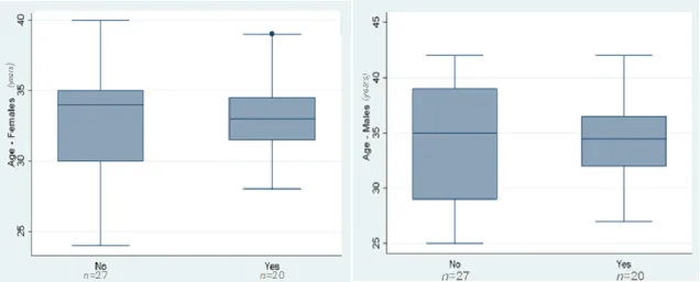
ARTICIPANTS AND SOCIAL HABITSThe study included 47 couples that underwent IVF treatments in CHVNG, between September 2012 and March 2013. The mean age of women was 32.9 ±3.5 years and of men was 34.4±4.6 years. The mean duration of infertility was 4.1±2.4 years.
Social habits (alcohol, cigarettes and drugs consumption, as well as, physical activity) of participants are summarized in Table 4.
Table 4| Socialhabits of the participants. (*For statistical analysis this variable was grouped in bigger groups. In alcohol consumption, the social was included in yes group. In physical activity the classes insufficiently active, active and very active were grouped in active.)
Code Age Infertility
Years of Inferlity Smoking habits Cigarettes/ day Alcohol consumption* Drugs consumption Chemicals/ raditation exposure Physical Activity*
4556 ♂ 37 Primary 2 Yes 0 - 5 Socialy No No Very Active
♀ 35 Primary 2 No 0 No No No Active
4839 ♂ 32 Primary 1 No 0 Socialy No No Inactive
♀ 32 Primary 1 No 0 No No No Inactive
4840 ♂ 35 Primary 4 Yes 15 - 20 Socialy No No Active
♀ 34 Primary 4 No 0 No No No Active
4841 ♂ 32 Primary 3 No 0 No No No Active
♀ 31 Primary 3 No 0 No No No Active
4642 ♂ 39 Primary 4 Yes 0 - 5 No No No Inactive
♀ 37 Primary 4 No 0 No No No Insuficiently Active
4643 ♂ 29 Primary 7 Yes 05 - 10 No No No Insuficiently Active
♀ 30 Primary 7 Yes 05 - 10 No No No Inactive
4644 ♂ 39 Primary 7 Yes 05 - 10 Socialy No Yes Inactive
♀ 28 Primary 7 Yes 10 - 15 No No No Inactive
4645 ♂ 34 Primary 8 Yes 0 Socialy No No Inactive
♀ 33 Primary 8 Yes 05 - 10 Socialy No No Insuficiently Active
4647 ♂ 35 Primary 11 Yes 0 - 5 Socialy No Yes Inactive
♀ 37 Primary 11 No 0 No No Yes Inactive
4648 ♂ 39 Primary 5 No 0 Socialy No No Very Active
♀ 37 Primary 5 No 0 No No No Inactive
4649 ♂ 35 Secondary 2 Yes 05 - 10 Socialy No No Insuficiently Active
Code Age Infertility Years of Inferlity Smoking habits Cigarettes/ day Alcohol consumption* Drugs consumption Chemicals/ raditation exposure Physical Activity* 4650 ♂ 37 Primary 6 No 0 No No No Inactive ♀ 34 Primary 6 No 0 No No No Inactive
4651 ♂ 36 Secondary 2 No 0 Socialy No No Inactive
♀ 34 Secondary 2 No 0 Socialy No No Inactive
4653 ♂ 40 Primary 4 No 0 No No No Active
♀ 39 Primary 4 No 0 No No No Inactive
4656 ♂ 28 Primary 5 No 0 No No No Inactive
♀ 27 Primary 5 No 0 No No No Inactive
4658 ♂ 42 Secondary 1 No 0 Socialy No No Inactive
♀ 34 Secondary 1 No 0 No No No Active
4659 ♂ 30 Primary 4 No 0 No No No Very Active
♀ 31 Primary 4 No 0 No No No Very Active
4660 ♂ 34 Primary 4 No 0 Socialy No Yes Inactive
♀ 34 Primary 4 No 0 No No No Inactive
4661 ♂ 42 Secondary 1 No 0 No No Yes Active
♀ 39 Secondary 1 No 0 No No No Active
4662 ♂ 39 Primary 3 No 0 No No No Inactive
♀ 35 Primary 3 No 0 No No No Inactive
4663 ♂ 27 Secondary 4 Yes 10 - 15 No No No Inactive
♀ 33 Secondary 4 No 0 No No No Inactive
4664 ♂ 40 Primary 3 Yes 15 - 20 No No No Inactive
♀ 24 Primary 3 Yes 15 - 20 No No No Inactive
4667 ♂ 33 Primary 5 Yes 10 - 15 Socialy No No Inactive
Code Age Infertility Years of Inferlity Smoking habits Cigarettes/ day Alcohol consumption* Drugs consumption Chemicals/ raditation exposure Physical Activity*
4669 ♂ 29 Primary 4 No 0 Socialy No No Active
♀ 27 Primary 4 No 0 Socialy No No Active
4670 ♂ 29 Primary 4 No 0 No No No Insuficiently Active
♀ 29 Primary 4 No 0 No No No Inactive
4671 ♂ 36 Secondary 3 No 0 Socialy No No Very Active
♀ 38 Secondary 3 Yes 05 - 10 Socialy No No Inactive
4672 ♂ 37 Primary 3 Yes 15 - 20 Socialy No No Active
♀ 35 Primary 3 Yes 15 - 20 No No Yes Inactive
4673 ♂ 31 Primary 3 Yes 15 - 20 Socialy No No Insuficiently Active
♀ 32 Primary 3 No 0 Socialy No No Active
4674 ♂ 33 Primary 4 Yes 0 - 5 Socialy No No Inactive
♀ 31 Primary 4 No 0 Socialy No No Inactive
4675 ♂ 40 Primary 4 No 0 Socialy No No Inactive
♀ 33 Primary 4 No 0 No No No Inactive
4826 ♂ 42 Primary 8 Yes 05 - 10 Socialy No No Inactive
♀ 38 Primary 5 Yes 05 - 10 Socialy No No Inactive
4827 ♂ 41 Primary 3 Yes 15 - 20 Yes Yes (canabis) No Active
♀ 35 Primary 3 Yes 0 - 5 No No No Active
4828 ♂ 35 Secondary 4 Yes 15 - 20 Socialy No No Very Active
♀ 35 Secondary 4 Yes 0 - 5 Socialy No No Active
4829
♂ 32 Secondary 11 No 0 No No Yes Insuficiently Active
♀ 32 Secondary 11 No 0 No No No Active
4830 ♂ 29 Primary 3 Yes 0 - 5 Socialy
Yes (cannabis)
Yes Active
Code Age Infertility Years of Inferlity Smoking habits Cigarettes/ day Alcohol consumption* Drugs consumption Chemicals/ raditation exposure Physical Activity*
4831 ♂ 37 Primary 3,5 Yes 0 - 5 Yes No No Active
♀ 35 Primary 3,5 No 0 No No No Insuficiently Active
4832 ♂ 28 Primary 2 No 0 No No No Active
♀ 25 Primary 2 No 0 No No No Inactive
4833 ♂ 29 Secondary 4 Yes 15 - 20 Socialy (cannabis) Yes No Active
♀ 34 Secondary 4 No 0 No No No Insuficiently Active
4834 ♂ 25 Primary 2 Yes 0 - 5 No No Yes Inactive
♀ 34 Primary 2 No 0 No No No Inactive
4835 ♂ 31 Primary 3 Yes 0 - 5 Socialy (cannabis) Yes Yes Inactive
♀ 30 Primary 3 No 0 No No No Inactive
4836 ♂ 35 Primary 5 Yes 15 - 20 No No Yes Very Active
♀ 34 Primary 5 No 0 No No No Active
4837 ♂ 29 Secondary 1 No 0 Socialy No Yes Active
♀ 32 Secondary 1 No 0 No No No Active
4838 ♂ 30 Primary 3 No 0 Yes No Yes Active
♀ 30 Primary 3 No 0 No No No Active
4839 ♂ 31 Primary 9 Yes 5 - 10 Yes No Yes Inactive
♀ 34 Primary 9 No 0 No No No Inactive
4840 ♂ 38 Primary 5 Yes 0 - 5 Socialy No No Insuficiently Active
♀ 40 Primary 5 No 0 Socialy No No Insuficiently Active
4841 ♂ 34 Primary 7 Yes 0 - 5 Yes No No Inactive
3.2. C
LINICAL DATAIn the couples studied 10 have a previous pregnancy (secondary infertility), the others had a primary infertility. The infertility cause was only masculine for 19 cases, only feminine for 10 cases, masculine and feminine for 12 cases and idiopathic for 6 cases. A GnRH antagonist was used in 25 of the cases and for the others was used a GnRH agonist (Table 5). Mean dose of recombinant FSH used was 1830.6±866.8 IU/ml and the mean duration of stimulation cycles was 12.2±2.0.
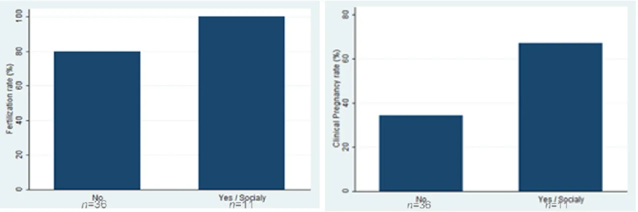
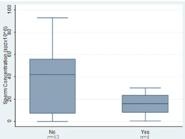
The cycles were performed with ejaculated sperm and the mean spermatozoa concentration was 33.4±27.7 million/ml. All cycles had positive pick-up with a mean number of 9.9±5.1 oocytes collected. The mean number of inseminated oocytes was 7.9±4.3 and the fertilization rate was 54.9%. Fertilization failed to occur in 7 cases, 5 ICSI cases and in 2 IVF cases.
From the cycles included in the study the embryo transfers was possible in 39 cases (21 ICSI cases and 18 IVF cases), with a mean number of 1.3±0.7 embryos per transfer. A total of 20 pregnancies were achieved (11 ICSI cases and in 9 IVF cases), which corresponded to a clinical pregnancy rate per transfer of 51.3%.The implantation rate was 37.3%.
In the pregnancies from these cycles, 12 births had already happened with a healthy single baby. From remained pregnancies 5 are still ongoing and 3 ended in a spontaneous abortion.