Elucidating the
role of Ndt80
transcription
factor in Candida
parapsilosis
virulence factors
regulation
Cláudia Patrícia Martins da Cruz
Mestrado em Biologia Celular e Molecular
Departamento de Biologia
Faculdade de Ciências da Universidade do Porto 2016
Orientador
Isabel M. Miranda, PhD
Todas as correções determinadas pelo júri, e só essas, foram efetuadas. O Presidente do Júri,
Dissertação para a candidatura ao grau de Mestre em Biologia Celular e Molecular submetida à Faculdade de Ciências da Universidade do Porto.
A presente dissertação foi orientada pela Doutora Isabel Miranda e foi realizada na Faculdade de Medicina da Universidade do Porto.
“We are trying to prove ourselves wrong as quickly as possible, because only in that way can we find progress.”
Agradecimentos
Em primeiro lugar, agradeço à Doutora Isabel Miranda, minha orientadora, por me ter aceitado como sua aluna de mestrado. Por todo o apoio incondicional, pela amizade, pela preocupação. Muito obrigada pelo entusiasmo e dedicação que sempre demonstrou, e que estiveram na génese de todo o meu empenho. Um último obrigado por ter contribuído para o meu crescimento, não só como cientista, mas também como pessoa. Um abraço apertado.
Agradeço também ao Professor Doutor Acácio Rodrigues por toda a simpatia e por me acolher no Serviço de Microbiologia da Faculdade de Medicina do Porto.
Um grande obrigado a todos os que fazem ou que já fizeram parte do Serviço de Microbiologia, por todos os momentos partilhados no laboratório. Por todos os sorrisos e apoio.
Um especial agradecimento à Joana Branco, por toda a ajuda e prontidão. Pela amizade.
À minha família, por me ter apoiado sempre e incondicionalmente, apesar de todas as adversidades. Pela paciência e sobretudo, por todo o esforço que fizeram para que eu pudesse estar aqui hoje. Prometo fazer tudo para que sempre se orgulhem de mim.
À Fátima e ao Joaquim, por serem incrivelmente dedicados e me acolherem sempre tão bem.
Ao Tiago, porque todas as palavras do mundo não seriam suficientes para te agradecer aquilo que representas para mim. Obrigada por seres incansável e por saberes sempre dar-me todo o apoio nos momentos mais acertados. Por estares sempre “ao” e “do” meu lado, incondicionalmente.
Sumário
Candida parapsilosis é um fungo oportunista emergente no ambiente hospitalar, afetando principalmente recém-nascidos e indivíduos imunocomprometidos. A adesão a superfícies inertes e dispositivos médicos, bem como a formação de biofilme em superfícies plásticas são duas das características relacionadas com a virulência deste microrganismo. Apesar do conhecimento acerca dos fatores de virulência associados à patogenicidade de C. parapsilosis ser limitado, esforços têm sido feitos no sentido de contrariar esta premissa. Diversos fatores de transcrição originalmente identificados em Candida albicans como reguladores-chave do programa de transcrição da filamentação e formação de biofilme, mostraram desempenhar funções semelhantes em C. parapsilosis. No entanto, outros moduladores importantes destes processos, tal como o Ndt80, ainda não foram caracterizados nesta espécie. O Ndt80 foi identificado pela primeira vez em Saccharomyces cerevisiae como um regulador do processo meiótico. Mais tarde, em C. albicans, este fator de transcrição mostrou ser essencial para a formação de biofilme, crescimento de hifas e expressão de genes que codificam para componentes da parede celular (por exemplo, ALS3, HWP1).
A deleção do gene NDT80 em C. parapsilosis, usando a ferramenta de disrupção génica SAT1 flipper, revelou alterações substanciais ao nível da morfogénese, adesão e desenvolvimento de biofilme. A estirpe mutante apresenta um aumento na capacidade de adesão a microesferas de polistireno e de formação de biofilme. Além disso, a deleção do gene alterou significativamente a morfologia celular, de leveduras a pseudohifas e células alongadas, o que se refletiu no crescimento em colónia, passando de lisas para rugosas. Por fim, estes resultados representam a primeira evidência do papel desempenhado pelo Ndt80 na regulação dos fatores de virulência de C. parapsilosis.
Palavras-chave:
Candida parapsilosis, adesão, formação de biofilme, morfogénese, virulência, middle sporulation element (MSE)Abstract
Candida parapsilosis is an emergent opportunistic fungus in hospital setting, that affects mainly neonates and immunocompromised individuals. Adherence to prosthetic materials as well as development of biofilms on plastic surfaces are among the biological attributes presumed to be related to its virulence capacity. Although the knowledge about the virulence factors associated with C. parapsilosis pathogenicity is limited, efforts have been made to counteract this assumption. Several transcription factors originally identified in Candida albicans as key regulators of the filamentous and/or biofilm growth transcriptional programs were found to play similar roles in C. parapsilosis. However, other important modulators of these processes, such as Ndt80, remain uncharacterised in this species. First identified in Saccharomyces cerevisiae as regulator of meiosis, Ndt80 was shown to play a distinct role in C. albicans, being an essential transcription factor for biofilm formation, hyphal growth and the proper expression of genes encoding cell wall components (e.g. ALS3, HWP1).
We knocked out C. parapsilosis NDT80 gene using the SAT1 flipper cassette and observed substantial alterations in morphogenesis, adhesion and biofilm growth. ndt80Δ mutant presents increased ability to adhere to polystyrene microspheres as well as to form biofilm. In addition, NDT80 disruption significantly changes cell morphology from yeast-shaped to pseudohyphal and elongated forms. As expected, this morphology switch produces colony changes, from wild type smooth colonies to crepe in ndt80Δ mutants. Thereby, these findings provide the first evidence for the direct role of Ndt80 in C. parapsilosis virulence factors regulation.
Keywords:
Candida parapsilosis, adhesion, biofilm formation, morphogenesis, virulence, middle sporulation element (MSE).
Table of contents
Agradecimentos I Resumo II Abstract III Table of contents IV List of Figures VIList of Tables VII
Abbreviations VIII
I. Introduction 1
1. C. parapsilosis: an emergent fungal pathogen 1
2. C. parapsilosis virulence factors 2
A. Morphological transition 3
B. Adhesion 5
C. Biofilm formation 7
D. Hydrolytic enzymes 9
E. Fatty acid synthesis and storage 10
3. Ndt80: a key regulator in C. albicans 11
4. Objectives 13
II. Materials and methods 14
1. Strains and growth conditions 14
2. Preparation of E. coli DH5α competent cells 14 3. Restriction, dephosphorylation and DNA cloning 15 4. Construction of C. parapsilosis mutant strains 15
5. DNA electrophoresis 18
6. E. coli DH5α transformation 19
7. C. parapsilosis transformation 19
8. Isolation of genomic DNA 19
9. SADH gene amplification and restriction 20
10. Adhesion assay 20
11. Biofilm assay 21
12. Microscopy 21
13. RNA extraction 21
14. cDNA synthesis and RT-qPCR 22
16. Statistical analysis 23
III. Results 24
1. Construction of C. parapsilosis mutant and complemented strains 24
2. Deleting NDT80 triggers morphogenesis 28
3. NDT80 deletion increases C. parapsilosis adhesion 29 4. NDT80 deletion promotes biofilm formation 30 5. NDT80 deletion alters the expression of biofilm-related genes 31 6. Biofilm-related genes contain Ndt80 binding motifs in their
promoters
31
7. BC014 and ndt80Δ mutant strains are C. parapsilosis sensu sticto 32
IV. Discussion and final conclusions 34
List of Figures
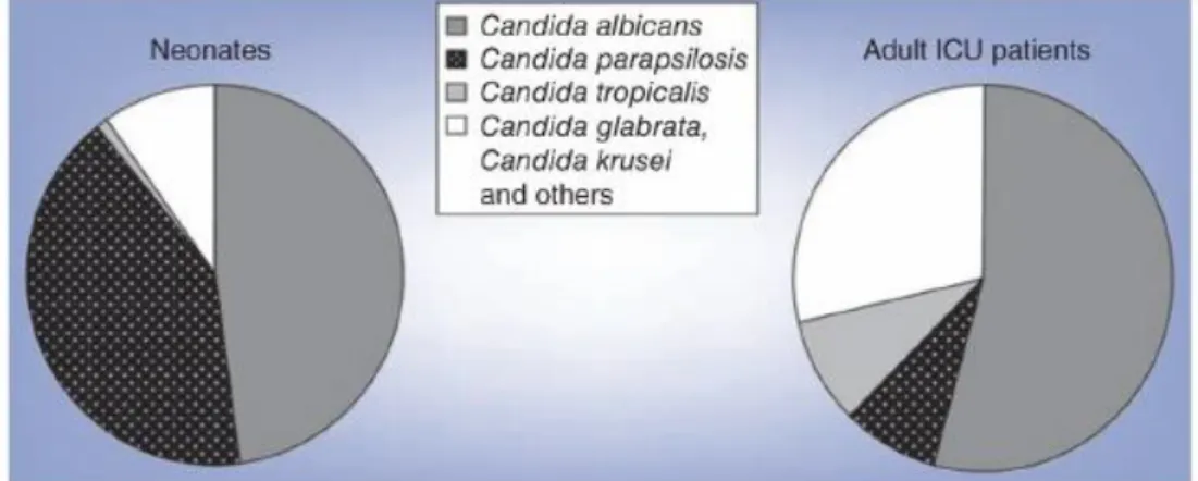
Figure 1. Distribution of Candida species by age. 2
Figure 2. Major morphologies of human fungal pathogens. 4 Figure 3. Phases of C. albicans biofilm formation. 8 Figure 4. C. albicans and C. parapsilosis MSE motifs. 12 Figure 5. Construction of the ndt80Δ mutant strain. 25 Figure 6. Differences in colony growth in YPD agar medium supplemented
with Nou 20 μg ml-1.
26
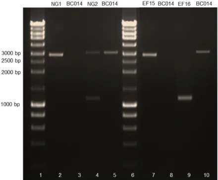
Figure 7. Deletion of NDT80 gene in C. parapsilosis. 26 Figure 8. Construction of ndt80Δ/NDT80 complemented strain. 27
Figure 9. Construction of ume6Δ mutant strain. 28
Figure 10. NDT80 deletion triggers morphogenesis in C. parapsilosis. 29
Figure 11. Deleting NDT80 increases adherence. 30
Figure 12. Deleting NDT80 increases biofilm formation. 30 Figure 13. Putative targets of Ndt80 transcription factor. 31
Figure 14. Identity of ndt80Δ mutant strain. 33
Figure 15. Representative model for the control of filamentous growth and biofilm formation by Ndt80.
List of Tables
Table 1. Primers used for construction of mutant and complemented strains. 17 Table 2. PCR programmes used for construction of mutant and
complemented strains.
18
Table 3. Primers used in RT-qPCR. 23
Table 4. Putative middle sporulation element (MSE) found in C. parapsilosis promoter genes.
Abbreviations
% – Percentage; µF – Microfarad; µg – Microgram; µl – Microlitre; µm – Micrometre; µM – Micromolar; A260 – Absorbance at 260 nanometres; A280 – Absorbance at 280 nanometres; Ap – Alkaline Phosphatase; ATP – Adenosine thriphosphate; bp – Base pair;BSA – Bovine Serum Albumin; CaCl2 – Calcium Chloride;
cDNA – Complementary deoxyribonucleic acid; Clo – Chloramphenicol;
CV – Crystal Violet;
DNA – Deoxyribonucleic acid; DNase – Deoxyribonuclease; dNTPs – Deoxyribonucleotide; DTT – Dithiothreitol;
EDTA – Ethylenediaminetetraacetic acid; fmol – Femtomole; g – Grams; g – Gravitational force; GO – Gene Ontology; h – Hour; kb – Kilobase pairs; KCl – Potassium Chloride; KOAc – Postassium Acetate; kV – Kilovolts;
LB – Luria-Bertoli; M – Molar;
min – Minutes; ml – Millilitre; mM – Millimolar;
MnCl2– Manganese Chloride;
MOPS – 3-(N-Morpholino)propanesulfonic acid sodium salt, 4-morpholinepropanesulfonic acid;
NaAc – Sodium Acetate; NaCl – Sodium Chloride;
NaMOPS – 3-(N-Morpholino)propanesulfonic acid sodium salt, 4-morpholinepropanesulfonic acid sodium salt;
ng – Nanogram; nm – Nanometres; Nou – Nourseothricin; ºC – Celsius degrees; OD550nm – Optical density at 550 nm; OD600nm – Optical density at 600 nm; ORF – Open Reading Frame; PBS – Phosphate Buffer Saline; PCR - Polymerase Chain Reaction; pmol – Picomole;
RT-qPCR – Quantitative Real-Time Polymerase Chain Reaction; RbCl2 – Rubidium Chloride;
RNA – Ribonucleic acid; RNase – Ribonuclease; rpm – Revolutions per minute;
RPMI – Roswell Park Memorial Institute; rRNA – Ribosomal RNA
s – Seconds;
SDS – Sodium Dodecyl Sulfate;
SOC – Buffer solution containing yeast extract, NaCl, glucose and MgCl2;
spp. – Species;
TAE – Buffer solution containing Tris base, acetic acid and EDTA; TE – Buffer solution containing Tris-HCl and EDTA;
TES – Buffer solution containing Tris-HCl, EDTA and SDS;
TFBI – Buffer solution containing KOAc, MnCl2, RbCl2, CaCl2 and glycerol;
Tris-HCl – Tris hydrochloride; U – Units;
UV – Ultraviolet light;
YPD – Yeast, Peptone and Dextrose; YPM – Yeast, Peptone and Maltose; Ω – Electrical resistance.
I. Introduction
1. C. parapsilosis: an emergent fungal pathogen
Over the last decades, Candida spp. represent the most common cause of invasive fungal infections worldwide, which can range from superficial to life-threatening disseminated candidiasis (Polke et al., 2015). Candidemia, i.e. bloodstream infections caused by Candida spp., is recognized as a major cause of hospital-acquired infections, accounting for a mortality of 15-35% for adults and 10-15% for neonates (Guinea, 2014; van Asbeck et al., 2009). The species of Candida causing this type of infection varies considerably according to the geographical region and hospital unit. Additionally, its frequency depends on several factors such as the predisposing conditions of infected patients, antifungal therapy received and hospital-related factors (Guinea, 2014). Common risk factors for the development of candidemia include central venous catheters, severe immunosuppression, extended treatment in intensive care units, among others (Polke et al., 2015). Although Candida albicans is still the most prevalent and common cause of candidemia globally, other non-albicans Candida spp., mainly Candida parapsilosis, Candida glabrata and Candida tropicalis have been assuming a relevant position in recent years (Pfaller et al., 2011; Trofa et al., 2008; van Asbeck et al., 2009). These species account for approximately 80% of all non-albicans Candida infections in European countries (Montagna et al., 2014).
C. parapsilosis was first described in 1928 and considered a relatively non-pathogenic yeast (Trofa et al., 2008; van Asbeck et al., 2009). This diploid microorganism is often isolated from several environmental sources (e.g. soil, seawater and domestic animals) and considered a human commensal (Trofa et al., 2008; van Asbeck et al., 2009). However, in the hospital setting this species emerged as an opportunistic fungal pathogen. Mainly due to its ability to adhere to and develop biofilms on intravascular devices and prosthetic materials, transmission through the hands of health care workers and gastrointestinal colonization (Dotis et al., 2012; Trofa et al., 2008), the use of parenteral nutrition and intravenous catheters are among the risk factors that contribute to the increased incidence of C. parapsilosis infections over the past decades (van Asbeck et al., 2009). In population-based studies, this fungus is considered the second most common cause of candidemia in Latin America, Asia and Southern Europe countries, including Portugal, representing 20% of Candida infections and accounting for a mortality rate of 28.5% (Costa-de-Oliveira et al., 2008; Hirai et al., 2014; Holland et al., 2014; Nucci et al., 2013; Pratikaki et al., 2011; Silva et al., 2009a; Trofa et al., 2008). C.
parapsilosis affects mostly immunocompromised individuals, patients requiring prolonged use of a central venous catheter, low birth weight neonates and children less than 24 months (Dotis et al., 2012; Guinea et al., 2014; Pammi et al., 2013; Trofa et al., 2008; van Asbeck et al., 2009). Interestingly, in pedriatic intensive care units, infections by C. parapsilosis are particularly concerning, accounting for a mortality rate of 10% (Pammi et al., 2013) (Figure 1).
Figure 1. Distribution of Candida species by age. Adapted from Chow et al. (2012).
In fact, in some hospitals, C. parapsilosis has surpassed C. albicans, becoming the leading fungal pathogen among children and neonates (Neu et al., 2009; Rodriguez et al., 2006). A multicentre study performed in 24 Spanish pedriatic intensive care units revealed that 75.7% of invasive Candida infections by C. parapsilosis occurred in children less than 2 years old (Jordan et al., 2014). Moreover, a surveillance study carried out between 2006 and 2010 in an Italian neonatal intensive care unit showed that C. parapsilosis was the second most frequent cause of infection (16.3%), being Pseudomonas aeruginosa (17%) the first and C. albicans representing 10.5% (Crivaro et al., 2015). According with Crivaro et al. (2015), the use of central line-, umbilical catheter and mechanical ventilation were associated with a higher risk of infection.
Considering epidemiological data, C. parapsilosis has consolidate its place as a nosocomial pathogenic fungus, therefore understanding its virulence behaviour will be imperative to i) design more suitable hospital care guidelines in order to prevent C. parapsilosis infections, and ii) the treatment outcome.
2. C. parapsilosis virulence factors
In conditions of decreased immune defences or microbiota imbalance, Candida spp. are considered one of the main human fungal pathogens. Virulence factors increase the pathogenic potential of these microorganisms by enhancing its ability to colonize and
invade the host and inclusively avoid immune defences. There is a whole interplay involving these fungal attributes that contribute to the development of disease in a human host and a failure of defence mechanisms that have evolved to avoid these infections (Chow et al., 2012). In this context, the characterization of virulence factors is an attempt to understand the infectious process and the pathogen-host relationship and explain differences in pathogenicity between fungal species among Candida spp. (Riceto et al., 2015).
C. albicans has been the main focus of the work related with Candida virulence, mostly due to its clinical importance as the most common cause of Candida infections worldwide and its unique virulence characteristics. However, as infections by other Candida spp. are increasing, more attention has been paid to non-albicans Candida spp., namely C. parapsilosis and C. glabrata, and significant differences from C. albicans have been noticed.
Virulence factors associated to C. parapsilosis include its capacity to adhere to a wide variety of substrates (biotic and abiotic surfaces), biofilm formation on medical indwelling devices, secretion of hydrolytic enzymes that causes tissue damage, fatty acid synthesis and storage and presumably the ability to switch morphology between yeast and filamentous growth (Chow et al., 2012; Singaravelu et al., 2014).
A. Morphological transition
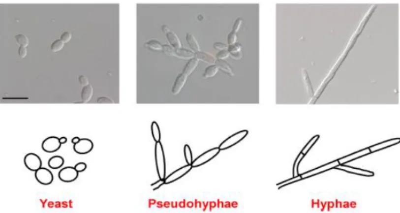
Fungal species can grow in three main cellular morphologies: yeast, pseudohyphae and hyphae (Figure 2). The hyphae as well as expression of hypha-specific genes is critical for virulence in several fungal pathogens, although its study focuses mostly on C. albicans (Thompson et al., 2011). However, both yeast and hyphal forms are believed to be important for pathogenicity. While the first is involved in colonization and rapid dissemination, the latter is associated with invasion of tissues and biofilm formation (Jacobsen et al., 2012; Zhu and Filler, 2010). Although C. parapsilosis do not form true hyphae, they are able to produce elongated cells called pseudohyphae (Ding and Butler, 2007; Kuhn et al., 2002). This morphotype is a potential virulence factor as it is essential for biofilm formation and presents a higher adherence ability to polystyrene and glass substrates than yeast forms (Abi-chacra et al., 2013). However, in this pathogen, little is known about the relationship between pseudohyphae and virulence.
Figure 2. Major morphologies of human fungal pathogens. (Top) Images of C. albicans cells visualized by differential
interference contrast (DIC) microscopy (bar = 10 µm). (Bottom) Schematic representation of each morphotype. Adapted from Thompson et al. (2011).
The ability to switch morphology from yeast to a filamentous form is triggered by a significant change in the transcriptional program, being linked not only to the expression of filament-related genes but also of genes encoding virulence factors that are not involved in filamentous growth per se, such as the hypha-associated cell wall proteins Als3 and Hwp1 (Mayer et al., 2013). Genes originally identified in C. albicans as part of the filamentous-growth transcriptional program have homologs in non-albicans Candida species, some of which display conserved functions (e.g. Ume6 and Efg1) (Banerjee et al., 2008; Carlisle et al., 2009; Connolly et al., 2013; Lackey et al., 2013; Stoldt et al., 1997). C. albicans strains lacking Ume6 transcription factor are attenuated for virulence and defective for hyphal growth in a mouse model of systemic candidiasis (Banerjee et al., 2008). Carlisle et al. (2009) demonstrated that UME6 expression levels influence C. albicans morphogenesis and its virulence potential. High levels of UME6 expression significantly increases hyphal formation and promotes virulence in vivo. Contrariwise, low levels of UME6 expression lead to the development of pseudohyphae (Carlisle et al., 2009). Similarly, Lackey et al. (2013) found that increased expression of UME6 in C. parapsilosis is sufficient to drive filamentous growth and filament-specific gene expression. This fact was also noticed for other non-albicans Candida species, such as C. tropicalis, suggesting that Ume6 is an evolutionary conserved regulator (Lackey et al., 2013). Consistent with this idea, Connolly et al. (2013), demonstrated that, as in C. albicans, C. parapsilosis Efg1 plays a central role in regulating morphology. Strains carrying deletions of CaEFG1 fail to undergo the transition from yeast to hyphal form in filament-inducing conditions and have reduced virulence in most infection models (Chamilos et al., 2006; Lo et al., 1997; Pukkila-Worley et al., 2009). Likewise, C. parapsilosis efg1Δ mutants increase the rate of morphological switching from “wrinkle” or “concentric” (elongated cells and pseudohyphae) to smooth (yeast-shaped cells)
colonies and have attenuated virulence in vivo (Connolly et al., 2013). Others important regulators of hyphal growth, such as Ndt80 (Sellam et al., 2010), remain uncharacterised in C. parapsilosis. In this context, there are still much work to do in identifying other regulators of morphogenesis, and, ultimately, understand its contribution for virulence in this species.
B. Adhesion
Adhesion is an important virulence attribute once it represents the first step for persistent colonization, biofilm formation and establishment of disease (Silva-Dias et al., 2015). The cell wall is the first site of contact between Candida and a substrate. This dynamic and complex structure is composed by polysaccharides, chitin, mannoproteins and covalent bound proteins, namely, proteins with a glycosylphosphatidylinositol (GPI) anchor (Als proteins) and Proteins with Internal Repeats (Pir proteins) (Chaffin et al., 1998; de Groot et al., 2013; Richard and Plaine, 2007). Mannoproteins (e.g. Hwp1) as well as GPI proteins (e.g. Als1 and Als3) are the main players in the initiation of the contact with a surface, having a significant role in the colonization, invasion and damage of host tissue as well as in the adhesion to inert surfaces (Chaffin, 2008; de Groot et al., 2013). In addition, these proteins are important for cell-cell adhesion, being required for biofilm formation as contact mediators that promote biomass accumulation and biofilm resilience (Fox and Nobile, 2012; Nobile et al., 2008). Because of its role in pathogenesis and importance as a potential target for antifungal therapy, the study of these cell wall components is of great interest (de Groot et al., 2013).
C. albicans adhesion is closely related to hyphal growth, since the contact of this fungus to a surface promotes filamentation and simultaneously induce the expression of hyphal-associated cell wall proteins, such as Als1, Als3 and Hwp1 (Jacobsen et al., 2012). In this species, ALS (Agglutinin-Like Sequence) gene family include eight members, ALS1-7 and ALS9, encoding large cell-surface glycoproteins (Cota and Hoyer, 2015; de Groot et al., 2013). Als proteins have similar structure displaying compensatory adhesion functions within its members (Hoyer et al., 2008; Zhao et al., 2005). However, the expression of each ALS gene appears to be host site-dependent, indicating protein-specific adhesive functions (de Groot et al., 2013). Als1 and Als3 are the two main proteins implicated in the virulence of C. albicans (Hoyer et al., 2008). Als1 appears to be important for adhesion to vascular endothelial cell monolayers. In contrast, deletion of ALS3 gene has large effects on adhesion to endothelial and buccal epithelial cells. In turn, HWP1, a well-characterized hypha-specific protein, is a critical adhesin for the binding of hyphae to host epithelial cells (Cuéllar-Cruz et al., 2012; Hoyer et al., 2008).
Moreover, Als1, Als3 and Hwp1 appear to be fundamental for biofilm development, playing complementary roles in this process (Nobile et al., 2006a; Nobile et al., 2006b; Nobile et al., 2008). Adhesion constitutes the essential first step for the formation of biofilm. Several studies demonstrate that deletion of either ALS1, ALS3 or HWP1 genes have severe effects upon biofilm growth (Cuéllar-Cruz et al., 2012; Nobile et al., 2006a; Nobile et al., 2006b) and compromise their roles as complementary adhesins for cell-cell adherence in this process (Nobile et al., 2008). Their expression is transcriptionally regulated by a number of transcription factors implicated in the biofilm formation, including Bcr1, Efg1 and Ndt80 (Dwivedi et al., 2011; Nailis et al., 2009; Nobile et al., 2006a; Nobile et al., 2012). Specifically, Ndt80 was shown to be required for the proper expression of HWP1 and ALS3 genes (Sellam et al., 2010).
Significant knowledge has been achieved regarding the components involved in C. albicans adherence ability. By contrast, this topic is poorly explored for other relevant species of Candida which can yield differences relatively to the first. For example, while C. albicans adheres to a large variety of materials compared with other Candida spp., (Cuéllar-Cruz et al., 2012) C. parapsilosis is frequently associated with pronounced capacity to adhere to plastic surfaces (Abi-chacra et al., 2013; Douglas, 2003; Kuhn et al., 2002). Compared with C. albicans, this species possesses considerable intraspecies variation of adhesion profiles (Silva-Dias et al., 2015).
Partial genomic microarrays used to determine transcriptional changes during biofilm growth in C. parapsilosis, identified a gene phylogenetically close to Hwp1 and orthologue of CaRBT1 (Rossignol et al., 2009). Its deletion revealed that CpRBT1 is required for biofilm formation (Rossignol et al., 2009). In addition, in silico analysis of the C. parapsilosis genome indicated the existence of five potential homologues of CaALS genes (Butler et al., 2009). Two putative Als-like proteins were found to possess the ability to bind to the extracellular matrix proteins, namely fibronectin, vibronectin and laminin (Kozik et al., 2015). However, to date, only one study focusing on the characterization of one of these putative adhesins was reported (Bertini et al., 2016). Syntenic to CaALS7 but with high sequence homology with CaAls3, Cpar2_404800, named CpAls7, was shown to be important for C. parapsilosis adhesion to host surfaces in vitro (Bertini et al., 2016).
C. Biofilm formation
Biofilms are an organized community comprised of a dense network of cells embedded in a polysaccharide-rich extracellular matrix (Cos et al., 2010; Donlan and Costerton, 2002). These structures are often associated with implanted medical devices and higher mortality rates, representing 65% of human infections (Blankenship and Mitchell, 2006; Cos et al., 2010). Microorganisms that grow in these communities exhibit unique phenotypic characteristics when compared to their planktonic counterparts (Ramage et al., 2009), as they have increased resistance to host defence mechanisms and antifungal therapy (Cos et al., 2010; Martinez and Fries, 2010), as well as, increased protection against environmental stresses or nutritional imbalances (Chandra and Mukherjee, 2015). In fact, several studies reported resistance of Candida biofilms to commonly used antifungals, including azoles, polyenes and echinocandins (Baillie and Douglas, 1999a; Chandra and Mukherjee, 2015; Katragkou et al., 2011). Katragkou et al. (2011) investigated the activity of two triazoles and two echinocandins alone or in combination with human phagocytes and concluded that at both conditions less damage was induced to C. parapsilosis biofilms compared with planktonic cells.
Biofilm formation is greatly influenced by nutrient availability and the substrate (Chandra and Mukherjee, 2015). Moreover, the ability to form such structure varies widely among the species of Candida and between strains. Silva et al. (2009b) characterized the biofilms of three non-albicans Candida species, namely C. parapsilosis, C. glabrata and C. tropicalis and noticed that the biofilm formed by the former was highly strain dependent. In addition, studies regarding C. parapsilosis have shown that this fungus has higher affinity for prosthetic materials than C. albicans (Nosek et al., 2009; Silva et al., 2011b), especially in high glucose media (Pereira et al., 2015).
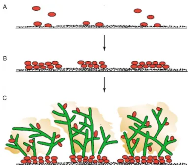
Fungal biofilms undergo multistep growth processes involving physical, chemical and biological changes (Nett and Andes, 2006). The formation of a biofilm is initiated by adherence of yeast cells to a surface (Figure 3A). Subsequently, attached cells multiply and form various cellular layers via cell-cell contact (Singaravelu et al., 2014) (Figure 3B). In C. albicans, cells undergo a morphological change from yeast to hyphae during this phase, which appear to be a prerequisite for a structured biofilm (Baillie and Douglas, 1999b; Ramage et al., 2002). Lastly, once the mature biofilm is formed it is covered by the extracellular matrix (Figure 3C). In comparison to C. albicans, the architecture of C. parapsilosis biofilm is much simpler. Because C. parapsilosis do not form true hyphae, its biofilm is solely composed by yeast and pseudohyphal forms (Ding and Butler, 2007; Kuhn et al., 2002). Nevertheless, pseudohyphal phenotypes were shown to form more
biofilm and have greater invasiveness into agar than yeast forms (Laffey and Butler, 2005). Additionally, contrary to C. albicans, its extracellular matrix is mainly composed by carbohydrate, with less protein content (Silva et al., 2009b).
Figure 3. Phases of C. albicans biofilm formation. (A) Adherence of yeast (red) by cell wall proteins to a substrate,
such as implanted medical devices or host surfaces. (B) Formation of basal layers of yeast cells and cell-cell adhesion.
(C) Completion of mature biofilm by formation of hyphal layers (green) and secretion of extracellular matrix containing
polysaccharides, carbohydrates and proteins (yellow) that surrounds both yeasts and hyphal forms. This structure protect cells against drugs and other toxic components. Adapted from Douglas (2003).
These communities are associated with a specific gene-expression pattern. The transcriptional changes that occur during the phases of biofilm growth are well established for C. albicans. During hyphal morphogenesis several adhesion molecules are expressed, for example, Als3 and Hwp1 proteins, which promote cell and cell-substrate binding (Pannanusorn et al., 2014). In fact, mutants of these proteins are defective in biofilm formation (Nobile et al., 2008). Many transcription factors are known to regulate these processes and consequently the development of biofilm in this species. Some of them are inclusively already described for C. parapilosis (Holland et al., 2014). Bcr1, Efg1 and Ace2 transcription factors are implicated on biofilm growth in both species (Finkel et al., 2012; Holland et al., 2014; Nobile et al., 2012; Pannanusorn et al., 2014; Ramage et al., 2002). Efg1 is also required for hyphal growth in C. albicans and involved in morphological switching in C. parapsilosis (Connolly et al., 2013; Stoldt et al., 1997; Sudbery, 2011). Other transcription factors appear to play a role in biofilm formation in only one species, e.g. Rob1 restricted to C. albicans and Gzf3 and Ume6 to C. parapsilosis (Holland et al., 2014; Nobile et al., 2012). However, the transcriptional regulator Ume6 shares a similar function with those of C. albicans at the morphological level (Banerjee et al., 2008; Carlisle et al., 2009; Lackey et al., 2013). Despite of the
increasing interest regarding C. parapsilosis virulence factors, many components remained to be characterized. This is the case of Ndt80, a major regulator of hyphal growth and biofilm formation in C. albicans (Nobile et al., 2012; Sellam et al., 2010).
D. Hydrolytic enzymes
The secretion of hydrolytic enzymes by Candida spp., including secreted aspartyl proteinases (Sap), phospholipases and lipases, is associated with fungal virulence. These proteins help fungi to adapt to different environmental conditions, to adhere to and invade host tissues and weaken the immune response (Modrzewska et al., 2016).
Sap enzymes are extensively studied and investigations have shown that they are important for virulence of both C. albicans and C. parapsilosis (Horvath et al., 2012; Naglik et al., 2003). C. albicans possesses ten Sap enzymes (Sap1-Sap10) localized mainly in the fungal cell wall and cytoplasm (Hube and Naglik, 2001; Modrzewska et al., 2016). They facilitate tissue adhesion and invasion through the degradation of host cell surface structure and intracellular substances and destruct cells and molecules of the host immune system (Hube and Naglik, 2001; Pichova et al., 2001; Trofa et al., 2008; Wu et al., 2013). The ability of C. albicans to invade and damage oral epithelium is reduced when treated with the aspartic protease inhibitor pepstatin A (Naglik et al., 2008). C. parapsilosis has three SAP genes, SAPP1, SAPP2 and SAPP3 and the later remains uncharacterized (Chow et al., 2012; Trofa et al., 2008). In addition, SAPP1 gene has undergone gene duplication (SAPP1a and SAPP1b) (Horvath et al., 2012). Gácser et al. (2007b) demonstrated that the epidermal and epithelial damage caused by C. parapsilosis in a human host tissue was reduced in the presence of the aspartic inhibitor pepstatin A, thus suggesting the involvement of CpSapp proteins in virulence. Later, deletion of SAPP1a and SAPP1b has demonstrated that these proteins are important for growth in human serum and survive within macrophages by inhibiting the fusion of phagolysosome (Horvath et al., 2012). Besides, studies demonstrated that Sapp1 has higher catalytic activity than Sapp2 (Hruskova-Heidingsfeldova et al., 2009). Interestingly, these proteins appear to be more important for localized invasive infections than bloodstream infections (Dagdeviren et al., 2005; Nemeth et al., 2013; Silva et al., 2009c).
Phospholipases are believed to be important for host damage and active penetration through disruption of cell membranes, which are mainly composed by phospholipids and proteins (Ghannoum, 2000) and adherence to epithelial cells (Dagdeviren et al., 2005). These enzymes hydrolyse ester linkages in glycerophospholipids (Trofa et al., 2008). In C. albicans, Pbl1 is the predominant
enzyme, responsible for most of extracellular phospholipase activity. Strains lacking PBL1 gene show attenuated virulence in an animal model of candidiasis (Ghannoum, 2000; Leidich et al., 1998). The role of phospholipases in C. parapsilosis virulence is less explored. Dagdeviren et al. (2005) established a positive correlation between phospholipase production and adherence of C. parapsilosis isolates. However, phospholipase activity seems to be highly variable among C. parapsilosis strains (D'Eca Junior et al., 2011; Gokce et al., 2007; Silva et al., 2009c).
In turn, secreted lipases are believed to be involved in the lipid digestion for nutrient acquisition and adhesion to host cells and tissues (Schaller et al., 2005). Ten lipase genes (LIP1-10) were identified in C. albicans (Yang et al., 2014) and deletion of the gene encoding Lip8 led to a reduction of the virulence in a mouse model of systemic infection (Gácser et al., 2007a). These enzymes are recognised to play an important role in host-pathogen interaction during C. parapsilosis infection (Toth et al., 2015). The disruption of both genes identified in this species, LIP1 and LIP2, caused a decrease of virulence in vivo and a reduction of the epithelial damage (Gácser et al., 2007b). In addition, Toth et al. (2014) demonstrated that C. parapsilosis secreted lipase is able to promote the survival of fungal cells in macrophages and mitigate the inflammatory response of the host. However, the mechanisms of action of these enzymes are not determined yet. These findings are clinically relevant since C. parapsilosis affect frequently patients receiving lipid-rich total parenteral nutrition (Trofa et al., 2008). In this context, lipases may be a potential target for future antifungal agent development.
E. Fatty acid synthesis and storage
The production of fatty acids, saturated and unsaturated, is critical for the generation and maintenance of cell membrane integrity. Fatty acid synthase (FAS), fatty acid desaturase (OLE) and fat storage-inducing protein (FIT) are important components in this pathway, being essential for normal yeast growth (Nguyen et al., 2012; Nguyen et al., 2011a; Nguyen et al., 2011b; Nguyen et al., 2009).
C. albicans strains lacking FAS2 gene were avirulent in a rat model (Zhao et al., 1997). In C. parapsilosis, the disruption of FAS2 gene resulted in attenuated virulence in a systemic murine infection model and impaired biofilm formation on polystyrene and silicone surfaces (Nguyen et al., 2009). Mutant cells were killed more efficiently by macrophages, probably due to changes in fatty acid composition that affect membrane fluidity and increase its susceptibility to reactive oxygen species secreted by these cells (Nguyen et al., 2009).
The disruption of OLE1 gene prevented cells to form hyphae and pseudohyphae in C. albicans and C. parapsilosis, respectively and consequently, decreased virulence in both species (Krishnamurthy et al., 2004; Nguyen et al., 2011a). In addition, C. parapsilosis ole1 mutants showed hypersensitivity to osmotic and oxidative stress as well as to human serum, probably due to the reduced content in unsaturated fatty acid, essential for cell membrane biosynthesis (Nguyen et al., 2011a). In S. cerevisiae, these enzyme is critical for cell viability through the regulation of unsaturated fatty acid production (Tehlivets et al., 2007).
In turn, C. parapsilosis strains lacking Fit2 enzyme also presented increased sensitivity to osmotic stress and attenuated virulence in a murine model of infection (Nguyen et al., 2011b). Mutant cells had a significant reduction in lipid droplets, which function as reservoir for fatty acids that are used for phospholipids biosynthesis, thus affecting membrane synthesis and the normal yeast growth (Nguyen et al., 2011b).
In general, the balance between saturated and unsaturated fatty acids is critical for membrane fluidity, affecting the function of numerous membrane systems of the cell and, ultimately, its ability to cause an infection (Nguyen et al., 2011a; Nguyen et al., 2011b; Nguyen et al., 2009).
3. Ndt80: a key regulator in C. albicans
The transcription factor Ndt80, a member of the NDT80/PhoG like DNA-binding family, is an important regulator of the progression of meiotic division in S. cerevisiae (Hepworth et al., 1998; Pierce et al., 2003), activating more than 150 middle sporulation genes during meiosis (Chu et al., 1998; Primig et al., 2000). In C. albicans, Ndt80 was first identified as a key modulator of azole drug sensitivity, being involved in the control of ergosterol biosynthesis (Sellam et al., 2009) and activation of the efflux pump Cdr1 (Chen et al., 2004). Nevertheless, genome-wide occupancy using Chip-chip method revealed that Ndt80p binds to promoter regions of 23% of C. albicans genes with diverse biological functions such as, carbohydrate metabolism, cell wall components and hyphal growth (Sellam et al., 2009). Remarkably, a large number of its target genes code for transcription regulators. Among those, CaNdt80p was found to bind promoters of several hyphal growth transcription factors, namely Cph2p, Efg1p, Ume6p and Flo8p, which in turn regulates filamentation-specific genes (Sellam et al., 2010; Sellam et al., 2009). Strains carrying deletion of CaNDT80 were unable to form hyphae in response to hypha-inducing conditions in vitro and to activate genes encoding cell wall components (HWP1, ECE1, RBT4, ALS3, HYR1, ALS1), exhibiting a marked loss of virulence in vivo (Sellam
et al., 2010). Additionally, Ndt80 is part of a network of six transcriptional regulators described by Nobile et al. (2012) as essential for biofilm development in C. albicans. Interestingly, Ndt80 is required for normal biofilm formation in vitro and in vivo, functioning as both an activator and repressor of its target genes (Nobile et al., 2012).
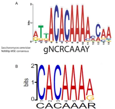
Structural studies demonstrated that the S. cerevisiae Ndt80 binds to the middle sporulation element (MSE) (Fingerman et al., 2004; Pierce et al., 2003). CaNdt80 MSE ACACAAA-3’) was found to be highly similar to those described for S. cerevisiae (5’-gNCRCAAAY-3’), suggesting a conservation of the Ndt80 binding site (Nobile et al., 2012; Sellam et al., 2009) (Figure 4A). In addition, Connolly et al. (2013) used Discriminative Regular Expression Motif Elicitation (DREME) program to identify conserved motifs and predicted the binding site of NDT80 in C. parapsilosis (5’-CACAAAR-3’) (Figure 4B), which closely resemble those of S. cerevisiae and C. albicans.
Figure 4. C. albicans and C. parapsilosis MSE motifs. (A) C. albicans MSE motif identified by Multiple EM for Motif
Elucidation (MEME) program. S. cerevisiae MSE consensus motif is shown on the bottom of the logo. Adapted from Sellam et al. (2009). (B) C. parapsilosis MSE motif predicted by DREME program. Adapted from Connolly et al. (2013).
Despite its importance for C. albicans virulence, the role of Ndt80 transcription factor has never been described for other clinically relevant Candida spp.
4. Objectives
Previous studies demonstrated that Ndt80 is an important regulator of the virulence process in C. albicans. However, their role in C. parapsilosis virulence was not investigated so far. Therefore, the main goal was to characterize the role of Ndt80 in C. parapsilosis virulence traits.
II. Materials and methods
1. Strains and growth conditions
C. parapsilosis strains were routinely grown in YPD medium (1% yeast extract, 2% peptone, 2% glucose) at 30°C with agitation (180 rpm) or YPD with 2% agar. For the selection of transformants, YPD agar plates were supplemented with nourseothricin (Nou; Werner BioAgents) at a final concentration of 200 µg ml-1. To recycle the SAT1
flipper cassette, transformants were incubated in YPM medium (1% yeast extract, 2% peptone, 2% maltose) overnight, with agitation (180 rpm). From this culture, approximately 100 cells were plated in YPD agar supplemented with Nou at final concentration of 20 μg ml-1 and incubated at 30ºC. After 48 h of incubation, two colonies
sizes were distinguishable. Smaller colonies were picked and stroked into fresh YPD agar plates. All C. parapsilosis strains were stored as frozen stocks in YPD medium with 40% glycerol, at -80°C. For phenotypic assays (adhesion and biofilm quantifications), strains were grown overnight in Sabouraud broth medium, at 30ºC with agitation (180 rpm).
Competent cells of Escherichia coli DH5α strain were used for cloning and construction of disruption and complementation cassettes. Cells were routinely grown in Luria-Bertani (LB) medium or LB with 2% agar. Transformants were grown in LB or LB agar plates supplemented with chloramphenicol (Clo; Sigma-Aldrich) at a final concentration of 25 μg ml-1. E. coli strains were stored at -80ºC in LB medium
supplemented with 20% glycerol and Clo (25 μg ml-1).
2. Preparation of E. coli strain DH5α competent cells
From an overnight culture (5 ml), 200 μl of cells were inoculated in 5 ml of LB medium and incubated at 37ºC, with agitation (180 rpm). After reaching the optical density (OD) at 550 nm (OD550nm) of 0.3, 4 ml of the culture were transferred to 100 ml
of LB and incubated in the same conditions until reaching the same OD550nm value.
Afterwards, cells were incubated on ice for 5 min and centrifuged at 1,503 g, for 5 min, 4ºC. The pellet was gently resuspended in 40 ml of cold TFBI solution (30 mM KOAc, 50 mM MnCl2, 100 mM RbCl2, 10 mM CaCl2, 15% glycerol) and centrifuged for 5 min at 664
g, 4ºC. Cells were then resuspended in 5 ml of cold TFBII solution (10 mM NaMOPS pH 7.0, 75 mM CaCl2, 10 mM RbCl2, 15% glycerol) and incubated on ice for 5 min. Lastly,
3. Restriction, dephosphorylation and DNA cloning
In a 30 μl reaction volume, about 3 μg of plasmid DNA was digested with 3 U of the respective restriction enzyme. Reactions with FastDigest enzymes (KpnI and ApaI; Fermentas) occurred in 1× FastDigest buffer, at 37ºC for 1 h. In contrast, 1× B buffer (10 mM Tris-HCl pH 7.5, 10 mM MgCl2, 0.1 mg/ml BSA) was added to reactions with
conventional enzymes, namely SacI and SacII (Fermentas). The mixture was incubated for approximately 16 h at 37ºC. Total plasmid digestions were verified by DNA electrophoresis in 1% agarose gel (see DNA electrophoresis section). The DNA was purified using the EasySpin DNA Gel Extraction kit (Citomed).
Afterwards, about 1 μg of digested plasmid was dephosphorylated with 1 U of Alkaline Phosphatase (Ap; Roche) in 1× Ap buffer (0.05 M Tris-HCl, 5 mM MgCl2, pH
8.5) for 1 h at 37ºC.
The insertion of DNA fragments into the respective plasmids (DNA cloning) was performed by T4 DNA Ligase enzyme (Fermentas). Approximately 23 fmol of plasmid and five to twenty times more number of moles of fragment were put together in 1× T4 DNA Ligase buffer (25 mM Tris-HCl pH 7.6, 5 mM MgCl2, 0.5 mM ATP, 0.5 mM DTT,
25% polietileno glycol-8000) and 5 U of enzyme, in a final volume of 20 μl. Reactions occurred at 22ºC for 1 h.
The inactivation of enzymes was carried out at 65ºC for 10 min.
4. Construction of C. parapsilosis mutant strains
For the construction of gene disruption and complementation cassettes, plasmid pCD8 containing a gene knockout system specifically constructed for C. parapsilosis, was used in this study (Ding and Butler, 2007). All sequences were retrieved from the Candida Genome Database (CGD; http://www.candidagenome.org/) (Inglis et al., 2012).
To knockout NDT80 gene (CPAR2_213640), 478 bp upstream and 460 bp downstream sequences of CpNDT80 were amplified using CpNDT80up_F and CpNDT80up_R primers (containing recognition sites for KpnI and ApaI) and CpNDT80down_F and CpNDT80down_R primers (containing recognition sites for SacII and SacI), respectively. PCR fragments were digested with the respective restriction enzymes and cloned in the same sites of pCD8, generating the plasmid pNG4 (see restriction, dephosphorylation and DNA cloning section). The entire cassette plus flanking regions were excised by digestion with FastDigest KpnI and SacI enzymes and transformed into C. parapsilosis BC014 (Silva et al., 2011a) after gel extraction. Integration of the disruption cassette at NDT80 locus was confirmed by PCR using a
primer 5’ to NDT80 (CpNDT80gen_F) and a primer inside the cassette (FLP_R). Primers CpNDT80gen_F and CpNDT80down_R were used to verify recycling of the cassette and deletion of the NDT80 gene. The same cassette was used to delete the two NDT80 alleles.
A similar method was used to delete UME6 gene (CPAR2_803820) in strains BC014 and EF16 (ndt80Δ mutant strain). Approximately 434 bp downstream and 706 bp upstream of the open reading frame were amplified using primer pairs CpUme6down_F/CpUme6down_R (containing recognition sites for KpnI and ApaI) and CpUme6up_F/CpUme6up_R (containing recognition sites for SacII and SacI), respectively. PCR products were digested and cloned in pCD8, generating plasmid pCC19 (see restriction, dephosphorylation and DNA cloning section). The entire SAT1 flipper cassette plus flanking regions were excised by digestion with KpnI and SacI, gel purified and transformed into C. parapsilosis strains (BC014 and EF16). Integration of the cassette at UME6 locus was confirmed by PCR using a primer 5’ to UME6 (CpUme6gen_F1) and a primer inside the cassette (CaFLP_R2).
In turn, to construct the complementation cassette, NDT80 gene (CPAR2_213640) sequence as well as its upstream and downstream regions (2,625 bp) were amplified using primers CpNDT80up_F2 and CpNDT80down_R2, containing recognitions sites for SacII and SacI, respectively, and cloned into plasmid pNG2, generating pCC17 (see restriction, dephosphorylation and DNA cloning section). The entire cassette was excised by digestion with FastDigest KpnI and SacI enzymes, gel purified and transformed into ndt80Δ mutant strain.
All primers were designed with Oligo Explorer 1.2 software (Gillanders et al., 2004) and are listed in Table 1. Sequences were amplified by PCR in a Realplex Mastercycler device (Eppendorf) using as template C. parapsilosis BC014 genomic DNA.
For amplification of upstream and downstream regions, PCR reactions were prepared as follows: 1× Dream Taq buffer, 0.2 mM dNTPs Mix, 0.5 µM forward and reverse primers, 0.1-1 μg of genomic DNA and 1 U Dream Taq DNA Polymerase (Fermentas). For NDT80 gene amplification, PCR reactions were prepared using 0.5 U NZYProof DNA Polymerase (NZYTech), 1× Reaction buffer, 0.5 mM dNTPs Mix, 0.5 µM forward and reverse primers and 0.01-0.5 µg of genomic DNA. In turn, for clone screening (confirmation of integration and recycling of each cassette), approximately 0.1-0.5 µg of genomic DNA was added to 12.5 µl NZYTaq 2× Green Master Mix (NZYTech) and 0.5 µM of forward and reverse primers. The respective PCR programmes are specified in Table 2.
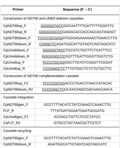
Table 1. Primers used for construction of mutant and complemented
strains.
Primer Sequence (5’ – 3’)
Construction of NDT80 and UME6 deletion cassettes
CpNDT80up_F GGGGGTACCGGCAATTTTGATTTTTGGGTTC CpNDT80up_R GGGGGGCCCGAGGCACCACCAGCAGTAGAGT CpNDT80down_F TCCCCGCGGGATGGGAGAAAAAACTGAACCTTG CpNDT80down_R CGAGCTCAGATGGCATTGTAGTCAGTAGCATC CpUme6down_F GGGGGTACCTGCGTCTGCTTCTCATTTCC CpUme6down_R GGGGGGCCCGGTTTGATTGGGTTGGTCTG CpUme6up_F TCCCCGCGGGGCTTGTGTCAGGTTCGGAT CpUme6up_R CCCGAGCTCTTTGTGGCTCTCTGTGCTTC
Construction of NDT80 complementation cassette
CpNDT80up_F2 TCCCCGCGGACCCTGACCTAACCATACAC CpNDT80down_R2 CCCGAGCTCGCAACAAGCGACAAGCAACA Cassette integration CpNDT80gen_F GCCTTTTACATCTATCGAAGTCAAACTTG FLP_R TTTATGATGGAATGAATGGGATG CpUme6gen_F1 ACGACCTATTCTCCCTATCC CaFLP_R2 GTGCCTACTAACGCTTGTCT Cassette recycling CpNDT80gen_F GCCTTTTACATCTATCGAAGTCAAACTTG CpNDT80down_R AGATGGCATTGTAGTCAGTAGCATC
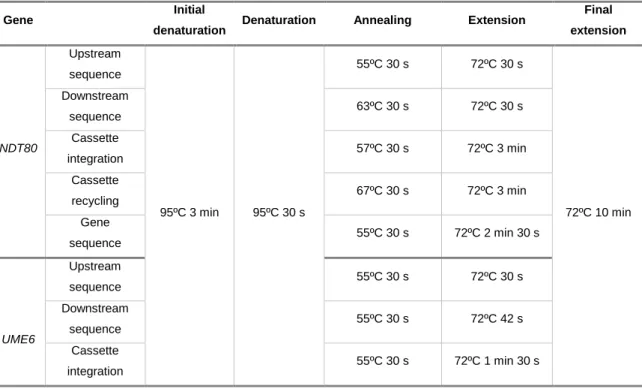
Table 2. PCR programmes used for construction of mutant and complemented strains.
Gene Initial
denaturation Denaturation Annealing Extension
Final extension NDT80 Upstream sequence 95ºC 3 min 95ºC 30 s 55ºC 30 s 72ºC 30 s 72ºC 10 min Downstream sequence 63ºC 30 s 72ºC 30 s Cassette integration 57ºC 30 s 72ºC 3 min Cassette recycling 67ºC 30 s 72ºC 3 min Gene sequence 55ºC 30 s 72ºC 2 min 30 s UME6 Upstream sequence 55ºC 30 s 72ºC 30 s Downstream sequence 55ºC 30 s 72ºC 42 s Cassette integration 55ºC 30 s 72ºC 1 min 30 s
PCR products were visualised by DNA electrophoresis in 1% agarose gel (see DNA electrophoresis section) and purified using a Qiaquick PCR purification kit (Qiagen). Plasmids were sequenced to confirm the integrity of the cloned regions and genes. Gel purification of digested cassette was carried out using the EasySpin DNA gel extraction kit (Citomed). Concentration and quality of DNA were assessed by spectrophotometry (A260/A280) in NanoDrop (Eppendorf).
5. DNA electrophoresis
DNA samples were prepared using 1× Loading buffer (0.25% bromophenol blue, 0.25% xylene cyanol, 30% glycerol). DNA electrophoresis was performed at 70 V (Power Pac Basic, Bio-Rad) in a horizontal electrophoresis system (Mini-Sub Cell GT, Bio-Rad). For DNA visualisation, agarose gel was prepared at 1% Agarose (NZYTech), dissolved in 1× TAE (0.04 M Tris, 1 M glacial acetic acid, 50 mM EDTA pH 8), containing ethidium bromide at final concentration of 0.02 μg ml-1.
For DNA extraction, gel was prepared at 2% UltraPure Grade Agarose (NZYTech), dissolved in 1× TAE. Ethidium bromide at final concentration of 0.02 μg ml -1 was added. DNA was observed through Image Lab software (Bio-Rad), using a Chemi
6. E. coli DH5α transformation
Five μl of ligation reaction were added to 200 μl of competent cells E. coli DH5α (see restriction, dephosphorylation and DNA cloning section). The suspension was incubated on ice for 30 min. A negative control, 200 μl of competent cells without plasmid was prepared in the same conditions. Cells were subjected to a heat shock at 42ºC for 90 s and placed on ice for 2 min. Subsequently, 800 μl of SOC (2% bacto-triptona, 0.5% yeast extract, 10 mM NaCl, 2.5 mM KCl, 2.0 mM glucose, 10 mM MgCl2) was added to
each microtube. Cells were incubated for 1 h at 37ºC with agitation (180 rpm) and approximately 50 μl of supernatant was plated on LB agar with Clo at final concentration of 25 μg ml-1. Plates were incubated for 16 h at 37ºC.
7. C. parapsilosis transformation
Transformation of C. parapsilosis strains was performed by electroporation as described by Ding and Butler (2007). Briefly, an overnight cell culture was diluted in 50 ml of YPD medium to an initial OD600nm of 0.2 and incubated at 30°C until reaching
approximately OD600nm of 2.0. Afterwards, cells were harvested at 5,000 g for 5 min at
room temperature and washed with 50 ml of sterile distilled water. The pellet was resuspended in 10 ml of Tris-EDTA buffer (pH 7.5) containing 10 mM DTT, and incubated at 30°C for 1 h with agitation (100 rpm). Yeast cells were then washed twice, first with 40 ml of cold water and second with 10 ml 1 M sorbitol. Centrifugations occurred at 5,000 g for 5 min, 4ºC. Subsequently, the pellet was resuspended in 125 μl of 1 M sorbitol. Approximately 1 μg of purified KpnI-SacI fragment, either disruption or complementation cassettes, was added to 50 μl of competent cells. The suspension was transferred to a 1 mm electroporation cuvette. The reaction was carried out at 1.25 kV, 25 µF of capacitance and 200 Ω of resistance using a Gene Pulser X-cell Electroporater (Bio-Rad). Afterwards, 950 μl of YPD containing 1 M sorbitol was immediately added and the mixture was incubated at 30°C for 4 h, with agitation. Cells were then collected and 100 μl were platted onto YPD plates supplemented with Nou at final concentration of 200 μg ml-1. Transformants were obtained after 48 h of incubation at 30°C.
8. Isolation of genomic DNA
C. parapsilosis genomic DNA was isolated from 10 ml of an overnight yeast culture, grown in YPD at 30°C, with agitation (180 rpm). Cells were initially harvested at 20,000 g for 5 min. The pellet was resuspended in 200 μl of lysis buffer (2% Triton X100,
1% sodium dodecyl sulfate (SDS), 100 mM Tris-HCl, pH 8.0, 1 mM EDTA), and 200 μl of phenol-choroform-isoamyl alcohol (25:24:1; Sigma-Aldrich). Glass beads were added (0.3 g). The mixture was lysed by vortexing (30 s) and incubation on ice (30 s) for a total period of 10 min. Thereafter, 200 μl of TE (1 mM EDTA, 10 Mm Tris-HCl, pH 8.0) was added to the lysate and the mixture was centrifuged at 20,000 g for 10 min, 4ºC. The aqueous phase was transferred to a new vial and 200 µl of phenol-choroform-isoamyl alcohol was added. Centrifugation occurred at 20,000 g for 5 min at 4ºC. This step was repeated twice. DNA precipitation occurred at -80ºC in 1 ml of absolute ethanol for 1 h. Samples were centrifuged at 20,000 g for 20 min, at 4ºC, washed with 500 µl of 70% ethanol and air-dried for approximately 1 h. Finally, DNA was resuspended in 50 μl of TE and treated with 0.001 mg l-1 RNase A (NZYTech) for 1 h at 37ºC.
9. SADH gene amplification and restriction
Mutant strains identity was confirmed through amplification and restriction of SADH gene, as described by Tavanti et al. (2005). The gene was amplified using primers
SADH_F (5’-GTTGATGCTGTTGGATTGT-3’) and SADH_R
(5’-CAATGCCAAATCTCCCAA-3’). PCR reactions were prepared as follows: 12.5 µl NZYTaq 2× Green Master Mix (NZYTech), 0.5 µM of forward and reverse primers and 0.1-0.5 µg of genomic DNA. PCR programme comprised a denaturation step at 95ºC for 2 min, followed by 30 cycles of 95ºC for 30 s, 50ºC for 30 s and 72ºC for 30 s, with a final extension step at 72ºC for 10 min. Approximately 10 µl of PCR products were digested with 10 U of FastDigest BanI enzyme (Fermentas) in 1× FastDigest buffer, for 30 min. PCR amplification fragments, as well as, digested products were visualised by DNA electrophoresis in 1% agarose gel (see DNA electrophoresis section).
10. Adhesion assay
The yeast adhesion ability was quantified by flow cytometry as described by Silva-Dias et al. (2012). Briefly, strains were grown overnight at 30°C in 20 ml of Sabouraud broth medium with agitation (180 rpm). Yeast culture was centrifuged at 10,000 g for 5 min, and washed twice with Phosphate Buffer Saline (PBS) (Sigma-Aldrich). After, cells were resupended in the same buffer and a cell concentration of 2×10⁶ cells ml-1 was obtained using a Densimat (bioMérieux) and McFarland standards.
About 2×10⁸ of 1 µm uncoated carboxylated highly green fluorescent polystyrene
microspheres (Molecular Probes) were added to the cell suspension. Suspensions were incubated at room temperature for 30 min, at 150 rpm. Samples were vortexed and
50,000 events were analysed using a FACS Calibur flow cytometer (BD Biosciences). Cell adhesion results are expressed as the percentage of cells with microspheres attached of at least three independent experiments, performed in triplicate.
11. Biofilm assay
After overnight growth at 37°C with agitation (180 rpm) in 30 ml of Sabouraud broth medium, cells were collected by centrifugation at 10,000 g for 5 min, washed with PBS and standardized to obtain a suspension of 1×10⁶ yeast cells ml-1 in RPMI-1640
medium, supplemented with L-glutamine and buffered with MOPS acid (Sigma-Aldrich). Posteriorly, 1 ml of each cell suspensions were placed into a 12-well polystyrene microplate and incubated for 24 and 48 h at 37°C. Following incubation, biofilms total biomass was quantified by Crystal Violet (CV) assay, previously described by Silva-Dias et al. (2015). Briefly, 1 ml of 99% methanol was added to each well and incubated at room temperature for 15 min. Then, methanol was removed and biofilms were air-dried at room temperature. Biofilms were stained with 1 ml of 0.02% of CV for 20 min. Each well was washed twice with distilled water and 1.5 ml of 33% acetic acid was added. The total biomass was measured spectrophotometrically at 590 nm using a UV-visible recording spectrophotometer (Shimadzu). Results are representative of at least three independent experiments, performed in triplicate.
12. Microscopy
Colony phenotypes were observed and photographed at 20× magnification using a Zeiss AxioCam HRc camera coupled to Zeiss dissecting microscope, after grown in YPD agar medium at 30ºC, for 48 h.
Images of cellular morphology were taken with a Zeiss Axioplan microscope coupled with an AxioVision image acquisition system (Zeiss), after stained with Calcofluor White (Sigma-Aldrich) and mounted on glass slides.
13. RNA extraction
RNA was extracted from a mid-log exponential phase growth culture as described by Kohrer and Domdey (1991), with some modifications. A C. parapsilosis culture was grown in 30 ml of YPD medium at 30°C until reaching an OD600 ranging from 0.6 to 0.8.
Cells were pelleted at 2,180 g for 3 min at room temperature and resuspended in acid phenol choloroform (5:1, Sigma-Aldrich) and TES buffer (10 mM Tris-HCl pH 7.5, 10 mM EDTA, 0.5% SDS). Approximately 500 µl of each solution was used per 25 OD units
(volume of culture × OD600nm). The suspension was vigorous vortexed for 20 s (at 20º
angle) to resuspend the pellet, incubated for 1 h at 65°C and vortexed every 10 min (10 s upright, 10 s at angle). After centrifugation (20,000 g, 20 min, 4ºC), two phenol extractions were carried out, first with 500 µl of phenol chloroform (5:1, pH 4.7) and second with 500 µl of phenol-chloroform-isoamyl alcohol (25:24:1, Sigma-Aldrich). Each extract was vigorous vortexed for 20 s and centrifuged for 10 min at 20,000 g and 4ºC. RNA precipitation occurred after adding 0.1 volumes of 3 M sodium acetate (NaAc) (pH 5.2) and 3 volumes of 100% ethanol, and incubation for periods of time longer than 30 min at -20°C. RNA pellet was then collected by centrifugation at 20,000 g for 5 min at room temperature, washed with 70% ethanol and resuspended in milliQ water. Concentration and quality of RNA samples were measured using Nanodrop (Eppendorf). Only samples yielding 28S rRNA/18S rRNA ratios ranging from 1.6 to 2.2 and showing no degradation were used in subsequent analyses. Each RNA extract was aliquoted and stored at -20ºC before analysis.
14. cDNA synthesis and RT-qPCR
First-strand cDNAs were synthesized from 1,000 and 100 ng of total RNA in a 20 µl reaction volume using a Maxima Reverse Transcriptase kit (Thermo Scientific) according to the manufacturer’s instructions. Briefly, about 100 pmol of Oligo(dT)18 and
0.5 mM of dNTPs Mix were added to RNA samples and tubes were incubated at 65ºC for 5 min. Following incubation on ice for 1 min, 4 µl of 5× RT buffer and 200 U of Maxima Reverse Transcriptase were added to the previous mixtures. Tubes were incubated for 30 min at 60ºC and then for 5 min at 85ºC. Subsequently, 5 U and 0.5 U of RNase H (NZYTech) were added to cDNAs of 1,000 and 100 ng RNA tubes, followed by incubation at 37ºC during 1 h and 20 min, respectively.
For gene expression quantifications by RT-qPCR, the reaction efficiency was assessed by a cDNA serial dilution (1:5) standard curve. The cDNA dilutions included ranged from the lowest to the highest expression levels of each target being quantified. Each dilution was assayed in triplicate. Gene-specific primers were design using Oligo Explorer 1.2 software (Gillanders et al., 2004) (Table 1). Each reaction contained 10 µM of each primer, 1× SensiFAST SYBR No-ROX Mix (Bioline), 2 µl of cDNA and milliQ water up to 20 µl. Five replicates per strain were analysed for each target gene. Reactions were performed in a Realplex Mastercycler instrument (Eppendorf), with the following program: first initial denaturation at 95ºC for 2 min, followed by 35 cycles of denaturation at 95ºC for 5 s, annealing at 60ºC for 10 s and extension at 72ºC for 15 s
(measuring point). The housekeeping gene TUB4 was used as the reference gene. Gene expression quantifications were performed with the Relative Expression Software (REST, Qiagen) (Pfaffl et al., 2002), using the standard mode.
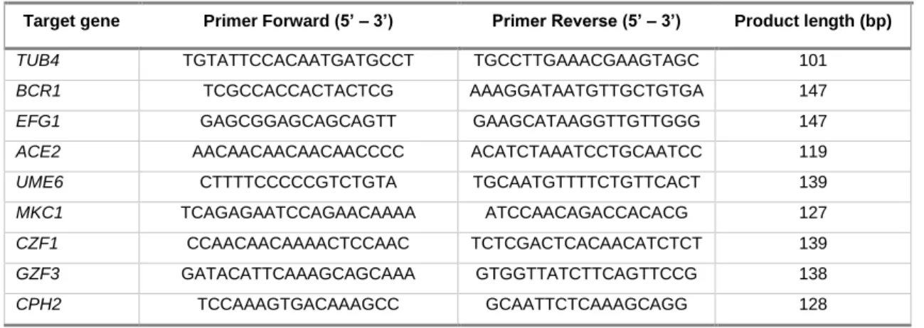
Table 3. Primers used in RT-qPCR and the respective amplified product length.
Target gene Primer Forward (5’ – 3’) Primer Reverse (5’ – 3’) Product length (bp)
TUB4 TGTATTCCACAATGATGCCT TGCCTTGAAACGAAGTAGC 101
BCR1 TCGCCACCACTACTCG AAAGGATAATGTTGCTGTGA 147
EFG1 GAGCGGAGCAGCAGTT GAAGCATAAGGTTGTTGGG 147
ACE2 AACAACAACAACAACCCC ACATCTAAATCCTGCAATCC 119
UME6 CTTTTCCCCCGTCTGTA TGCAATGTTTTCTGTTCACT 139
MKC1 TCAGAGAATCCAGAACAAAA ATCCAACAGACCACACG 127
CZF1 CCAACAACAAAACTCCAAC TCTCGACTCACAACATCTCT 139
GZF3 GATACATTCAAAGCAGCAAA GTGGTTATCTTCAGTTCCG 138
CPH2 TCCAAAGTGACAAAGCC GCAATTCTCAAAGCAGG 128
15. Sequence analysis
Sequences from CPH2, GZF3, CZF1, MCK1, UME6, ACE2, EFG1 and BCR1 of C. parapsilosis CDC317 plus 1,000 base pairs (bp) upstream and downstream were downloaded from the CGD (http://candidagenome.org) (Inglis et al., 2012). To identify putative binding sites in these genes, a search for the C. parapsilosis MSE consensus motif (5’-CACAAAR-3’) was performed in the promoter regions (1,000 bp upstream of the start codon) using NCBI Blast tool.
16. Statistical analysis
The significance of differences was determined by two-tailed Student’s t-test with 95% confidence intervals, calculated with Microsoft Excel 2016. Differences were considered statistically significant if p-value inferior to 0.05.
III. Results
1. Construction of C. parapsilosis mutant and complemented
strains
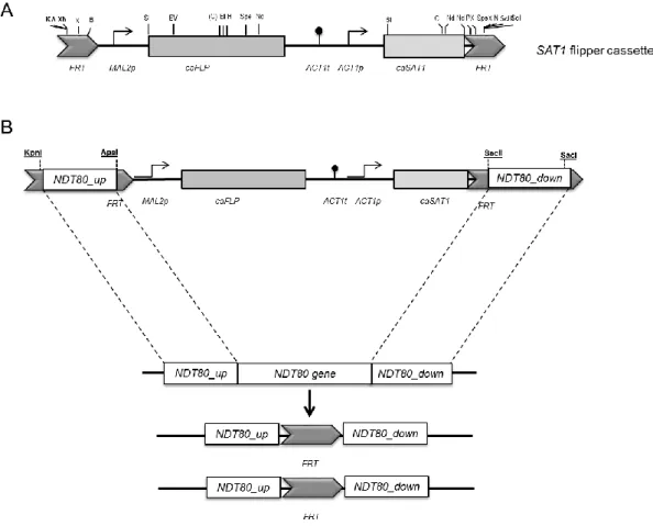
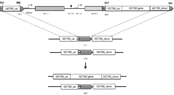
To gain insight into the role of Ndt80 in C. parapsilosis virulence attributes, a homozygous ndt80Δ mutant (EF16 strain) was generated in a clinical isolate background, BC014 strain (Silva et al., 2011a). The deletion was carried out using a gene specific disruption cassette (pNG4) based on the recyclable nourseothricin-resistant marker previously described (Ding and Butler, 2007). Specifically, this method relies on the use of a cassette that contains a C. albicans-adapted Nou resistant gene (CaSAT1) under the control of the C. parapsilosis actin promoter (CpACT1) for the selection of positive transformants (Figure 5A). In addition, the tool also contains the C. albicans-adapted FLP gene (CaFLP), whose expression is driven by a C. parapsilosis maltose inducible promoter (CpMAL2) (Figure 5A). This gene codifies for a Flp recombinase enzyme that recognizes FRT (Flippase Recognition Target) regions flanking the cassette, being responsible for its excision from the genome (Ding and Butler, 2007).
Integration into the target locus is specific and occurs by homologous recombination between the flanking sequences of the cassette (Ding and Butler, 2007; Reuß et al., 2004) (Figure 5B). Once C. parapsilosis is a diploid organism, it is necessary two separate rounds of transformation and excision to generate a mutant strain lacking both copies of CpNDT80 gene (Figure 5B). Homozygous knock-out mutants of C. parapsilosis wild type strain differ from their parental strain only by the absence of a functional target gene.