Protein in
Caulobacter crescentus
Carmen Fernandez-Fernandez, Diego Gonzalez, Justine Collier*
Department of Fundamental Microbiology, Faculty of Biology and Medicine, University of Lausanne, Lausanne, Switzerland
Abstract
DnaA is a conserved essential bacterial protein that acts as the initiator of chromosomal replication as well as a master transcriptional regulator inCaulobacter crescentus. Thus, the intracellular levels of active DnaA need to be tightly regulated during the cell cycle. Our previous work suggested that DnaA may be regulated at the level of its activity by the replisome-associated protein HdaA. Here, we describe the construction of a mutant DnaA protein [DnaA(R357A)]. The R357 residue in the AAA+domain of theC. crescentusDnaA protein is equivalent to the R334 residue of theE. coliDnaA protein, which is required for the Regulatory Inactivation of DnaA (RIDA). We found that the expression of the DnaA(R357A) mutant protein inC. crescentus, but not the expression of the wild-type DnaA protein at similar levels, causes a severe phenotype of over-initiation of chromosomal replication and that it blocks cell division. Thus, the mutant DnaA(R357A) protein is hyper-active to promote the initiation of DNA replication, compared to the wild-type DnaA protein. DnaA(R357A) could not replace DnaA in vivo, indicating that the switch in DnaA activity once chromosomal replication has started may be an essential process in C. crescentus.We propose that the inactivation of DnaA is the main mechanism ensuring that chromosomal replication starts only once per cell cycle. We further observed that the R357A substitution in DnaA does not promote the activity of DnaA as a direct transcriptional activator of four important genes, encoding HdaA, the GcrA master cell cycle regulator, the FtsZ cell division protein and the MipZ spatial regulator of cell division. Thus, the AAA+ domain of DnaA may play a role in temporally regulating the bifunctionality of DnaA by reallocating DnaA molecules from initiating DNA replication to transcribing genes within the unique DnaA regulon ofC. crescentus.
Citation:Fernandez-Fernandez C, Gonzalez D, Collier J (2011) Regulation of the Activity of the Dual-Function DnaA Protein inCaulobacter crescentus. PLoS ONE 6(10): e26028. doi:10.1371/journal.pone.0026028
Editor:Martin G. Marinus, University of Massachusetts Medical School, United States of America
ReceivedJuly 20, 2011;AcceptedSeptember 15, 2011;PublishedOctober 14, 2011
Copyright:ß2011 Fernandez-Fernandez et al. This is an open-access article distributed under the terms of the Creative Commons Attribution License, which permits unrestricted use, distribution, and reproduction in any medium, provided the original author and source are credited.
Funding:This work was supported by the Swiss National Science Foundation (SNSF) fellowship 31003A_122541 and the SNSF R’Equip grant 316030_121391 (SNSF: http://www.snf.ch). The funders had no role in study design, data collection and analysis, decision to publish, or preparation of the manuscript.
Competing Interests:The authors have declared that no competing interests exist.
* E-mail: [email protected]
Introduction
Faithful chromosomal replication requires regulatory networks that ensure the precise coordination of DNA replication with other cell cycle events. The prokaryotic DnaA protein or the eukaryotic origin replication complex (ORC) control the onset of DNA replication. DnaA and several ORC proteins are ATP-binding proteins that contain AAA+ (ATPase associated with diverse cellular activities) domains of very similar structures, suggesting mechanistic conservation throughout evolution [1,2]. Binding of ATP to these proteins is thought to induce conformational changes that activate these proteins to promote the initiation of DNA replication at the correct time of the cell cycle. Besides its function as an initiator of bacterial chromosome replication, DnaA is also a transcription factor that binds to many promoters to regulate their activities in diverse bacterial species [3,4]. It is however still unclear whether oscillations in the levels of DnaA-ATP influence the timing of gene transcription in most cases [5,6].
In theEscherichia coliprokaryotic model, DnaA proteins bind to DnaA boxes located in the origin of replication, but only the ATP-bound form (DnaA-ATP) can initiate replication [1]. Soon after the initiation of DNA replication, the ATPase activity of the AAA+
domain of DnaA is stimulated by the essential Hda protein, resulting in the hydrolysis of the ATP bound to DnaA and thus in the regulatory inactivation of DnaA (RIDA) [1,7,8]. Hda
supposedly needs to interact with the b-clamp of the DNA polymerase III loaded onto DNA to promote the inactivation of DnaA [9]. Although there exists several other mechanisms that regulate the timing of the initiation of DNA replication inE. coli [1], the RIDA system seems to be one of the most important [10,11]. Consistent with this model, it was shown that ATPase defective mutants of DnaA, andhda-deficient cells, display over-initiation phenotypes inE. coli [7,12,13]. The use of replisome-associated proteins that act as negative regulators of initiation appears quite conserved in bacteria, as indicated by the discoveries of the YabA protein inBacillus subtilis[14,15] or the HdaA protein inCaulobacter crescentus[16].
swarmer and pre-divisional cells [17,18]. A gradient of active CtrA establishes the asymmetry of the initiation of DNA replication between the two future daughter cells before cell division [22]. Active CtrA is however undetectable for about one third of the cell cycle during the stalked cell stage [23]. During the time when no CtrA is detectable, other factors restrict the initiation of chromosomal replication to only once per cell cycle. We previously identified HdaA as one of these factors, showing that the HdaA protein is necessary to ensure that chromosomal replication does not restart a second time during the same cell cycle [16], indicating that HdaA may stimulate the inactivation of DnaA once DNA replication has initiated, by a mechanism similar to the RIDA system inE. coli. To further address this possibility, we describe here the creation of a mutant DnaA protein inC. crescentus, with an R357A substitution in its AAA+ domain. We found that the expression of this protein caused severe over-initiation of chromosomal replication and blocked cell division inC. crescentus, unlike the expression of the wild-type DnaA, indicating that DnaA(R357A) is hyper-active as an initiator of DNA replication. We also showed that DnaA(R357A) cannot substitute for DnaA, indicating that the inactivation of DnaA by its AAA+domain may be an essential process inC. crescentus, to restrict the initiation of DNA replication to only once per cell cycle. We also showed that, surprisingly, the R357A substitution in DnaA does not promote the activity of DnaA as a direct transcriptional activator of fourC. crescentus genes, encoding HdaA, the GcrA master cell cycle regulator, and the FtsZ cell division protein and the MipZ spatial regulator of cell division, that are all required for normal cell cycle progression.
Results
Mutagenesis of a conserved arginine residue in the AAA+
domain of theC. crescentus DnaA protein
Assuming that ATP-DnaA is the active form that initiates chromosomal replication in a bacterial species, DnaA mutants that can bind ATP but that are defective in hydrolysis of the bound ATP may cause over-initiation because the ATP-bound form of DnaA accumulates. InE. coli, such a mutant DnaA protein was constructed by a R334A amino-acid substitution [12]. The highly conserved R334 residue lays in the Box VIII sensor 2 motif of the AAA+ domain of DnaA (Fig.1). In vitro and in vivoexperiments demonstrated that this mutant protein can not be inactivated by the RIDA mechanism in E. coli, and thus that its expression at
moderate levels results in over-initiation of chromosomal replica-tion, unlike the wild-type DnaA protein [12].
Our previous discovery of the HdaA protein [16], which is homologous to theE. coliHda protein, suggested that RIDA may also occur inC. crescentus, and that this mechanism may restrict the initiation of chromosomal replication to only once per cell cycle in this particular bacterial species. To examine this possibility, we generated a mutant DnaA protein, named DnaA(R357A). The R357A substitution in the AAA+ motif of theC. crescentus DnaA protein is equivalent to the previously characterized R334A mutation in theE. coliDnaA protein (Fig.1). When expressed from the xylose-inducible xylX promoter on a medium copy number plasmid for four hours in rich medium containing xylose, we observed by immunoblot analysis that the levels of DnaA or DnaA(R357A) were approximately four-fold higher than the natural levels of DnaA expressed from its native locus in the wild-type strain (Fig.2). Hence, DnaA(R357A) was expressed at moderate levels that were comparable to the levels of the wild-type DnaA protein expressed from the same vector. We used these conditions to investigate and compare the effects of the expression of DnaA or DnaA(R357A) on cell morphology and chromosome content, and on the expression of four important genes directly activated by DnaA.
Moderate expression of DnaA(R357A), but not DnaA, blocks cell division
When DnaA was expressed upon xylose induction for four hours or longer time periods from a medium-copy number plasmid in the JC366 strain, we observed by phase contrast microscopy that cells had a quite normal morphology (Fig.3). This observation shows that a four-fold over-expression of DnaA is not preventing cell division. We however still noticed that JC366 cells tended to divide at lower cell mass, giving slightly smaller daughter cells than JC919 cells containing the empty vector.
Next, we analyzed the phenotypes associated with the over-expression of the mutant DnaA(R357A) protein. Phase contrast microscopy four hours after the addition of the xylose inducer showed that JC367 cells expressing DnaA(R357A) were signifi-cantly longer than cells over-expressing DnaA and than cells containing the empty control vector (Fig.3). When cells were grown in xylose-containing medium for longer time periods, many cells became very filamentous (Fig.3), indicating a severe reduction in the efficiency of cell division upon expression of DnaA(R357A).
Figure 1. Amino-acid sequence homology between the C. crescentus and the E. coli DnaA proteins. The upper schematic shows the four typical domains of DnaA proteins [1]. The AAA+domain
contains three conserved Walker motifs indicated as WA, WB, and WC. The bottom schematic shows the homology between the box VIII motif in the AAA+domains of theC. crescentusandE. coliDnaA proteins. Amino acid positions are indicated with numbers. R357 from the C. crescentusDnaA protein and R334 from theE. coliDnaA protein are indicated by a black arrow.E. c.indicatesE. coli, whileC. c.indicatesC. crescentus.
doi:10.1371/journal.pone.0026028.g001
Figure 2. DnaA and DnaA(R357A) are expressed at moderate levels.Strains NA1000 (WT), JC366 (containing pJSX-DnaA) and JC367 [containing pJSX-DnaA(R357A)] were grown to exponential phase in PYE medium plus 0.2% glucose (PYEG) and 0.3% xylose was added (PYEGX) to half of the culture for four hours before cells were collected for immunoblot analysis using antibodies raised against DnaA, HdaA and GcrA.
Expression of DnaA(R357A) leads to over-initiation of chromosomal replication
We previously showed that cells slightly over-expressing DnaA from an integrated second copy of thednaAgene under the control of thexylX promoter do not over-initiate DNA replication [16]. We confirmed this result when DnaA was expressed at higher levels from the same promoter on our medium-copy number plasmid (Fig.2). We performed flow-cytometry experiments using a fluorescent dye that labels the DNA, to observe the number of chromosomes in single cells after a rifampicin treatment. We found that only (8.7+/-2.6)% of the cells contained more than two chromosomes, which is just three-fold higher than the control strain containing the empty vector [(2.9+/-0.4)%] (Fig.4A&B). Cells not treated with rifampicin prior to the flow cytometry analysis also had a profile quite comparable to that observed for the control strain (Fig.S1). Measurements of the ratio ofCoritoter (the replication terminus region) by quantitative-polymerase chain reaction (Q-PCR) from genomic DNA extracted from a population of these cells also confirmed that these cells do not over-initiate chromosomal replication (Fig.4C). Collectively, our data demonstrates that an over-expression of DnaA from a medium-copy number plasmid is not significantly affecting the precise once-per-cell-cycle control of the initiation of chromosome replication inC. crescentus. We nevertheless noticed that cells over-expressing DnaA more often accumulated two complete copies of the chromosome after rifampicin treatment, compared to the
control cells (Fig.4A&B). This observation may indicate that chromosomal replication starts a little earlier in cells over-expressing DnaA than in control cells.
To determine whether the over-expression of DnaA(R357A) affects the initiation of chromosomal replication, we also performed flow cytometry experiments with JC367 cells. In accordance with our predictions, we observed that (69.4+/ -2.9)% of the cells expressing DnaA(R357A) for four hours contained more than two chromosomes after rifampicin treatment (Fig. 4A&B). In fact, many cells even contained numerous incomplete chromosomes per cells after rifampicin treatment. When we performed flow cytometry experiments without a rifampicin treatment, we observed a profile drastically shifted towards a higher amount of DNA content per cell, compared to what we observed with the control strain or with the strain that over-expresses the wild-type DnaA protein at similar levels (Fig.S1). To confirm that these cells over-initiated DNA replica-tion, we also measured theCori/terratio by Q-PCR from genomic DNA extracted from these cells. Consistent with our single cell flow cytometry results, we observed that theCori/terratio in cells expressing DnaA(R357A) is about two fold higher than that of cells expressing DnaA as similar levels (Fig.4C). All together, these results indicate that DnaA(R357A) can initiate new rounds of chromosomal replication, but in an uncontrolled manner, suggesting that DnaA(R357A) is hyper-active for its function as an initiator of chromosomal replication. It is very likely that
Figure 3. A moderate overproduction of DnaA(R357A), but not DnaA, blocks cell division.Representative phase contrast images from strains JC919 containing the pJS14 empty vector (Panel A), JC366 containing pJSX-DnaA (Panel B) and JC367 containing pJSX-DnaA(R357A) (Panel C) grown to exponential phase in PYE medium plus 0.2% glucose (PYEG) before 0.3% xylose was added (PYEGX) to half of the culture for four hours. Cells maintained in PYEGX for twenty hours are also shown in the bottom panel. All images are at the same scale.
DnaA(R357A) remains bound to ATP, and thus active for replication initiation, at all times of the cell cycle ofC. crescentus, as it is the case for the DnaA(R334A) protein inE coli.
DnaA(R357A) cannot replace DnaA(WT)in vivo
We previously demonstrated that the HdaA protein is essential for normal cell cycle progression inC. crescentusand that aDhdaA strain can not be constructed in the absence of an extra copy of the hdaAgene expressedin trans[16]. These results suggested that the switch of DnaA activity promoted by HdaA after chromosome
replication has initiated could be essential for the viability ofC. crescentus. If this prediction is correct, it should be very difficult, or impossible, to construct a strain carrying thednaA(R357A)allele as the sole copy of dnaA on the C. crescentus chromosome. We attempted to construct such a strain by two different and complementary approaches.
First, we tried to construct a [DdnaA::V PxylX::dnaA(R357A)] strain by generalized transduction using bacteriophageWCR30. Two phage lysates were prepared from strain GM2471 containing theDdnaA::Vmutation and strain JC125 containing aDCC1613::V
Figure 4. A moderate overproduction of DnaA(R357A), but not DnaA, leads to severe over-initiation of chromosomal replication. (A) Cells overproducing DnaA(R357A) accumulate extra chromosomal copies after rifampicin treatment. Representative profiles obtained by flow cytometry analyses of cells from strains JC919 (containing the pJS14 empty vector), JC366 (containing DnaA) and JC367 [containing pJSX-DnaA(R357A)] grown to exponential phase in PYE medium plus 0.2% glucose (PYEG) before 0.3% xylose was added (PYEGX) to half of the culture for four hours. Cells were treated with rifampicin for three hours prior to fixing and staining with Vybrant DyeCycle orange. The horizontal axis indicates the number N of complete chromosomes: 1N, 2N or more than 2N (+). The vertical axis indicates the number of cells. (B) Quantification of the results of flow cytometry experiments as shown in A. The percentage represents the average proportion of cells containing the indicated number of chromosomes per cell. Results are the averages of data from three independent experiments. The standard deviations are also indicated (SD). (C) The graph shows the relativeCori/terratio obtained from Q-PCR on chromosomal DNA extracted from strains JC919 (containing the pJS14 empty vector), JC366 (containing pJSX-DnaA) and JC367 [containing pJSX-DnaA(R357A)]. Cells were grown to exponential phase in PYE medium plus 0.2% glucose before 0.3% xylose was added for four hours. Values were normalized to a control strain, which corresponds to the wild-type strain (NA1000) grown in PYE medium and treated with rifampicin for three hours. Results are the average of data from four independent experiments and standard deviations were always lower than 0.5%.
mutation. The JC125 strain has no apparent phenotype and grows like the wild-type strain: it was used as a control for transduction efficiency. TheVcassette confers resistance to spectinomycin and streptomycin. Both phage lysates were used for transduction assays using the NA1000 wild-type strain and the JC323 and JC324 strains containing a copy of the dnaAor thednaA(R357A) gene, respectively, under the control of the xylX promoter at thexylX locus. Upon selection on spectinomycin and streptomycin containing plates, we isolated numerous DCC1613::V transduc-tants in the three different genetic backgrounds, regardless of the presence of the xylose inducer (Table 1), demonstrating that transduction efficiency is not significantly affected by the expression of dnaAor dnaA(R357A). We also isolated numerous DdnaA::Vmutant colonies from our transduction assay into JC323, but only on xylose-containing plates (Table 1). This result demonstrated that the wild-type allele of dnaA expressed from the xylX promoter is sufficient to complement the unviable DdnaA::V mutation. In contrast, no DdnaA::V mutant colonies were isolated from the transduction assays into the wild-type strain (Table 1), confirming that dnaA is essential for viability in C. crescentus [24]. Similarly, we were not able to isolate DdnaA::V mutant colonies from the transduction assay into the JC324 strain expressing DnaA(R357A) in the presence of the xylose inducer on PYE plates (Table 1). This result suggested that DnaA(R357A) can not replace DnaAin vivo.
Second, we tried to exchange the native dnaA allele for the dnaA(R357A) allele by double recombination using the suicide plasmid pNPTS138-DnaA(R357A) carrying the dnaA(R357A) allele. We systematically recovered the dnaA allele on the chromosome after re-excision of the plasmid (data not shown), indicating again that thednaA(R357A)allele is probably not viable as the sole copy ofdnaAon theC. crescentuschromosome.
Given that the DnaA(R357A) protein is probably functional to initiate chromosomal replication as indicated by our previous findings (Fig.4), these last genetic evidences suggest that the switch in DnaA activity mediated by its AAA+ domain is an essential process inC. crescentusand may also explain why the HdaA protein is essential for normal cell cycle progression inC. crescentus.
DnaA(R357A) is not more active than DnaA to promote the transcription of four importantC. crescentusgenes
DnaA is not only the initiator of chromosomal replication in nearly all bacteria, but it also acts as an important transcriptional regulator by directly binding to promoters in multiple bacterial species. In B. subtilis, for example, it was estimated that DnaA directly regulates the transcription of about 50 different genes [4], while DnaA directly regulates the transcription of minimum 13 genes in C. crescentus [3,16,25]. One of the still outstanding questions regarding DnaA activity as a transcription factor is
whether the nucleotide bound to DnaA often influences its binding and activity at DnaA-regulated promoters in diverse bacterial species [2].
We investigated whether the DnaA(R357A) protein may also be hyper-active to promote transcription from four well-characterized DnaA-activated promoters inC. crescentus. These are: (i) thehdaA promoter, controlling the expression of the HdaA repressor of the initiation of chromosomal replication [16]; (ii) thegcrApromoter, controlling the expression of the GcrA master regulator of theC. crescentuscell cycle [3,25,26]; (iii) theftsZpromoter, controlling the expression of the FtsZ cell division protein [3,27,28]; (iv) themipZ promoter, controlling the expression of the MipZ spatial regulator of cell division [3,29]. We chose these four promoters because they have a quite different structure with respect to the position and the number of DnaA boxes that they contain. Indeed, the gcrAand mipZ promoters contain only one DnaA box, the ftsZpromoter contains two DnaA boxes, and thehdaApromoter contains up to six DnaA boxes [16]. In addition, the four genes that we selected are essential or required for normal cell cycle progression inC. crescentus.
We first compared by immunoblot analysis the amounts of GcrA and HdaA that accumulated in cell extracts from the wild-type strain and from the strain that over-expresses DnaA. We found that GcrA and HdaA accumulated at higher levels in DnaA over-expressing cells than in wild-type cells, although the effect was more pronounced for HdaA than for GcrA (Fig.2). HdaA accumulated at similar levels in cells expressing DnaA(R357A) compared to cells expressing DnaA (Fig.2). In contrast, GcrA accumulated at lower levels in cells constitutively expressing DnaA(R357A) compared to cells expressing DnaA (Fig.2). Altogether, these observations suggested that DnaA(R357A) is not more active than DnaA to promote the transcription of at least certain genes that belong to the direct DnaA regulon.
To confirm that the activity of DnaA as a transcriptional regulator of our four selected genes is not increased by the R357A mutation, we introduced a low copy number plasmid carrying a transcriptional fusion between the hdaA promoter, the gcrA promoter, theftsZpromoter or the mipZ promoter and thelacZ gene [16,25,26], into the strains that over-express DnaA or DnaA(R357A). We measured b-galactosidase activities as an indication of promoter activities (Fig.5). Four hours after xylose addition into the medium, we observed that b-galactosidase activities from each promoter that we tested were higher in cells over-expressing DnaA than in wild-type cells. Consistent with previous data [16,25], these results confirm that DnaA activates the transcription of all four genes. Next, we compared the effect of the over-expression of DnaA or DnaA(R357A) on the activities of these four promoters. Surprisingly, we observed that the activation of the transcription from these four promoters was never higher
Table 1.DnaA(R357A) cannot complement for the loss of DnaA.
DdnaA::Vtransductants DCC1613::Vtransductants
PYEG PYEX PYEG PYEX
NA1000 - - + +
NA1000 PxylX::dnaA - + + +
NA1000 PxylX::dnaA(R357A) - - + +
We used bacteriophageWCR30 to transduce theDdnaA::Vor theDCC1613::Valleles into strains NA1000, JC323 and JC324 on PYE medium plus 0.2% glucose (PXEG) or on PYE medium plus 0.3% xylose (PYEX). The+symbol indicates that minimum 400 transductant colonies were isolated. The – symbol indicates that no transductant colonies were isolated.
when DnaA(R357A) rather than DnaA was over-expressed (Fig.5). These interesting results indicate that DnaA(R357A) is not more efficient than DnaA to activate the transcription of these four important genes. Thus, the R357A mutation in DnaA uncouples the ability of DnaA to initiate DNA replication from its activity as a transcription factor regulating minimum four genes, only leading to increased activity in the initiation of DNA replication. In addition, we observed that DnaA(R357A) is even less efficient than DnaA to activate the transcription of gcrA, ftsZ and mipZ (Fig.5B&C&D), suggesting that the switch in the activity of DnaA that takes place at the time when DNA replication is initiated, may promote the expression of these genes.
Discussion
In this study, we showed that the DnaA(R357A) mutant protein in C. crescentus retains its ability to promote the initiation of chromosomal replication in vivo, and is even hyper-active as an initiator compared to the wild-type DnaA protein, as indicated by the severe over-replication phenotype of cells that over-express DnaA(R357A) (Fig.4). In addition, we showed that the DnaA(R357A) protein cannot replace DnaA (Table 1), suggesting that the inactivation of the initiator DnaA is an essential process in C. crescentus. In contrast, we observed that the activity of DnaA(R357A) as a transcription factor that stimulates the transcription of four genes is not higher than that of DnaA (Fig.5), indicating that the AAA+ domain of DnaA may not inactivate DnaA as a transcriptional regulator of these genes inC. crescentus. Below, we discuss the role of the AAA+domain of DnaA in the regulation of both activities of DnaA in the control of theC. crescentuscell cycle.
Model for a RIDA system inC. crescentus
The R357A substitution in the AAA+motif of theC. crescentus DnaA protein is equivalent to the previously characterized R334A mutation in theE. coliDnaA protein that inhibits RIDA and the intrinsic ATPase activity of DnaAin vivoandin vitro[12,30]. It is thus likely that the R357 residue in the AAA+ domain of theC. crescentusDnaA protein participates in the hydrolysis of an ATP bound to DnaA, to inactivate DnaA immediately following the initiation of chromosome replication (Fig.6). Consistent with this model, theC. crescentusDnaA(R357A) protein would be bound to ATP at all times of the cell cycle, as it is the case for theE. coli DnaA(R334A) protein. Then, DnaA-ATP would initiate chromo-some replication whenever and wherever active CtrA is absent, leading to C. crescentus cells that have undergone additional chromosome replication initiations like we observed (Fig.4). The
Figure 5. Effect of DnaA and DnaA(R357A) on the transcription of four DnaA-regulated genes. (A) Effect on the transcription of hdaA, encoding the inhibitor of DnaA. The graph shows the relativeb-galactosidase activities from placZ290-hdaAP(WT) in strains
JC964 (containing pJS14), JC973 (containing pJSX-DnaA) and JC974 [containing pJSX-DnaA(R357A)].(B) Effect on the transcription of gcrA, encoding a master transcriptional regulator. The graph shows the relative b-galactosidase activities from placZ290-gcrAP in strains JC971 (containing pJS14), JC972 (containing pJSX-DnaA) and JC975 [containing pJSX-DnaA(R357A)].(C) Effect on the transcrip-tion offtsZ,encoding the cell division protein FtsZ.The graph shows the relative b-galactosidase activities from placZ290-ftsZP in strains JC1006 (containing pJS14), JC1007 (containing pJSX-DnaA) and JC1008 [containing pJSX-DnaA(R357A)].(D) Effect on the transcrip-tion ofmipZ,encoding a spatial regulator of FtsZ assembly.The graph shows the relativeb-galactosidase activities fromplacZ290-ftsZP in strains JC1037 (containing pJS14), JC1038 (containing pJSX-DnaA) and JC1039 [containing pJSX-DnaA(R357A)]. In each case, cells were grown to exponential phase in PYE medium plus 0.2% glucose, before 0.3% xylose was added for 4 hours. Error bars indicate the standard deviations.
C. crescentusHdaA protein may be a functional homolog of theE. coli Hda protein, stimulating the ATPase activity of the AAA+
domain of DnaA when bound to the replisome. In agreement with this model, we observed that the phenotype of HdaA-depleted cells [16] resembles that of DnaA(R357A) over-expressing cells (Fig. 3 and 4), and that the HdaA protein dynamically co-localizes with the replisome [16]. Our results suggest that the inactivation of the initiator DnaA by the hydrolysis of the ATP bound to DnaA by its AAA+domain is an essential process inC. crescentus, as we were not able to replace the wild-type dnaA allele by the mutant
dnaA(R357A)allele on theC. crescentuschromosome (Table 1 and data not shown). This could then explain why the HdaA protein is also essential for normal cell cycle progression inC. crescentus[16]. When DnaA(R357A) was expressed together with DnaA in strains such as JC367 or JC324, DnaA was probably competing with DnaA(R357A) when binding the Cori prior to the initiation of chromosomal replication, thereby maintaining cells alive by the inactivation of the wild-type subset of the multiple DnaA molecules bound to theCoriafter the initiation of chromosomal replication.
The intracellular levels of DnaA fluctuate during theC. crescentus cell cycle, being the most abundant at the time when chromosomal replication is initiated during the swarmer-to-stalked cell transition [25,31]. Both the transcription of thednaAgene and the proteolysis of the DnaA protein are temporally regulated, to ensure that DnaA accumulates the most at the right time of the cell cycle [31,32,33,34]. Our data suggest that the proteolysis of DnaA in stalked cells is not sufficient to inhibit the initiation of chromosomal replication after the first round of replication has started, and that the activity of DnaA also needs to be down-regulated at that moment of the cell cycle for normal cell cycle progression. Consistent with this proposal, we found that the constitutive expression of a DnaA(R357A) mutant protein resulted in severe over-initiations of chromosomal replication (Fig.4), while the constitutive expression of DnaA did not. Jonaset al.recently reported that the over-production of an M2-tagged DnaA protein resulted in over-initiation of chromosomal replication in C. crescentus[35], unlike what we observed when we over-produced the native DnaA protein. This difference may be attributable to the artificial addition of the M2 epitope to the DnaA protein by Jonas et al., or to differences in growth conditions that may promote DnaA accumulation at higher levels.
CtrA and the RIDA system have complementary
functions in the control of the initiation of chromosomal replication inC. crescentus
The CtrA master regulator is conserved in alpha-proteobac-teria but is not found in entero-bacalpha-proteobac-teria likeE. coli. InC. crescentus, it acts as an inhibitor of the initiation of chromosomal replication that is present and active in swarmer cells and in the swarmer compartment of pre-divisional cells (Fig.6B). Thus, CtrA essentially inhibits replication by binding to the origin of non-replicating chromosomes [19,22,36]. Accordingly, we observed that (11.3+/-1.6)% of the cells expressing DnaA(R357A) still contained only one chromosome by flow cytometry analysis after rifampicin treatment (Fig.3). This observation suggests that CtrA is still able to inhibit the initiation of DNA replication in the presence of the hyper-active DnaA(R357A) mutant protein in swarmer cells. HdaA protein co-localizes with the replisome assembled at the replication forks once DNA replication is ongoing [16]. In E. coli, Hda needs to interact with the active replisome to stimulate the AAA+ domain of DnaA and thereby promote the inactivation of DnaA [9]. By analogy, we propose that the C. crescentusRIDA system inactivates DnaA as soon as chromosomal replication has started, and maybe until the end of the replication process (Fig.6B). This coincides with the period of the cell cycle when active CtrA is poorly abundant or absent, suggesting that RIDA and the activity of CtrA have complemen-tary roles in controlling the timing of the initiation of chromosomal replication. We propose that active CtrA deter-mines when the initiation of chromosomal replication can start, while the RIDA system ensures that chromosomal replication starts only once per cell cycle inC. crescentus.
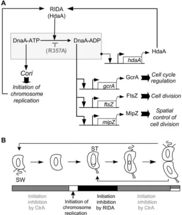
Figure 6. Control of the initiation of chromosomal replication inC. crescentus. (A) Model for a RIDA system inC. crescentus.A schematic connecting the regulation of the activity of DnaA with processes regulated by DnaA is shown. We propose that DnaA associates with ATP soon after its synthesis and thus promotes the initiation of chromosomal replication in the absence of active CtrA. Once replication is initiated, the ATPase activity of DnaA is stimulated by HdaA assembled onto the replisome, to hydrolyse the ATP bound to DnaA into ADP, resulting in the inactivation of DnaA as an initiator of DNA replication (RIDA). ThehdaA,gcrA, ftsZandmipZpromoters are all directly activated by DnaA. DnaA-ADP may be more efficient than DnaA-ATP when activating the gcrA, ftsZ and mipZ promoters. The effect of the R357A mutation in DnaA is also shown on the schematic (accumulation of DnaA-ATP).(B) Model for the temporal control of the initiation during theC. crescentuscell cycle.A schematic of theC. crescentuscell cycle is shown. Theta structures inside the cells indicate replicating DNA. SW indicates swarmer cells. ST indicates stalked cells. In swarmer cells, active CtrA is abundant and it blocks the initiation of DNA replication. Once CtrA is degraded during the swarmer-to-stalked cell transition, active DnaA initiates DNA replication. Immediately after, DnaA is inactivated by a RIDA process involving HdaA and the replisome, thus preventing the start of new rounds of replication in stalked cells. In predivisional cells, active CtrA re-accumulates, to block the initiation of DNA replication in the future swarmer progeny. Overall, CtrA determines when and where the initiation of DNA replication can start, while the RIDA system ensures that it starts only once per cell cycle.
Is the RIDA system involved in the transcriptional control of DnaA-regulated genes inC. crescentus?
Besides its function as the initiator of chromosomal replica-tion, DnaA is also a master transcriptional regulator in C. crescentus [3]. DnaA activates the transcription of about forty different genes at the swarmer-to-stalked cell transition, by directly binding to minimum thirteen different promoters. Genes whose expression is directly regulated by DnaA encode proteins involved in multiple processes essential for cell cycle progression. Examples are the FtsZ protein required for cell division, the NrdB protein involved in nucleotide synthesis, and the cell polarity factor PodJ.
To explore the potential role of the AAA+domain of DnaA in the regulation of the activity of DnaA as a transcription factor, we compared the effect of the over-expression of DnaA or DnaA(R357A) on the activities of several promoters driving the expression of proteins involved in the RIDA system, in cell cycle regulation and in cell division inC. crescentus. Interestingly, the selected genes are not regulated by DnaA in E. coli. We found that DnaA(R357A) is not more active than DnaA to activate the transcription of each selected genes (Fig.5), suggesting that ATP is not more active than DnaA-ADP when acting as a transcription factor regulating the expression of these genes (Fig.6). Thus, the DnaA mutant we generated decouples the activity of DnaA as a transcription factor regulating several genes from its activity as an initiator of DNA replication.
We also observed that DnaA(R357A) was less efficient than DnaA in activatinggcrA,ftsZandgcrAtranscription (Fig.5B&C&D). This effect is particularly striking for thegcrAandmipZpromoters, which are not significantly activated by the expression of DnaA(R357A), compared to the control strain containing an empty vector (Fig.5B). One possible interpretation is that these three promoters may be more efficiently activated by DnaA-ADP than by ATP (Fig.6). Genes specifically activated by DnaA-ADP may then wait for the initiation of DNA replication to be activated by DnaA, thereby coordinating the initiation of DNA replication with the expression of specific genes. Consistently, the transcription ofgcrAis at its maximum in stalked cells, right after the initiation of DNA replication [25,26]. Also, GcrA activates the transcription of genes whose products are involved in the elongation of DNA replication and in chromosome segregation [26]. Thus, it may be logical for these genetic modules to be expressed only after the initiation of DNA replication. We propose that the AAA+ domain of DnaA plays a role in temporally regulating the bifunctionality of DnaA by reallocating DnaA molecules from initiating DNA replication to transcribing genes within the DnaA regulon.
The extent to which the regulation of the activity of DnaA may affect the regulation of other genes directly controlled by DnaA remains unkown inC. crescentusand future detailed investigations studying the regulation of each gene will be required to determine in which cases the AAA+ domain of DnaA may regulate the activity of DnaA as a transcription factor. In other bacterial species, there are now several examples of DnaA-regulated genes whose expression was shown to depend on both the concentration and the nucleotide-bound state of DnaA [2,5,37,38], but the role of the nucleotide bound to DnaA in the regulation of the activity of DnaA as a transcription factor still remains poorly understood in most cases [2]. Thus, the full extend to which DnaA is utilized to regulate the timing of gene expression during the bacterial cell cycle is an interesting avenue for future research andC. crescentusis an ideal model to study such questions.
Materials and Methods
Bacterial strains and growth conditions
C. crescentusstrains were grown in PYE complex media [39] at 28uC. Plasmids and strains used are listed in Table 2 and Table 3. Antibiotics used forC. crescentusliquid cultures include rifampicin (15mg/mL), chloramphenicol (1mg/mL), kanamycin (5mg/mL) and oxytetracycline (1mg/mL). Antibiotics used for E. coli liquid cultures include chloramphenicol (20mg/mL), kanamycin (30mg/mL) and oxytetracycline (12mg/mL). Plas-mids were mobilized fromE. coliS17-1 [40] intoC. crescentusby bacterial conjugation or introduced by electro-transformation. BacteriophageWCR30 was used for general transduction intoC. crescentus.
Plasmids constructions
Construction of pDnaA(R357A). Oligos 59
-CCCATATGA-CCATGAAGGGCGGGGTTGCC-39, 59
-CCGGATCCTTAG-CCCCGCAGCTTGCGCGT-39, 59
-CACCGACAGCGTCGC-CGAGCTGGAAGGC-39 and 59-
GCCTTCCAGCTCGGCG-ACGCTGTCGGTG-39 were used to create a mutated dnaA coding sequence [dnaA(R357A)] from a two-step PCR amplifica-tion procedure using CB15N genomic DNA. The corresponding PCR product was cloned into pCR-BluntII-TOPO (Invitrogen).
Construction of the pX-DnaA(R357A) plasmid. pDnaA (R357A) was digested by NdeI and BamHI to extract the mutant dnaAinsert, which was cloned into NdeI-BamHI-digested pXGFP4C1 (with thegfpgene eliminated).
Construction of the pJSX-DnaA plasmid. Oligos 59
-AAG-GTACCCAGCCGATCAGGCGGAACTGG-39 and 59-
CCG-GATCCTTAGCCCCGCAGCTTGCGCGT-39 were used to
amplify PxylX::dnaA from pX-DnaA. The PCR product was digested by KpnI and BamHI, and cloned into KpnI-BamHI-digested pJS14.
Construction of the pJSX-DnaA(R357A) plasmid. same as construction of pJSX-DnaA, except that pX-DnaA(R357A) was used as template DNA for the PCR amplification.
Construction of the pNPTS138-DCC1613::Vplasmid. The CC1613downstream region was PCR-amplified using primers 59
-CCGGATCCCCGCGAATCTCGCACTGA-39 and 59
-CCGG-GCTAGCGCGGCGATTGGCGAGGTG-39. The 500 base pair
product was digested withNheI andBamHI, and cloned into aNhe I-BamHI-digested pNPTS138 plasmid, giving pNPTS138- CC-1613down. The CC1613 upstream region was PCR-amplified
using primers 59
-GGTAAGCTTCGCGCCGCGACAGGCCTG-GG-39 and 59
-CCGGATCCGGCGAGACGAGAATTCATG-GCG-39. The 500 base pair product was then digested with HindIII and BamHI, and cloned into BamHI-HindIII-digested pNPTS138-CC1613down, giving the pNPTS138-DCC1613 plasmid. The 2 kb BamHI fragment from pBOR, containing the SpecR/StrepR cassette (V), was ligated into BamHI-digested pNPTS138-DCC1613.
Construction of the pNPTS138-dnaA(R357A) plasmid. The dnaA(R357A) coding sequence was PCR-amplified from pDna-A(R357A) using primers 59
-GGTAAGCTTACCATGAAGGGC-GGGGTTGCC-39and 59
-ACGCGCAAGCTGCGGGGCTAAG-GATCCGG-39. The product was then digested with BamHI and HindIII, and cloned intoBamHI-HindIII-digested pNPTS138.
Construction of the placZ290-ftsZP plasmid. The ftsZ promoter region was PCR-amplified using primers 59-
GGAA-TTCCAGCCAGCTGGCCGGTGTGC-39 and 59
Construction of the placZ290-mipZP plasmid. ThemipZ promoter region was PCR-amplified using primers 59
-CGGA-ATTCTCGGGGCCTCCACGCAAACTGG-39 and 59
-AACT-GCAGGGCTCGGATCCTTCTGCGTCGC-39. The product
was then digested with EcoRI and PstI, and cloned into EcoRI-PstI-digested placZ290.
Strain constructions
Construction of the JC323 and JC324 strains: Plasmids pX-DnaA and pX-pX-DnaA(R357A) were integrated into the xylX promoter [41] of strain NA1000 by single integration events, giving strains JC323 and JC324 respectively.
Construction of the JC125 strain. To integrate plasmid pNPTS138-DCC1613::V into the C. crescentus chromosome by single homologous recombination, plasmid pNPTS138-D CC16-13::Vwas introduced into strain NA1000 by conjugation, selecting for kanamycin-resistant colonies with the plasmid integrated at the CC1613 locus by PCR. The resulting strain was grown to stationary phase in PYE medium lacking kanamycin. Cells were plated on PYE + sucrose 3% and incubated at 28uC. Single colonies were picked and transferred in parallel onto plain PYE plates and PYE plates containing kanamycin. Kanamycin-sensitive and SpecR/StrepRcolonies, which had lost the integrated plasmid due to a second recombination event, were then checked for the presence of theDCC1613::Vallele by colony PCR.
Immunoblot analysis
DnaA, GcrA and HdaA proteins were resolved on 10%, 15% and 12% SDS/PAGE, respectively [42]. Gels were electrotrans-fered to a PVDF membrane (Millipore). Immunodetection was performed with polyclonal antibodies. Anti-HdaA and anti-GcrA sera were diluted 1:2000, anti-rabbit-conjugated to horse-radish peroxidase (Sigma Aldrich) and anti-DnaA sera were diluted 1:10000. A chemiluminescent reagent (PerkinElmer, Wellesley, MA) and Kodak (Rochester, NY) Bio-Max MR films were used. Images were processed with Photoshop (Adobe, Mountain View, CA), and relative band intensities were determined by using ImageJ software version 1.43.
Flow cytometry analysis
Rifampicin-treated cells were fixed and stained with the DNA-binding Vybrant DyeCycle Orange (Invitrogen), as previously described [43]. Rifampicin treatment of cells blocks the initiation of chromosomal replication, but allows ongoing rounds of replication to finish. Fixed cells were analyzed using a FACS-Calibur (BD Biosciences, Erembodegem, Belgium) cytometer, equipped with an air-cooled argon laser (488 nm). Flow cytometry data were acquired using the CellQuest software. 30000 cells were analyzed from each biological sample. To quantify the results (Fig.4B), the proportion of cells having 1N, 2N or .2N chromosomes was estimated on the basis of the fluorescence area (FL2-A) given by the flow cytometer for each cell. The data were normalized so that the fluorescence value for the maximum of the 1N peak is equal for all data sets. The average difference N between the 2N and the 1N peak maximum was estimated from representative data sets. In each data set, all cells whose fluorescence is greater than 0.5N and smaller than 1.5N fall in the 1N category; all cells whose fluorescence is greater than 1.5N and smaller than 2.5N fall in the 2N category; all cells whose fluorescence is greater that 2.5N fall in the.2N category.
Q-PCR to measureCori/ter ratios
Cells were grown to exponential phase in PYE medium plus 0.2% glucose, before 0.3% xylose was added for 4 hours. Cells were harvested and chromosomal DNA was extracted using Wizard Genomic DNA Purification Kit (Promega), 2ml of lysozyme (20 mg/ml) were used instead of Cell Lysis Solution. The following primer sets were used for the Q-PCR reaction: Cori_fwd (59 -CGCGGAACGACCCACAAACT-39) and Cori_rev (59 -CAGCC-GACCGACCAGAGCA-39) targeting a region close to the origin (Cori) ; Ter_fwd (59-CCGTACGCGACAGGGTGAAATAG-39) and Ter_rev (59-GACGCGGCGGGCAACAT-39), targeting a region close to the terminus (ter). Reactions were run using SYBR Green Supermix (KAPA biosystems) and a Mx3005P Instrument (Stratagene) in a volume of 20ml, containing 10ml of Supermix, 2ml of each pair of primers (concentration 4mM) and 8ml of DNA. Reactions were run in 4 replicates. Efficiency of each primer set was calculated by performing a standard curve with 4 different dilutions of primers. For quantification of the results, a calibrator-normalized relative analysis was performed using MxPro qPCR Software (Stratagene) for determining the relative abundance of the chromosomalCoriandtersites in each of the samples. The results were normalized to theCori/terratio of the wild-type control strain (NA1000) treated with rifampicin for three hours, whoseCori/ter ratio is expected to be close to 1.
Microscopy
Cells were immobilized using a thin layer of media + 1% agarose. Phase contrast microscopy images were taken with a
Table 2.Strains.
Strains Description Reference/source
Escherichia coli
TOP10 Cloning strain Invitrogen
S17 RP4, Tc::Mu Km::Tn7 [40]
Caulobacter crescentus
NA1000 Synchronizable derivative of wild-type strain
[45]
GM2471 NA1000DdnaA::V PxylX::dnaA
[24]
JC125 NA1000DCC1613::V This study
JC919 NA1000 pJS14 This study
JC366 NA1000 pJSX-DnaA This study
JC367 NA1000 pJSX-DnaA(R357A) This study
JC323 NA1000 PxylX::dnaA This study
JC324 NA1000 PxylX::dnaA(R357A) This study
JC964 JC919 placZ290-hdaAP(WT) This study
JC973 JC366 placZ290-hdaAP(WT) This study
JC974 JC367 placZ290-hdaAP(WT) This study
JC971 JC919 placZ290-gcrAP This study
JC972 JC366 placZ290-gcrAP This study
JC975 JC367 placZ290-gcrAP This study
JC1006 JC919 placZ290-ftsZP This study
JC1007 JC366 placZ290-ftsZP This study
JC1008 JC367 placZ290-ftsZP This study
JC1037 JC919 placZ290-mipZP This study
JC1038 JC366 placZ290-mipZP This study
JC1039 JC367 placZ290-mipZP This study
Plan-Apochromat 100X/1.45 oil Ph3 objective on an AxioImager M1 microscope (Zeiss) with a cascade 1K EMCCD camera (Photometrics) controlled through Metamorph 7.5 (Universal Imaging, Downingtown, PA). Images were processed using Adobe Photoshop and Metamorph 7.5.
Promoter activity assays
The b-galactosidase activity of strains containing pLacZ290 derivatives was assayed from exponential phase cultures in PYE media, as previously described [44]. b-galactosidase activities represent the average of minimum three independent assays.
Supporting Information
Figure S1 A moderate overproduction of DnaA(R357A), but not DnaA, leads to severe over-initiation of chromo-somal replication. Representative profiles obtained by flow cytometry analyses of cells from strains JC919 (containing the pJS14 empty vector), JC366 (containing pJSX-DnaA) and JC367
[containing pJSX-DnaA(R357A)] grown to exponential phase in PYE medium plus 0.2% glucose (PYEG) before 0.3% xylose was added (PYEGX) to half of the culture for four hours. Cells were fixed and stained with Vybrant DyeCycle orange. The horizontal axis indicates the number N of complete chromosomes: 1N, 2N or more than 2N (+). The vertical axis indicates the number of cells. (DOC)
Acknowledgments
We thank Katharina Eich for helpful discussions, Sean Murray for the critical reading of this manuscript, and Anaı¨s Meyer and Gael Close for technical assistance.
Author Contributions
Conceived and designed the experiments: JC CFF DG. Performed the experiments: JC CFF DG. Analyzed the data: JC CFF DG. Contributed reagents/materials/analysis tools: JC CFF DG. Wrote the paper: JC CFF DG.
References
1. Mott ML, Berger JM (2007) DNA replication initiation: mechanisms and regulation in bacteria. Nat Rev Microbiol 5: 343–354.
2. Scholefield G, Veening JW, Murray H (2011) DnaA and ORC: more than DNA replication initiators. Trends Cell Biol 21: 188–194.
3. Hottes AK, Shapiro L, McAdams HH (2005) DnaA coordinates replication initiation and cell cycle transcription inCaulobacter crescentus. Mol Microbiol 58: 1340–1353.
4. Goranov AI, Katz L, Breier AM, Burge CB, Grossman AD (2005) A transcriptional response to replication status mediated by the conserved bacterial replication protein DnaA. Proc Natl Acad Sci U S A 102: 12932–12937. 5. Speck C, Weigel C, Messer W (1999) ATP- and ADP-dnaA protein, a molecular
switch in gene regulation. Embo J 18: 6169–6176.
6. Olliver A, Saggioro C, Herrick J, Sclavi B (2010) DnaA-ATP acts as a molecular switch to control levels of ribonucleotide reductase expression inEscherichia coli. Mol Microbiol 76: 1555–1571.
7. Kato J, Katayama T (2001) Hda, a novel DnaA-related protein, regulates the replication cycle inEscherichia coli. Embo J 20: 4253–4262.
8. Katayama T, Kubota T, Kurokawa K, Crooke E, Sekimizu K (1998) The initiator function of DnaA protein is negatively regulated by the sliding clamp of theE. colichromosomal replicase. Cell 94: 61–71.
9. Su’etsugu M, Shimuta TR, Ishida T, Kawakami H, Katayama T (2005) Protein associations in DnaA-ATP hydrolysis mediated by the Hda-replicase clamp complex. J Biol Chem 280: 6528–6536.
10. Riber L, Olsson JA, Jensen RB, Skovgaard O, Dasgupta S, et al. (2006) Hda-mediated inactivation of the DnaA protein and dnaA gene autoregulation act in concert to ensure homeostatic maintenance of theEscherichia colichromosome. Genes Dev 20: 2121–2134.
11. Camara JE, Breier AM, Brendler T, Austin S, Cozzarelli NR, et al. (2005) Hda inactivation of DnaA is the predominant mechanism preventing hyperinitiation of Escherichia coli DNA replication. EMBO Rep 6: 736–741.
12. Nishida S, Fujimitsu K, Sekimizu K, Ohmura T, Ueda T, et al. (2002) A nucleotide switch in the Escherichia coli DnaA protein initiates chromosomal replication: evidnece from a mutant DnaA protein defective in regulatory ATP hydrolysis in vitro and in vivo. J Biol Chem 277: 14986–14995.
Table 3.Plasmids.
Plasmids Description Reference/source
pCR-BluntII-TOPO Cloning plasmid Invitrogen
pDnaA(R357A) dnaA(R357A)in pCR-BluntII-TOPO This study
pJS14 Mid-copy number replicating plasmid J. Skerker, unpublished
pJSX-DnaA dnaA(WT)under the control of thexylXpromoter in pJS14 [16]
pJSX-DnaA(R357A) dnaA(R357A)under the control of thexylXpromoter in pJS14 This study
pXGFP4C1 Integrating plasmid D. Alley, unpublished
pX-DnaA dnaA(WT)under the control of thexylXpromoter in pXGFP4C1 [16]
pX-DnaA(R357A) dnaA(R357A)under the control of thexylXpromoter in pXGFP4C1 This study
pNPTS138 Integrating plasmid containing thesacBgene D. Alley, unpublished
pHP45V Vector carrying a SpecR/StrepRcassette (V) [46]
pBOR 2-kb EcoRI fragment from pHP45Vligated into EcoRI-digested pBluescript C. Stevens, unpublished
pNPTS138-DnaA(R357A) dnaA(R357A)cloned into pNPTS138 This study
pNPTS138-DCC1613::V The regions upstream and downstream of theCC1613coding sequence flanking an Vcassette, cloned into pNPTS138
This study
placZ290 Low-copy number plasmid to create transcriptional fusions withlacZ [47]
placZ290-hdaAP(WT) lacZgene under the control of the wild-typehdaApromoter in placZ290 [16]
placZ290-gcrAP lacZgene under the control of the wild-typegcrApromoter in placZ290 [25,26]
placZ290-ftsZP lacZgene under the control of theftsZpromoter in placZ290 This study
placZ290-mipZP lacZgene under the control of themipZpromoter in placZ290 This study
13. Camara JE, Skarstad K, Crooke E (2003) Controlled initiation of chromosomal replication inEscherichia colirequires functional Hda protein. J Bacteriol 185: 3244–3248.
14. Noirot-Gros MF, Dervyn E, Wu LJ, Mervelet P, Errington J, et al. (2002) An expanded view of bacterial DNA replication. Proc Natl Acad Sci U S A 99: 8342–8347.
15. Hayashi M, Ogura Y, Harry EJ, Ogasawara N, Moriya S (2005) Bacillus subtilis YabA is involved in determining the timing and synchrony of replication initiation. FEMS Microbiol Lett 247: 73–79.
16. Collier J, Shapiro L (2009) Feedback control of DnaA-mediated replication initiation by replisome-associated HdaA protein inCaulobacter. J Bacteriol 191: 5706–5716.
17. Collier J, Shapiro L (2007) Spatial complexity and control of a bacterial cell cycle. Curr Opin Biotechnol 18: 333–340.
18. Curtis PD, Brun YV (2010) Getting in the loop: regulation of development in
Caulobacter crescentus. Microbiol Mol Biol Rev 74: 13–41.
19. Quon KC, Yang B, Domian IJ, Shapiro L, Marczynski GT (1998) Negative control of bacterial DNA replication by a cell cycle regulatory protein that binds at the chromosome origin. Proc Natl Acad Sci U S A 95: 120–125. 20. Laub MT, Chen SL, Shapiro L, McAdams HH (2002) Genes directly controlled
by CtrA, a master regulator of theCaulobactercell cycle. Proc Natl Acad Sci U S A 99: 4632–4637.
21. Quon KC, Marczynski GT, Shapiro L (1996) Cell cycle control by an essential bacterial two-component signal transduction protein. Cell 84: 83–93. 22. Chen YE, Tropini C, Jonas K, Tsokos CG, Huang KC, et al. (2011) Spatial
gradient of protein phosphorylation underlies replicative asymmetry in a bacterium. Proc Natl Acad Sci U S A 108: 1052–1057.
23. Domian IJ, Quon KC, Shapiro L (1997) Cell type-specific phosphorylation and proteolysis of a transcriptional regulator controls the G1-to-S transition in a bacterial cell cycle. Cell 90: 415–424.
24. Gorbatyuk B, Marczynski GT (2001) Physiological consequences of blocked
Caulobacter crescentus dnaAexpression, an essential DNA replication gene. Mol Microbiol 40: 485–497.
25. Collier J, Murray SR, Shapiro L (2006) DnaA couples DNA replication and the expression of two cell cycle master regulators. Embo J 25: 346–356. 26. Holtzendorff J, Hung D, Brende P, Reisenauer A, Viollier PH, et al. (2004)
Oscillating global regulators control the genetic circuit driving a bacterial cell cycle. Science 304: 983–987.
27. Kelly AJ, Sackett MJ, Din N, Quardokus E, Brun YV (1998) Cell cycle-dependent transcriptional and proteolytic regulation of FtsZ inCaulobacter.Genes Dev 12: 880–893.
28. Quardokus E, Din N, Brun YV (1996) Cell cycle regulation and cell type-specific localization of the FtsZ division initiation protein inCaulobacter. Proc Natl Acad Sci U S A 93: 6314–6319.
29. Thanbichler M, Shapiro L (2006) MipZ, a spatial regulator coordinating chromosome segregation with cell division inCaulobacter. Cell 126: 147–162.
30. Nakamura K, Katayama T (2010) Novel essential residues of Hda for interaction with DnaA in the regulatory inactivation of DnaA: unique roles for Hda AAA Box VI and VII motifs. Mol Microbiol 76: 302–317.
31. Cheng L, Keiler KC (2009) Correct timing ofdnaAtranscription and initiation of DNA replication requires trans translation. J Bacteriol 191: 4268–4275. 32. Zweiger G, Shapiro L (1994) Expression ofCaulobacter dnaAas a function of the
cell cycle. J Bacteriol 176: 401–408.
33. Collier J, McAdams HH, Shapiro L (2007) A DNA methylation ratchet governs progression through a bacterial cell cycle. Proc Natl Acad Sci U S A 104: 17111–17116.
34. Gorbatyuk B, Marczynski GT (2005) Regulated degradation of chromosome replication proteins DnaA and CtrA inCaulobacter crescentus. Mol Microbiol 55: 1233–1245.
35. Jonas K, Chen YE, Laub MT (2011) Modularity of the bacterial cell cycle enables independent spatial and temporal control of DNA replication. Curr Biol 21: 1092–1101.
36. Marczynski GT, Shapiro L (2002) Control of chromosome replication in
caulobacter crescentus. Annu Rev Microbiol 56: 625–656.
37. Olliver A, Saggioro C, Herrick J, Sclavi B (2010) DnaA-ATP acts as a molecular switch to control levels of ribonucleotide reductase expression inEscherichia coli. Mol Microbiol 76: 1555–1571.
38. Gon S, Camara JE, Klungsoyr HK, Crooke E, Skarstad K, et al. (2006) A novel regulatory mechanism couples deoxyribonucleotide synthesis and DNA replication inEscherichia coli. EMBO J 25: 1137–1147.
39. Ely B (1991) Genetics ofCaulobacter crescentus. Methods Enzymol 204: 372–384. 40. Simon R PU, Puhler A (1983) A broad host range mobilization system for in vivo genetic engeneering: Transposon mutagenesis in gram negative bacteria. Bio/ Technology 1: 784–790.
41. Meisenzahl AC, Shapiro L, Jenal U (1997) Isolation and characterization of a xylose-dependent promoter fromCaulobacter crescentus. J Bacteriol 179: 592–600. 42. Sambrook J, Fritsch EF, Maniatis T (1989) Molecular Cloning: a Laboratory
Manual. Cold Spring Harbor, NY: Cold Spring Harbor Laboratory Press. 43. Lesley JA, Shapiro L (2008) SpoT regulates DnaA stability and initiation of
DNA replication in carbon-starved Caulobacter crescentus. J Bacteriol 190: 6867–6880.
44. Miller JH (1972) Expreriments in Molecular Genetics. Cold Spring Harbor, NY: Cold Spring Harbor Laboratory Press.
45. Evinger M, Agabian N (1977) Envelope-associated nucleoid fromCaulobacter crescentusstalked and swarmer cells. J Bacteriol 132: 294–301.
46. Prentki P, Krisch HM (1984) In vitro insertional mutagenesis with a selectable DNA fragment. Gene 29: 303–313.
![Figure 2. DnaA and DnaA(R357A) are expressed at moderate levels. Strains NA1000 (WT), JC366 (containing pJSX-DnaA) and JC367 [containing pJSX-DnaA(R357A)] were grown to exponential phase in PYE medium plus 0.2% glucose (PYEG) and 0.3% xylose was added (PYE](https://thumb-eu.123doks.com/thumbv2/123dok_br/16282481.184728/2.918.473.832.823.973/figure-expressed-moderate-strains-containing-containing-exponential-glucose.webp)