Expressão do receptor ativador de NF-kBETA (Rank), Rank ligante (RANKL), e Osteoproterina (OPG) em sítios de reparo ósseo de ratos diabéticos
60
0
0
Texto
(2) Fernanda Penna Lima Guedes de Amorim. EXPRESSÃO DO RECEPTOR ATIVADOR DE NF-κ κBETA (RANK), RANK LIGANTE (RANKL) E OSTEOPROTEGERINA (OPG) EM SÍTIOS DE REPARO ÓSSEO DE RATOS DIABÉTICOS. Dissertação submetida ao Programa de Pós-Graduação em Ciências Médicas da Faculdade de Medicina da Universidade de Brasília, como requisito parcial para a obtenção do Grau de Mestre. Área de concentração: Medicina. Orientadora: Profª. Drª. Tarcília Aparecida da Silva.. BRASÍLIA – DF 2007.
(3) DEDICATÓRIA. Dedico esse estudo ao meu filho Eduardo, cujo nascimento me inspirou na realização desse sonho. Que esse trabalho sirva de exemplo da importância da busca do conhecimento para seu desenvolvimento pessoal. O anseio pela descoberta do novo há de estar presente nas suas realizações de vida.. ii.
(4) AGRADECIMENTOS. Ao querido Rodrigo, meu companheiro, agradeço por aceitar viver comigo meus sonhos, compartilhar comigo alegrias e tristezas, incentivando-me a prosseguir. Obrigada pela confiança, respeito, dedicação e por todos os ensinamentos científicos. E desculpe-me a ausência em alguns momentos.. A meus pais pelo amor incondicional, pelo incentivo e pelo ensinamento de que o conhecimento é a principal conquista que devemos objetivar na vida, para que possamos melhorar como pessoa.. À minha irmã pelo exemplo de dedicação à pesquisa.. Ao meu irmão pela alegria de sua existência.. À professora Tarcília Aparecida da Silva pelo acolhimento, confiança e paciência. Obrigada pelos ensinamentos científicos.. À professora Aline Carvalho Batista pelo carinho e dedicação.. Ao amigo Sandoval Felicíssimo Diniz pelo companheirismo na realização de todo o projeto de pesquisa.. À veterinária Gabriela Mariângela Farias de Oliveira e à aluna Tarciane por todo apoio na etapa experimental da pesquisa.. Aos funcionários José Tavares dos Santos, pelos cuidados aos animais da pesquisa, e Edvaldo Batista Teles, pelo apoio técnico de radiologia.. Às estagiárias Édelyn Cristina Nunes Silva e Renata Ribeiro de Souza pela ajuda valiosa no preparo das peças para análise histopatológica.. À mestranda Helenisa e aos técnicos Erildo Ribeiro da Silva e Simone de Fátima Cruz iii.
(5) pelo apoio técnico na imunohistoquímica.. Ao doutorando Sócrates Sousa Ornelas e prof. Kansaki pelos ensinamentos e ajuda incondicional em toda etapa de biologia molecular.. Aos amigos André Leite, Paulo Furtado, Hugo Caracas por todo apoio na etapa de radiologia e análise das imagens.. Ao funcionário do departamento de pós-graduação Alessandro, que me orientou sempre que precisei.. À minha secretária Denise, que resolveu os problemas relacionados com minha ausência durante os dois anos desta caminhada.. Aos animais da pesquisa. Sem eles nada poderia ter sido realizado.. iv.
(6) RESUMO. Os mecanismos envolvidos na modificação do reparo ósseo em diabéticos ainda permanece pouco elucidado. Assim, esse estudo investigou a expressão de reguladores do metabolismo ósseo receptor ativador de NFκB (RANK), RANK ligante (RANKL) e osteoprotegerina (OPG), através da imunohistoquímica e RT-PCR, em sítios de fratura óssea de ratos diabéticos. Foram realizadas fraturas ósseas fechadas em tíbias esquerdas de ratos controle e com diabetes induzido pelo aloxano. A análise histomorfométrica dos sítios de fratura após 7 dias revelaram que os ratos diabéticos (db) apresentaram menor formação óssea e de cartilagem em comparação com o grupo controle. Paralelamente, o número de células RANK e RANKL positivas foi reduzido no grupo diabético. Além disso, células OPG positivas apresentaram-se. significantemente. diminuídas no grupo diabético comparado ao grupo controle (p=0,05). Entretanto, a razão RANKL/OPG foi similar no grupo controle (0,074) e diabético (0,099) nesse período. Após 14 dias, o número de células RANKL e OPG positivas e a expressão de RNAm desses marcadores foi maior no grupo controle (p=0.008). Apesar de menores níveis, a razão RANKL/OPG no grupo diabético (1,29) foi maior do que no grupo controle (0,90), o que sugere o favorecimento dos mecanismos de reabsorção óssea. Os resultados obtidos demonstram a expressão de marcadores da atividade de formação/remodelação de tecidos duros em sítios de fratura. Modificações no balanço da expressão de RANKL/OPG pode contribuir para o retardo do reparo de fraturas associado ao estado diabético.. Palavras- chave: RANK, RANKL, OPG, diabetes, reparo ósseo.. v.
(7) ABSTRACT. To clarify the mechanisms of altered bone reapair in diabetic state, we investigate the RANK, RANKL and OPG expression by immunohistochemistry and RT-PCR in the fracture sites of diabetic rats. A closed fracture was performed on the anatomical left tibia in rats either healthy or made diabetic by alloxan. Histomorphometric analysis of fracture site at 7 days after fracture revealed that diabetic rats (db) have significantly lesser bone and cartilage formation at fracture site in comparison with controls. Parallel with this, the number of RANK and RANKL positive cells were slighly decreased in db group. Furthermore, OPG+ cells were significantly lower in db than control (p=0.05). However, the RANKL/OPG ratio was similar in control (0.074) and db (0.099) at this time. At day 14, the numbers of RANKL and OPG positive cells and the mRNA expression for these markers were increased in control group (P=0.008). Despite lower levels, the RANKL/OPG ratio in db group (1.29) was greater than in controls (0.90) what suggets a favoring of pro-resorptive pathways. Our results demonstrate the expression of consistent markers of hard tissue formation/remodeling activities in sites of fractures. The imbalance of RANKL/OPG expression may contribute to the delay of fracture repair during diabetes course.. Key words: RANK, RANKL, OPG, diabetes, facture repair. vi.
(8) LISTA DE ABREVIATURAS E SIGLAS. AGEs. Produtos finais avançados da glicosilação. BMP. Proteínas morfogenéticas ósseas. CGRP. Calcitonin gene-related peptide. EGF. Fator de crescimento epidérmico. FGF. Fator de crescimento dos fibroblastos. GAGs. Glicosaminoglicanos. GM-CSF. Fator estimulador de colônia de granulócitos-macrófagos. GD. Grupo diabético. IGF. Fator de crescimento semelhante à insulina. IAPP. Islet amyloid polypeptide. IFN-γ. Interferon –γ. IL. Interleucina. LIF. Fator inibidor de leucócitos. M-CSF. Fator estimulante de colônia de macrófago. MGP. Fetuin-matrix Gla protein complex. MMPs. Metaloproteinases. NOD. Camundongos diabéticos não-obesos. OCIF. Fator inibidor de osteoclastos. ODF. Fator de diferenciação dos osteoclastos. OPG. Osteoprotegerina. OPGL. Osteoprotegerina ligante. OSM. Oncostatina M. PDGF. Fator de crescimento derivado de plaquetas. PGF-2. Prostaglandina. PTH. Paratormônio. PTHrP. PTH related protein. RANK. Receptor Ativador de Nf-κBeta. RANKL. Receptor Ativador de Nf-kBeta Ligante. TGF. Fator transformador do crescimento. TGF. Fator de crescimento transformador. TNF. Fator de necrose tumoral. TNFR. Receptores de fator de necrose tumoral vii.
(9) TRANCE. TNF-related activation induced cytokine. TRAP. Fosfatase ácida resistente ao tartarato. VDR. Vitamina D3 (1,25(OH)2D3). VEGF. Fator de crescimento vascular endotelial. viii.
(10) SUMÁRIO. AGRADECIMENTOS----------------------------------------------------------------------------iii RESUMO -------------------------------------------------------------------------------------------v ABSTRACT----------------------------------------------------------------------------------------vi LISTA DE ABREVIATURAS E SIGLAS ---------------------------------------------------vii 1 INTRODUÇÃO --------------------------------------------------------------------------------10 2 OBJETIVOS ------------------------------------------------------------------------------------18 3 ARTIGO : Imbalance of RANK, RANKL and OPG expression during tibial fracture repair in diabetic rats -----------------------------------------------------------------------------19 4 CONSIDERAÇÕES FINAIS -----------------------------------------------------------------37 5 CONCLUSÕES --------------------------------------------------------------------------------43 6 REFERÊNCIAS BIBLIOGRÁFICAS ------------------------------------------------------44 7 ANEXOS ---------------------------------------------------------------------------------------58. ix.
(11) 1- INTRODUÇÃO. O Diabetes mellitus é a doença endócrina mais comum em todas as populações mundiais, em todas as faixas etárias, e sua prevalência está crescendo drasticamente. Nos Estados Unidos, essa desordem metabólica afeta aproximadamente 15,7 milhões de indivíduos, o que representa 5,9% da população (PORTUESE & ORCHARD, 1995; ABDULWASSIE & DHANRAJANI, 2002; HUAFEI et al., 2005). Segundo as estimativas e projeções mundiais apresentadas por Zimmet et al. (2001), em 2010 teremos 236 milhões de diabéticos em todo o mundo; no Brasil as estimativas são de 11 milhões (MALERBI et al., 1992; MANDRUP-POULSEN, 1998; FARZAD et al., 2002).. Existem duas formas da doença: a forma primária ou idiopática, conhecida como diabetes mellitus insulino-dependente ou diabetes tipo 1, e a segunda forma, denominada diabetes mellitus não insulino-dependente ou diabetes tipo 2 (IKEGAMI & OGIHARA, 1996; TUOMINEN et al., 1999). Aproximadamente 10% a 20% dos indivíduos diabéticos são do tipo 1 (ABDULWASSIE & DHANRAJANI, 2002). Ambos os tipos da doença estão associados a alta morbidade e mortalidade (FARZAD et al., 2002). Pacientes diabéticos apresentam, em média, redução de 5 a 10 anos na expectativa de vida, dependendo do tempo do diagnóstico e da duração da doença (MANDRUP-POULSEN, 1998).. O diabetes representa uma complexa síndrome, pois a alta concentração de glicose no sangue irá resultar em complicações sistêmicas secundárias, tais como doenças cardiovasculares; retinopatias; nefropatias; neuropatias; suscetibilidade à infecções; atraso na cicatrização tecidual, com destaque para atraso no reparo de fraturas ósseas,. 10.
(12) entre outras. O diabetes é ainda causa importante de amputação de extremidades sem trauma prévio e a principal causa de insuficiência renal (FIORELLINI & NEVINS, 2000; ABDULWASSIE & DHANRAJANI, 2002; FARZAD et al., 2002; ALMEIDA et al. 2002; SIVAN- LOUKIANOVA et al., 2003).. Vários aspectos da fisiopatologia das alterações ósseas desenvolvidas em decorrência do diabetes vêm sendo estudados. Dentre eles, citam-se: alterações funcionais nos leucócitos polimorfonucleares (OLIVER & TERVONEN, 1994; GORCHON et al.,1994; BOUMA et al., 2005), macrófagos e fibroblastos (WETZLER et al., 2000), com prolongamento da fase inflamatória; diminuição da angiogênese e da produção. de. fator. de. crescimento. vascular. endotelial. (VEGF). (SIVAN-. LOUKIANOVA et al., 2003; GALIANO et al., 2004) diminuição da biossíntese de colágeno (MANOUCHEHR-POUR et al., 1981; RAMAMURTHY & GOLUB, 1983; OLIVER & TERVONEN, 1994; CLARO et al., 2005) e glicosaminoglicanos (GAGs); formação de produtos finais avançados da glicosilação (AGEs) (FIORELLINI & NEVINS, 2000; SANTANA et al., 2003; HAYNES, 2004); alterações na síntese protéica; deficiência na mineralização óssea (GIGLIO et al., 2000; SHYNG et al., 2001; SIQUEIRA, 2003; PADULA et al., 2003; MARGONAR et al., 2003; FOLLAK, 2004; FOLLAK, 2005; FACCHINI et al., 2005); redução do turnover ósseo (VERHAEGHE et al., 1989; CLARO et al., 2005); diminuição do número de osteoblastos e osteoclastos (WETZLER et al., 2000; ALMEIDA et al. 2002; KOPMAN et al., 2005).. É importante observar que o diabetes tipo 1 e tipo 2 apresentam repercussões diferentes no metabolismo ósseo. Enquanto a ação do diabetes tipo 1 no osso consiste em osteopenia, aumento no risco de fratura e significante atraso na consolidação das fraturas (MACEY et al., 1989; SIQUEIRA, 2003; PADULA et al., 2003;MARGONAR. 11.
(13) et al., 2003; FOLLAK, 2004; CLARO et al., 2005; STROTMEYER et al., 2005; HONGBING et al., 2006;), a presença de perda óssea no diabetes tipo 2 não está clara, e entende-se que essa forma de diabetes não está associada à osteopenia (JEFFCOATE et al., 2004; THRAILKILL et al., 2005; HONGBING et al., 2006). Ressalta-se ainda que a insulina exerça importante papel no controle dos níveis de glicose e modulação dos fatores de crescimento esquelético, estimulando diretamente a síntese de matriz osteoblástica e, indiretamente, o fator de crescimento semelhante à insulina-1 (IGF-1) (KWON et al., 2005). Thrailkill et al. (2005) estudaram a neoformação óssea em fraturas de camundongos diabéticos não obesos (NOD) e observaram que a insulina aparentemente tem a capacidade de prevenir a perda de volume do calo ósseo e do osso trabecular. Beam et al. (2002) também mostraram que não há diferença histomorfométrica na cicatrização de fraturas ósseas entre ratos espontaneamente diabéticos sob terapia com insulina e o grupo controle (BALSHI et al., 1999; FIORELLINI et al., 1999; FIORELLINI & NEVINS, 2000; SIQUEIRA et al., 2003; MARGONAR et al., 2003; FOLLAK et al., 2004; FOLLAK, 2005; KWON et al., 2005).. Os novos conhecimentos sobre a fisiologia das células ósseas no diabetes incluem o estudo da participação de citocinas e fatores de crescimento. Alterações significativas nos níveis das citocinas e de seus receptores solúveis e antagonistas específicos, no ambiente celular ósseo, têm sido observados tanto sistêmica como localmente (JEFFCOATE, 2004). As citocinas são proteínas com peso molecular de 6.000M a 60.000M que regulam as respostas imunológica e inflamatória do hospedeiro, como também a cicatrização de feridas, hematopoiese e muitos outros processos biológicos. Em relação ao tecido ósseo, as citocinas podem modificar a remodelação óssea, atuando na formação e na reabsorção. A reabsorção é iniciada por fatores sistêmicos ou locais e. 12.
(14) a continuidade do processo depende de fatores locais nos quais as citocinas exercem importante papel como mediadoras. Algumas citocinas apresentam maior envolvimento nos processos fisiopatológicos de remodelamento ósseo. Dentre as que estimulam a reabsorção óssea, citam-se: fator de necrose tumoral alfa (TNF-α) (HAYNES, 2004); interleucina-1 (IL-1); IL-6; IL-11; o fator estimulador de colônia de granulócitosmacrófagos (GM-CSF); o fator de crescimento transformador alfa (TGF-α), fator de crescimento epidérmico (EGF) e IL-11 estimulam a formação dos osteoclastos e a reabsorção óssea, tanto in vitro como in vivo (MOHAN & BAYLINK, 1991). A IL-1 e a IL-6 também aumentam a proliferação e atividade dos osteoblastos. Os fatores de crescimento semelhantes à insulina I e II (IGF-I e II), fator de crescimento transformador beta (TGF-β), fator de crescimento derivado de plaquetas (PDGF), fator de crescimento dos fibroblastos (FGF) e proteínas morfogenéticas ósseas (BMP) atuam como promotores da formação, enquanto o TNF e interferon –γ (IFN-γ) agem como inibidores da formação óssea (MOHAN & BAYLINK, 1991; TEITELBAUM, 2007).. O paratormônio (PTH) é um dos principais reguladores da remodelação óssea. Juntamente com a IL-1, TNF-α e calcitriol (1.25-(OH)2D3), atua nas células precursoras do estroma dando origem aos osteoblastos. Essas células precursoras liberam IL-6 e IL-11, que agem sobre o preosteoclasto estimulando sua diferenciação. Esse processo é complexo, envolvendo a participação das citocinas, fatores de crescimento e inter-relação dos osteoblastos e osteoclastos. O PTH estimula também a síntese de IGF-1 e TGF-β pelos osteoblastos, favorecendo a formação óssea (TEITELBAUM, 2007).. Com relação ao calcitriol, este é produzido por células renais, monócitos, células T, macrófagos e osteoblastos. O calcitriol, nos osteoblastos, aumenta a expressão do. 13.
(15) receptor da IL-1 e a produção de IL-6 (LIU et al., 2006), assim como a produção de matriz protéica, tendo ação também na diferenciação final dos osteoclastos (PINOMONTES et al., 2004).. Em 1995, Suda et al propuseram um modelo hipotético para explicar o papel das células do estroma/osteoblastos que quando estimulados por fatores sistêmicos ou locais seriam capazes de induzirem a formação dos osteoclastos. As células do estroma/ osteoblastos apresentam os seguintes receptores: vitamina D3 (1,25(OH)2D3) (VDR); sistema proteína quinase A; glicoproteína 130. Esses receptores são ativados por diferentes estímulos, ou seja, o VDR pelo calcitriol; o sistema quinase A pelo PTH, PTHrP (PTH related protein), IL-1, TNF e prostaglandina (PGF-2); enquanto a via da glicoproteína130 pela IL-6, IL-11, oncostatina M (OSM) e fator inibidor de leucócitos (LIF) (LIU et al., 2006). Quando esses receptores são estimulados, as células expressam um fator de diferenciação dos osteoclastos, chamado Receptor Ativador de NFk-B Ligante (RANKL), que se liga à membrana do osteoclasto. Outros nomes para o RANKL podem ser fator de diferenciação dos osteoclastos (ODF), TNF-related activation induced cytokine (TRANCE) ou osteoprotegerina ligante (OPGL) (MOSCHEN et al., 2005; HOFBAUER et al., 2000; GONZÁLEZ, 2000; LACEY et al., 1998; YASUDA et al., 1998).. O RANKL é um peptídeo com 317 aminoácidos e pertencente à família de fatores de necrose tumoral, podendo se apresentar em duas formas: solúvel e ligado à membrana. O RANKL se liga ao RANK (receptor activator de NFκ-B) presente nas células progenitoras dos osteoclastos e na presença de fator estimulante de colônia de macrófago (M-CSF), estimula a diferenciação e atividade dos osteoclastos (LACEY et al. 1998; YASUDA et al. 1998; HOFBAUER et al. 2000; GONZÁLEZ, 2000;. 14.
(16) HAYNES, 2004; ROGERS & EASTELL; 2005). Camundongos deficientes de RANKL não apresentam formação de osteoclastos e desenvolvem severa osteopetrose, além de defeito dos nódulos linfáticos e células B. Em animais normais, ao injetar RANKL solúvel, verificou-se a rápida indução de hipercalcemia associada à perda óssea (LACEY et al. 1998).. O RANKL tem sido relatado como mediador de várias doenças: artrite reumatóide (STOLINA et al., 2005); osteoporose pós-menopausa; osteoporose induzida por glicocorticóide; osteólises peri-protéticas; hiperparatireoidismo; doença de Paget; mieloma ósseo; metástases ósseas e infecções periodontais (MOSCHEN et al., 2005; HAYNES, 2004; ANANDARAJAH & SCHWARZ, 2006).. O RANKL exerce sua ações via interação com o receptor RANK, o qual é uma proteína de membrana com 616 aminoácidos, da superfamília dos receptores de fator de necrose tumoral (TNFR), expressa primariamente em células precursoras dos osteoclastos, células B e T, células dendríticas e fibroblastos (HSU et al. 2006; ANANDARAJAH & SCHWARZ, 2006). Camundongos que não produzem RANK desenvolvem osteopetrose e defeito na maturação das células B e T (LU et al., 2006; HSU et al. 2006).. A interação entre RANKL e seu receptor RANK nos osteoclastos é controlada pela osteoprotegerina (OPG) ou fator inibidor de osteoclasto (OCIF) (LIN et al., 2006; BEETON et al., 2006). A OPG é uma proteína de 401 aminoácidos pertencente à superfamília dos receptores de fator de necrose tumoral (TNFR). É um receptor solúvel e age inibindo a ligação do RANKL ao RANK, impedindo, assim, o recrutamento, a proliferação e a ativação dos osteoclastos (BOYLE et al., 2003; THÉOLEYRE et al., 2006). Em camundongos, a expressão de RNA mensageiro de OPG tem sido. 15.
(17) demonstrada em vários tecidos, como: osso, pele, fígado, pulmão, coração, células endoteliais, rins, estômago, intestino, glândula tireóide, cérebro e medula espinhal (HOROWITZ et al., 2001; KNUDSEN et al., 2003; ANANDARAJAH & SCHWARZ, 2006).. A OPG neutraliza os efeitos biológicos de RANKL, pois inibe a diferenciação de osteoclastos, suprime a ativação dos osteoclastos maduros e induz sua apoptose (HOROWITZ et al., 2001; NAKAMICHI et al., 2007). Animais que não produzem OPG apresentam severa osteoporose pelo aumento da atividade osteoclástica e subseqüente reabsorção óssea, calcificação da aorta e das artérias renais (BROWNER et al., 2001; YAMAZAKI & SASAKI, 2005; NAKAMICHI et al., 2007). Injeções subcutâneas de OPG na área abdominal de mulheres pós-menopausa rapidamente reduziram os marcadores do turnover ósseo (HSU et al., 2006). Além disso, a administração de OPG em animais com artrite induzida experimentalmente, protegeu-os da perda óssea, enquanto os animais que não receberam OPG apresentaram extensa perda óssea e de cartilagem (KONG et al., 1999). Ainda não está clara, entretanto, a associação entre o conteúdo mineral ósseo, o risco de fratura e a concentração plasmática dessa molécula (HOFBAUER & SCHOPPET., 2001; BOWNER et al., 2001; KNUDSEN et al., 2003; ANANDARAJAH & SCHWARZ, 2006). O balanço do sistema OPG/RANKL controla o remodelamento ósseo (ROGERS & EASTELL; 2005; KWAN et al., 2006; RUCCI et al., 2007). Alterações na concentração dessas moléculas induzidas por variações dos níveis de outras citocinas, administração de glicocorticóides ou deficiência de estrogênio podem induzir aumento do número e da atividade dos osteoclastos, determinando perda de massa óssea (HOFBAUER et al., 2000; GONZÁLEZ, 2000).. 16.
(18) Recentes estudos têm mostrado baixos níveis do RANKL e OPG em sítios de reabsorção óssea em pacientes diabéticos (DUARTE et al., 2007). Em contraste, outros autores associam a alta expressão desses fatores ao estado diabético (GALLUZZI et al., 2005; SUZUKI et al., 2005; DUARTE et al., 2007; HIE et al., 2007; KAYAL et al., 2007). Existem controvérsias acerca da atividade osteoclástica associada ao diabetes mellitus e até o momento ainda não está esclarecido se ocorre inibição (HE et al., 2004) ou aumento da atividade osteoclástica (HIE et al., 2007; KAYAL et al., 2007). Embora o diabetes tenha sido associado à perda óssea, a falta de consenso acerca do mecanismo que leva à menor formação óssea e ao atraso no reparo ósseo no diabetes tipo 1 indica a necessidade de futuras investigações. Como o papel do sistema OPG/RANK/RANKL no controle do remodelamento ósseo tem sido bem documentado, o presente estudo pretende investigar a expressão dessas moléculas em sítios de fraturas ósseas de animais com diabetes induzido por aloxano. Sendo assim, justifica-se a realização do presente estudo, pois os dados obtidos poderão ser úteis para a compreensão dos mecanismos envolvidos na deficiência de consolidação óssea em diabéticos, contribuindo na busca de estratégias terapêuticas que possam dar suporte aos protocolos tradicionais de tratamento.. 17.
(19) 3- OBJETIVOS. Os objetivos deste estudo são:. 1. Avaliar a expressão do Receptor Ativador de NF-kBeta (RANK), RANK ligante (RANKL) e Osteoprotegerina (OPG), em sítios de reparo ósseo de ratos diabéticos;. 2. Correlacionar à expressão de RANK, RANKL e OPG ao processo de consolidação de fraturas fechadas em tíbia de ratos diabéticos induzidos por aloxano.. 18.
(20) 4- ARTIGO (enviado para publicação sob as normas do Journal of Orthopedic Research). Imbalance of RANK, RANKL and OPG expression during tibial fracture repair in diabetic rats. Running title: RANK, RANKL and OPG expression in diabetic rats. Fernanda Penna Lima Guedes de Amorima; Sócrates Souza Ornelasb, Sandoval Felicíssimo Diniza Aline Carvalho Batistac Tarcília Aparecida da Silvad∗. a. Department of Pathology, Faculty of Medicine, University of Brasília; Brasília,. Distrito Federal, Brazil; b. Department of Molecular Pathology, Faculty of Medicine, University of Brasília;. Brasília, Distrito Federal, Brazil; c. Department of Stomatology (Oral Pathology), Dental School, Universidade Federal de. Goiás, Goiânia, Goiás, Brazil; d. Department of Oral Pathology and Surgery, Faculty of Dentistry, Universidade Federal. de. Minas. Gerais,. Belo. Horizonte,. ∗. Minas. Gerais,. Brazil;. Corresponding author: Tarcília Aparecida da Silva Mailing address: Departamento de Clínica, Patologia e Cirurgia Odontológicas, Faculdade de Odontologia, Universidade Federal de Minas Gerais, Av. Antônio Carlos 6627, CEP 31.270-901, Belo Horizonte, Minas Gerais, Brazil. Phone: 55 31 3499-2476 (voice); 55 31 3499-2430 (Fax). E-mail: tarcilia@odonto.ufmg.br. 19.
(21) Abstract To clarify the mechanisms of altered bone reapair in diabetic state, we investigate the RANK, RANKL and OPG expression by immunohistochemistry and RT-PCR in the fracture sites of diabetic rats. A closed fracture was performed on the anatomical left tibia in rats either healthy or made diabetic by alloxan. Histomorphometric analysis of fracture site at 7 days after fracture revealed that diabetic rats (db) have significantly lesser bone and cartilage formation at fracture site in comparison with controls. Parallel with this, the number of RANK and RANKL positive cells were slighly decreased in db group. Furthermore, OPG+ cells were significantly lower in db than control (p=0.05). However, the RANKL/OPG ratio was similar in control (0.074) and db (0.099) at this time. At day 14, the numbers of RANKL and OPG positive cells and the mRNA expression for these markers were increased in control group (P=0.008). Despite lower levels, the RANKL/OPG ratio in db group (1.29) was slightly greater than in controls (0.90) what suggets a favoring of pro-resorptive pathways. Our results demonstrate the expression of consistent markers of hard tissue formation/remodeling activities in sites of fractures. The imbalance of RANKL/OPG expression may contribute to the delay of fracture repair during diabetes course.. Key words: RANK, RANKL, OPG, diabetes, facture repair. 20.
(22) Introduction It has been recognized a relationship between diabetes and the delayed healing of fractures and bone defects in human and animal models [1-6]. Several mechanisms have been proposed to explain the greater incidence of the delayed healing and nonunion of fractures in diabetes. These include the reduction in the blood supply and angiogenesis [7]; more severe inflammatory response [8]; decrease in collagen synthesis [1, 9]; disturbance in the mineralization process [5, 6] and the imbalance between bone resorption by osteoclasts and bone deposition by osteoblasts [10]. In this setting, the receptor activator of NF-kappaB (RANK), RANK ligand (RANKL) and osteoprotegerin (OPG) provide the cellular and molecular basis for osteoblast-osteoclast cross-talks, which are crucial during bone remodeling. RANK belongs to the tumor necrosis factor (TNF) receptor superfamily and is activated by RANKL, a homotrimeric, TNF-like cytokine. RANK is present on the surface of osteoclast cell precursors, where its interaction with RANKL induces the cells’ terminal differentiation into osteoclasts, thus increasing bone breakdown [11, 12]. The secreted, soluble receptor OPG interrupts this activation by binding directly to RANKL [11-13]. An imbalance of RANK/RANKL/OPG system has been observed in osteoporosis, osteopetrosis, rheumatoid arthritis and periodontal diseases [13, 14]. Recent studies have been showed lower levels of RANKL and OPG at bone resorption sites in diabetic patients [14]. In contrast, a higher expression of these factors has been associated with diabetic state by others [14-18]. Controversy also surrounds the osteoclastic activity in that it has not been defined whether an inhibition [10] or increased osteoclastic activity [17, 18] is associated with diabetes mellitus. Although the diabetes has been associated with a net loss of bone, the lack of consensus regarding the mechanism of diminished bone formation and delayed bone. 21.
(23) repair in type 1 diabetes point to the need for further investigation into the impact of diabetes on bone. As the roles of OPG/RANK/RANKL system in controlling bone remodeling have been well documented, we investigate in this study the expression of these proteins at bone fracture sites in a model of alloxan-induced diabetes. Our results suggest that the imbalance of RANKL/OPG expression may have a role on the delay of fracture repair during diabetes course.. Methods Animals The Animal Care Committee at University of Brasília approved all procedures performed. In total, 54 skeletally mature, male, 3-month-old Wistar rats (Rattus norvegicus albinus) weighing 250 to 350 g were used for the study. Rats were randomly assigned to the diabetic or control group. Alloxan-induced diabetes model Diabetes was induced by the intraperitoneal injection of 150 mg kg-1 of monohydrated alloxan (Sigma Chemical Co., St. Louis, MO, USA) dissolved in sterile 0.9% saline solution. Rats were kept on fasting prior to alloxan injection. After 12 hours of alloxan administration, a 10% glucose solution was offered to the animals to prevent hypoglycemia [19, 20]. After 72 hours, blood samples were collected from the tail vein of the animals for evaluation of the plasma glucose levels by the glucose-oxidase enzymatic method using Accu-Chek Advantage (Boehringer Mannheim, IN, USA). Animals presenting glucose levels above 200 mg/dL (the normal levels of serum glucose in Rattus novergicus ranges from 50 to 135 mg/dL). The examinations were repeated every 7 days to confirm the maintenance of the glucose levels. The animals presenting reversion of the signs of diabetes, ie, presenting glucose levels below 200. 22.
(24) mg/dL, were excluded from this study. The pancreas was obtained in the two surgical times from control and diabetic animals for routine histopathological analysis, five animals for each group. Tibia fracture and Specimen Preparation Using general anesthesia with ketamine (200 mg kg-1) and xylazine (10 mg kg-1), closed fracture was inflicted manually on the middle of the left tibia. The traces of all fractures were similar, with no exposure to the outer environment; as previously described [21]. All procedures were performed by one operator. The animals were sacrificed at 7 and 14 days following the fracture. For the histological studies, the entire tibia was used after disarticulating it from the knee and ankle. The specimens were fixed in 10% buffered formaldehyde for 24 h, delcalcified in 1% nitric acid for 24 hours and embedded in paraffin blocks. Approximately 30 sections, 5-µm thick, were available from each paraffin block. Approximately 6 hematoxylin and eosin-stained sections with the largest areas of fracture callus surrounding the site of injury were selected from each paraffin block for use in subsequent analyses. Microscopic analysis was performed by two pathologists who were blinded to the identity of the specimen. Histomorphometry Three representative slides were selected from each animal for histomorphometric analysis. Measurements of the areas of fibrous connective, cartilaginous and bone tissues at fracture callus were averaged in six fields selected by systematic sampling in a stepwise manner, moving the microscope stage from left to right and then down and across in order to avoid measuring the same area again. The area of each tissue at 25x magnification was obtained by drawing around the boundaries using the software Image J. (National. Institutes. of. 23. Health,. USA)..
(25) Immunohistochemistry For immunostaining, paraffin-embedded tissues sectioned at 5 µm and collected in serial sections on glass slides coated with 2% 3-aminopropyltriethylsilane (Sigma Chemicals, St. Louis, MO) were deparaffinized and dehydrated. Sections were then incubated with 3% hydrogen peroxide and immersed in citrate buffer, pH 6.0, for 20 minutes at 95°C. Sections were then blocked by incubation with 3% normal goat serum at room temperature, for 20 minutes and slides were incubated with the following primary rabbit polyclonal antibodies: anti-OPG (H-249, sc11383, Santa Cruz Biotechnology, Santa Cruz, CA) diluted 1:200; anti-RANK (H-300, sc9072, Santa Cruz) diluted 1:200; anti-RANKL (FL-327, sc9073, Santa Cruz) diluted 1:150; at 4°C, overnight, in a humidified chamber. After washing in TBS, the sections were treated with a labeled streptavidin-biotin (LSAB) kit (K0492, Dako, Carpinteria, CA). The sections were then incubated in 3,3’-Diaminobenzidine (K3468, Dako) for 2 to 5 minutes and stained with Mayer´s hematoxylin. The specimens of peripheral giant cell granuloma were employed as positive controls for all markers. Negative controls were obtained by the omission of primary antibodies and substitution of primary antibodies by non-immune rabbit serum (X0902, Dako). The number of cells was evaluated in sites of fracture by two independent examiners blinded to the stans of groups. The number of positive cells was counted in five representative and consecutive microscopic high-power fields (x100) using the integration graticule (4740680000000, Carl Zeiss, Göttingen, Germany), at this magnification each field had an area of 0.0961 mm2. Results were expressed as the mean of positive cells per mm2 ± standard deviation (SD) of n observations.. 24.
(26) RT-PCR Tibia samples (five for each group) were immersed in TRIZOL reagent (Gibco BRL, Life Technologies, Rockville, MD, USA) and stored at –70°C. RNA Extraction was performed after addition of 200 µL of chloroform followed by centrifugation. RNA was recovered from the aqueous phase by precipitation with isopropyl alcohol. The RNA stored. in. 70%. ethanol. at. -20°C. until. use.. Total. RNA was. quantified. spectrophotometrically, and the integrity of the RNA preparations was examined by agarose gel electrophoresis. Complementary DNA (cDNA) was synthesized using 100 ng of RNA through a reverse transcription reaction (Superscript II, Gibco). The standard PCR conditions were 95°C (10 minutes), and then 40 cycles of 94°C (1 minute), 54°C (1 minute), and 72°C (2 minutes). The primers sequences used were as follows: OPG. forward. 5’GCCAACACTGATGGAGCAGAT3’;. OPG. reverse. 5’TCTTCATTCCCACCAACTGATG3’;. RANK forward. 5’CGTCCTGCTCCTCTTCATCTCT3’;. RANK. 5’CCCTGAGGACTCCTTATTTCCA3’;. reverse. RANKL forward. 5’GCTCACCTCACCATCAATGCT3’;. RANKL reverse. 5’GGTACCAAGAGGACAGACTGACTTTA3’;. GAPDH forward. 5’AAATTCCATGGCACCGTCAA3’;. GAPDH reverse. 5’AGGGATCTCGCTCCTGGAA3’.. For each run, water was the negative control. The reaction product was quantified by measuring the densities of bands using a Spot-denso-program with Alpha Ease software (Alpha Innotech, San Leandro, CA), and values representing the average density per area unit were plotted. Glyceraldehyde-3-phosphate dehydrogenase (GAPDH) was used as the reference gene.. 25.
(27) Statistical analysis Data are expressed as means± standard deviation (SD) of n samples. Comparative analysis was performed using the non-parametric Mann-Whitney test. All analysis were made using the SPSS 12.0 (SPSS Inc. Chicago, IL, USA) statistical software package and probability values of less than 0.05 were considered as statistically significant.. Results Alloxan-induced diabetes As expected, blood-glucose levels were significantly (P < 0.01) higher in diabetic rats than controls at all time points before the surgery and postoperatively. Furthermore, pancreatic islets under the action of alloxan showed disorganization, reduction of sized and loss of architeture in comparison with controls. Treatment with alloxan did not significantly affected body weight throughout the experimental period. Histomorphometric Findings On day 7, diabetic rats have significantly lesser bone (4.2 ± 2.6; 1.9 ± 0.8, respectively for control and diabetic) cells per mm² (P=0.068) and cartilage (6.9 ± 3.3; 3.4 ± 1.4; respectively for control and diabetic) (P=0.045) and greater fibrous connective (8.5 ± 1.7; 14.1 ± 1.2, respectively for control and diabetic) (P=0.006) tissue formation at fracture site in comparison with controls. Similar results were found at day 14. Results concerning relative data of morphometric analysis (values were corrected in relation to the total analysed area) showed the mostly identical outline of absolute values for both groups. RANK, RANKL and OPG expression at tibia-fracture sites Immunohistochemical reactivity for RANK, RANKL, and OPG was detected in the cytoplasm and membrane of connective and bone tissue cells. The presence of. 26.
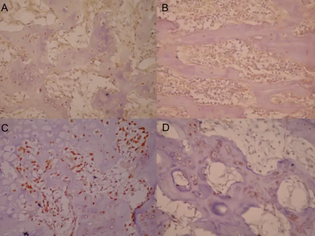
(28) positive fibroblasts, endothelial cells, bone surrounding cells, osteocytes, bone marrow cells and chondrocytes was observed (Figure 1). At day 7, the number of RANK (299.7 ± 139.5) and RANKL (7.5 ± 2.5) positive cells per mm2 were slighly decreased in db group when compared with controls (362.1 ± 128.1; 32.4 ± 10.9, respectively for RANK and RANKL). Furthermore, OPG+ cells were significantly lower in db (75.6 ± 35.6) than control (442.8 ± 163.1) (P=0.05). The RANKL/OPG ratio was similar in control (0.074) and db (0.099) at this time. A significant decrease in the the number of RANK positive cells at day 14 was observed in both groups in relation to day 7 (P<0.05). Although not statistically significant, we also observed a tendency of control group to have less OPG at day 14. At day 14, the numbers of RANK positive cells were similar in controls (58.8 ± 35.3) and db (74.51 ± 43.45). Although no significant differences were observed concerning the numbers of RANKL and OPG immunostained cells in both groups, we observed a tendency of db group have lesser positive cells for RANKL (29.9 ± 19.9; 6.9 ± 2.7; respectively for db and control groups) and OPG (137.4 ± 46.0; 72.1 ± 26.0, respectively for db and control groups). In line with these results, RT-PCR analysis revealed a significantly higher mRNA expression of RANK, RANKL and OPG in control when compared with db group. However, the RANKL/OPG ratio was greater in db group (1.29) than in controls (0.90) (Figure 2). No positive staining was observed when primary antibodies were omitted or substituted by PBS or non-immune rabbit serum. Furthermore, an intense immunostaining for RANKL, RANK, and OPG was observed in the giant cell granuloma, used as positive controls (data not shown).. 27.
(29) Discussion The impact of type 1 diabetes on bone is reflected by a significant delay in fracture healing. Consistent with previous data [1-6] we found that alloxan-induced diabetes delays fracture healing in a rat model of closed fracture. The effects of diabetes on bone have generally been attributed to impairment in osteoblast function observed in animals and humans [10, 17]. A number of studies have reported that type 1 diabetes alters bone remodeling by reducing the formation of new bone, leading to osteopenia. This has been shown by a decrease in bone mineral density in human and alterations in the formation of new bone in animal studies [10, 22-25]. In this study, we obtained comparable results regarding immunostaining and mRNA expression. The immunohistochemical reactivity for RANK, RANKL, and OPG was detected in the cytoplasm and membrane of connective and bone tissue cells in accordance with previous studies [26]. In parallel with decreased bone formation at fracture site of diabetic rats at day 7, we also found a reduced expression at protein and mRNA level of RANK, RANKL and OPG at sites of fracture. Given that the osteoclast maturation and activity (and bone homeostasis) are regulated in vivo by RANK-RANKL and OPG levels of expression [11-13] our results suggest a decreased osteoclastic recruitment and differentiation at bone fracture sites of diabetic rats at day 7. In accordance with our findings, it has been previously showed a low osteoclastogenesis in diabetic mice in a model of bacteriastimulated bone loss [10] and low bone resorption activity in diabetic individuals [27]. At 14 days of fracture, although we also observed reduced levels of RANK, RANKL and OPG at fracture sites of diabetic, the RANKL/OPG ratio was significantly higher in diabetic group suggesting an increase of resorptive activity. Our results are similar to that observed in periodontitis-associated bone resorption of diabetic patients, in which a. 28.
(30) reduced expression of RANKL and OPG, but an augmented RANKL/OPG ratio was observed in diabetic individuals [14]. The increased markers of osteoclast function such as urine excretions of calcium, hydroxyproline, and deoxypyridinoline [16, 28, 29] and expression of Tartarate-resistant acid phosphatase (TRAP), cathepsin K and RANKL [17, 18] were reported in subjects with diabetes. On the other hand, normal activity of bone resorption was also reported. [30]. Recently, it has been suggested that impaired fracture healing in diabetes is characterized by increased rates of cartilage resorption [18]. Different from our results, these authors demonstrated a high expression of RANKL at fracture site of diabetic animals. However, they did not determine the OPG levels at these sites. Besides the role of RANK-RANKL-OPG system in bone resorption, current evidence indicates that interactions between members of the TNF family and their specific receptors (TNFRs) are influential in several functions, including immune cell regulation [13, 31] induction of leukocytes transmigration [32], prevention of apoptosis of endothelial cells [33]. In this setting, it is possible that these factors have an effect on bone remodeling by also affecting cell proliferation, inflammatory cell migration, vascularity or the delaying of apoptosis. Thus, these aspects need to be further explored in diabetes. One potential explanation for our results is that there is reduced coupling of bone resorption and formation in the diabetic group after bone fracture, compared with robust coupling that occurs in normoglycemic littermates. The reduced coupling may be a result of decreased expression of RANK, RANKL and OPG. These results might be helpful in minimizing the effect of diabetic state in bone consolidation.. 29.
(31) Acknowledgment The authors are grateful to Gabriela Mariângela Farias de Oliveira, Édelyn Cristina Nunes Silva and Renata Ribeiro de Souza for their hepful technical assistance. This work was supported by Conselho Nacional de Desenvolvimento Científico e Tecnológico (CNPq).. References 1. Macey LR, Kana SM, Jingushi S, Terek RM, Borretos J, Bolander ME. 1989. Defects of early fracture-healing in experimental diabetes. J Bone Joint Surg Am 71:722-733. 2. Funk JR, Hale JE, Carmines D, Gooch HL, Hurwitz SR. 2000. Biomechanical evaluation of early fracture healing in normal and diabetic rats. J Orthop Res 18:126-132. 3. Follak N, Kloting I, Ganzer D, Merk H. 2003. Scanning electron microscopic examinations on retarded bone defect healing in spontaneously diabetic BB/O(ttawa)K(arlsburg) rats. Histol Histopathol 18:111-120. 4. Follak N, Kloting L, Wolf E, Merk H. 2004. Delayed remodeling in the early period of fracture healing in spontaneously diabetic BB/OK rats depending on the diabetic metabolic state. Histol Histopathol 19:473-486. 5. Follak N, Kloting I, Wolf E, Merk H. 2004. Histomorphometric evaluation of the influence of the diabetic metabolic state on bone defect healing depending on the defect size in spontaneously diabetic BB/OK rats. Bone 35:144-152. 6. Follak N, Kloting I, Merk H. 2005. Influence of diabetic metabolic state on fracture healing in spontaneously diabetic rats. Diabetes Metab Res Rev 21:288-296.. 30.
(32) 7. Dib SA, Russo EMK, Chacra AR. Tratado de endocrinologia clínica. São Paulo: Ed. Rocca; 1992. 8. Wetzler C, Kampfer H, Stallmeyer B, Pfeilschifter J, Frank S. 2000. Large and sustained induction of chemokines during impaired wound healing in the genetically diabetic mouse: prolonged persistence of neutrophils and macrophages during the late phase of repair. J Invest Dermatol 115:245-253. 9. Gooch HL, Hale JE, Fujioka H, Balian G,. Hurwitz SR. 2000. Alterations of cartilage and collagen expression during fracture healing in experimental diabetes. Connect Tissue Res 41:81-91. 10. He H, Liu R, Desta T, Leone C, Gerstenfeld LC, Graves DT. 2004. Diabetes causes decreased osteoclastogenesis, reduced bone formation, and enhanced apoptosis of osteoblastic cells in bacteria stimulated bone loss. Endocrinology 145:447-452. 11. Lacey DL, Timms E, Tan HL, Kelley MJ, Dunstan CR, Burgess T, Elliott R, Colombero A, Elliott G, Scully S, Hsu H, Sullivan J, Hawkins N, Davy E, Capparelli C, Eli A, Qian YX, Kaufman S, Sarosi I, Shalhoub V, Senaldi G, Guo J, Delaney J, Boyle WJ. 1998. Osteoprotegerin ligand is a cytokine that regulates osteoclast differentiation and activation. Cell 93:165-176. 12. Yasuda H, Shima N, Nakagawa N, Yamaguchi K, Kinosaki M, Mochizuki S, Tomoyasu A, Yano K, Goto M, Murakami A, Tsuda E, Morinaga T, Higashio K, Udagawa N, Takahashi N, Suda T. 1998. Osteoclast differentiation factor is a ligand for osteoprotegerin/osteoclastogenesis-inhibitory factor and is identical to TRANCE/RANKL. Proc Natl Acad Sci U S A 95:3597-3602. 13. Boyle WJ, Simonet WS, Lacey DL. 2003. Osteoclast differentiation and activation. Nature 423:337-342.. 31.
(33) 14. Duarte PM, Neto JB, Casati MZ, Sallum EA, Nociti FH Jr. 2007. Diabetes modulates gene expression in the gingival tissues of patients with chronic periodontitis. Oral Dis 13:594-599. 15. Galluzzi F, Stagi S, Salti R, Toni S, Piscitelli E, Simonini G, Falcini F, Chiarelli F. 2005. Osteoprotegerin serum levels in children with type 1 diabetes: a potential modulating role in bone status. Eur J Endocrinol 153:879-885. 16. Suzuki K, Kurose T, Takizawa M, Maruyama M, Ushikawa K, Kikuyama M, Sugimoto C, Seino Y, Nagamatsu S, Ishida H. 2005. Osteoclastic function is accelerated in male patients with type 2 diabetes mellitus: the preventive role of osteoclastogenesis inhibitory factor/osteoprotegerin (OCIF/OPG) on the decrease of bone mineral density. Diabetes Res Clin Pract 68:117-125. 17. Hie M, Shimono M, Fujii K, Tsukamoto I. 2007. Increased cathepsin K and tartrateresistant acid phosphatase expression in bone of streptozotocin-induced diabetic rats. Bone. 18. Kayal RA, Tsatsas D, Bauer MA, Allen B, Al-Sebaei MO, Kakar S, Leone CW, Morgan EF, Gerstenfeld LC, Einhorn TA, Graves DT. 2007. Diminished bone formation during diabetic fracture healing is related to the premature resorption of cartilage associated with increased osteoclast activity. J Bone Miner Res 22:560568. 19. Katsumata K, Katsumata Jr K, Katsumata Y. 1992. Protective effect of diltiazem hydrochloride on the occurrence of alloxan- or streptozotocin-induced diabetes in rats. Horm Metab Res 24:508-510. 20. Szkudelski T. 2001. The mechanism of alloxan and streptozotocin action in B cells of the rat pancreas. Physiol Res 50:537-546.. 32.
(34) 21. An YH, Friedman RJ. Animal Models in Orthopedic Research, Boca Raton: CRC press; 1999. 22. Horcajada-Molteni MN, Chanteranne B, Lebecque P, Davicco MJ, Coxam V, Young A, Barlet JP. 2001. Amylin and bone metabolism in streptozotocin-induced diabetic rats. J Bone Miner Res 16:958-965. 23. Levin ME, Boisseau VC, Avioli LV. 1976. Effects of diabetes mellitus on bone mass in juvenile and adult-onset diabetes. N Engl J Med 294:241-245. 24. Tuominen JT, Impivaara O, Puukka P, Rönnemaa T. 1999. Bone mineral density in patients with type 1 and type 2 diabetes. Diabetes Care 22:1196-200. 25. Krakauer JC, McKenna MJ, Buderer NF, Rao DS, Whitehouse FW, Parfitt AM. 1995. Bone loss and bone turnover in diabetes. Diabetes 44:775-782. 26. Silvestrini G, Ballanti P, Patacchioli F, Leopizzi M, Gualtieri N, Monnazzi P, Tremante E, Sardella D, Bonucci E. 2005. Detection of osteoprotegerin (OPG) and its ligand (RANKL) mRNA and protein in femur and tibia of the rat. J Mol Histol 36:59-67. 27. Guarneri MP, Weber G, Gallia P, Chiumello G. 1993. Effect of insulin treatment on osteocalcin levels in diabetic children and adolescents. J Endocrinol Invest 16:505509. 28. Olmos JM, Perez-Castrillon JL, Garcia MT, Garrido JC, Amado JA, GonzalezMacias J. 1994. Bone densitometry and biochemical bone remodeling markers in type 1 diabetes mellitus. Bone Miner 26:1–8. 29. Herrero S, Calvo OM, García-Moreno C, Martín E, San Román JI, Martín M, García-Talavera JR, Calvo JJ, del Pino-Montes J. 1998. Low bone density with normal bone turnover in ovariectomized and streptozotocin-induced diabetic rats. Calcif Tissue Int 62:260–265.. 33.
(35) 30. Miazgowski T, Czekalski S. 1998. A 2-year follow-up study on bone mineral density and markers of bone turnover in patients with long-standing insulindependent diabetes mellitus. Osteoporos Int 8:399-403. 31. So T, Lee SW, Croft M. 2006. Tumour necrosis factor/tumour necrosis factor receptor family members that positively regulate immunity. Int J Hematol 83:1-11. 32. Zauli G, Corallini F, Bossi F, Fischetti F, Durigutto P, Celeghini C, Tedesco F, Secchiero P. 2007. Osteoprotegerin increases leukocyte adhesion to endothelial cells both in vitro and in vivo. Blood 110:536-543. 33. Kobayashi-Sakamoto M, Hirose K, Nishikata M, Isogai E, Chiba I. 2006. Osteoprotegerin protects endothelial cells against apoptotic cell death induced by Porphyromonas gingivalis cysteine proteinases. FEMS Microbiol Lett 264:238-245.. 34.
(36) Figure 1 Representative immunohistochemical reactivity for RANKL (A, B) and OPG (C, D) at sites of tibia fracture of control (A, C) and alloxan-induced diabetic rats (B, D) at 14 days. [Immunohistochemical staining, original magnification x 400].. 35.
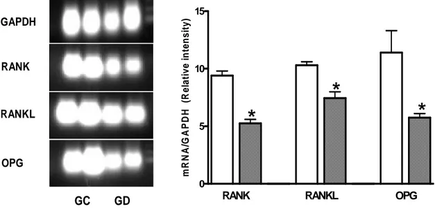
(37) 15 mR N A/G A PD H (R elative intensity). GAPDH. RANK. RANKL. OPG. 10. * *. *. 5. 0. GC. RANK. GD. RANKL. OPG. Figure 2 RANK, RANKL and OPG mRNA expression in bone callus of controls (GC) and alloxan-induced diabetic rats (GG) at 14 days after tibia fracture. Total RNA was extracted from bone callus tissue and expression of RANK, RANKL and OPG was analyzed by RT-PCR. Lanes 1-2 represent controls and lanes 3-4 represent diabetic animals. Bars represent the mean ± standard deviation (SD) of densitometric analysis of RANK, RANKL and OPG mRNA normalized by GAPDH levels per lane. The experiment is representative of two.*P<0.05 compared to each control. Control (white bars) and alloxan-induced diabetic (squared bars).. 36.
(38) 5- CONSIDERAÇÕES FINAIS. O diabetes tipo 1 está associado a várias desordens da saúde esquelética, incluindo diminuição da densidade óssea (FACCHINI et al., 2005), aumento do risco de osteoporose (THRAILKILL et al., 2005; GALLUZZI et al., 2005) e risco de fraturas, assim como retardo na cicatrização óssea e na capacidade regenerativa (THRAILKILL et al., 2005). Dados prévios da literatura (MACEY et al., 1989; FUNK et al., 2000; FOLLAK et al., 2003; FOLLAK et al., 2004a; FOLLAK et al., 2004b; FOLLAK et al., 2005; HONGBING et al., 2006) foram coincidentes ao verificado nesse estudo com relação ao atraso do reparo de fraturas fechadas em ratos com diabetes induzido pelo aloxano.. A consolidação de fraturas ósseas é uma complexa cascata de eventos celulares, e sua regulação é pouco conhecida (GIORDANO et al., 2000). A relação entre consolidação de fraturas e diabetes tipo 1 é complexa (PADULA et al., 2003). Vários trabalhos foram realizados com intuito de explicar a relação entre diabetes e reparo ósseo. Alterações nos mecanismos de resposta imune dos pacientes diabéticos, tais como a inibição da capacidade fagocitária dos neutrófilos associada a alterações vasculares, podem comprometer o processo de reparo das fraturas (CLARO et al., 2005). Dificuldades na fase inicial da cicatrização das fraturas também têm sido associadas à inibição da proliferação celular, que pode estar relacionada à diminuição da expressão do fator de crescimento derivado de plaquetas (PDGF) e dos níveis de fatores de crescimento semelhantes à insulina I e II (IGF-I e II), fator de crescimento transformador beta (TGF-β) e fator de crescimento de fibroblastos (FGF) (TYNDALL et al., 2003).. 37.
(39) Além disso, distúrbios na regulação dos níveis de metaloproteinases (MMPs) e a formação de AGEs, característicos do estado diabético, resultam em alterações de solubilidade do colágeno (SIQUEIRA et al., 2003), levando a alterações estruturais e funcionais do tecido conjuntivo (RAMAMURTHY & GOLUB, 1983; KOPMAN et al.,2005). Os efeitos do diabetes no osso podem ser ainda atribuídos ao desequilíbrio na função dos osteoblastos, o que se explica pela diminuição da expressão dos fatores de transcrição que regulam a diferenciação destas células (LACEY et al., 1998; YASUDA et al., 1998; BOYLE et al., 2003; HE et al., 2004; GALLUZZI et al., 2005; SUZUKI et al., 2005; THRAILKILL et al., 2005; DUARTE et al., 2007; HIE et al., 2007) e pelo aumento da apoptose das células precursoras de osteoblastos (VERHAEGHE et al, 1997; THRAILKILL et al., 2005). Paralelamente, alguns estudos têm mostrado que no diabetes tipo 1 observa-se redução de marcadores séricos correlacionados com a formação óssea, tais como osteocalcina (MCCRACKEN et al., 2000; THRAILKILL et al., 2005; KOPMAN et al., 2005; HONGBING et al., 2006) e fosfatase alcalina (THRAILKILL et al., 2005). Em acréscimo, a alteração do remodelamento ósseo pela redução na formação de novo osso leva à osteopenia (VERHAEGHE et al.,1989; HONGBING et al., 2006). Estudos prévios demonstraram diminuição da densidade mineral óssea e alterações de formação de novo osso nos humanos e em animais (HE et al., 2004; HORCAJADA-MOLTENI et al., 2001; LEVIN et al., 1976; TUOMINEN. et al., 1999; KRAKAUER et al., 1995; SIQUEIRA et al., 2003). As razões para menor densidade mineral óssea em diabetes tipo 1 ainda não são conhecidas (FACCHINI et al., 2005). Krakauer et al. (1995), entretanto, especula-se que exista redução na formação óssea durante o período de pico da massa óssea. Em oposição, Tuominen et al. (1999), ao examinar pacientes com diabetes tipo 1 e tipo 2. 38.
(40) com mais de 30 anos, sugerem que no diabetes tipo 1 ocorre maior perda de massa óssea, e a causa seria devido ao diabetes tipo 1 provocar efeitos profundos na fisiologia da remodelação óssea comparado ao diabetes tipo 2. No presente estudo, os resultados obtidos pela avaliação imunohistoquímica e RTPRC foram semelhantes. A reatividade imunohistoquímica de RANK, RANKL e OPG foi detectada no citoplasma e membrana de células dos tecidos conjuntivo e ósseo, assim como observado em estudos prévios (SILVESTRINI et al., 2005). Paralelamente à diminuição da formação óssea no sítio de fratura dos ratos diabéticos após 7 dias foi também verificada a redução da expressão protéica e dos níveis de RANK, RANKL e OPG nos sítios de fratura. Sabendo-se que a maturação dos osteoclastos e atividade (e homeostase óssea) são reguladas, in vivo, por níveis de expressão de RANK-RANKL e OPG (LACEY et al., 1998; YASUDA et al., 1998; BOYLE et al., 2003), os resultados encontrados sugerem a diminuição do recrutamento e diferenciação osteoclástica nos sítios de fratura óssea nos ratos diabéticos no sétimo dia. Corroborando os achados desse estudo, trabalhos prévios demonstraram redução da osteoclastogênese em camundongos diabéticos em modelo de perda óssea induzida pela inoculação de bactérias (HE et al., 2004) e ainda menor atividade de reabsorção óssea em indivíduos diabéticos (GUARNERI et al., 1993). Após 14 dias da fratura, apesar de observado também redução nos níveis de RANK, RANKL e OPG nos sítios de fratura dos diabéticos, a razão RANKL/OPG foi significativamente maior no grupo diabético sugerindo aumento da atividade de reabsorção. Esses resultados são similares aos observados na perda óssea associada à periodontite em pacientes diabéticos, nos quais houve redução da expressão de RANKL e OPG, mas aumento da razão RANKL/OPG (DUARTE et al., 2007).. 39.
(41) A expressão de RANKL e OPG durante o reparo de fraturas tem sido estudada em modelos animais. Picos da expressão de OPG têm sido verificados no período de 24 horas até 7 dias após a fratura, coincidindo com o pico de formação de cartilagem. A expressão de RANKL, entretanto, atinge seu pico entre o terceiro dia e o décimo quarto dia após a fratura, quando os níveis de OPG estão diminuindo (ROGERS & EASTELL, 2005). O estudo da cicatrização de fraturas em humanos após fratura traumática da tíbia mostrou diminuição da concentração sérica de OPG, sendo a menor concentração de OPG observada 8 semanas após a fratura. A correlação negativa entre a concentração sérica de OPG e os marcadores de formação óssea como a osteocalcina e a porção Nterminal do propeptídeo de colágeno tipo I foi também observada. Esses dados sugerem que a razão RANKL/OPG está envolvida no processo de reparo de fraturas, apesar de não demonstrarem necessariamente causa e efeito. Deve-se notar também que camundongos que não expressam RANK apresentam normal cicatrização de fraturas (ROGERS & EASTELL, 2005). A expressão de OPG e RANKL é modulada, direta ou indiretamente, por um grande número de fatores, incluindo TNF-α, IL-1 e IL-17, outras interleucinas, esteróides (SWANSON et al., 2006), hormônio do crescimento, IGF-1, M-CSF, óxido nítrico, leptina (LAMGHARI et al., 2006), fetuin-matrix Gla protein (MGP) complex, hormônios da tireóide e paratireóide, vitamina D e lipoproteínas (JEFFCOATE, 2004). Mais significantemente a expressão é afetada pela calcitonina e hormônios relacionados, calcitonin gene-related peptide (CGRP) e islet amyloid polypeptide (IAPP) (JEFFCOATE, 2004). A relação entre a expressão tecidual e a concentração na circulação sangüínea é incerta (ROGERS & EASTELL, 2005), entretanto, tem-se sido verificado que as. 40.
(42) concentrações séricas de RANKL e OPG apresentam-se elevadas em doenças associadas ao aumento do turnover ósseo (JEFFCOATE, 2004). O aumento de marcadores da função osteoclástica excretados na urina, como cálcio, hidroxiprolina e deoxipiridinolina (OLMOS et al., 1994; HERRERO et al., 1998; SUZUKI et al., 2005) e a expressão de fosfatase ácida resistente ao tartarato (TRAP), catepsina K e RANKL (HIE et al., 2007; KAYAL et al., 2007) foram reportados em indivíduos com diabetes. Por outro lado, estudos relatam atividade normal de reabsorção óssea nestes indivíduos (MIAZGOWSKI & CZEKALSKI, 1998). Recentemente, foi sugerido por alguns autores que o retardo da cicatrização de fraturas nos diabéticos é caracterizado pelo aumento das taxas de reabsorção de cartilagem (KAYAL et al., 2007). Esses autores demonstraram a alta expressão de RANKL nos sítios de fratura dos animais diabéticos, divergindo dos resultados obtidos no presente estudo. Entretanto, os níveis de OPG nesses sítios não foram verificados por estes autores. KNUDSEN et al. (2003), observaram um aumento de OPG no plasma dos pacientes com diabetes tipo 2. Esta observação está de acordo com os resultados encontrados no estudo de BOWNER et al. (2001) que relatou aumento das concentrações de OPG em pacientes diabéticos. Estes autores também verificaram que os níveis de OPG plasmático estavam significantemente aumentados apenas nos pacientes com complicações microvasculares, sugerindo que o aumento de OPG no plasma pode indicar perigo microvascular. Além do papel do sistema RANK/RANKL/OPG na reabsorção óssea, evidências atuais indicam que interações entre os membros da família de fatores de necrose tumoral (TNF) e seus receptors específicos (TNFRs) influenciam várias funções, incluindo regulação das células do sistema imune (BOYLE et al., 2003; SO et al.,. 41.
(43) 2006), indução de transmigração de leucócitos (ZAULI et al., 2007), prevenção da apoptose de células endoteliais (KOBAYASHI-SAKAMOTO et al., 2006). Nesse cenário, é possível que esses fatores que tenham efeito no remodelamento ósseo por afetarem a proliferação celular, a migração de células inflamatórias, vascularização ou apoptose. Assim, esses aspectos necessitam ser melhor explorados no diabetes. Uma possível explicação para os resultados obtidos neste estudo é que há redução conjunta da reabsorção e formação óssea no grupo diabético após fratura óssea, comparado à forte atividade metabólica observada nos animais normoglicêmicos. A redução conjunta pode ser resultado da menor expressão de RANK, RANKL e OPG. Esses resultados podem ser úteis para futuras terapias na tentativa de minimizar o efeito do estado diabético na consolidação óssea.. 42.
(44) 6-CONCLUSÕES A partir dos resultados obtidos e, com base na metodologia utilizada, podese concluir que: a) O diabetes induzido pelo aloxano atrasa significativamente o processo de reparação de fraturas ósseas; b) O estado diabético interfere com a expressão dos fatores reguladores da remodelação óssea RANK, RANKL e OPG em sítios de fratura; c) O atraso no reparo de fraturas observado nos ratos diabéticos parece estar associado à redução na expressão das proteínas RANK, RANKL e OPG causando um desequilíbrio nos níveis de RANKL e OPG nos sítios de reparo ósseo.. 43.
(45) 7- REFERÊNCIAS BIBLIOGRÁFICAS. 1. ABDULWASSIE, H.; DHANRAJANI, P.J. Diabetes mellitus and dental implants: a clinical study. Implant Dentistry 11 (2002) 83-85. 2. AN, Y.H.; FRIEDMAN, R.J. Animal Models in Orthopedic Research, Boca Raton: CRC press (1999). 3. ANANDARAJAH, A.P.; SCHWARZ, E. M. Anti-RANKL Therapy for inflammatory bone disorders: Mechanisms and potencial clinical applications. Journal of Cellular Biochemistry 97 (2006) 226-232. 4.. BALSHI, T.J.; WOLFINGER, G. J. Dental Implants in the diabetic patient: a retrospective study. Implant Dentistry 8 (1999) 355-358.. 5. BEAM, H.A.; PARSONS, J.R.; LIN, S.S. The effects of blood glucose control upon fracture healing in the BB Whistar rat with diabetes mellitus. J. Orthop. Res 20 (2002) 1210-1216. 6.. BEETON, C.A.; BORD, S.; IRELAND, D.; COMPSTON J.E. Osteoclast formation and bone resorption are inhibited by megakaryocytes. Bone 39 (2006) 985-990.. 7.. BOUMA, G.; NIKOLIC, T.; JOJANNEKE, M.C. Coppens, Cornelia G. van Helden-Meeuwsen, Pieter J. M. Leenen, Hemmo A. Drexhage, Silvano Sozzani and Marjan A. Versnel. NOD mice have a severly impaired ability to recruit leukocytes into sites of inflammation. Eur. J. Immunol. 35 (2005) 225-235.. 8. BOYLE, W.J.; SIMONET, W.S.;, LACEY, D.L. Osteoclast differentiation and activation. Nature 423 (2003) 337-342. 9. BROWNER, W.S.; LUI, L.; CUMMINGS, S.R. Associations of serum osteoprotegerin levels with diabetes, stroke, bone density, fractures, and. 44.
(46) mortality in elderly women. The Journal of Clinical Endocrinology & Metabolism 86 (2001) 631-637. 10. CLARO, F. A.; LIMA, J. R. S.; SALGADO, M. A. C.; GOMES, M. F. Porous Polyethylene for tissue engineering applications in diabetic rats treated with calcitonin: histomorphometric analysis. Int. J. Oral Maxillofac. Implants 20 (2005) 211-219. 11. DIB, S.A.; RUSSO, E.M.K.; CHACRA, A.R. Tratado de endocrinologia clínica. São Paulo: Ed. Rocca (1992). 12. DUARTE, P.M.; NETO, J.B.; CASATI, M.Z.; SALLUM, E.A.; NOCITI, F.H. JR. Diabetes modulates gene expression in the gingival tissues of patients with chronic periodontitis. Oral Dis 13 (2007) 594-599. 13. FARZAD, P.; ANDERSSON, L.; NYBERG, J. Dental implant treatment in diabetic patients. Implant Dentistry 11 (2002) 262-267. 14. FIORELLINI, J. P.; NEVINS, M. L.; NORKIN, A.; WEBER, H. P.; KARIMBUX N. Y. The effect of insulin therapy on osseointegration in a diabetic rat model. Clinical Oral Implants Research 10 (1999) 362-268. 15. FIORELLINI, J. P.; NEVINS, M.N. Dental implant considerations in the diabetic patient. Periodontology 23 (2000) 73-77. 16. FOLLAK, N.; KLOTING, I.; GANZER, D.; MERK, H. Scanning electron microscopic examinations on retarded bone defect healing in spontaneously diabetic BB/O(ttawa)K(arlsburg) rats. Histol Histopathol 18 (2003) 111-120. 17. FOLLAK, N.; KLOTING, I.; MERK, H. Influence of diabetic metabolic state on fracture healing in spontaneously diabetic rats. Diabetes Metab Res Rev 21(2005) 288-296.. 45.
(47) 18. FOLLAK, N.; KLOTING, I.; WOLF, E.; MERK, H. Histomorphometric evaluation of the influence of the diabetic metabolic state on bone defect healing depending on the defect size in spontaneously diabetic BB/OK rats. Bone 35 (2004)a 144-152. 19. FOLLAK, N.; KLOTING, L.; WOLF, E.; MERK, H. Delayed remodeling in the early period of fracture healing in spontaneously diabetic BB/OK rats depending on the diabetic metabolic state. Histol Histopathol 19 (2004)b 473-486. 20. FUNK, J.R.; HALE, J.E.; CARMINES, D.; GOOCH, H.L.; HURWITZ, S.R. Biomechanical evaluation of early fracture healing in normal and diabetic rats. J Orthop Res 18 (2000) 126-132. 21. GALIANO,. R.. D.;. TEPPER,. O.M.;. PELO,. C.R.;. BHATT,. K.A.;. CALLAGHAN, M.; BASTIDAS, N.; BUNTING, S.; STEINMETZ, H.G.; GURTNER, G.C. Topical vascular endothelial growth factor accelerates diabetic wound healing through increased angiogenesis and by mobilizing and recruiting bone marrow-derived cells. American Journal of Pathology 164 (2004) 19351947. 22. GALLUZZI, F.; STAGI, S.; SALTI, R.; TONI, S.; PISCITELLI, E.; SIMONINI, G.; FALCINI, F.; CHIARELLI, F. Osteoprotegerin serum levels in children with type 1 diabetes: a potential modulating role in bone status. European Journal of Endocrinology 153 (2005) 879-885. 23. GIGLIO, M. J.; GIANNUNZIO, G.; OLMEDO, D.; GUGLIELMOTTI, M.B. Histomorphometric study of bone healing around laminar implants in experimental diabetes. Implant Dentistry 9 (2000) 143-147.. 46.
(48) 24. GIORDANO, V., GIORDANO, M., KNACKFUSS, I. G. Fatores de crescimento e diferenciação ósseos. Efeitos sobre o processo de consolidação de fratura: presente e futuro. Rev. Bras. Med 57 (2000) 1018-1028. 25. GOOCH, H.L.; HALE, J.E.; FUJIOKA, H.; BALIAN, G.; HURWITZ, S.R. Alterations of cartilage and collagen expression during fracture healing in experimental diabetes. Connect Tissue Res 41(2000) 81-91. 26. GOOVA, M.T.; LI J.; KISLINGER, T.; QU, W.; LU, Y.; BUCCIARELLI, L.G. Blockade of receptor for advanced glycation end-products restores effective wound healing in diabetic mice. Am J Pathol 159 (2001) 513-525. 27. GUARNERI, M.P.; WEBER, G.; GALLIA, P.; CHIUMELLO, G. Effect of insulin treatment on osteocalcin levels in diabetic children and adolescents. J Endocrinol Invest 16 (1993) 505-509. 28. HAYNES, D.R. Bone Lysis and inflammation. Inflammation Research 53 (2004) 596-600. 29. HE, H.; LIU, R.; DESTA, T.; LEONE, C.; GERSTENFELD, L.C.; GRAVES, D.T. Diabetes causes decreased osteoclastogenesis, reduced bone formation, and enhanced apoptosis of osteoblastic cells in bacteria stimulated bone loss. Endocrinology 145(2004) 447-452. 30. HERRERO, S.; CALVO, O.M.; GARCÍA-MORENO, C.; MARTÍN, E.; SAN ROMÁN, J.I.; MARTÍN, M.; GARCÍA-TALAVERA, JR.; CALVO, J.J.; DEL PINO-MONTES, J. Low bone density with normal bone turnover in ovariectomized and streptozotocin-induced diabetic rats. Calcif Tissue Int 62(1998) 260–265.. 47.
(49) 31. HIE, M.; SHIMONO, M.; FUJII, K.; TSUKAMOTO, I. Increased cathepsin K and tartrate-resistant acid phosphatase expression in bone of streptozotocininduced diabetic rats. Bone (2007) 32. HONGBING, H. E.; RONGKUN, LIU.; TESFAHUN, D.; CATALDO, L.; GERSTENFELD, L. C.; GRAVES, D. T. Diabetes causes decreased osteoclastogenesis, reduced bone formation, and enhanced apoptosis of osteoblastic cells in bacteria stimulated bone loss. Endocrinology 145 (2004) 447-452. 33. HORCAJADA-MOLTENI, M.N.; CHANTERANNE, B.; LEBECQUE, P.; DAVICCO, M.J.; COXAM, V.; YOUNG, A.; BARLET, JP. Amylin and bone metabolism in streptozotocin-induced diabetic rats. J Bone Miner Res 16 (2001) 958-965. 34. HSU, Y.; NIU, T.; TERWEDOW, H. A.; XU, X.; FENG, Y.; LI, Z.; BRAIN, J. D.; ROSEN, C.J.; LAIRD, N.; XU, X. Variation in genes involved in the RANKL/RANK/OPG bone remodeling pathway are associated with bone remodeling pathway are associated with bone mineral density at different skeletal sites in men. Hum Genet 118 (2006) 568-577. 35. IKEGAMI, H.; OGIHARA, T. Genetics of insulin-dependent diabetes mellitus. Endocrine Journal 43 (6) (1996) 605-613. 36. JAVITT, J.C.; CHIANG, Y. Economic impact of diabetes.. In: National. Diabetes Data Group, ed. Diabetes in America, MD: National Institutes of Health, (1995) 601-611. 37. JEFFCOATE, W. Vascular calcification and osteolysis in diabetic neuropathyis RANK-L the missing link? Diabetologia 47 (2004) 1488-1492.. 48.
(50) 38. KATSUMATA, K.; KATSUMATA, JR. K.; KATSUMATA, Y. Protective effect of diltiazem hydrochloride on the occurrence of alloxan- or streptozotocin-induced diabetes in rats. Horm Metab Res 24 (1992) 508-510. 39. KAYAL, R.A.; TSATSAS, D.; BAUER, M.A.; ALLEN, B.; AL-SEBAEI, M.O.; KAKAR, S.; LEONE, C.W.; MORGAN, E.F.; GERSTENFELD, L.C.; EINHORN, T.A.; GRAVES, D.T. Diminished bone formation during diabetic fracture healing is related to the premature resorption of cartilage associated with increased osteoclast activity. J Bone Miner Res 22(2007) 560-568. 40. KNUDSEN, S.T.; FOSS, C. H.; POULSEN, P. L.; ANDERSEN, N. H.; MOGENSEN, C. E.; RASMUSSEN, L.M. Increased plasma concentrations of osteoprotegerin in type 2 diabetic patients with microvascular complications. European Journal of Endocrinology 149 (2003) 39-42. 41. KOBAYASHI-SAKAMOTO, M.; HIROSE, K.; NISHIKATA, M.; ISOGAI, E.; CHIBA, I. Osteoprotegerin protects endothelial cells against apoptotic cell death induced by Porphyromonas gingivalis cysteine proteinases. FEMS Microbiol Lett 264 (2006) 238-245. 42. KOPMAN, J. A.; KIM, D. M.; RAHMAN, S. S.; ARANDIA, J. A.; KARIMBUX, N.Y.; FIORELLINI, J. P. Modulating the effects of diabetes on osseointegration with aminoguanidine and doxycycline.. J Periodontol 76. (2005) 614-620. 43. KRAKAUER, J.C.; MCKENNA, M.J.; BUDERER, N.F.; RAO, D.S.; WHITEHOUSE, F.W.; PARFITT, A.M. Bone loss and bone turnover in diabetes. Diabetes 44 (1995) 775-782.. 49.
Imagem
Outline
Documentos relacionados
In this study, we investigated the effect of geraniin on OLC- induced bone resorption in vitro and the mRNA and protein expression of OPG and RANKL in rat osteoblasts.. MATERIAL
No Capítulo 2 são apresentadas a revisão da literatura e a fundamentação teórica, nanocompósitos, bionanocompósitos, breve histórico das nanopartículas, nanopartículas
[r]
Effects of IL-10 and glucose on expression of OPG and RANKL in human periodontal ligament fibroblasts
The effects of interleukin-10 (IL-10) and glucose on mRNA and protein expression of osteoprotegerin (OPG), and its ligand, receptor activator of nuclear factor-kB ligand (RANKL),
Receptor activator of nuclear factor kappa B ligand (RANKL) expres- sion is induced in osteoblasts, activated T-cells, synovial fibroblasts and bone marrow stromal cells, and
In the glucocorti- coid-induced osteoporosis the RANK/RANKL/ /OPG role was described: glucocorticoids stimu- late RANKL expression by osteoclasts and inhibit OPG synthesis,
Abstract: The last Brazilian guidelines on melanoma were published in 2002. Development in diagnosis and treatment made updating necessary. The coordinators
As tecnologias da informação (TI), por sua vez, têm dado um enorme contributo ao desenvolvimento e à diversidade de sistemas de suporte à decisão de crédito. Não obstante
