RANK, RANKL and osteoprotegerin in
arthritic bone loss
Departamento de Reumatologia, Faculdade de Medicina, Universidade de São Paulo, São Paulo, SP, Brasil
M.C. Bezerra, J.F. Carvalho, A.S. Prokopowitsch and R.M.R. Pereira
Abstract
Rheumatoid arthritis is characterized by the presence of inflamma-tory synovitis and destruction of joint cartilage and bone. Tissue proteinases released by synovia, chondrocytes and pannus can cause cartilage destruction and cytokine-activated osteoclasts have been implicated in bone erosions. Rheumatoid arthritis synovial tissues produce a variety of cytokines and growth factors that induce monocyte differentiation to osteoclasts and their prolifera-tion, activation and longer survival in tissues. More recently, a major role in bone erosion has been attributed to the receptor activator of nuclear factor kappa B ligand (RANKL) released by activated lymphocytes and osteoblasts. In fact, osteoclasts are markedly activated after RANKL binding to the cognate RANK expressed on the surface of these cells. RANKL expression can be upregulated by bone-resorbing factors such as glucocorticoids, vitamin D3, interleukin 1 (IL-1), IL-6, IL-11, IL-17, tumor necrosis factor-α, prostaglandin E2, or parathyroid hormone-related pep-tide. Supporting this idea, inhibition of RANKL by osteoprotegerin, a natural soluble RANKL receptor, prevents bone loss in experi-mental models. Tumor growth factor-ß released from bone during active bone resorption has been suggested as one feedback mech-anism for upregulating osteoprotegerin and estrogen can increase its production on osteoblasts. Modulation of these systems pro-vides the opportunity to inhibit bone loss and deformity in chronic arthritis.
Correspondence R.M.R. Pereira
Departamento de Reumatologia FM, USP
Av. Dr. Arnaldo, 455, Sala 3107 01246-903 São Paulo, SP Brasil
Fax: +55-11-3066-7490 E-mail: [email protected] or [email protected]
Publication supported by FAPESP.
Received May 10, 2004 Accepted December 9, 2004
Key words
• Osteoprotegerin •RANK/RANKL •Arthritis
•Bone loss
•Erosion
Introduction
Bone remodeling involves the synthesis of bone matrix by osteoblasts and its resorp-tion by osteoclast cells. These cells originate from different lineages and maturation pro-cesses: the osteoblasts differentiate from mesenchymal stem cells while the osteo-clasts arise from hematopoietic monocyte/
macrophage precursors.
The mechanisms of cartilage destruction in rheumatoid arthritis (RA) have been de-scribed, but the bone erosion mechanisms have only recently been studied. In this respect, there is strong evidence for the role of osteoclasts in bone erosion in RA (1-3).
(M-CSF), tumor necrosis factor alpha (TNF-α), interleukin-1 (IL-1), and parathyroid hor-mone-related peptide are known to induce monocyte/macrophage differentiation to os-teoclasts. The major factor responsible for osteoclast cell differentiation has been cloned and identified as receptor activator of nuclear factor kappa B ligand (RANKL) (4-6).
Receptor activator of nuclear factor kappa B ligand
The rankl gene encodes a TNF
super-family molecule, RANKL of 316 amino acids (38 kDa) plus three RANKL subunits form the functional molecule. It is formed as a membrane-anchored molecule and can then be released from the cell after proteolytic cleavage by the metalloprotease desintegrin TNF-α convertase (7). RANKL is highly expressed on osteoblast/stromal cells, primi-tive mesenchymal cells surrounding the car-tilaginous anlagen and hypertrophying chon-drocytes (4). In addition to playing a role in the differentiation of osteoclasts from their precursor cells, RANKL also promotes in-creased activity and survival of these cells by an anti-apoptotic effect (4,7).
There are transgenic animal models in-cluding knockout of the rankl gene, which develop severe osteopetrosis (8). These ani-mals have a complete block in osteoclast development that can be restored after the reintroduction of the rankl gene into bone marrow progenitor cells (9).
Receptor activator of nuclear factor kappa B
The receptor activator of nuclear factor kappa B (RANK) is a member of the TNF receptor superfamily expressed on the sur-face of hematopoietic osteoclast progeni-tors, mature osteoclasts, chondrocytes, mammary gland epithelial cells, and tropho-blast cells (10,11). It is a transmembrane heterotrimer containing three molecular
in-tracellular domains (I, II and III). In vitro, binding of RANK with its cognate RANKL results in osteoclastogenesis by monocyte/ macrophage progenitor differentiation to osteoclasts and the activation of mature os-teoclasts (11).
Activation of the RANK receptor on the osteoclast surface triggers intracellular sig-nals mediated by the interaction of intracellu-lar I, II and III domains and adapter proteins, TNF receptor-associated factors (TRAF). These TRAF-binding domains of the RANK molecule are important for the RANK-de-pendent induction of nuclear factor kappa B and c-Jun NH2-terminal kinase activities.
RANKL also activates the anti-apoptotic serine/threonine kinase Akt/PKB through a signaling complex involving c-Src and TRAF6. c-Src and TRAF6 interact with each other and with RANK after receptor engagement and deficiency of c-Src or addi-tion of Src family kinase inhibitors blocks RANK-mediated Akt/PKB activation in os-teoclasts (12).
Osteoprotegerin
Osteoprotegerin (OPG) is a protein with homology to members of the TNF receptor family and is produced and released by acti-vated osteoblast cells (13). OPG functions as a soluble decoy receptor for RANKL and competes with RANK for RANKL binding. Consequently, OPG is an effective inhibitor of osteoclast maturation and osteoclast acti-vation in vitro and in vivo (8,13).
High systemic levels of OPG in OPG transgenic mice cause osteopetrosis, as also observed in rankl knockout mice. As ex-pected, OPG-deficient mice display severe osteoporosis associated with a high inci-dence of fractures, indicating that the level of bone mass correlates with the levels of OPG in mice (14).
of osteoclasts and bone metabolism. All fac-tors that inhibit or increase bone resorption via osteoclasts act via regulation of RANKL-RANK and/or OPG-RANKL-RANKL interactions (Figure 1).
RANK/RANKL/OPG and the immune system
In addition to its role in osteoclast devel-opment, RANKL was found to be expressed in activated T-cells, lymph nodes, spleen, thymus, intestinal lymphoid patches, and immature CD4/CD8 thymocytes (5,6). RANK is expressed on the surface of dendritic cells, mature T-cells and hematopoietic precur-sors, and its interaction with RANKL can induce Bcl-XL expression, CD40 expression
and IL-12 production in dendritic cells; more-over, RANK/RANKL interaction can pro-duce proliferation of T-cells activated by dendritic cells (5,6,16). In contrast to the CD40/CD40L system, RANK/RANKL sig-naling does not affect the expression of surface molecules, and maximum levels of RANKL are attained 48 h after the initial activation of T-cells (and sustained for 96 h), while CD40L is rapidly expressed and down-regulated (17). This suggests that CD40/ CD40L interactions might control the initial phases of the response, while RANK/RANKL might act at later time points. RANK and RANKL are also important factors in lym-phoid tissue development and in maturation of T- and B-cell precursors in bone marrow. Like RANK, OPG can be found on the surface of dendritic cells. It has been sug-gested that RANK/RANKL interactions might regulate dendritic cell functions, T-cell acti-vation and T-cell/dendritic cell communica-tion in vitro (5), and it is possible that OPG modulates these interactions, participating in the regulation of these phases of the immune response. OPG decreases the production of cytokines (IL-6 and IL-11) in response to dendritic cell stimulation by RANKL, and decreases the generation of cytokines (IL-12
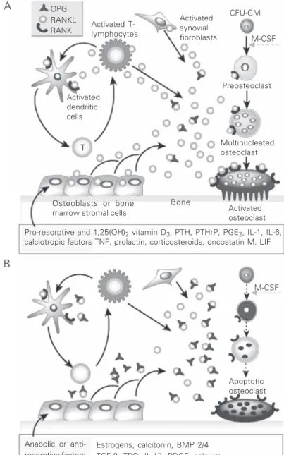
Figure 1. Hormonal control of bone resorption. Schematic representation of the mechan-ism of action of A, pro-resorptive and calcitropic factors and B, anabolic and anti-osteoclastic factors. Receptor activator of nuclear factor kappa B ligand (RANKL) expres-sion is induced in osteoblasts, activated T-cells, synovial fibroblasts and bone marrow stromal cells, and RANKL subsequently binds to its specific membrane-bound receptor activator of nuclear factor kappa B (RANK), thereby triggering a network of tumor necrosis factor (TNF) receptor-associated factor-mediated kinase cascades that promote osteoclast differentiation, activation and survival. Conversely, osteoprotegerin (OPG) expression is induced by factors that block bone catabolism and promote anabolic effects. OPG binds and neutralizes RANKL, leading to a block in osteoclastogenesis and decreased survival of pre-existing osteoclasts. CFU-MG = colony-forming units-granulocyte-macrophage; M-CSF = macrophage colony-stimulating factor; PTH = parathyroid hormone; PTHrP = PTH-related peptide; PGE2 = prostaglandin E2; IL = interleukin; LIF = leukemia inhibitor factor;
BMP 2/4 = bone morphogenetic proteins 2 and 4; TGF-ß = transforming growth factor ß; TPO = thrombopoietin; PDGF = platelet-derived growth factor. (Reproduced, with permis-sion, from Ref. 15).
Pro-resorptive and 1,25(OH)2 vitamin D3, PTH, PTHrP, PGE2, IL-1, IL-6,
calciotropic factors TNF, prolactin, corticosteroids, oncostatin M, LIF Activated
T-lymphocytes
Activated synovial fibroblasts OPG
RANKL RANK
Activated dendritic cells
Preosteoclast M-CSF CFU-GM
Multinucleated osteoclast
Activated osteoclast Osteoblasts or bone
marrow stromal cells
Bone
M-CSF
Apoptotic osteoclast
Anabolic or anti-resorptive factors
Estrogens, calcitonin, BMP 2/4 TGF-β, TPO, IL-17, PDGF, calcium T
A
and IL-15) by proliferating T-cells (18). It has been shown that RANKL can increase the number and persistence of anti-gen-presenting dendritic cells in vivo (16). Moreover, RANKL treatment increases anti-gen-specific primary T-cell responses and appears to be an important factor in memory T-cell responses (16), probably by increas-ing and/or changincreas-ing the production of cyto-kines such as IL-12. However, none of these molecules (RANKL, RANK, OPG) seems to play an essential role in T-cells or dendritic cells that cannot be compensated for by other molecules such as CD40/CD40L, al-though they can influence some aspects of lymphocyte and dendritic cell functions (19). The expression of these molecules can be influenced by sex hormones (10,20), so it is possible that they control sex-specific differ-ences in immunity and could be involved in the higher incidence of some autoimmune diseases in women, for example. Surpris-ingly, in vivo studies have shown T-cells to be crucial to the mechanism by which estro-gen deficiency induces bone loss. T-cells from ovariectomized mice produce increased amounts of TNF which augments RANKL. Roggia et al. (21) showed that ovariectomy induced rapid bone loss in wild-type mice but failed to do so in TNF-deficient mice. Thus, the true function of RANK/RANKL/OPG in the immune system remains to be elucidated. Several cytokines (TNF-α, IL-1, IL-11, IL-17, and others) with regulatory effects on immune function appear to contribute to bone homeostasis by enhancing bone re-sorption. These cytokines have already been identified in the rheumatoid synovium and may promote osteocartilaginous resorption by stimulating osteoclastic mediators (22). Synovial tissue provides a source of RANKL that could influence osteoclastogenesis. Fur-thermore, synovial fibroblasts and T-lym-phocytes (3) from patients with RA produce mRNA and protein (2) for RANKL. Synovial tissues may also provide a source of osteo-clast precursor cells since macrophages
iso-lated from RA differentiate into osteoclasts in the presence of M-CSF plus RANKL. Cells digested from RA synovial tissue samples generate osteoclasts, tartrate-resistant acid phosphatase (TRAP)-positive multinucleated cells that form resorption pits on dentin slices. The formation of these resorption pits is inhibited by OPG. In addition, the number of resorption pits strongly correlates with the ratios ofRANKL/OPG mRNA levels.Given the important role of RANK, RANKL and OPG in bone metabolism and immune func-tion, it has been suggested that the RANK/ RANKL/OPG system and these cytokines may work together in order to cause bone resorption via regulation of the RANKL/OPG ratio (23). Several lines of evidence have shown that some of these cytokines can influence the expression of RANKL and OPG. For example, IL-1, IL-11, IL-17,
TNF-α, PTHrP, and prostaglandin E2 can increase
RANKL mRNA expression by T-cells, and PTHrP and prostaglandin E2 can decrease
OPG mRNA expression in these cells (22,24). Moreover, IL-6 can induce RANKL mRNA expression in cultures of stromal cells from rodents (25), and IL-1 and TNF-α can cause stromal cells and osteoblasts to produce IL-7, which induces osteoclastogenesis via the RANKL system (26).
IL-17 is a crucial cytokine for osteoclas-tic bone resorption and may parosteoclas-ticipate in the development of cartilage destruction in RA patients (27,28). The levels of this cytokine were significantly higher in RA patients and anti-IL-17 antibody significantly inhibited osteoclast formation induced by culture medium of RA synovial tissues. IL-1 is another important cytokine involved in syn-ovial inflammation and bone resorption. Chabaud and Miossec (29) demonstrated that the combination of TNF-α, 1 and IL-17 receptors was more effective in inhibiting bone resorption than each cytokine alone in RA synovium and bone explants.
with psoriatic arthritis have a marked in-crease in osteoclast precursors and readily form osteoclasts in vitro without exogenous RANKL or M-CSF (30).
However, a counter-regulatory mechan-ism by which activated T-cells can inhibit the osteoclastogenesis induced by RANKL has been recently described (31). This mechan-ism involves the participation of interferon-γ
(IFN-γ), which interferes with the signal generated by RANKL. IFN-γ reduces TRAF-6 expression in a selective manner, causing an inhibition of the transcription pathways induced by RANKL and, therefore, decreas-ing osteoclast formation.
It has already been demonstrated that production of RANKL by activated T-cells can directly control osteoclastogenesis and bone remodeling in vitro and in vivo, and these effects can be blocked by the adminis-tration of OPG (16). So, systemic activation of T-cells can lead to bone loss, indicating that T-cells are important mediators of bone loss in vivo. T-cells are probably not re-quired for normal bone homeostasis, since mutant mice lacking T-cells still have normal bone cavities and tooth eruption (32). How-ever, it seems that chronic activation of T-cells can affect bone remodeling by RANKL production and chronic glucocorticoid ad-ministration can lead to bone loss, probably by inducing RANKL expression and de-creasing OPG production (33). These find-ings provide a framework for local or sys-temic bone loss associated with diseases that involve the immune system. Inhibition of RANKL function via OPG might prevent bone destruction in these diseases.
RANKL and arthritis
Focal or diffuse bone loss represents a major unsolved problem in RA. This is a condition of gradual joint destruction related to chronic inflammation with T-cell activa-tion. The skeletal complications of RA con-sist of focal bone erosions and periarticular
osteoporosis at sites of active inflammation and generalized bone loss with reduced bone mass. Local bone loss in the affected joints frequently results in life-long crippling. In this disease characterized by both inflamma-tion and bone destrucinflamma-tion, interacinflamma-tions be-tween the RANKL/RANK/OPG system and T-cells may partly explain the lesions.
Arthritis in humans and in animal models is characterized by synovial inflammation, erosion of bone and cartilage, severe joint pain, and ultimately life-long crippling. For example, in Lewis rats, experimental induc-tion of arthritis by subcutaneous injecinduc-tion of bacterial products in an adjuvant leads to severe inflammation of bone marrow and of the soft tissues surrounding joints, accom-panied by extensive local bone and cartilage destruction, loss of bone mineral density, and crippling (34). In rats, this condition, called adjuvant-induced arthritis, mimics many of the clinical and pathologic features of human RA.
treated with OPG at the onset of the disease did not show any signs of clinical crippling.
Recent dosing data obtained for male Lewis rats showed that OPG preserves ar-ticular bone in arthritic joints in a dose- and schedule-dependent manner, halts bone ero-sion when given at any point during the course of arthritis, and produces sustained anti-erosive activity after a short course (36). Moreover, a single OPG bolus subcu-taneously injected at the onset of the disease eliminated osteoclasts, preserved bone min-eral density for 7 days, and prevented bone erosions for 4 days. No OPG dosage or regimen alleviated weight loss, inflamma-tion, or periosteal osteophyte production. These data indicate that a single OPG injec-tion will inhibit joint erosions for several days, and that OPG treatment is most effec-tive in protecting joint integrity when initiated early during the disease (37).
Alteration of cartilage structures leading to cartilage collapse constitutes a critical step in arthritic joint destruction. In untreated arthritic rats, partial or complete erosion of the cartilage is observed in both the central and peripheral regions of joint surfaces. In striking contrast, neither cartilage erosion nor matrix degeneration in the centers of joint surfaces occurred in OPG-treated animals (35,36). OPG could protect the cartilage by maintaining the overlying cartilage from the inflammatory cell infiltrates in the bone mar-row. RANK, RANKL and OPG expression have been recently observed in normal carti-lage (38). The functional relevance of RANKL-RANK expression in chondrocyte physiology is not known. Thus, in adjuvant-induced arthritis inhibition of RANKL activ-ity by OPG can prevent cartilage destruction, a critical, irreversible step in the pathogenesis of arthritis.
Arthritis can occur in the absence of T-cells (39). Using in situ hybridization of inflamed rat joints and isolation of different cell populations from these joints, Kong et al. (35) demonstrated that RANKL is indeed
expressed in lymphocytes, macrophages, and especially in synoviocytes. Similarly, genetic ablation of RANKL also does not prevent inflammation in an antibody-medi-ated model of arthritis using the K/BxN serum transfer model (40). Multinucleated TRAP-positive osteoclast-like cells were abundant in resorption lacunae in areas of bone erosion in arthritic control mice, and were completely absent in arthritic rankl knockout mice, demonstrating the absolute requirement for RANKL in osteoclastogen-esis in this serum transfer model of inflam-matory arthritis (41). Cartilage damage was still observed in both arthritic rankl knockout mice and arthritic control mice but a trend toward milder cartilage damage in the rankl
-/-mice was noted. Thus, it appears that RANKL is not required for cartilage destruction but clearly plays a still unidentified modulatory role (41).
IL-1 and, to a lesser extent, TNF-α are critical mediators of antibody-induced ar-thritis in the serum transfer model (42). Importantly, inhibition of RANKL via OPG at the onset of disease prevents bone erosion and has a beneficial effect on cartilage de-struction without affecting inflammation in a TNF-α-induced arthritis model (43,44). Furthermore, a significant reduction of os-teoclast numbers was seen in animals treated with OPG. TNF-α-triggered joint destruc-tion is critically dependent on RANKL ex-pression and OPG alone or in combination with bisphosphonates is an effective thera-peutic tool for the prevention of TNF-medi-ated destruction of bone by reducing the number of bone-resorbing cells in the in-flamed tissue (44).
de-struction were evaluated (44-46). Schett et al. (45) investigated systemic bone changes in human TNF transgenic mice which spon-taneously developed severe inflammatory arthritis and osteoclast blockage with OPG. Osteodensitometry revealed a decrease in trabecular bone mineral density (-37%) and histomorphometry revealed a dramatic loss of bone volume (-85%) compared with wild-type controls. OPG completely blocked
TNF-α-mediated bone loss by increasing bone mineral density (+89%) and bone volume (+647%).Zwerina et al. (46) investigated the efficacy of single and combined blockade of TNF, IL-1 and RANK pathways on synovial inflammation, bone erosion and cartilage destruction in a TNF-driven arthritis model. Synovial inflammation was inhibited by anti-TNF (-51%), but not by IL-1Ra or OPG monotherapy. The combination of anti-TNF with either IL-1Ra (-91%) or OPG (-81%) was additive and almost completely blocked inflammation. Bone erosion was effectively blocked by anti-TNF (-79%) and OPG (-60%), but not by IL-1Ra monotherapy. The combination of anti-TNF and IL-1Ra, however, completely blocked bone erosion (-98%) and was the most effective double combination therapy in preventing cartilage destruction (-80%).
RANKL is also expressed in collagen-induced arthritis in which focal collections of osteoclasts are prominent at sites of bone destruction (47). After induction of col-lagen-induced arthritis in Dark Agouti rats, short-term OPG treatment at the onset of the disease effectively prevented joint destruc-tion, even though it had no impact on the inflammatory aspects of collagen-induced arthritis. In arthritic joints, OPG treatment depleted osteoclast numbers by over 75% and diminished bone erosion scores by over 60%. Although cartilage loss was also re-duced by OPG, the effects on cartilage were again lower than those on bone (48). Similar to this rat model, it has been recently sug-gested that the RANKL-RANK system plays
an important role in osteoclastogenesis in both local and systemic osteolytic lesions in autoimmune type II collagen-induced arthri-tis in mice (49). These studies provide evi-dence for the role of osteoclasts (and of RANKL) in the pathogenesis of bone erosion in arthritis in several animal models of arthri-tis with distinct pathogenetic mechanisms.
adjuvant arthritis models and suggest that RANKL is the principal mediator of bone destruction in human arthritis.
Perspectives
The association between resorption and the elevation of the RANKL/OPG ratio sug-gests that the recombinant OPG may be beneficial in a number of conditions. A recent study on postmenopausal women confirmed that OPG reduced bone resorption in vivo: a single monthly injection decreased deoxypy-ridinoline levels by 80% (56). OPG might help to combat inflammation-induced bone resorption in patients with RA.
In all experimental animal models studied thus far, inhibition of RANKL had no appar-ent effect on inflammation, but completely prevented bone loss and partially protected cartilage in all arthritis models studied thus far. The connections between cytokine
pro-duction by inflammatory cells and subse-quent activation of the RANKL/RANK sys-tem point to a unifying paradigm for the entire spectrum of skeletal pathology in RA. Thus, whereas inhibition of TNF and IL-1 using soluble receptor antagonists to some extent prevents inflammation and bone loss in patients with arthritis (57-59), inhibition of the downstream RANKL effectors via OPG or other drugs should prevent bone destruc-tion and cartilage damage in patients with RA irrespective of the initial trigger. Whether inhibition of RANKL will also be beneficial for other forms of arthritis, in particular osteoarthritis, remains to be seen. RANKL inhibition appears to be the most rational and advisable strategy to prevent bone destruc-tion in multiple diseases, to possibly eradicate major human diseases such as osteoporosis, periodontal disease and rheumatoid arthritis that affect millions of people (60).
References
1. Bromley M & Wooley DE (1984). Chondrocytes and osteoclasts at subchondral sites of erosions in the rheumatoid joint. Arthritis and Rheumatism, 27: 968-975.
2. Gravallese EM, Manning C, Tsay A, Naito A, Pan C, Amento E & Goldring SR (2000). Synovial tissue in rheumatoid arthritis is a source of osteoclast differentiation factor. Arthritis and Rheuma-tism, 43: 250-258.
3. Romas E, Bakharevski O, Hards DK, Kartsogiannis V, Quinn JMW, Ryan PF, Martin TJ & Gillespie MT (2000). Expression of osteoclast differentiation factor at sites of bone erosion in collagen-induced arthritis. Arthritis and Rheumatism, 43: 821-826.
4. Lacey DL, Timms E, Tan HL et al. (1998). Osteoprotegerin ligand is a cytokine that regulates osteoclast differentiation and activation.
Cell, 93: 165-176.
5. Anderson DM, Maraskovsky E, Billingsley WL, Dougall WC, Tometsko ME, Roux ER, Teepe MC, DuBose RF, Cosman D & Galibert L (1997). A homologue of the TNF receptor and its ligand enhance T-cell growth and dendritic-cell function. Nature, 390: 175-179.
6. Wong BR, Rho J, Arron J et al. (1997). TRANCE is a novel ligand of the tumor necrosis factor family that activates c-Jun N-terminal kinase in T-cells. Journal of Biological Chemistry, 272: 25190-25194.
7. Lum L, Wong BR, Jasien R, Becherer JD, Erdjument-Bromage H, Schlondorff J, Tempst P, Choi Y & Blobel CP (1999). Evidence for a role of a tumor necrosis factor-alpha (TNF-alpha)-converting enzyme-like protease in shedding of TRANCE, a TNF family
mem-ber involved in osteoclastogenesis and dendritic cell survival.
Journal of Biological Chemistry, 274: 13613-13618.
8. Kong YY, Yoshida H, Sarosi I et al. (1999). OPGL is a key regulator of osteoclastogenesis, lymphocyte development and lymph-node organogenesis. Nature, 397: 315-323.
9. Li J, Sarosi I & Yan XO (2000). RANK is the intrinsic hematopoietic cell surface receptor that controls osteoclastogenesis and regula-tion of bone mass and calcium metabolism. Proceedings of the National Academy of Sciences, USA, 97: 1566-1571.
10. Fata JE, Kong YY, Li J, Sasaki T, Irie-Sasaki J & Moorehead RA (2000). The osteoclast differentiation factor osteoprotegerin-ligand is essential for mammary gland development. Cell, 103: 41-50. 11. Hsu, H, Lacey DL, Dunstan CR, Solovyev I, Colombero A & Timms
E (1999). Tumor necrosis factor receptor family member RANK mediates osteoclast differentiation and activation induced by osteoprotegerin ligand. Proceedings of the National Academy of Sciences, USA, 96: 3540-3545.
12. Wong BR, Besser D, Kim N, Arron JR, Vologodskaia M, Hanafusa H & Choi Y (1999). TRANCE, a TNF family member, activates Akt/PKB through a signaling complex involving TRAF6 and c-Src. Molecular Cell, 4: 1041-1049.
13. Simonet WS, Lancey DL & Dunstan CL (1997). Osteoprotegerin: a novel secreted protein involved in the regulation of bone density.
Cell, 89: 309-319.
14. Bucay N, Sarosi I & Dunstan CR (1998). Osteoprotegerin-deficient mice develop early onset osteoporosis and arterial calcification.
15. Boyle WJ, Simonet WS & Lacey DL (2003). Osteoclast differentia-tion and activadifferentia-tion. Nature, 423: 337-342.
16. Josien R, Li HL, Ingulli E, Sarma S, Wong BR, Vologodskaia M, Steinman RM & Choi Y (2000). TRANCE, a tumor necrosis factor family member, enhances the longevity and adjuvant properties of dendritic cells in vivo. Journal of Experimental Medicine, 191: 495-502.
17. Roy M, Waldschmidt T, Aruffo A, Ledbetter JA & Noelle RJ (1993). The regulation of the expression of gp39, the CD40 ligand, on normal and cloned CD4+ T-cells. Journal of Immunology, 151: 2497-2510.
18. Josien R, Wong BR, Li HL, Steinman RM & Choi Y (1999). TRANCE, a TNF family member, is differentially expressed on T cell subsets and induces cytokine production in dendritic cells. Journal of Immunology, 162: 2562-2568.
19. Jones DH, Kong YY & Penninger JM (2002). Role of RANKL and RANK in bone loss and arthritis. Annals of the Rheumatic Diseases, 61: ii-32-ii-39.
20. Saika M, Inoue D, Kido S & Matsumoto T (2001). 17-Beta-estradiol stimulates expression of osteoprotegerin by a mouse stromal cell line, ST-2, via estrogen receptor-alpha. Endocrinology, 142: 2205-2212.
21. Roggia C, Gao Y, Cenci S, Weitzmann MN, Toraldo G, Isaia G & Pacifici R (2001). Up-regulation of TNF-producing T-cells in the bone marrow: a key mechanism by which estrogen deficiency induces bone loss in vivo. Proceedings of the National Academy of Sciences, USA, 20: 13960-13965.
22. Goldring SR & Gravallese EM (2000). Pathogenesis of bone ero-sions in rheumatoid arthritis. Current Opinion in Rheumatology, 12: 195-199.
23. Saidenberg-Kermanac’h N, Cohen-Solal M, Bessis N, De Vernejoul MC & Boissier MC (2004). Role of osteoprotegerin in rheumatoid inflammation. Joint Bone Spine, 71: 9-13.
24. Suda T, Takahashi N, Udagawa N, Jimi E, Gillepsie MT & Martin TJ (1999). Modulation of osteoclast differentiation and function by the new member of the tumor necrosis factor receptor and ligand families. Endocrine Reviews, 20: 345-357.
25. Kotake S, Sato K, Kim KJ, Takahashi N, Udagawa N, Nakamura I, Yamaguchi A, Kishimoto T, Suda T & Kashiwazaki S (1996). Inter-leukin-6 and soluble interleukin 6 receptors in the synovial fluids from rheumatoid arthritis patient are responsible for osteoclast-like cell formation. Journal of Bone and Mineral Research, 11: 88-95. 26. Weitzmann MN, Cenci S, Rifas L, Brown C & Pacifici B (2000). Interleukin-7 stimulates osteoclast formation by up-regulating the T-cell production of soluble osteoclastogenic cytokines. Blood, 96: 1873-1878.
27. Kotake S, Udagawa N, Takahashi N et al. (1999). IL-17 in synovial fluids from patients with rheumatoid arthritis is a potent stimulator of osteoclastogenesis. Journal of Clinical Investigation, 103: 1345-1352.
28. Van Bezooijen RL, Van Der Wee-Pals L, Papapoulos SE & Lowik CW (2002). Interleukin 17 synergises with tumor necrosis factor alpha to induce cartilage destruction in vitro. Annals of the Rheu-matic Diseases, 61: 870-876.
29. Chabaud M & Miossec P (2001). The combination of tumor necro-sis factor alpha blockade with interleukin-1 and interleukin-17 blockade is more effective for controlling synovial inflammation and bone resorption in an ex vivo model. Arthritis and Rheumatism, 44: 1293-1303.
30. Ritchlin CT, Haas Smith SA, Li P, Hicks DG & Schwarz EM (2003). Mechanisms of TNF-alpha- and RANKL-mediated
osteoclastogen-esis and bone resorption in psoriatic arthritis. Journal of Clinical Investigation, 111: 821-831.
31. Takayanagi H, Ogasawara K, Hida S et al. (2000). T-cell-mediated regulation of osteoclastogenesis by signaling cross-talk between RANKL and IFN-gamma. Nature, 408: 600-605.
32. Nakashima T, Wada T & Penninger JM (2003). RANKL and RANK as novel therapeutic targets for arthritis. Current Opinion in Rheu-matology, 15: 280-287.
33. Vidal NO, Brandstrom H, Jonsson KB & Ohlsson C (1998). Osteoprotegerin mRNA is expressed in primary human osteoblast-like cells: down-regulation by glucocorticoids. Journal of Endocri-nology, 159: 191-195.
34. Bendele A, McComb J, Gould T, MaAbee T, Semello G, Chlipala E & Guy M (1999). Animal models of arthritis: relevance to human disease. Toxicologic Pathology, 27: 134-142.
35. Kong YY, Feige U, Sarosi I et al. (1999). Activated T-cells regulate bone loss and joint destruction in adjuvant arthritis through osteoprotegerin ligand. Nature, 402: 304-309.
36. Campagnuolo G, Bolon B & Feige U (2002). Kinetics of bone protection by recombinant osteoprotegerin therapy in Lewis rats with adjuvant arthritis. Arthritis and Rheumatism, 46: 1926-1936. 37. Bolon B, Campagnuolo G & Feige U (2002). Duration of bone
protection by a single osteoprotegerin injection in rats with adju-vant-induced arthritis. Cellular and Molecular Life Sciences, 59: 1569-1576.
38. Komuro H, Olee T, Kuhn K, Quach J, Brinson DC, Shikhman A, Valbracht J, Creighton-Achermann L & Lotz M (2001). The osteoprotegerin/receptor activator of nuclear factor kappaB/recep-tor activakappaB/recep-tor of nuclear fackappaB/recep-tor kappaB ligand system in cartilage.
Arthritis and Rheumatism, 44: 2768-2776.
39. Benoist C & Mathis D (2000). A revival of the B cell paradigm for rheumatoid arthritis pathogenesis? Arthritis Research, 2: 90-94. 40. Kouskoff V, Korganow AS, Duchatelle V, Degott C, Benoist C &
Mathis D (1996) Organ-specific disease provoked by systemic autoimmunity. Cell, 87: 811-822.
41. Pettit AR, Ji H, von Stechow D, Muller R, Goldring SR, Choi Y, Benoist C & Gravallese EM (2001). TRANCE/RANKL knockout mice are protected from bone erosion in the K/BxN serum transfer model of arthritis. American Journal of Pathology, 159: 1689-1699. 42. Ji H, Pettit A, Ohmura K, Ortiz-Lopez A, Duchatelle V, Degott C, Gravallese E, Mathis D & Benoist C (2002). Critical roles for interleu-kin 1 and tumor necrosis factor alpha in antibody-induced arthritis.
Journal of Experimental Medicine, 196: 77-85.
43. Keffer J, Probert L, Cazlaris H, Georgopoulos S, Kaslaris E, Kioussis D & Koelias G (1991). Transgenic mice expressing human tumor necrosis factor: a predictive genetic model of arthritis. EMBO Journal, 10: 4025-4031.
44. Redlich K, Hayer S, Maier A et al. (2002). Tumor necrosis factor alpha-mediated joint destruction is inhibited by targeting osteo-clasts with osteoprotegerin. Arthritis and Rheumatism, 46: 785-792.
45. Schett G, Redllich K, Zwerina J et al. (2003). Osteoprotegerin protects against generalized bone loss in tumor necrosis factor-transgenic mice. Arthritis and Rheumatism, 48: 2042-2051. 46. Zwerina J, Hayer S, Tohidast-Akrad M et al. (2004). Single and
combined inhibition of tumor necrosis factor, interleukin-1, and RANK pathways in tumor necrosis factor-induced arthritis. Effects on synovial inflammation, bone erosion, and cartilage destruction.
Arthritis and Rheumatism, 50: 277-290.
(2002). Increase in expression of receptor activator of nuclear factor κB at sites of bone erosion correlates with progression of inflammation in evolving collagen-induced arthritis. Arthritis and Rheumatism, 46: 3055-3064.
48. Romas E, Sims NA, Hards DK, Lindsay M, Quinn JW, Ryan PF, Dunstan CR, Martin TJ & Gillespie MT (2002). Osteoprotegerin reduces osteoclast numbers and prevents bone erosion in col-lagen-induced arthritis. American Journal of Pathology, 161: 1419-1427.
49. Mori H, Kitazawa R, Mizuki S, Nose M, Maeda S & Kitazawa S (2002). RANK ligand, RANK, and OPG expression in type II collagen-induced arthritis mouse. Histochemistry and Cell Biology, 117: 283-292.
50. Kotake S, Udagawa N, Hakoda M et al. (2001). Activated human T-cells directly induce osteoclastogenesis from human monocytes: possible role of T-cells in bone destruction in rheumatoid arthritis patients. Arthritis and Rheumatism, 44: 1003-1012.
51. Ziolkowska M, Kurowska A, Radzikowska A et al. (2002). High levels of osteoprotegerin and soluble receptor activator of nuclear factor kappa B ligand in serum of rheumatoid arthritis patients and their normalization after anti-tumor necrosis factor alpha treatment.
Arthritis and Rheumatism, 46: 1744-1753.
52. Haynes DR, Crotti TN, Loric M, Bain GI, Atkins GI & Findlay DM (2001). Osteoprotegerin and receptor activator of nuclear factor κB ligand (RANKL) regulate osteoclast formation by cells in the human rheumatoid arthritic joint. Rheumatology, 40: 623-630.
53. Takayanagi H, Iizuka H, Juji T, Nakagawa T, Yamamoto A, Miyazaki T, Koshihara Y, Oda H, Nakamura K & Tanaka S (2000). Involve-ment of receptor activator of nuclear factor κBligand/osteoclast
differentiation factor in osteoclastogenesis from synoviocyte in rheumatoid arthritis. Arthritis and Rheumatism, 43: 259-269. 54. Itonaga I, Fujikawa Y, Sabokbar A, Murray DW & Athanasou NA
(2000). Rheumatoid arthritis synovial macrophage-osteoclast dif-ferentiation is osteoprotegerin ligand-dependent. Journal of Pa-thology, 192: 97-104.
55. Takayanagi H, Oda H, Yamamoto S, Kawaguchi H, Tanaka S, Nishikawa T & Koshihara Y (1997). A new mechanism of bone destruction in rheumatoid arthritis; synovial fibroblasts induce os-teoclastogenesis. Biochemical and Biophysical Research Commu-nications, 240: 279-286.
56. Bekker PJ, Holloway D, Nakanishi A, Arrigh HM, Leese PT & Dunstan CR (2001). Osteoprotegerin (OPG) has potent and sub-stantial anti-resorptive activity in post menopausal women. Journal of Bone and Mineral Research, 16: 348-360.
57. Fye KH (1999). New treatments for rheumatoid arthritis. Available and upcoming slow-acting antirheumatic drugs. Postgraduate Medicine, 106: 82-85.
58. Graninger WB & Smolen JS (2001). One-year inhibition of tumor necrosis factor-alpha: a major success or a larger puzzle? Current Opinion in Rheumatology, 13: 9-13.
59. Oliveri MB, Mautalen CA, Rodriguez Fuchs CA & Romanelli MC (1991). Vertebral compression fractures at the onset of acute lymphoblastic leukemia in a child. Henry Ford Hospital Medical Journal, 39: 45-48.