Abstract—Introduction: The application of fissure sealants is considered to be an important primary prevention method used in dental medicine. However, one of the most common reasons of dental caries development in teeth with fissure sealants is due to the formation of microleakages. The association between various dental biomaterials may limit the major disadvantages and limitations of biomaterials functioning in a complementary manner. The present study consists in the incorporation of a cariostatic agent – silver diamine fluoride (SDF) – in a resin-based fissure sealant followed by the study of release kinetics by spectrophotometry analysis of the association between both biomaterials and assessment of the inhibitory effect on the growth of the reference bacterial strain Streptococcus mutans (S. mutans) in an in vitro study. Materials and Methods: An experimental in vitro study was designed consisting in the entrapment of SDF (Cariestop® 12% and 30%) into a commercially available fissure sealant (Fissurit®), by photopolymerization and photocrosslinking. The same sealant, without SDF was used as a negative control. The effect of the sealants on the growth of S. mutans was determined by the presence of bacterial inhibitory halos in the cultures at the end of the incubation period. In order to confirm the absence of bacteria in the surface of the materials, Scanning Electron Microscopy (SEM) characterization was performed. Also, to analyze the release profile of SDF along time, spectrophotometry technique was applied. Results: The obtained results indicate that the association of SDF to a resin-based fissure sealant may be able to increase the inhibition of S. mutans growth. However, no SDF release was noticed during the in vitro release studies and no statistical significant difference was verified when comparing the inhibitory halo sizes obtained for test and control group. Conclusions: In this study, the entrapment of SDF in the resin-based fissure sealant did not potentiate the antibacterial effect of the fissure sealant or avoid the immediate development of dental caries. The development of more laboratorial research and, afterwards, long-term clinical data are necessary in order to verify if this association between these biomaterials is effective and can be considered for being used in oral health management. Also, other methodologies for associating cariostatic agents and sealant should be addressed.
Nélio Veiga is with the Health Sciences Institute of the Universidade Católica Portuguesa and the Centre for Interdisciplinary Research in Health (CIIS) of the Universidade Católica Portuguesa (corresponding author, Estrada da Circunvalação, 3504-505, Viseu; phone: 00351 966454933; e-mail: nelioveiga@gmail.com).
Paula Ferreira is with the Chemical Process Engineering and Forest Products Research Centre, Chemical Engineering Department, University of Coimbra (e-mail: pcnferreira@gmail.com).
Tiago Correia and Ilídio J. Correia are with the Health Sciences Research Centre – Health Sciences Faculty, Beira Interior University (e-mail: trc2003@gmail.com, icorreia@ubi.pt).
Maria J. Correia is with the Health Sciences Institute of the Universidade Católica Portuguesa and the Centre for Interdisciplinary Research in Health (CIIS) of the Universidade Católica Portuguesa (e-mail: mariacorrei@gmail.com).
Carlos Pereira and Odete Amaral are with CI&DETS, Polytechnic Institute of Viseu (e-mail: carlospereiraviseu@gmail.com, mopamaral@gmail.com).
Keywords—Biomaterial, fissure sealant, primary prevention, silver diamine fluoride, S. mutans.
I.
INTRODUCTIONHE development of dental cariesis a highly prevalent oral disease worldwide [1]. This susceptibility is related with the physical size and individual morphology of pits and fissures of the tooth surface, where microorganisms can cohabit, causing more difficulties of the oral hyigiene procedures of these areas, allowing greater amount of dental plaque retention [2]. This is considered as being an important risk factor because the accumulation of bacterial biofilms is responsible for oral disease development, such as dental caries and periodontitis [3]. Fissure sealant application in pits and fissures of the occlusal surface of the tooth permits the creation of a physical barrier for the bacterial biofilm accumulation in these specific anatomical areas of the tooth [4], [5]. Fissure sealants application in high susceptible tooth decay areas is one of the primary preventive measures to minimize the risk of dental caries development, reducing its incidence in pits and fissures, preventing to carry out more invasive dental fillings with silver amalgam or filling resins [1], [6], [7]. A study developed by Hiiri et al. evidences the superiority fissure sealants over fluoride varnish application as a primary prevention method to avoid dental caries development. [8].
A key problem of fissure sealants is the high risk of microleakage development sometime after their application. This situation may happen due to chewing forces applied during time and also because of polymerization shrinkage of resin-based fissure sealants that might facilitate the formation of gaps between the material and the tooth surface, providing space for bacterial invasion and proliferation [9]. This microleakage may lead to dental plaque accumulation, which in contact with enamel, can turn into a carious lesion [10].
The reassessment of fissure sealants should be made each six months in order to avoid the development of dental caries in teeth that have microleakage on the fissure sealant applied. This reassessment is especially important in particular cases of patients with high risk of developing dental caries and insufficient oral health behaviors [11].
It would be important to have a fissure sealant which offers a genuinely antimicrobial and antibiofilm efficacy that would be a considerable clinical benefit. Such a material could reduce recurrent decay providing an antibacterial seal, protecting the enamel surface of the tooth [12].
T
Antibacterial Effect of Silver Diamine Fluoride
Incorporated in Fissure Sealants
Various attempts have been made in order to reduce plaque accumulation on the surface of restorative and preventive materials by the incorporation of bacterial agents [3]. A study developed by Li et al. demonstrated that the incorporation of an antibacterial agent (DMAE-CB) may influence the antibacterial effect and properties [9]. In this study, it was found that the combination of an antibacterial agent in fissure sealants did not affect its main properties and also does not increase the risk of microleakage. Various studies have demonstrated the antibacterial beneficial combination between a bactericidal and / or bacteriostatic agent and an adhesive system, and other dental biomaterials, with a reduces risk of affecting its chemical and physcial properties [13].
Another proven to be efficient primary prevention method consists in the application of fluoride on the enamel surface of the teeth. Fluoride is well known to reduce dental caries by reducing the solubility and enhancing remineralization of dental enamel by the incorporation of available fluoride into the tooth hard tissue structure during the presence of the organic acids [14]. Studies showed that the incorporation of fluoride components in biomaterials may increase their antibacterial effect. [8,15-18]. However, there is still some debate among researchers about whether a sealant containing fluoride actually prevents dental caries more effectively than does a sealant without fluoride [9].
The association of an antibacterial agent with certain dental biomaterials may increase the inhibitory effect on the growth of bacterial strains, in this case, of S. mutans [9,13,19,20]. Nowadays, nanotechnology has become a key research field that has brought beneficial applications in medicine and healthcare. One of the most advanced methods applied in healthcare consists in the development of releasing controlled systems of silver particles, which increases the therapeutical antibacterial effect of silver ion particles [21,22]. Silver is a natural antibacterial agent and has properties that permits its use in medicine, namely the fact of being non-toxic for the human cells, long-term bactericide effect and stability at high temperatures and low volatility [22].
Silver has been used in various situations like water purification, wound treatments, bone prothesis, surgical orthopedic reconstruction, development of heart functioning devices and even has been incorporated in textile made for hospital clothing in order to decrease the risk of nosocomial infections [23,24].
Some studies demonstrate the possible association of silver ion particles with filled resins applied in dental medicine which increases the antibacterial effect and assures a better protection against the development of recidivating dental caries [25].
We can also find a biomaterial that associates the advantages of sodium fluoride and silver nitrate: SDF. This is a biomaterial with proven anti-cariostatic properties that can stop the development of dental caries in the hard tissue of the tooth with a mechanism that consists in the self-stimulation of the calcified and sclerotic dentin formed by the silver ion [19, 26-28].
Other studies show that the synergism established between the fluoride and the silver increases the enamel resistance against the organic acids produced in the oral cavity and that is
responsible for the dental tissue demineralization, which gives this biomaterial the possibility of becoming an important primary prevention method for deciduous and permanent teeth in the future [21,29]. SDF has also been shown to inhibit bacterial growth, mainly against cariogenic strains of S. mutans [30].
In the present study, SDF was incorporated by physical entrapment in a resin-based fissure sealant. The prepared system was afterwards analyzed by assessing its inhibitory effect on the growth of S. mutans strain.
II. MATERIALS AND METHODS
The purpose of this work was to assess the efficiency of incorporating SDF in a selected resin-based fissure sealant. S. mutans was used as model bacteria, since it is considered to be the primary responsible for the initiation of dental caries as well as for the progression of an established lesion [3]. This study was developed in three different stages:
Stage 1: Samples with SDF were prepared. Two commercially available SDF solutions with different concentrations were used: 12% (Cariestop 12%) and 30% (Cariestop 30%). Samples were prepared by adding the same concentration of cariostatic to the sealant (10%), meaning that the only variable was the initial SDF solution concentration. Once prepared, the mixtures were photopolymerized under UV irradiation for 30s. Samples without incorporation of SDF were prepared directly from the sealant to be further used as a negative control. At the end of this phase, three groups of samples were obtained: resin fissure sealant (sample A); resin fissure sealant with SDF 12% solution (Sample B) and resin fissure sealant with SDF 30% solution (Sample C).
Stage 2: Assessment of the inhibitory effect on the bacterial strain S. mutans growth in the presence of the samples prepared during stage 1. The bacteria were cultured in Brain Heart Infusion broth, at 37ºC, anaerobically, for 24 hours. S.
mutans (1.0 x10⁸ colony-forming units (CFU/mL) was
inoculated in an agar plate with the different materials. After 24 hours, the inhibitory halos were analyzed and measured, as previously described in literature [31]. Furthermore, in order to confirm the absence of bacteria at the surface of the materials, SEM analysis was also performed [32].
Stage 3: Analysis of the release profile of SDF from samples was performed by spectrophotometric technique. In a first approach, a scanning of the SDF solutions was performed in the range of 200 to 500 nm of the electromagnetic spectrum. The wavelength in which the maximum absorbance value was registered and established as the one to be used during samples analyses. Afterwards, 0.5g of each sample were individually immersed in distilled water and placed in an oven at 37ºC. At predetermined times, the samples were removed from the oven and the absorbance of the incubation medium was measured at previously established wavelength.
III. RESULTS
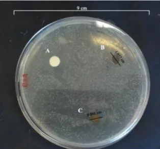
The obtained results demonstrate that the materials have inhibited the bacterial growth after 24 hours. The determination of the inhibitory halos surrounding the samples was assessed. In the sample containing the resin-based fissure sealant with 12 % SDF solution (sample B) the inhibitory halo
had a diameter of 1.06 cm while the sample containing the resin-based fissure sealant with 30 % SDF solution (sample C) had a 0.89 cm inhibitory halo (Fig 1). Therefore, no statistically significant differences were found between the inhibitory halos obtained for samples B and C.
Fig. 1 - Evaluation of the inhibitory effect of SDF on S. mutans: A) Resin-based fissure sealant without SDF incorporated (control); B) based fissure sealant with 12 % SDF solution and C)
Resin-based fissure sealant with 30 % SDF solution.
Moreover, SEM images of the surfaces of the materials were also acquired in order to fully characterize bacterial growth on the materials surface (Fig. 2). As it can be seen, no bacterial growth was noticed on the surfaces of the materials. The authors found that the cured sealant containing SDF had an inhibitory effect on the growth of S. mutans. Although, the control sample (sample A) did not show any detectable growth of the tested bacteria. Further studies have to be done in order to check the antibacterial activity of the produced materials. Release of SDF from the samples produced during stage 1 was studied by using a spectrophotometric approach. For that purpose, a scan in the range between 200 and 500 nm of the electromagnetic spectrum was performed in order to determine the correct wavelength to be used during further analyses. The obtained values of absorbance indicated that maximum absorbance occurred at 300 nm.
As previously described, 0.5 g of each sample were incubated in distilled water at 37ºC in order to evaluate the delivery of SDF from the polymeric matrix. The analyses were performed by measuring the incubation medium’s absorbance at 300 nm. However, the obtained results indicated that no measurable amount of the drug was released. Such may be attributed to the highly rigid structure of the photopolymerized sealant which is crosslinked by UV radiation.
Fig 2 - SEM images of the surfaces of the materials in the presence of S. mutans (S. mutans samples A, B and C) and in the absence of it
(control samples A, B and C) after 24 hour for the resin fissure sealant (sample A); resin fissure sealant with SDF 12% solution
(Sample B) and SDF with SDF 30% solution (Sample C).
IV. DISCUSSION
Providing dental materials with antibacterial properties has been attempted to prevent the formation and development of dental caries [9]. New biomaterials with optimized antibacterial properties are crucial for their future application in communities where oral health behaviors are not well implemented, in order to avoid the increase of oral disease development.
It is essential to understand that the research described in this paper, reports preliminary results of an initial phase of biomaterial development that needs to be complemented in a near future. The results obtained in the present study do not demonstrate significant differences in the inhibitory effect of S. mutans growth between the control sample and the samples with SDF incorporated into the fissure sealant. Also, the SDF release profile from the fissure sealant could not be determined through the spectrophotometric method. However, forthcoming studies will allow the authors to optimize entrapment methodology as well as quantification techniques. As an example, HPLC may be used to quantify SDF release allowing detection of lower amounts of the drug.
Moreover, the antibacterial activity determination of the resin-based fissure sealant and SDF was made only with the bacterial strain S. mutans, since in literature it is described that S. mutans can be easily isolated from dental plaque samples and are considered as being the main bacterial strain involved in the initiation of dental caries [26,33]. Further bacterial strains can be tested in the near future to characterize antimicrobial effect on samples.
SDF is effective in arresting enamel and dentin caries and inhibiting cariogenic biofilm formation [26]. Therefore, the main goal of this study was to provide information concerning the potential clinical performance of the sealant with antibacterial activity, when SDF was added to its production [9,13,19,20].
The methodology applied presents considerable limitations mainly related with the reproductibility of the physiological and biological conditions of oral cavity. Due to these limitations, the results obtained during in vitro studies cannot be extrapolated to an in vivo clinical pattern due to the fact of not being able to reflect the effect of the biomaterial when applied in clinical situations. However, more laboratorial tests have to be done for a complete characterization of the materials performed.
Another point that has to be taken into account as a disadvantage of the use of SDF to arrest caries, is that the lesions will be stained black. It has been suggested that when carious dentin is treated with SDF, silver phosphate is formed and precipitated [30,34].
However, previous studies have demonstrated that SDF application is effective for dental caries arrest, as an alternative for more complex restorative treatments and do not cause adverse effects [3,28].
In addition, it was also described that the incorporation of the SDF in the crosslinked structure of the fissure sealant permits the presence of a high concentration of silver and fluoride ions, which can inhibit the growth of different cariogenic bacteria that are responsible for biofilm formation [34,35]. However, and also due to the highly complex crosslinked structure of the cured fissure sealant, the release of SDF is precluded.
Another issue that has to be taken into account in future studies are the systemic symptoms (argyria) that can occur after topical use of silver. In some cases, silver also produces a localized discoloration of various tissues [3].
V. CONCLUSIONS
In this study, the entrapment of SDF in the resin-based fissure sealant did not show to be an effective method to potentiate the antibacterial effect of the fissure sealant, and also avoid the development of dental caries. Further laboratorial research and, afterwards, long-term clinical data is necessary in order to verify if SDF impregnation in fissure sealants is effective and can be considered advantageous in the field of oral health management. Further research may allow the identification of other dental biomaterials that can prevent and reduce the risk of dental caries development and protect the tooth from bacterial infections.
REFERENCES
[1] [1]. A. Ahovuo-Saloranta, H. Forss, T. Walsh, A. Hiiri, A. Nordblad, M. Mäkelä, et al. "Sealants for preventing dental decay in the permanent teeth," Cochrane Database Syst Rev., vol. 3, pp. CD001830, 2013. [2] [2]. P. Axelsson P, "Preventive Materials, Methods and Programs," 1st
ed. Slovakia: Quintessence Books, 2004.
[3] [3]. R. Burgers, A. Eidt, R. Frankenberger, M. Rosentritt, H. Schweikl, G. Handel, et al. "The anti-adherence activity and bactericidal effect of microparticulate silver additives in composite resin materials," Arch
Oral Biol., vol. 54, pp. 595-601, 2009.
[4] [4]. J. Avinash, C. Marya, S. Dhingra, P. Gupta, S. Kataria, H. Meenu. "Pit and fissure sealants: na unused caries prevention tool," J Oral
Health Comm Dent., vol. 4, no. 1, pp. 1-6, 2010.
[5] [5]. S. Daniel, S. Harfst, R. Wilder, B. Francis, S. Mitchell. "Mosby´s Dental Hygiene: Concepts, Cases and Competencies," 2nd ed. St. Louis, USA: Mosby Elvesier, 2008.
[6] [6]. G. Rose. "Sick individuals and sick populations. Reiteration," Int J
Epidemiol., vol. 30, pp. 427-32, 2001.
[7] [7]. A. Pereira, V. Pardi, F. Mialhe, C. Meneghim, G. Ambrosano. "A 3-year clinical evaluation of glass-ionomer cements used as fissure sealants," Am J Dent., vol. 16, no. 1, pp. 23-7, 2003.
[8] [8]. A. Hiiri, A. Ahovuo-Saloranta, A. Nordblad, M. Makela. "Pit and fissure sealants versus fluoride varnishes for preventing dental decay in children and adolescents," Cochrane Database Syst Rev., vol. 3, pp. CD003067, 2010.
[9] [9]. F. Li, F. Li, D. Wu, S. Ma, J. Gao, Y. Li, et al. "The effect of an antibacterial monomer on the antibacterial activity and mechanical properties of a pit-and-fissure sealant," J Am Dent Assoc., vol 142, no. 2, pp. 184-93, 2011.
[10] [10]. A. Koyuturk, A. Kusgoz, M. Ulker, C. Yeşilyurt. "Effects of mechanical and thermal aging on microleakage of different fissure sealants," Dent Mater J., vol. 27, no. 6, pp. 795-801, 2008.
[11] [11]. Irish Oral Health Services Guideline Initiative. "Pit and Fissure Sealants: Evidence-based guidance on the use of sealants for the prevention and management of pit and fissure caries," Guideline and
supplementary data available at:
http://ohsrc.ucc.ie/html/guidelines.html2010. doi:10.1038/sj.ebd.6400591.
[12] [12]. E. Hook, O. Owen, C. Bellis, J. Holder, D. O`Sullivan, M. Barbour. "Development of a novel antimicrobial-releasing glass ionomer cement functionalized with chlorhexidine hexametaphosphate nanoparticles," J Nanobiotechnol., vol. 12, pp. 3, 2014.
[13] [13]. Y. Xiao, S. Ma, J. Chen, Z. Chai, F. Li, Y. Wang. "Antibacterial activity and bonding ability of an adhesive incorporating an antibacterial monomer DMAE-CB," J Biomed Mater Res B Appl Biomater., vol. 90, no. 2, pp. 813-7, 2009.
[14] [14]. W. Siqueira, M. Bakkal, Y. Xiao, J. Sutton, F. Mendes. "Quantitative proteomic analysis of the effect of fluoride on the acquired enamel pellicle," PloS ONE., vol. 7, no. 8, pp. e42204, 2012.
[15] [15]. J. Loyola-Rodriguez, F. Garcia-Godoy. "Antibacterial activity of fluoride release sealants on mutans streptococci," J Clin Pediatr Dent., vol. 20, no. 2, pp. 109-11, 1996.
[16] [16]. S. Matalon, B. Peretz, R. Sidon, E. Weiss, H. Slutzky. "Antibacterial properties of pit and fissure sealants combined with daily fluoride mouth rinse," Pediatr Dent., vol. 32, no. 1, pp. 9-13, 2010. [17] [17]. T. Morphis, J. Toumba. "Fluoride pit and fissure sealants: a
review," Int J Paediatr Dent., vol. 10, no. 2, pp. 90-8, 2000.
[18] [18]. S. Naorungroj, H. Wei, R. Arnold, E. Swift, R. Walter. "Antibacterial surface properties of fluoride containing resin-based sealants," J Dent., vol. 38, no. 5, pp. 387-91, 2010.
[19] [19]. T. Alves, C. Silva, N. Silva, E. Medeiros, A. Valença. "A Antimicrobial activity of fluoridated products on biofilm-forming bactéria: na in vitro study," Pesq Bras Odontoped Clin Integr, João
Pessoa., vol. 10, no. 2, pp. 209-16, 2010.
[20] [20]. Q. Feng, J. Wu, G. Chen, F. Cui, T. Kim, J. Kim. "A mechanistic study of the antibacterial effect of silver ions on Escherichia coli and Staphylococcus aureus," J Biomed Mater Res., vol. 52, pp. 662-8, 2000. [21] [21]. A. Rosenblatt, T. Stamford, R. Niederman. "Silver Diamine Fluoride: A Caries ''Silver-Fluoride Bullet," J Dent Res., vol. 88, pp. 116-25, 2009.
[22] [22]. R. Kumar, H. Munstedt. "Silver ion release from antimicrobial polyamide/silver composites," Biomaterials., vol. 26, pp. 2081-8, 2005. [23] [23]. M. Kostić, N. Radić, B. Obradović, S. Dimitrijević, M. Kuraica,
P. Škundrić. "Silver-Loaded Cotton/Polyester Fabric Modified by Dielectric Barrier Discharge Treatment," Plasma Processes Polym., vol. 6, no. 1, pp. 58-67, 2009.
[24] [24]. W. Jung, H. Koo, K. Kim, S. Shin, S. Kim, Y. Park. "Antibacterial Activity and Mechanism of Action of the Silver Ion in Staphylococcus aureus and Escherichia coli," Appl Environ Microbiol. vol. 74, no. 7, pp. 2171-8, 2008.
[25] [25]. K. Yamamoto, S. Ohashil, M. Aonol, T. Kokuboz, I. Yamada, J. Yamauchid. "Antibacterial activity of silver ions implanted in filler on oral streptococci," J Dent Mater., vol. 12, pp. 227-9, 1996.
[26] [26]. S. Shah, V. Bhaskar, K. Venkataraghavan, P. Choudhary, M. Ganesh, K. Trivedi. "Efficacy of silver diamine fluoride as an
antibacterial as well as antiplaque agent compared to fluoride varnish and acidulated phosphate fluoride gel: an vivo study," Indian J Dent
Res., vol. 24, no. 5, pp. 575-81, 2013.
[27] [27]. R. Ditterich, M. Romanelli, M. Rastelli, G. Czlusniak, D. Wambier. "Diamino Fluoreto de Prata: uma revisão de literatura," Biol
Saúde Ponta Grossa., vol. 12, no. 2, pp. 45-52, 2006.
[28] [28]. R. Yee, C. Holmgren, J. Mulder, D. Lama, D. Walker, W. Helderman. "Efficacy of Silver Diamine Fluoride for Arresting Caries Treatment," J Dent Res., vol. 88, pp. 644-8, 2009.
[29] [29]. J. Llodra, A. Rodriguez, B. Ferrer, V. Menardia, T. Ramos, M. Morato. "Efficacy of silver diamine fluoride for caries reduction in primary teeth and first permanent molars of schoolchildren: 36-month clinical trial," J Dent Res., vol. 84, no. 8, pp. 721-4, 2005.
[30] [30]. J. Peng, M. Botelho. "Silver compounds used in dentistry for caries management: A review," J Dent., vol. 40, pp. 531-41, 2012. [31] [31]. S. Kim, D. Shin. "Antibacterial effect of self-etching adhesive
systems on Streptococcus mutans," Restor Dent Endod., vol. 39, n0. 1, pp. 32-8, 2014.
[32] [32]. T. Chaitra, R. Subba, G. Devarasa, T. Ravishankar. "Microleakage and SEM analysis of flowable resin used as a sealant following three fissure preparation techniques – an in vitro study," J Clin
Pediatr Dent. vol. 35, no. 3, pp. 277-82, 2011.
[33] [33]. M. Mahesh, B. Mithun, G. Prashant, V. Reddy, M. Usha, C. Madura, et al. "Antibacterial properties of fluoride releasing glass ionomer cements (GICs) and pit and fissure sealants on Streptococcus mutans," Int J Clin Pediatr Dent. vol. 3, no. 2, pp. 93-6, 2010.
[34] [34]. M. Mei, Q. Li, C. Chu, E. Lo, L. Samaranayake. "Antibacterial effects of silver diamine fluoride on multi-species cariogenic biofilm on caries," Ann of Clin Microbiol Antimicrob., vol. 12, pp. 4, 2013. [35] [35]. G. Knight, J. McIntyre. "The effect of silver fluoride and
potassium iodide on the bond strength of auto cure glass ionomer cement to dentine," Aust Dent J., vol. 51, no. 1, pp. 42-5, 2006.