Cyanobacteria
from Cape Verde
Islands: a
contribution to
the diversity and
biotechnological
potential
Bruna Lopes Tavares Silva
Biologia e Gestão da Qualidade da Água
Departamento de Biologia
2019
Orientador
Professor Doutor Vítor Vasconcelos Professor Catedrático
Faculdade de Ciências da Universidade do Porto (FCUP)
Centro Interdisciplinar de Investigação Marinha e Ambiental (CIIMAR)
Co-Orientador
Professora Doutora Maria do Rosário Martins Professora Adjunta
Escola Superior de Saúde, Politécnico do Porto (ESS-IPP)
Todas as correções determinadas pelo júri, e só essas, foram efetuadas. O Presidente do Júri,
Acknowledgements
I would like to thank some people who were important for the accomplishment of this work.
My sincere appreciation goes to my Co supervisor Professor Rosário Martins for the total support, availability, knowledge transmitted, collaboration, guidance, suggestions for planning and carrying out the work.
My appreciations go to my supervisor Vitor Vasconcelos for giving me the opportunity to do my dissertation at the Blue Biotechnology and Ecotoxicology (BBE) laboratory where I could expand my scientific knowledge.
I must also acknowledge the contribution and support of João Morais and Guilherme Hentschke for their great contribution from the beginning and for giving me the opportunity to learn.
My special thanks go to the whole staff of the LEGE (CIIMAR) for providing me with all the support I needed especially to Ana Raquel Silva, Mariana Reis, Leonor Ferreira, João Silva and Jorge Neves.
I would like to thank my Parents Natalia and José for the love, sharing and unconditional support. My brothers Natalicio Martins, Mauricio Martins sister Mitsa Silva, my cousin Lourdes and my friend Keila I thank you for your understanding, generosity and joy. A great appreciation goes to Bruno for all support and love.
This work was done in the framework of the project ALGAVALOR - MicroALGAs: integrated production and valorization of biomass and its various applications, supported by the PORTUGAL 2020 through the European Regional Development Fund (ERDF).
This work was done in the framework project H2020 RISE project EMERTOX— Emergent Marine Toxins in the North Atlantic and Mediterranean: New Approaches to Assess their Occurrence and Future Scenarios in the Framework of Global Environmental Changes—Grant Agreement No. 778069.
This work was supported by the FCT Project UID/Multi/04423/2019 and by the Atlantic Interreg Project – EnhanceMicroAlgae - High added-value industrial opportunities for microalgae in the Atlantic Area (EAPA_338/2016).
Abstract
Cyanobacteria are widespread prokaryotic photosynthetic organisms with an extreme capability to withstand different environment conditions. This adaptive capacity is attributed to the production of secondary compounds with antioxidant, photoprotective and humectant properties among others. In recent years marine cyanobacteria extracts and compounds revealed potential for biotechnology, namely in the pharmacological field. The isolation of marine cyanobacteria from different latitudes and its preservation ex situ is thus of crucial importance in order to trace their bioactive potential. In this work, coastal marine samples were collected from beaches of two Cape Verde Islands, Santo Antão and São Vicente. The objective was to contribute to the study of the cyanobacteria diversity of the archipelago and to isolate strains in order to infer about its biotechnological potential. From a total of 25 samples collected in São Vicente and 3 samples collected in Santo Antão, 20 cyanobacteria strains were isolated using both solid and liquid Z8 medium. Isolates were identified by morphometry and molecular tools, through the sequencing of the 16S rRNA gene. All the isolated strains are filamentous non-heterocystous forms. The phylogenetic analysis reveals strains that might belong to new genera, which contributes to enrich the diversity that occurs within this group of organisms.
To infer about the bioactive potential, strains were cultured in large scale for biomass production and methanolic extracts were prepared and tested for cytotoxicity using the normal keratinocytes cell line HaCat, the normal endothelial cell line hCMEC/D3, the hepatic carcinoma cell line HepG2, the colon carcinoma cell line RKO and the breast carcinoma cell line T47D. Cellular viability was evaluated by the 3-(4.5-dimethylthiazol-2-yl)-2.5-diphenyltetrazolium bromide (MTT) assay after 24 and 48 hours of exposure to the extracts at a final concentration of 100, 75, 50, 25, 12,5 and 6,25 µg mL-1. The MTT
results revealed both toxic and no toxic responses revealing strains with potential for further fractionation. Among those the strain JM1B and JM5A induced selective cytotoxicity against the T47D cell line, revealing potential as anticancer. The strains PL1 and PL2 induced selective toxicity against the keratinocytes cell line, while strains VV5, VV3, VV3B and VV13 induced a proliferation of the same cell line. In both cases results revealed the potential of the strains for skin care applications.
Resumo
As cianobactérias são organismos procarióticos fotossintéticos com uma capacidade extrema de suportar diferentes condições ambientais. Essa capacidade adaptativa é atribuída à produção de compostos secundários com propriedades antioxidantes, fotoprotetoras e hidratantes, entre outras. Nos últimos anos, os extratos e compostos de cianobactérias marinhas têm revelado potencial para aplicação em biotecnologia, principalmente no campo farmacológico. O isolamento de cianobactérias marinhas de diferentes latitudes e sua preservação ex situ reveste-se assim de grande importância no sentido de avaliar o seu potencial bioativo. Este trabalho teve como base amostras recolhidas de zonas costeiras de duas ilhas de Cabo Verde, Santo Antão e São Vicente. O objetivo foi contribuir para o estudo da diversidade de cianobactérias do arquipélago e isolar estirpes para estudo sobre seu potencial biotecnológico. De um total de 25 amostras recolhidas em São Vicente e 3 amostras recolhidas em Santo Antão, conseguiu-se o isolamento de 20 estirpes usando meio Z8 sólido e líquido. Os isolados foram identificados por morfometria e ferramentas moleculares, através da sequenciação do gene 16S rRNA. Todas as estirpes isoladas são formas filamentosas não formadoras de heterocistos. A análise filogenética revela estirpes que podem pertencer a novos géneros, o que contribui para enriquecer a diversidade que ocorre dentro deste grupo de organismos.
Para avaliação do potencial bioativo, os isolados foram cultivados em larga escala para produção de biomassa e preparados extratos metanólicos. Os extratos foram testados quanto à citotoxicidade usando a linha celular normal de queratinócitos HaCat, a linha celular endotelial normal hCMEC / D3, a linha celular de carcinoma hepático HepG2, a linha celular RKO de carcinoma do cólon e linha celular T47D de carcinoma da mama. A viabilidade celular foi avaliada pelo ensaio de brometo de 3 (4,5dimetiltiazol2il) -2,5-difeniltetrazólio (MTT) após 24 e 48 horas de exposição aos extratos, numa concentração final de 100, 75, 50, 25, 12, 5 e 6,25 µg mL-1. Os resultados do MTT
revelaram respostas de citotoxicidade e proliferação seletiva, permitindo a seleção de algumas estirpes para posterior fracionamento. Entre estas, as estirpes JM1B e JM5A induziram citotoxicidade seletiva contra a linhagem de células T47D, revelando potencial anticancerigeno. As estirpes PL1 e PL2 induziram toxicidade seletiva contra a linha celular de queratinócitos, enquanto as estirpes VV5, VV3, VV3B e VV13 induziram uma proliferação da mesma linha celular. Nos dois casos, os resultados revelam o potencial das estirpes para aplicações cutâneas.
Palavras chave: Cabo Verde; Cianobactérias; Diversidade; Compostos naturais; Bioatividade.
Índice
Acknowledgements ... i
Abstract ... iii
List of Tables ... vii
List of Figures ... viii
List of Abbreviations ... i
1. Introduction ... 2
1.1. Cape Verde Archipelago ... 2
1.2. Cyanobacteria ... 3
1.2.1. Cyanobacteria taxonomy ... 4
1.2.2. Cyanobacteria isolation and preservation ... 6
1.2.3. Cyanobacteria bioactive compounds ... 7
2. Objectives ... 10
3. Material and Methods ... 11
3.1. Sampling ... 11
3.2. Isolation of cyanobacteria strains ... 13
3.3. Identification of cyanobacteria strains ... 13
3.3.1. Morphological characterization ... 13
3.3.2. Molecular characterization – DNA extraction and PCR conditions . 14 3.3.2.1. DNA extraction ... 14
3.3.2.2. PCR amplification and analysis of products ... 14
3.3.2.3. Sequencing, sequence alignment and phylogenetic analysis .. 16
3.4. Cyanobacteria culture and extraction ... 17
3.5. Cytotoxicity analysis ... 19
3.5.1 Cell culture ... 19
3.5.2 Cytotoxicity assay – MTT assay ... 19
4. Results and Discussion ... 21
4.1. Isolated strains and identification ... 21
4.2. Phylogenetic analysis ... 24
4.3. Bioactivity ... 28
5. Conclusions ...36
List of Tables
Table 1 - Main characteristics and genus of the 5 orders/subsections of the phylum cyanobacteria. ... 5 Table 2 - Marine cyanobacteria compounds with bioactive potential (anticancer, anti-inflammatory, antioxidant, antiparasitic, photoprotective and whitening). ... 8 Table 3 - Sampling sites and environmental samples collected from São Vicente and santo Antão islands (data kindly provided by João Morais, BBE-CIIMAR) ... 12 Table 4 - Primer pairs used for amplification of the 16S rRNA gene ... 14 Table 5 - Mastermix components of the PCR reaction ... 15 Table 6 - PCR conditions used for the primer pair used to amplify the 16S rRNA gene ... 15 Table 7 - Cell lines included in the study ... 19 Table 8 - Isolated strains ... 22 Table 9 - Best Blast results obtained for each isolate based on the molecular marker 16S rRNA ... 24 Table 10 - Summary of the cell viability results obtained with the MTT assay for each of the cyanobacteria strains and cell lines. +++ cell viability higher than 100%; ++ cell viability 100%-80%, + cell viability 80-70% and - cell viability lower than 70%. ... 35
List of Figures
Figure 1 - Map of Cape verde archipelago ... 3
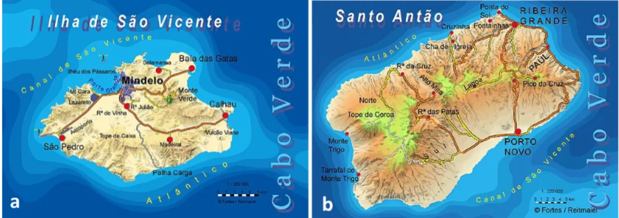
Figure 2 - Samples locations in são Vicente (Baia das Gatas, Praia da Laginha, Cova da Inglesa, Salamensa, Calhau) and Santo Antão (Ponta do Sol). ... 11
Figure 3 - Cyanobacteria cultures after 25 days growth. ... 17
Figure 4 - Extraction system from lyophilized cyanobacterial material (according to the MeOH extraction SOP V12.2019 protocol from BBE.CIIMAR). ... 18
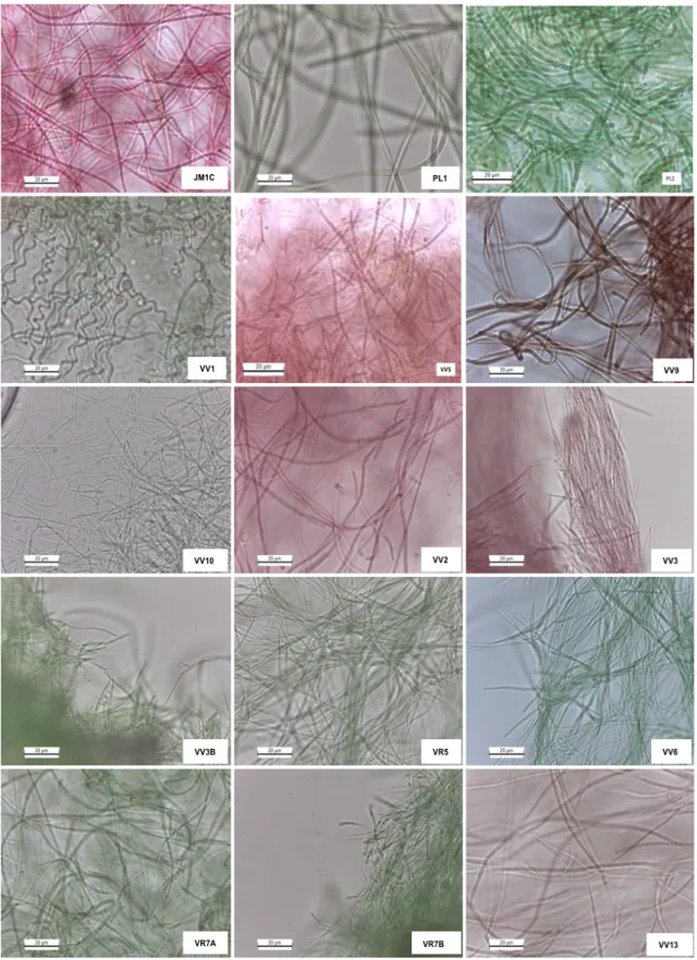
Figure 5 - Photomicrographs of the cyanobacteria isolates. ... 23
Figure 6 - Phylogenetic distribution based on the 16S rRNA of isolates (red) with the respective Best Hits. ... 27
Figure 7 - Cellular viability of cells exposed to the extract of strain JM1C. ... 28
Figure 8 - Cellular viability of cells exposed to the extract of the strain JM1B. ... 29
Figure 9 - Cellular viability of cells exposed to the extract of the strain PL1 ... 29
Figure 10 - Cellular viability of the cells exposed to the extract of the strain PL2 ... 29
Figure 11 - Cellular viability of the cells exposed to the extract of the strain VV9 ... 30
Figure 12 - Cellular viability of the cells exposed to the extract of the strain VV5 ... 30
Figure 13 - Cellular viability of the cells exposed to the extract of the strain JM5A ... 30
Figure 14 - Cellular viability of the cells exposed to the extract of the strain VV3 ... 31
Figure 15 - Cellular viability of the cells exposed to the extract of the strain VV3B ... 31
Figure 16 - Celular viability of the cells exposed to the extract of the strain VR7A ... 31
Figure 17 - Cellular viability of the cells exposed to the extract of the strain VR7B ... 32
List of Abbreviations
ATCC - American Type Culture Collection BBE – Blue Biotechnology Ecotoxicology
BLASTn-Basic Local Alignment Search Tool for nucleotides
CIIMAR - Interdisciplinary Centre of Marine and Environmental Research CVA - Cape Verde Archipelago
DMEM Glutamax – Glutamax Dulbecco’s Modified Eagle Medium DMSO - Dimethyl sulfoxide
dNTPs - Deoxynucleotides
gDNA-Genomic deoxyribonucleic acid EDTA Ethylenediamine tetraacetic acid HaCat – Human keratinocytes cell line hCMEC/D3 – Human endothelial cell line HepG2-Liver hepatocellular carcinoma cell line
LEGEcc Blue Biotechnology and Ecotoxicology Culture Collection MAAs-Mycosporine-like amino acids
MTT - 3-(4,5-dimethylthiazole-2-yl)-2,5-diphenyltetrazolium bromide MgCl2 - Magnesium dichloride
NCBI - National Center for Biotechnology Information
PCR -Polymerase Chain Reaction PBS - Phosphate-buffered saline 16S rRNA- RNA ribosomal 16S RKO-Colon carcinoma cell line SCY - Scytonemin
1. Introduction
1.1. Cape Verde Archipelago
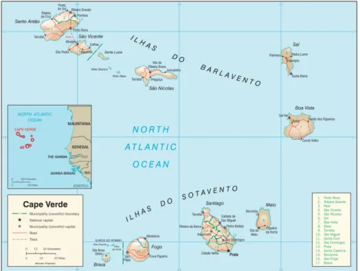
Cape Verde Archipelago (CVA) is a group of ten volcanic islands located in the Tropical Eastern Atlantic, around the African coast (Figure 1). The archipelago main islands are split into two groups composed by six northern Barlavento (windward) Isles and four southern Sotavento isles. The origins of the archipelago are thought to be associated with hotspot activity impinging on the old oceanic lithosphere. Together with the Canary Islands, Madeira and Azores, Cape Verde belong to the Macaronesia biogeographic region [1]. This region is an important transitional climatic zone that encompasses a large part of the eastern north Atlantic, which is influenced by the semi-permanent Azores high-pressure system, prevailing north-easterly trade winds and the surrounding ocean, The Cape Verde climate ranges from tropical dry to semi-desert, mainly governed by seasonal changes in the location of the Azores anticyclone, the Intertropical Convergence Zone and arid Atlantic air masses [2].
Cape Verde precipitation is limited, and typically confined to the raining season (August-October). The two major influences on Cape Verde precipitation are the African Monsoon and African Easterly waves. As such, decadal changes in Cape Verde precipitation mirror precipitation trends across the West African Sahel, as they share the same generation mechanism [2].
São Vicente, one of the Barlavento (windward) islands, has an extension of 227 km2.
The depth of the sea floor between São Vicente and Santa Luzia does not reach 50 m, whereas between São Vicente and Santo Antão it reaches 500 m. Its outline resembles that of a rhombus with a major (E–W) diagonal of about 24 km and a minor (N–S) diagonal of about 16 km [3].
Santo Antão is the most north-western island of the CVA and the second largest of the ten main islands, with an area of 779 km2. Santo Antão consists of alkaline volcanic rocks
that vary in composition from nefelinite and basanite to phonolite, as for many volcanic oceanic islands [4]. The island has a rugged relief with the highest point at 1979 m. North-easterly trade winds ensure a humid climate and high erosion rates in the north and north-western parts of Santo Antão, which has many deeply dissected valleys where older successions of composite lava flows and associated dyke swarms are well exposed.
Figure 1 - Map of Cape verde archipelago
1.2. Cyanobacteria
Cyanobacteria are prokaryotic organisms with photosynthetic capacity but that are able to use also anaerobic respiration and to fix atmospheric nitrogen. In the marine environment cyanobacteria are considered the main producer of the phytoplankton community food chain as they produce 50% of the biomass [5]. Also known as cyanophyta or blue-green algae, cyanobacteria have a great physiological and
morphological variability, which might be the base for their global distribution [6]. In fact, cyanobacteria have a wide distribution and can be found in aquatic environments as well as in terrestrial environments and even in extreme conditions such as a high salinity [7] high and
low temperature [8]. In order to survive to such extreme conditions through a long evolution period cyanobacteria have developed mechanisms of adaptation that include the production of sundry secondary metabolites [9], with antioxidant, photoprotective, moisturizing, allelopathic and toxic properties, which includes pigments, polysaccharides, fatty acids and peptides [10].
The bioactive properties of cyanobacteria, the high growth rates and the ability to grow autotrophically which makes their cultivation simple and cost-effective, have attracted an increasing interest by a wide range of industries, such as the food industry, the cosmetic industry, the pharmacological industry, the energetic industry and agriculture. In fact cyanobacteria have been considered a rich source of compounds for use as food additives, cosmetics, antitumor, antibacterial and antiviral drugs, biofuels as well as organic fertilizers [11].
The high interest in cyanobacteria has led to further research on strains from different habitats to obtain value-added strains for industry and agriculture. This research involves collecting samples in the wild, isolation, identification and culture. In order to maintain the isolated strains, biologic specimens must be deposited in collections, being then available for investigation.
1.2.1. Cyanobacteria taxonomy
The cyanobacterial phylum is very diversified in terms of morphology, including unicellular, colonial and filamentous forms. In some filamentous strains a cell differentiation is presented by the production of specialized cells involved in nitrogen fixation, the heterocysts, and resting cells that can resist to environmental stress, the akinetes. Some genera also produce small fragments with different size and shape, the hormogonia, used for dispersal and host infection [12].
Traditionally, cyanobacteria were classified according the International Code of Botanical Nomenclature due to the ability to perform oxygenic photosynthesis and the similarity to the structure of eukaryotic chloroplasts. However, due to their prokaryotic nature cyanobacteria are also classified according to the International Code of Nomenclature of Bacteria. Currently the two aproaches prevail, the botanical [13] and the bacterial [14].
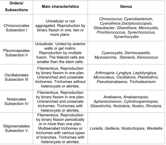
According to the botanical classification cyanobacteria are classified into five Orders while according to the Bacterial approach cyanobacteria are classified into five Subsections. The five orders and subsections broadly coincide as described in Table 1, where the main characteristics are presented.
Although the traditional classification and identification of cyanobacteria has been based on morphological characteristics, the advances in molecular biology led to the use of molecular markers. A combination of different approaches has thus been lately adopted, which includes not only the morphological description of the specimens but also a genotypic analysis, namely by using the small subunit ribosomal RNA, the 16S rRNA gene [15]. This new polyphasic approach makes it possible to overcome some difficulties in morphology-only identification due to the small size of many strains and morphological changes during strain cultivation or long-term maintenance in the laboratory [15].
Table 1 - Main characteristics and genus of the 5 orders/subsections of the phylum cyanobacteria.
Orders/
Subsections Main characteristics Genus
Chroococcales Subsection I
Unicellular or not aggragated. Reproduction by
binary fission in one, two or more plans.
Chroococcus, Cyanobacterium, Cyanothece,Dactylococcopsis, Gloeobacter, Gloeothece, Microcystis,
Prochlorococcus, Synechococcus, Synechocystis
Pleurocapsales Subsection II
Unicellular. United by exterior walls or gel matrix. Reproduction by multiple fusion. The tilakoid cells are
smaller than the stem cells.
Cyanocystis, Dermocarpella, Myxosarcina, Stanieria, Xenococcus
Oscillatoriales Subsection III
Filamentous. Reproduction by binary fission in one plan.
Unbranched and uniseriate trichomes. Trichomes without
heterocysts or akintes.
Arthrospira, Lyngbya, Leptolyngbya, Microcoleus, Oscillatoria, Planktothrix,
Pseudoanabaena, Trichodesmium
Nostocales Subsection IV
Filamentous. Reproduction by binary fission in one plan.
Unbranched and uniseriate trichomes. Trichomes with
heterocysts or akintes.
Anabaena, Anabaenopsis, Aphanizomenon, Cylindrospermopsis, Gloeotrichia, Nodularia, Nostoc, Rivularia,
Stigonematales Subsection V
Filamentous. Reproduction by binary fission periodically
or in more than one plan. Multiseriated trichomes or trichomes with various types of branches. Trichomes with
heterocysts or akintes.
1.2.2. Cyanobacteria isolation and preservation
Although for several years cyanobacteria were found to be difficult or even impossible to isolate, Allen (1968) and Huges (1958) proved that it was possible to isolate and maintain ex-situ cyanobacteria in laboratory culture, for several years [16],[17].To initiate the process of cyanobacteria isolation, environmental samples are collected, and their origin noted. Upon arrival to the laboratory, it is critical to evaluate the sample material content in order to identify cyanobacteria that have capacity to grow in specific medium, since the characteristics that an organism must develop in a given environment are used as selective factors in the isolation process. This evaluation is carefully performed with the aid of a microscope [18]. After, it is crucial the choice of the most appropriate medium to their successful isolation. The medium used for enrichment is generally liquid and have a nutritional composition that enables the development of specific organisms to the detriment of competitors. These media may be selective enrichment media, if it involves the addition of inhibitory substances (eg antibiotics, antifungals) that suppress the growth of most organisms with the exception of the organism to be isolated [19]. Following the collection, identification, and characterization of organisms, environmental samples should be enriched with the selected medium.
In addition to isolation in liquid media, cyanobacteria can also be isolated using solid media such by inoculum streak and streak seeding [18] or selecting organisms by micromanipulation, a technique that requires practice and patience [20], or a combination of these different techniques. The micromanipulation is performed with the aid of an optical microscope and consists of the selection of the organism of interest, ideally present in liquid medium, using microcapillaries [20]. The obtained cultures should be monitored and successively picked until isolation is confirmed [20].
After isolation and classification, strains must be preserved in controlled culture conditions in order to ensure the ex-situ preservation. This ex-situ preservation of microorganisms and the public access to microbial specimens is the aim of the microbial domain of Biological Resource Centres (mBRC), the so-called microbial Culture Collections.
Blue Biotechnology and Ecotoxicology (BBE) Culture Collection (LEGE-CC) http://lege.ciimar.up.pt/ is a biological resource center of the Interdisciplinary Marine and Environmental Research Center (CIIMAR) Portugal. The collection currently comprises about 1000 of cyanobacteria and microalgae strains collected from different sites, mainly in mainland Portugal including Madeira and Azores Islands, but also elsewhere (e.g. Australia, Brazil, Colombia, Morocco, Mexico). LEGE CC strains are derived from many
environments, 93% of those are from aquatic environments (2% hypersaline, 46% marine, 11% brackish and 34% freshwater), 3% from terrestrial environments and 4% with unknown origin. They are also producers of secondary metabolites, such as microcystin, that have toxic effects on human beings
[22]
. One of the main purposes of LEGE-CC is to provide living organisms, and/or related information, to third parties for use in research and development activities. It is therefore essential that strains existing (or to be included) in the LEGE-CC crop collection are properly isolated and appropriately characterized in order to interest potential users [21].1.2.3. Cyanobacteria bioactive compounds
A main reason why the study of cyanobacteria is so attractable is the production of bioactive compounds with diverse biotechnological applications. Cyanobacteria bioactive compounds are mainly secondary metabolites. These are metabolites produced by an organism in which a role in primary functions such as growth or reproduction has not been found [23]. They are responsible for umpteen functions such as allelopathy [24] and defense [25], although they are not absolutely required for the survival of the organism.
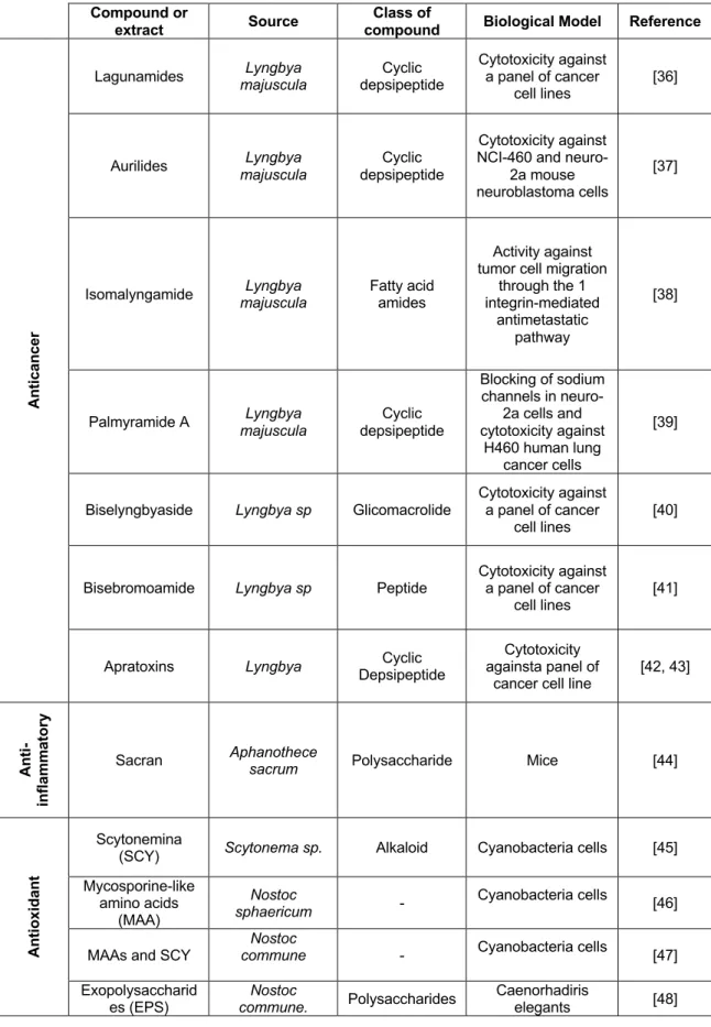
The high biodiversity of cyanobacteria in nature is reflected also in a high chemical diversity and bioactive potential. The range of bioactive secondary compounds from cyanobacteria includes alkaloids, peptides, lipopeptides, amino acids and fatty acids [26], being cyanobacteria considered a good source of compounds for biotechnology applications, namely for pharmaceutical and therapeutic application [27],[28],[29],[30]. In fact, in the last few years cyanobacteria have gained a lot of attention being considered one of the most promising sources of natural compounds with antibacterial [31], antiviral [32], antifungal, anti-inflammatory, algicide [33], anticancer [34, 35], and antiparasitic activity [26] (Table 2)
Table 2 - Marine cyanobacteria compounds with bioactive potential (anticancer, anti-inflammatory, antioxidant, antiparasitic, photoprotective and whitening).
Compound or
extract Source
Class of
compound Biological Model Reference
An ti ca n ce r
Lagunamides majuscula Lyngbya depsipeptide Cyclic Cytotoxicity against a panel of cancer cell lines
[36]
Aurilides majuscula Lyngbya depsipeptide Cyclic
Cytotoxicity against NCI-460 and
neuro-2a mouse neuroblastoma cells
[37]
Isomalyngamide majuscula Lyngbya Fatty acid amides
Activity against tumor cell migration
through the 1 integrin-mediated
antimetastatic pathway
[38]
Palmyramide A majuscula Lyngbya depsipeptide Cyclic
Blocking of sodium channels in neuro-2a cells and cytotoxicity against H460 human lung cancer cells [39]
Biselyngbyaside Lyngbya sp Glicomacrolide
Cytotoxicity against a panel of cancer
cell lines [40]
Bisebromoamide Lyngbya sp Peptide
Cytotoxicity against a panel of cancer
cell lines [41]
Apratoxins Lyngbya Depsipeptide Cyclic againsta panel of Cytotoxicity cancer cell line
[42, 43] An ti -in fla m m at o ry
Sacran Aphanothece sacrum Polysaccharide Mice [44]
An ti o xi d an t Scytonemina
(SCY) Scytonema sp. Alkaloid Cyanobacteria cells [45] Mycosporine-like amino acids (MAA) Nostoc sphaericum - Cyanobacteria cells [46]
MAAs and SCY
Nostoc
commune - Cyanobacteria cells [47]
Exopolysaccharid es (EPS) Nostoc commune. Polysaccharides Caenorhadiris elegants [48]
Table 2 (Cont.) Compound or extract Source Class of compound Biological Model Reference An ti p ar as it ic (L ei sh m an ia
) Venturamide B Oscillatoria sp. Cyclic peptides Leishmania
donovani [49]
Gallinamide A Schizothrix sp. Peptide L. donovani [49]
An ti p ar as it ic (M al ar ia
) Lagunamide A majusucla Lyngbya depsipeptides Cyclic Plasmodium falciparum [50]
Venturamide A Oscillatoria sp Cyclic hexapeptides P. falciparum [51]
An ti p ar as it ic (T ri p an o ss o m a)
Venturamide B Oscillatoria sp. Cyclic peptides Tripamossona. Cruzi [49]
Dragonamide E majusucla Lyngbya Lipopeptides L. donovani [52]
Ph o to Pr o te ct io n SCY Arthrospira platensis Alkaloid Cyanobacterial cells [45]
MAAs and SCY Lyngbya sp. _ Cyanobacterial cells [53]
Wh it en in g Chloroform-methanol extract Oscilnorlatoria agardhii - Tyrosinase activity assay [54] C-phycocyanin Spirulina Pigment B16F10 [55]
2. Objectives
The main aim of this work was to contribute to the study of the biodiversity and the biotechnological potential of cyanobacteria collected from Cape Verde Islands. The environmental samples included in this study were collected from two islands of the archipelago, São Vicente and Santo Antão. Samples were collected by members of the Blue Biotechnology and Ecotoxicology Group (BBE) from the Centre of Marine and Environmental Research (CIIMAR).
Based on this main goal, specific tasks were performed, which included:
1. Isolation of cyanobacteria strains using both solid and liquid Z8 medium;
2. Identification of the isolated cyanobacteria using a morphological and a molecular approach;
3. Culture of isolated cyanobacteria strains for biomass production. 4. Preparation of cyanobacteria extracts;
5. Evaluation of the bioactive potential of cyanobacteria extracts:
a. evaluation of the anticancer potential using cancer cell lines and normal cell lines.
3. Material and Methods
3.1. Sampling
The biological environmental samples were collected in April 2018 in two Cape Verde Islands, São Vicente and Santo Antão. In São Vicente, samples were collected in Baía das Gatas, Praia da Laginha, Cova da Inglesa, Salamansa and Calhau. In Santo Antão samples were collected in Ponta do Sol (Figure 2; Table 1).
Figure 2 - Samples locations in são Vicente (Baia das Gatas, Praia da Laginha, Cova da Inglesa, Salamansa,
Calhau) and Santo Antão (Ponta do Sol).
The collected material consisted in rock scrapes along the coast and sea water samples. After collection the material was stored in thermal bags and kept refrigerated until processing.
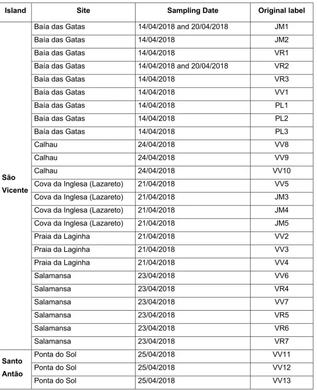
Table 3 - Sampling sites and environmental samples collected from São Vicente and santo Antão islands (data kindly provided by João Morais, BBE-CIIMAR).
Island Site Sampling Date Original label
São Vicente
Baía das Gatas 14/04/2018 and 20/04/2018 JM1
Baía das Gatas 14/04/2018 JM2
Baía das Gatas 14/04/2018 VR1
Baía das Gatas 14/04/2018 and 20/04/2018 VR2
Baía das Gatas 14/04/2018 VR3
Baía das Gatas 14/04/2018 VV1
Baía das Gatas 14/04/2018 PL1
Baía das Gatas 14/04/2018 PL2
Baía das Gatas 14/04/2018 PL3
Calhau 24/04/2018 VV8
Calhau 24/04/2018 VV9
Calhau 24/04/2018 VV10
Cova da Inglesa (Lazareto) 21/04/2018 VV5
Cova da Inglesa (Lazareto) 21/04/2018 JM3
Cova da Inglesa (Lazareto) 21/04/2018 JM4
Cova da Inglesa (Lazareto) 21/04/2018 JM5
Praia da Laginha 21/04/2018 VV2 Praia da Laginha 21/04/2018 VV3 Praia da Laginha 21/04/2018 VV4 Salamansa 23/04/2018 VV6 Salamansa 23/04/2018 VR4 Salamansa 23/04/2018 VV7 Salamansa 23/04/2018 VR5 Salamansa 23/04/2018 VR6 Salamansa 23/04/2018 VR7 Santo Antão Ponta do Sol 25/04/2018 VV11 Ponta do Sol 25/04/2018 VV12 Ponta do Sol 25/04/2018 VV13
3.2. Isolation of cyanobacteria strains
The isolation of cyanobacteria from environmental samples was performed using both liquid and solid Z8 culture medium, supplemented with 25g/L NaCl [56], in aseptic conditions. All the glass material used was autoclaved (Tuttnauer 3870 ELV-D, The Netherlands) for 20 min at 121°C. The preparation of the medium and the entire isolation process was carried out in a laminar flow chamber (Telstar Bio II Advance, Spain) previously disinfected with 70% alcohol and exposed to UV light for 30 minutes before
each use.
For solid culture medium, agarose was added to Z8 medium to a concentration of 1,2%. The mixture was spread on sterile polystyrene Petri dishes and after solidification, the Petri dishes were stored aseptically for further use.
The solid Z8 medium was the primary method used to isolate cyanobacteria from the environmental samples. All the media for the raw environmental samples were prepared with cycloheximide, at a concentration of 0.025%, to prevent the growth of eukaryotic microorganisms.
3.3. Identification of cyanobacteria strains
3.3.1. Morphological characterization
To morphologically characterize the isolates, strains were observed, annotated and photographed using an optical microscope (Leica DMLB) coupled to a camera (Leica ICC50 HD) with the Leica Qwin Color computer program (Leitz, Germany) at a magnification of 1000X. For each isolate, morphometric measurements were also made and the mean dimensions (length, width) of the vegetative cells of filaments recorded. Length and width were measured in 20 cells, being the cells from different individuals of each strain.
3.3.2. Molecular characterization – DNA extraction and PCR
conditions
3.3.2.1. DNA extraction
To extract the gDNA from the isolated strains, a small amount of biomass was collected in 1.5 mL Eppendorf tubes, in the flow chamber. Tubes were centrifuged at 10000 X g for 1 minute to concentrate the biomass and remove the liquid in excess. The biomass was then stored at -20 °C for posterior extraction of DNA.
Extraction of gDNA was performed using the Genetic DNA Mini-Kit Purelink (Invitrogen, USA), using the protocol for extracting DNA from gram-negative bacterial cells, according to the manufacturer’s instructions. Isolated DNA was stored in 1.5 mL Eppendorf tubes at a temperature of -20 ° C.
To confirm that the DNA was successfully extracted an electrophoresis in Agarose gel (UltrapureTM Agarose, Invitrogen, USA) at 1% concentration in Tris-Acetate EDTA buffer
solution (TAE Ultrapuretm, Invitrogen, USA) at 40mM Tris-acetate and 1mM EDTA, was performed. Gel was stained with 3µL of SYBRsafe (Invitrogen, USA).
Briefly, 3 μl of extracted DNA mixed with 0.5 μl of loading buffer were loaded onto the gel, 1 μl molecular marker (1Kb Plus DNA Ladder-Invitrogen) was loaded. The gel was run at 85V for 20 minutes and visualized and photographed on the GEL DOCTM Molecular Imager® transilluminator with Image Lab ™ software (USA).
3.3.2.2. PCR amplification and analysis of products
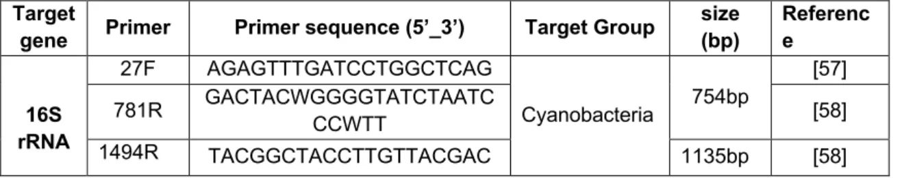
The molecular identification of isolates was based on the 16S rRNA gene. Information on primers is described in Table 4.
Table 4 - Primer pairs used for amplification of the 16S rRNA gene
Target
gene Primer Primer sequence (5’_3’) Target Group
size (bp) Referenc e 16S rRNA 27F AGAGTTTGATCCTGGCTCAG Cyanobacteria 754bp [57] 781R GACTACWGGGGTATCTAATC CCWTT [58] 1494R TACGGCTACCTTGTTACGAC 1135bp [58]
359F GGGGAATYTTCCGCAATGGG [57]
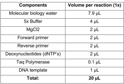
PCR reactions consisted in 1x GoTaq buffer, 2.5 mM for MgCl2, 1 µM for each primer, 0.5 mM for the dNTP mix, 0.5 U for GoTaqR Flexi DNA polymerase. The final volume of each reaction was 20 µL with 1 µL of DNA template (Table 5).
Table 5 - Mastermix components of the PCR reaction
Components Volume per reaction (1x)
Molecular biology water 7.9 µL
5x Buffer 4 µL MgCl2 2 µL Forward primer 2 µL Reverse primer 2 µL Deoxynucleotides (dNTP’s) 2 µL Taq Polymerase 0.1 µL DNA template 1 µL Total: 20 µL
The PCR reactions were performed in a Biometra T-Professional Standard Gradient Thermocycler (Germany) as described in table 6.
Table 6 - PCR conditions used for the primer pair used to amplify the 16S rRNA gene
Target
gene Primer Pair
PCR reaction References
Initial
Denaturation PCR cycles extension Final
[59] RNA 16S 27F/359F 95ºC 2 mn 35 Cycles 72 ºC 5 mn 95ºC 2 mn 55ºC 45 s 72ºC 1 mn 781R/1494R
After amplification, all PCR products were analyzed and visualized via electrophoresis in an agarose gel ((Ultrapuretm Agarose, Invitrogen, USA) at a concentration of 1.5% which was stained with 3µL of SYBRsafe (Invitrogen, USA). The selected voltage and running time were 85 V and 20 minutes. The agarose gel was visualized and photographed in
the transilluminator Molecular Imager® GEL DOCTM with the software Image LabTM(USA).
3.3.2.3. Sequencing, sequence alignment and phylogenetic
analysis
All the samples that exhibited positive PCR results (i.e presence of the gene) were selected for sequencing. The 16S rRNA gene was firstly amplified and sequenced for all the isolates obtained, in order to identify the cyanobacteria and thus assess the diversity of the samples. For sequencing purposes, the PCR preparation involved duplicating the number of reactions for each isolated sample, in order to have enough amplified product to enable sequencing. So, the final volume of PCR product per sample loaded into the 1.5% agarose gel was 40 µL. The electrophoresis was runed at 90 V for 60 minutes, then the amplified fragments was observed in the CSMICRODOC system (Cleaver scientific, UK) transilluminator coupled with a Canon PowerShot G9 camera. The bands with the expected size were excised from the gel and collected to be purified using the Nztech – genes & enzymes (NZYGelpure, Portugal) purification kit, following the manufacturer’s instructions. To check the efficacy of the DNA purification, an electrophoresis (100V; 20 minutes) was run in a 1% agarose gel, and 2 µL of the purified DNA was mixed with 1 µL of loading buffer. All the purified PCR products and the respective pair of primers was sent to GATC Biotech (Germany) to be sequenced. The forward and reverse sequences (i.e. 5’ and 3’) obtained from the same PCR product were examined in the bioinformatic software Geneious (v.8) and assembled together (de novo assembly), their chromatograms were analyzed to check the quality of the sequences and to determine if further sequences were needed to form a consensus sequence (i.g. if the quality was bad on either one of the forward or reverse sequences). The sequences usually be trimmed at the extremities due to bad quality. Then, the consensus sequences of each strain isolate were compared with the sequences in the GenBank® database using the BLAST®n (Basic Local Alignment Search Tool for nucleotides) tool available in the NCBI (National Center for Biotechnology Information) and compared with other cyanobacterial sequences in the GenBank® database to check for similarities and to help in their identification. To settle the phylogenetic relation of the isolated strains to other reference strains, the sequences were aligned by ClustalW, and the GTR+G+I evolutionary model was selected by MEGA 7.0. The robustness of
Maximum Likelihood (ML) trees was estimated by bootstrap percentages, using 1000 replications in MEGA 7.0.
3.4. Cyanobacteria culture and extraction
For stock and culture, the cyanobacteria strains were inoculated in 500 mL and 4 liters of Z8 medium (with 25g/L NaCl) respectively. Cultures in 4L medium were maintained under an aeration system. All the procedures were performed in controlled temperature conditions (25 °C) and luminance (light cycles / dark 14h / 10h). When cultures reached the exponential growth stage, after approximately 30 days of the inoculation, visually recognized by the high concentration of biomass (Figure 2), the cultures were harvested by filtration.
Figure 3 - Cyanobacteria cultures after 25 days growth.
Before lyophilization, the concentrated biomass was stored at – 80ºC. The frozen biomass was lyophilized for 7 days at -48ºC (Telstar LyoQuest) under reduced pressure (0.1 mbar with condenser at -47 ºC) and stored at -20ºC until further use.
A smaller stock culture of all cyanobacteria strains was maintained during this work in order to provide biomass to inoculate in the 500 mL medium. This stock culture was
performed by removing a small amount of biomass of the culture and placing it in flasks with 100 ml of Z8 medium (25g/L NaCl).
The cyanobacterial extracts were performed with lyophilized material. The extraction was started by breaking the lyophilized biomass using a mortar and porcelain pestle, in small portions at a time, transferring the grounded biomass to an Erlenmeyer flask. After, 50 ml of methanol were added, and the flask placed in the ultrasonic bath for 5 minutes (taking care not to heat above 30 ºC). The flask was rested for a few minutes, letting the particulate matter settle to the bottom. The supernatant was filtered using a vacuum filtering system as describe in the Fig. 4. This procedure was repeated twice using 25 mL of methanol. The final extract was concentrated in a round flask on a rotary evaporator at 30 ºC, under a control pressure. The concentrated extract was transferred to a pre-weighed 20 mL vial, by filtering it through a Pasteur pipette with a cotton filter (to capture any non-soluble material). Finally, the solvent was evaporated in a rotary evaporator and the flask was left in high vacuum to thoroughly dry. The glass vial was weighted again, and the total mass of the crude extract calculated. The dry extract was dissolved at the concentration of 10 mg/mL using DMSO and stored at -20 ºC.
Figure 4 - Extraction system from lyophilized cyanobacterial material (according to the MeOH
3.5. Cytotoxicity analysis
3.5.1 Cell culture
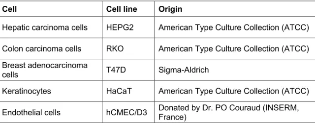
The bioactivity of the extracts was assessed using normal and cancer cell lines (Table 7).
Table 7 - Cell lines included in the study
Cell Cell line Origin
Hepatic carcinoma cells HEPG2 American Type Culture Collection (ATCC) Colon carcinoma cells RKO American Type Culture Collection (ATCC) Breast adenocarcinoma
cells T47D Sigma-Aldrich
Keratinocytes HaCaT American Type Culture Collection (ATCC)
Endothelial cells hCMEC/D3 Donated by Dr. PO Couraud (INSERM, France)
Cells were cultured in DMEM Glutamax medium (Dulbecco’s Modified Eagle Medium DMEM GlutaMAXTM – Merck, Germany), supplemented with 10% (v/v) fetal bovine serum (Gibco, USA), 0,1% Amphotericin B (Gibco, Germany) and 1% of penicillin-streptomycin (Pen-Strep 100 IU/ml and 10 mg/ml, respectively) (Gibco, Germany) in a humidified atmosphere containing 5% CO2 andat 37ºC. Culture medium was renewed
every two days. At 80-90% cell confluence, adherent cells were washed with phosphate-buffered saline (PBS) (Gibco, Germany) and enzymatically released with TrypLE express enzyme (1x) (Gibco, Denmark).
3.5.2
Cytotoxicity assay – MTT assay
The viability of cells exposed to the cyanobacteria extracts was evaluated by using the 3- (4,5 dimethylthiazole-2-yl)-2,5-diphenyltetrazolium bromide (MTT) assay. The MTT is
a yellow tretazole soluble in water that is reduced by mitochondrial enzymes to purple formazan crystals that are insoluble in water. The reduction of MTT to formazan is directly proportional to the mitochondrial activity and consequently cell viability ( [61].
For the cytotoxic assays cells were seeded in 96-well culture plates at a density of 2,5 x 10⁴cells mL-1 for keratinocytes, 1 x 10⁵ cells mL-1for endothelial cells and 3.3 x 10⁴ cells
mL-1 for T47D, RKO and HepG2 cells. After 24 hours of adhesion, cells were exposed to
100 μL fresh medium supplemented with the extracts to a final concentration of 100, 75, 50, 25, 12,5 and 6,25 µg mL-1 for a period of 24 and 48 hours. After each incubation time,
an aliquot of 20 μL MTT solution (1 mg mL-1) was added to each well in medium and
incubated at 37 ºC for 3h. After the incubation time, the medium was carefully aspirated, and the purple colored formazan salts dissolved in 100 μL DMSO. Absorbance was read at 550 nm in using a Synergy HT Multi-detection microplate reader (Biotek, Bad Friedrichshall, Germany) operating by GEN5TM software.
4. Results and Discussion
4.1. Isolated strains and identification
The isolation procedure, as well as time consuming, does not isolate all the diversity in a sample collected at a given location. A total of twenty isolates were obtained (Table 8 and Figure 5). All the isolated were obtained by petri dish spreading. From the total, nineteen strains were isolated from São Vicente island and one strain was isolated from Santo Antão. Due to time constraints deadlines only Z8 medium was used. Although Z8 medium is a nutrient rich medium and widely used in the isolation of cyanobacteria, it may not have facilitated the isolation of some groups, namely coccoid forms and heterocystous forms. In this sense other media may be used in the continuation of the work, such as culture medium BG110.
For the identification of the isolates, a polyphasic approach was followed. These included morphological characteristics such as the presence of specialized cells and cell size, and molecular tools such as the amplification and sequencing of the 16S rRNA gene [62].
As we can see in the Table 8 the morphological diversity of cyanobacteria is quite variable, however, it is verified that the isolates obtained from different places of the Islands have some peculiar and distinct characteristics. All the isolates are filamentous and no specialized cells such as heterocysts are present.This might be due to the faster growth occurring in Z8 medium, which seemed to favor the growth of non-heterocystous strains.
There are isolates that have filaments with rounded end cells, others that have slightly arched filaments at the ends and other cellular constrictions at wall level. Still others have thin filaments that coil, some in the shape of a ball. The isolates color ranged from green, dark green, brownish to reddish. By the color it was possible to conclude that the strains JM1C, VV2, VV3 and VV5 are rich in phycoerythrin responsible for the reddish color (Figure 5). These may be interesting to produce the red pigment that can potential be use as a natural colorant for the food and cosmetic industry.
Table 8 - Isolated strains, morphological characteristics.
Sampling site Original Label
Isolate code
Color in
culture Morphological characteristics
Cell size (Width x Length ) µm
São Vicente
Baía das Gatas JM1 JM1B Cyano Filaments with sheath
1,2±0,1x1,3±0,3 JMIC Reddish Filaments with trichomes constricted at cross walls 1,6±0,2 x 1,6±0,3
B. das Gatas PL1 PL1 Cyano Filaments with sheath and terminal cells rounded 2,4±0,5 x 2,7±0,8
B. das Gatas PL2 PL2 Cyano Filaments with sheath 2,3±0,5 x 4,7±1,4
B. das Gatas PL3 PL3 Cyano Filaments with sheath 2,3±0,2 X 3,0±1,0
B. das Gatas VV1 VV1 Cyano Filaments with sheath 1,4±0,2 x 1,4±0,2
Calhau VV9 VV9 Dark brown Filaments with some helical filaments, forming tuft like
aggregates 1,8±0,2 x 1,5±0,3
Calhau VV10 VV10 Cyano Thin, coiled filaments wrapped in mucilage 1,1±0,2 x 1,4 ±0,4
Cova da Inglesa VV5 VV5 Brownish Filaments growing homogeneously, but also forming
aggregates 1,5±0,3 x 1,5±0,3
C. da Inglesa JM5 JM5A Cyano Filaments with sheath 1,2±0,3 X 1,1±0,2
Praia da Laginha VV2 VV2 Reddish Trichomes slightly constricted at cross walls 1,8±0,3 X 1,4±0,3
P. da Laginha VV3
VV3 Reddish Thin, coiled filaments wrapped in mucilage 1,2±0,3 x 1,2±0,3
VV3A Green olive Filaments growing homogeneously, but also forming aggregates 1,4±0,2 x 1,4±0,2 VV3B Dark green Filaments growing homogeneously, but also forming
aggregates 1,1±0,18 x 1,2±0,3
P. da Laginha VV4 VV4 Reddish Trichomes slightly constricted at cross walls 1,7± 0,3 X 1,6±0,2
Salamansa VR5 VR5 Cyano Filaments forming a smooth biofilm 1,1± 0,2 x 1,2±0,2
Salamansa VV6 VV6 Cyano Filaments growing homogeneously, but also forming
aggregates 1,2±0,1 x 1,4±0,3
Salamansa VR7 VR7A Cyano Filaments growing homogeneously also forming aggregates 1,6±0,3 x 1,6±0,4
VR7B Dark green Thin, coiled filaments wrapped in mucilage 1,3±0,3 x 2,2±1,0
Santo Antão
Ponta do Sol VV13 VV13 Reddish
4.2.
Phylogenetic analysis
The identification of the cyanobacteria isolates by a molecular approach was based in the 16S rRNA gene sequences. In Table 9 it is presented the best BLAST results obtained from Genbank for the consensus sequence of sixteen of the isolates. The sequences of the BLAST results from the isolates of this study and a subset of 56 sequences representing cyanobacterial diversity (obtained from the online database Cyanotype) were integrated into the phylogenetic tree shown in Figure 6.
Table 9 - Best Blast results obtained for each isolate based on the molecular marker 16S rRNA
Isolate
Code Molecular Identification (ID) (top cyano hit) % ID
Sequence Length bp
JM1B Filamentous cyanobacterium ESFC-1 clone 4_5A 16S ribosomal RNA gene 96.45% 733 JM1C Leptolyngbya sp. RS02 16S ribosomal RNA gene 99.8% 1363 PL1 Oscillatoria sp. S8 16S ribosomal RNA gene 99.72% 1053 VV1 Nodosilinea sp. LEGE 11468 16S ribosomal RNA gene 99.62% 1330 PL2 Geitlerinema sp. PCC 7105 small subunit ribosomal RNA gene, partial sequence 99.49% 1369 VV9 Leptolyngbya sp. PCC 7376, complete genome 99.26% 1355 VV10 Filamentous cyanobacterium 73-2 16S ribosomal RNA gene 97.69% 1382 JM5A Nodosilinea sp. LEGE 11468 16S ribosomal RNA gene 99.26% 1079 VV2 Leptolyngbya sp. RS02 16S ribosomal RNA gene, partial sequence 99.22% 1279 VV3 Phormidium angustissimum SABC020801 16S ribosomal RNA gene 96.71% 1370 VV3A Filamentous cyanobacterium 73-2 16S ribosomal RNA gene 99.98% 1372 VV3B Filamentous cyanobacterium 73-2 16S ribosomal RNA gene 99.13% 1375 VV6 Filamentous cyanobacterium 73-2 16S ribosomal RNA gene 99.13% 1380 VR7A Filamentous cyanobacterium 73-2 16S ribosomal RNA gene 99.12% 1366 VR7B Nodosilinea sp. LEGE 11468 16S ribosomal RNA gene, partial sequence 99.62% 1042 VV13 Leptolyngbya ectocarpi LEGE 11474 16S ribosomal RNA gene 97.73% 1189
In the total of 20 isolated strains, 16 could be analyzed through molecular tools while for VV5, VR5, PL3 and VV4 only morphological characteristics were able to be determined.
The ML tree shows the sequences from the isolates and reference strains of cyanobacteria, in order to verify the diversity of the obtained isolates.
The phylogenetic tree consisted of 25 clusters (Figure 6), which were defined by being monophyletic groups with a bootstrap value of 70% or higher and had to include at least 1 isolate strain or 1 reference strain. An exception was made to the Cluster Y and X which had a bootstrap value lower than 50% but was defined as a cluster because it is a monophyletic group that includes the isolate strain. Clade N (Figure 6) includes PL1 and PL2, being supported by a high bootstrap value (100%) and is close related (boostrap 94%) to Lyngbya cf. confervoides AY599507, which cannot be assigned as a reference strain for Lyngbya. Because of that, this clade is worthy to be described as a new cyanobacterial genus in further studies.
Clade S (Figure 6) also does not include a reference strain and is phylogenetically strongly supported (bootstrap 100%). It includes the isolated strain VV9 identified, which is phylogenetically close related to Leptolyngbya PCC7376 and the respective best hit that is an unidentified 4Oscillatoria_rosea_IAM. This clade is also worthy to be described as a new cyanobacterial genus in further studies.
The clade T is identified as Jaaginema, and includes the isolate obtained in this study JM1B the best hit strain to Jaaginema strain JaaginemaspPsrJGgm 14 and JaaginemaspIkpSMP32 which are non-heterocyst filamentous, usually of very simple and thin morphology and without large distinctive characters [13] and one JQ013010_Filamentouscyanobacte.
The Clade X was subdivided into 2 subclades (Figure 6) subclade X1 included comprises 5 isolated strains (VV10,VV6,VV3A,VV3B,VR7A, VV3) all identified as belonging to not already described genera. The subclade X2 (figure 6) was integrated by 3 isolates strains VV13, JM1C and VV2 identified as a Leptothoe_TAU_MAC_161 and Leptolyngbya_sp_HBC2,it is important to refer that the VV13,JM1C and VV2 beside they are phylogenetically positioned in the same clade, their morphological characteristics are very similar even to they were collected from different places. Clade Y (figure 6) comprised of 3 isolate strains VR7B,JM5A the Best hit strains Nodosilinea and Leptolyngbya_sp_LEGE_0 and VV1 the Best hit strains Leptolyngbya_sp_CENA15 and Haloleptolyngbyaalcali with a low bootstrap value (<50 %) this clade was only defined to illustrate the close proximity of the isolates.
In a general analysis we verified the presence of genera of common cyanobacteria in marine benthonic environments such as the genus Leptolyngbya. The most striking result was the possibility that most strains belong to new filamentous genera. In order support the 16S rRNA-based classification of cyanobacteria other molecular tools may be suggested such as the DNA-dependent RNA polymerase gamma subunit (rpoC1), and and an in-depth morphological evaluation using electron microscopy. Being new genera, this work allows to contribute to the knowledge of cyanobacteria by revealing unreported cyanobacterial diversity.
4.3.
Bioactivity
The potential bioactivity of the extracts was assayed against cancer and normal cell lines. For the evaluation of the anticancer potential, three cancer cell lines representative of major cancers affecting humans were selected, hepatic carcinoma cells HepG2, colon carcinoma cells RKO and breast adenocarcinoma cells T47D. Since the potential utility of the cyanobacteria extracts as anticancer drugs depends on the degree of selective toxicity on tumor cells, sparing normal cells, two normal cells were selected, the keratinocytes cell line HaCat and the endothelial cell line hCMEC/D3. The selection of these two normal cell lines was also based in the potential for skin care application that the cyanobacteria strains might revealed since keratinocytes are the major epidermis cells and endothelial cells are major blood vessels cells. The cytotoxicity of the cyanobacteria extracts was assayed by the MTT assay. The MTT assay is a widely used cytotoxic assay that measures cellular viability and thus, indirectly, cell proliferation. The assay was performed with the extract of the isolated strains at concentrations of 100μg mL-1; 75μg mL-1; 50μg / mL-1; 25μg mL-1; 12.5μg mL-1 and 6μg mL -1 and exposure times
of 24 and 48 hours. In all the assays the solvent control consisted in 1% DMSO in cell culture medium. Values are expressed as mean ± SD, n=3. Results obtained are presented in Figures 6-18 and a summary is presented in Table 10.
Figure 8 - Cellular viability of cells exposed to the extract of the strain JM1B.
Figure 9 - Cellular viability of cells exposed to the extract of the strain PL1
Figure 11 - Cellular viability of the cells exposed to the extract of the strain VV9
Figure 12 - Cellular viability of the cells exposed to the extract of the strain VV5
Figure 14 - Cellular viability of the cells exposed to the extract of the strain VV3
Figure 15 - Cellular viability of the cells exposed to the extract of the strain VV3B
Figure 17 – Cellular viability of the cells exposed to the extract of the strain VR7B
Figure 18 - Cellular viability of the cells exposed to the extract of the strain VV13
By analyzing the results, we can see that the results showed both a reduction and increase in cells viability. The strain JM1C was the most toxic one inducing a decrease in cell viability in all tested cell lines, especially after 48 hours incubation (Figure 7). This reduction was particularly evident in the endothelial cells and in the keratinocytes cell line. Although no selectivity between the different cell lines is evident, the cytotoxicity registered reflects the presence of bioactive compounds with potential anticancer effect. Since only one crude extract has been tested and it represents a mixture of compounds, this strain is interesting for the isolation of compounds which, once isolated, may exhibit differential bioactivity in different cell lines ( colour in Table 10).
The strain JM1B is particularly interesting since different responses were obtained with the three different cancer cell lines. The extract of this strain induced a higher reduction of the cellular viability of the breast adenocarcinoma cell line T47D, especially after 48
hours incubation, with a reduction of nearly 70% in cell viability at all extract concentration (Figure 8). Although a reduction in cell viability of the normal endothelial cells hCMEC/D3 was also registered, this was between 30% and 40% for the 100 μg mL -1,50 μg mL-1,25 μg mL-1 and only 20% and 12% for the 12,5 μg mL-1,and 6,25 μg mL-1
concentrations respectively. The higher cytotoxicity of the extract to the T47D cell line in all concentrations compared to the slight cytotoxicity of the extract to the hCMEC/D3 cell line at the two lower concentrations tested, indicate the selectivity of the extract concerning the cancer cell line and the normal cell line. Also, the lower cytotoxicity registered for the hepatic carcinoma cell line HepG2 and the colon adenocarcinoma cell line RKO, indicates selectivity among cancer cell lines. This cyanobacteria strain is, in this study, one of the strains that further extract fractionation should be performed in order to isolate bioactive compounds with potential anticancer interest ( colour in Table 10)..
The extract of the strain PL1 revealed no cytotoxicity for the cancer cell lines HepG2 and RKO and the normal cell line hCMEC/D3, however, a reduction between 50% and 40% was obtained with the keratinocytes cell line after 48 hours incubation (Figure 9). The same pattern was registered with the extract of the strain PL2 where only a slight decrease in cell viability at the concentrations higher than 25 μg mL-1 was registered in
the cancer cell lines and a reduction of cell viability between 40-50% was obtained with the keratinocytes cell line (Figure 10). Although to a lesser extent, the results of cytotoxicity with the strain VV9 are similar with the two previous strains. This strain induced no toxicity or a slight toxicity against the cancer cell lines and the endothelial cells and a slightly higher cytotoxicity against the keratinocytes (Figure 11).
With the strain VV5, a reduction in cell viability, that reached 70%, was registered particularly after 48 hours incubation and especially for cells RKO and T47D (Figure 12). On the oposite, an increase in cell viability was registered for the keratinocytes HaCat. The same pattern was obtained with strains VV3 (Figure 14), VV3B (Figure 15) and VV13 (Figure 18). In addition to JM1C and JM1B these are also interesting strains to continuing for the isolation of bioactive compounds, in this case with anticancer potential and potential for the use on skin care products ( Colour in Table 10). Concerning keratinocytes these are the major constituents of the epidermis which is the superficial layer of the skin and the primary defense against exogeneous factors such as UV radiation, pollution and cold [63]. In order to protect against these exogenous factors, skin regenerative actions occur, such as the proliferation of keratinocytes. The cyanobacteria strains that induced keratinocytes proliferation are thus interesting for the isolation of compounds with this potential. With these four cyanobacteria strains the
increase in keratinocytes viability was in fact evident. With the VV3 strain a percentage of cell viability up to 140% in the concentration of 100 μg mL-1 ,120% at the concentration
of 50 μg mL-1,12,5 μg mL-1,6,25μg mL-1 and up to 80% in the concentration of 75 μg mL -1 for 24 hours after the exposure and for 48 the cell viability was up 160% for all
concentrations was obtained (Figure 13). An increase of viability was also observed for a VV3B strain in the keratinocytes which obtained a percentage of cell viability up 100% in the concentration of 100 μg mL-1,120% of 75 μg mL-1 and then up to 140% the
concentration of 50 μg mL-1,25 μg mL-1,12,5 μg mL-1,and 6,25 μg mL-1 for 24 hours and
which obtained a percentage of cell viability up to 80% at the concentration of 100 μg mL-1,50 μg mL-1,25 μg mL-1 ,100% at the concentration of 75 μg mL-1 and then 120% at
the concentration of 12,5 μg mL-1,6,25 μg mL-1(Figure 14) for 48 hours. For the VV5
strain as well in keratinocytes an increase of viability was obtained which at the concentration of 100 μg mL-1and 50 μg mL-1 a percentage up to 100% was obtained and
120% at the concentration of 75 μg mL-1,25 μg mL-1,12,5 μg mL-1,6,25 μg mL-1 for 24
hours and up to 140% for all the extract concentration after 48 hours(Figure 11) Even as VV3 strain we can see an increase of the viability of the cell after 48 hours of the exposure. For a VV13 strain also we can see that the highest the cell viability was observed in the keratinocytes which displayed a percentage of cell viability up to 100% at the concentration of 100 μg mL-1, 120% at the concentration of 75 μg mL-1,50 μg mL -1,160% at the concentration of 25 μg mL-1,12,5 μg mL-1,6,25 μg mL- for 24 hours and for
48 a percentage of cell viability up to 80% for the concentration 100 μg mL-1,up to 100%
for the concentration of 75 μg mL-1,50 μg mL-1 and up 25 μg mL-1,12,5 μg mL-1,6,25 μg
mL-1(Figure 17).
The cytotoxicity results obtained with the strains JM5A and VR7A were similar since the extracts affected more the T47D cells following the HepG2 cells and with no cytotoxicity against the RKO cells. As for strain JM1B, these results point to a selective toxicity of the extracts towards different cancer cell lines being thus also interesting for the isolation of potential anticancer compounds ( . colour in Table 10).
Considering the endothelial cells hCMEC/D3, strains PL1, PL2 and VV9 induced cell proliferation (Figures 9, 10 and 11). This result is also interesting concerning skin care, since skin is a higly irrigated organ being irrigation closely linked to skin regeneration [64].
In general, there was some selectivity of the extracts, both in the whole of the cancerous lines and normal lines and between the normal lines and the cancerous lines. These results support the presence of compounds with bioactive activity that will be interesting to explore.
Table 10 - Summary of the cell viability results obtained with the MTT assay for each of the cyanobacteria strains and cell lines. +++ cell viability higher than 100%; ++ cell viability 100%-80%, + cell viability 80-70% and - cell viability lower than 70%. Isolates 100 µg mL-1 75 µg mL-1 50 µg mL-1 25 µg mL-1 12.5 µg mL-1 6.25 µg mL-1 24h 48h 24h 48h 24h 48h 24h 48h 24h 48h 24h 48h RKO JM1C - - - - JM1B +++ +++ +++ ++ +++ ++ +++ ++ +++ ++ +++ ++ PL1 +++ ++ +++ ++ +++ ++ +++ +++ +++ +++ +++ ++ PL2 +++ - +++ - +++ ++ +++ +++ +++ ++ ++ ++ VV9 +++ +++ +++ +++ +++ +++ +++ +++ +++ +++ ++ - VV5 +++ - ++ - ++ - ++ - ++ - ++ - JM5A ++ ++ ++ ++ +++ +++ +++ +++ +++ +++ +++ +++ VV3 ++ - ++ - ++ - ++ - ++ - ++ - VV3B + - + - ++ - ++ - ++ - ++ - VR7A +++ +++ +++ +++ +++ +++ +++ +++ +++ +++ +++ +++ VR7B +++ +++ +++ +++ +++ +++ +++ +++ +++ +++ ++ +++ VV13 ++ - ++ - ++ - ++ - ++ - ++ - HepG2 JM1C - - - + - JM1B ++ + + ++ ++ ++ ++ ++ ++ + ++ + PL1 +++ +++ +++ +++ +++ +++ +++ +++ +++ +++ +++ +++ PL2 +++ + +++ ++ +++ ++ +++ +++ +++ +++ +++ ++ VV9 +++ ++ +++ ++ +++ ++ +++ ++ +++ +++ +++ +++ VV5 ++ ++ +++ ++ ++ ++ ++ + ++ + ++ + JM5A - ++ - +++ - +++ + +++ + +++ ++ +++ VV3 +++ + ++ + ++ + +++ ++ ++ + ++ ++ VV3B ++ + ++ + ++ + +++ ++ +++ ++ +++ +++ VR7A - +++ - +++ - +++ - +++ + +++ + +++ VR7B +++ ++ +++ ++ +++ ++ +++ ++ - +++ - +++ VV13 ++ ++ ++ ++ ++ ++ ++ ++ ++ ++ ++ + T47D JM1B + - - - + - + - + - + - VV3 + - + - + - ++ - ++ - ++ - VV3B ++ - ++ - ++ - ++ - +++ - +++ - VV5 + - + - ++ - ++ - ++ - ++ - JM5A - - + - ++ - +++ - +++ - +++ - VR7A + - + - ++ - ++ - +++ - +++ - hCMEC/D3 JM1C - - - ++ - ++ - JM1B - - - + - - - + + + ++ ++ PL1 ++ ++ +++ ++ +++ +++ +++ +++ +++ +++ +++ +++ PL2 +++ ++ +++ ++ +++ ++ +++ +++ +++ +++ +++ +++ VV9 +++ ++ +++ ++ ++ ++ +++ +++ +++ ++ +++ ++ VV5 - - + - - - + - - - - - JM5A - - - + - + + ++ ++ ++ ++ +++ VV3 - - - ++ - ++ - ++ - VV3B - - - + - + + ++ + VR7B ++ ++ ++ ++ ++ ++ +++ +++ +++ +++ +++ +++ VV13 - - - - HaCat JM1C - - - - PL1 - - + - ++ - ++ - ++ - ++ - PL2 - - - - ++ - ++ - ++ - - - VV9 + - ++ ++ + +++ ++ +++ + ++ ++ ++ VV5 +++ +++ +++ +++ +++ +++ +++ +++ +++ +++ +++ +++ VV3 +++ +++ +++ +++ +++ +++ +++ +++ +++ +++ +++ +++ VV3B +++ ++ +++ +++ +++ +++ +++ +++ +++ +++ +++ +++ VR7B ++ ++ ++ + ++ ++ ++ ++ ++ +++ ++ ++ VV13 +++ ++ +++ +++ +++ +++ +++ +++ +++ +++ +++ +++