1. Reumatologia, Hospital Garcia de Orta 2. Neurologia, Hospital Garcia de Orta
*Both authors contributed equally to this work
mained asymptomatic, without evidence of SLE activ-ity, during four years, when he noted self-limited (less than 10 minutes) episodes of vertical binocular diplop-ia and holocrandiplop-ial, non-pulsatile headache, without any precipitating factor. He did not seek for medical obser-vation until suddenly developing right central facial palsy and numbness of the right side of the body. At hospital admission, 45 minutes later, only discrete right central facial palsy persisted. INR was therapeutic and cranium CT had no acute alterations. Brain MRI showed no acute vascular lesions, but demonstrated white matter hyperintensities, microvascular in nature, and pallidum microbleeds. He completely recovered from facial palsy within the first 24 hours and was dis-charged under the same treatment. Nonetheless, one month later he suddenly developed gait instability with preferential deviation to the right, which spontaneous-ly resolved within 10 minutes, and holocranial, pressuretype headache, without warning signs. Neurolo -gical examination was unremarkable. Laboratory showed slightly supratherapeutic INR. Cranium CT had no alterations, but brain MRI demonstrated an acute ischemic stroke at right postcentricity gyrus. Echocardiograms (transthoracic and transesophageal) and transcranial and carotid Doppler ultrasonography were unremarkable. In a patient with APS and recurrent thrombosis despite therapeutic anticoagulation, aspirin and rituximab (RTX; 1g twice, two weeks apart) were added, without new thrombotic events one year after first RTX cycle.
CASE 2
A 32-years-old female, with five-year history of SLE, was admitted with binocular, horizontal diplopia, associated with nausea, vomiting, vertigo and right hea -ring loss. One week before she reported fever and bi-lateral, frontal, pressure type headache. She had documented renal, articular, mucocutaneous and im-munological involvement (negative antiphospholipid antibodies; aPL) and was under treatment with HCQ
ACTA REUMATOL PORT. 2019;44:312-316
How challenging can neuropsychiatric systemic lupus
erythematous be? - experience from a tertiary care centre
Duarte AC1*, Barata Silvério T2*, Sousa S1, Ribeiro AC2, Gonçalves P1, Cordeiro A1
INTRODUCTION
Neuropsychiatric Systemic Lupus Erythematous (NPSLE) is a generic definition that includes a wide range of neurological and psychiatric manifestations of Systemic Lupus Erythematous (SLE). Its prevalence is highly variable (15 to 95%)1 and strongly influenced
by American College Rheumatology (ACR) nomencla-ture2.
Early recognition of NPSLE manifestations is of ex-treme importance due to treatment and prognosis im-plications. Herein, we report four patients with NPSLE followed in Rheumatology and Neurology Departments of Hospital Garcia de Orta.
CASE SERIES CASE 1
A 40-years-old male, with three-year history of SLE was admitted to hospital with sudden dysarthria, without any precipitating factor. He had documented hemato-logical involvement and serositis, associated with antiphospholipid syndrome (APS; positive lupus anticoa -gulant, alveolar hemorrhage, mitral and aortic valves regurgitation with need for valve replacement and re-nal disease with thrombotic microangiopathy). Cur-rently treated with hydroxychloroquine (HCQ), aza-thioprine (AZA; 2mg/kg/day) and warfarin. At hospital admission, only moderate dysarthria was noted. Cra-nium computed tomography (CT) was normal, but brain MRI demonstrated an acute ischemic stroke in-volving the ascending branches of the middle cerebral artery. Echocardiogram was unremarkable and labora-tory showed infratherapeutic international normalized ratio (INR). The patient clinically improved was dis-charged under an increased dose of warfarin. He
re-and AZA (2mg/kg/day). Neurological examination at hospital admission revealed limitation in left eye’s ab-duction, suggesting left VI nerve palsy. Cranium CT was normal and brain MRI demonstrated focal pons’ paramedian hypersignal in T2-weightened sequences and enhancement of left III and VI cranial nerve, after gadolinium administration. Audiogram revealed right, neurosensorial auditory acuity loss and laboratory showed decreased C3 and C4 complement fractions and increased anti-dsDNA antibodies and inflamma-tory markers. CSF disclosed 86 leucocytes/L (mainly polymorphonuclear), protein 63 mg/dL and normal glucose and septic meningitis was assumed and treat-ed with ceftriaxone, ampicillin and acyclovir. Nonethe-less, CSF turned out to be sterile and neurotropic virus research negative. NPSLE was eventually diagnosed and she started IV methylprednisolone (1g/day, 3 days), followed by prednisolone (PDN; 1mg/kg/day, in association with mycophenolate mofetil (MMF; dose titration to 2g/day). One month later, VI nerve palsy persisted and she referred onset of dysgraphia and mo-tor discoordination during daily living activities. Neu-rological examination revealed left dysmetria and gait instability, without preferential lateral deviation. Brain
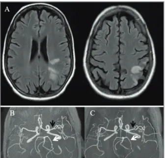
MRI suggested cerebral vasculitis (Figure 1A and 1B) and new CSF disclosed 5 leucocytes/L, protein 29 mg/dL and normal glucose with 65 mg/dL. At this point RTX (1g, twice) was given in association with cy-clophosphamide (CYC; 500mg), two weeks apart. Brain MRI one month after first RTX administration revealed improvement of vasculitic lesions (Figure 1C).
CASE 3
A 39-year old female, with 13-year history of SLE with mucocutaneous, articular, lung, renal, haematological and immunological (triple positive aPL) involvement, under treatment with HCQ, PDN 10mg/day and MMF 1.5g/day, presented with holocranial, pressure type headache without warning signs, blurred vision and generalized tonic-clonic seizure. At admission she was markedly hypertensive (225/149 mmHg) and her neu-rological examination was unremarkable. Cranium CT and laboratory were normal. She was started on hy-potensive treatment with complete recovery within 3 days and no complementary evaluation was per-formed.
Six months later she was readmitted with fever and holocranial, non-pulsatile headache. Laboratory showed haemolytic anaemia, thrombocytopenia, raised inflammatory markers, decreased C3 comple-ment fraction, normal anti-dsDNA antibody and wors-ening proteinuria. Cranium CT was normal and CSF disclosed 230 leucocytes/L (mainly polymorphonu-clear), protein 176 mg/dL and normal glucose with 60 mg/dL. Septic meningitis was assumed and she stopped MMF and started ceftriaxone, ampicillin and vancomycin. However, fever persisted (despite sterile CSF) and hyporeflexive paraparesis with total sensory loss below D10 developed. MRI demonstrated in-tradural-extramedullary hematoma D9-L3 secondary to traumatic lumbar puncture, which was successful-ly drained.
During postoperative period, she remained mildly hypertensive (medium blood pressure above 100-115 mmHg) and at the third day developed generalized tonic-clonic seizures. Brain MRI demonstrated asym-metric bilateral occipital cortical and subcortical white matter hyperintensities in T2-fluid attenuated inver-sion recovery sequence, suggesting posterior reversible encephalopathy syndrome (PRES). Electroencephalo-gram (EEG) showed teta slow wave activity located to right posterior temporal lobe, without epileptiform acti vity. Hypotensive treatment was readjusted and leve tiracetam was added, with subsequent blood
pres-FIGURE 1. A (MRI in T2 FLAIR sequence) shows multiple ischemic lesions in different vascular territories, suggesting cerebral vasculitis. B (MRI in TOF sequence) shows segmental narrowing and irregularity of left middle (black arrow) and left posterior arteries (white arrow), with caliber improvement one month later (C), after treatment with cyclophosphamide and rituximab
sure control and no seizure recurrence.
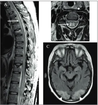
One month after surgery, despite intensive rehabi -litation, motor and sensory alterations persisted, with onset of urinary retention. New MRI showed complete resolution of occipital oedema and holocord abnormal T2 hyperintense signal from conus medullaris into brainstem, with associated swelling, in relation to lon-gitudinally extensive transverse myelitis (LETM; Fi gure 2A and 2B). Infectious and metabolic causes were ex-cluded and aquaporin-4 (AQP4) and anti-myelin oligodendrocyte glycoprotein (anti-MOG) an-tibodies were negative. NPSLE was assumed and she received methylprednisolone (1g/day, 3 days), followed by PDN (1mg/Kg/day) and intravenous human im-munoglobulin (IVIg; 1g/kg/day for 2 days), due to per-sistent infection of open surgical scar. IVIg was kept monthly for 6 months, in association with HCQ and PDN 10mg/day, with resolution of MRI medullar fin -dings and progressive neurological recovery, except for neurogenic bladder.
Twelve months after finishing IVIg she was read-mitted with posterior, tension type headache, binocu-lar horizontal diplopia and sudden loss of conscious-ness. At admission she was slightly hypertensive (156-75 mmHg), with neurological examination showing bilateral papilledema. Laboratory was unremar kable. Brain MRI suggested a new episode of PRES (Fi -gure 2C).
PRES recurrence without immunosuppression or re-markable hypertension and LETM were considered NPSLE manifestations and she restarted monthly IVIg, due to recurrent cystitis in a patient with neurogenic bladder. After 5 months of IVIg no PRES recurrence occurred.
CASE 4
A previously healthy 36-years-old female with four--month history of vasculitic lesions of fingers and in-flammatory poliarthralgias of hands, wrists and shoul-ders presented to a rheumatologist, who treated her with PDN 15mg/day and asked for complementary in-vestigation. One week later, she suddenly developed auditory and visual hallucinations, delusional ideas of persecutory content, disorganized speech and motor agitation, which eventually led her to attempt suicide. At hospital admission, she was febrile, with psy-chomotor agitation and disorganized speech, but without meningeal signs. Cranium CT was normal and to -xicological examination was negative. CSF disclosed 8 leucocytes/L, protein 35 mg/dL and normal glucose,
with negative culture and neurotropic virus research. Brain MRI and EEG were normal. At the same time, complementary investigation showed decreased C3 and C4 complement fractions, positive antinuclear an-tibodies with positive anti-ds DNA and anti-ribosomal P protein (anti-Rib-P) antibodies, negative aPL, nega-tive anti-AQP4, anti-MOG and anti-NMDA antibo dies. NPSLE was assumed and the patient treated with IV methylprednisolone (1g/day, 3 days), followed by PDN (1mg/kg/day), in association with CYC (6 monthly pulses of 0.5-0.75 g/m2body surface) and
antipsy-chotics, with complete neurologic recovery. Nine months after stopping CYC she remained asymp-tomatic, only under HCQ.
DISCUSSION
NPSLE is a complex entity, which comprise a wide range of neuropsychiatric signs/symptoms attributable to SLE. Risk factors associated with NPSLE include gene ralized SLE activity/cumulative damage, previous
FIGURE 2. A and B (MRI in T2 FSE sequence) show abnormal hyperintense signal of spinal cord (arrow), at C4 axial level (A) and extending longitudinally through thoracic spinal cord (B). C (MRI in T2 FLAIR sequence) shows bilateral occipital cortical and subcortical white matter hyperintensities (arrowhead), compatible with posterior reversible encephalopathy syndrome
or concurrent major NPSLE events and aPL3.
In 1999, ACR developed a standard nomenclature and case definitions for 19 neuropsychiatric syndromes that are known to occur in SLE2, including 12 central
nervous system and 7 peripheral nervous system. How-ever, these case definitions are not specific for SLE and other neurologic conditions reported in SLE patients, like NMO4, PRES5and small fiber neuropathy6, were
excluded. More recently, Bortoluzzi et. al developed a new attribution model7that allowed a confident correct
attribution of neuropsychiatric syndromes deemed as SLE.
Diagnostic approach of NPSLE should be similar for non-SLE patients presenting with the same manifesta-tions 8 and mimickers like infections, drug-induced side
effects, metabolic abnormalities (including alcoholrelated) and malignancies have to be excluded. A gene -ral assessment of SLE activity may contribute to attri-bution to SLE 8.
Some autoantibodies have been implicated in spe-cific NPSLE manifestations9,with aPL being largely re
-cognized as risk factors for cerebrovascular ischemic events10. Nonetheless, they have also been associated
with other NPSLE manifestations, such as chorea, movement disorders, cognitive dysfunction and myelopathy11–14. Psychosis has been associated with
anti-Rib-P antibodies15 and anti-endothelial cell
antibodies8. Anti-glutamate receptor antibodies
(ant-NMDA and anti-NR2) have been linked to cogni-tive dysfunction16,17and anti-AQP4 and anti-MOG
an-tibodies to NMO4.
Brain MRI remains the gold standard imaging exam in NPSLE 18, although position emission tomography
can be useful in patients with neurological manifesta-tions and normal MRI, by evaluating brain metabolic activity19.
Management of patients with NPSLE includes symp-tomatic treatment and specific therapy dictated by the underlying pathophysiological process3,20. For instance,
immune-mediated manifestations of patients with gen-eralized lupus activity must be treated with steroids either alone or in association with other immunosuppressive drugs. Intravenous CYC associated with me -thyl prednisolone demonstrated superiority compared to methylprednisolone alone21and is the drug
recom-mended for severe NPSLE. Other immunosuppressants, like AZA and MMF, can be used as main tenance therapy after CYC or in cases of mild NPSLE20,22,23. In patients
with severe refractory NPSLE, RTX, IVIg, plasma ex-change and autologous hematopoietic stem cell
trans-plantation can also be considered3,20. In case 2, cranial
palsy was considered a mild NPSLE manifestation and therefore treated with MMF. However, as the patient developed one month later acute and severe cerebral vasculitis, we decided to switch treatment to CYC and RTX. In case 4, the seve rity of NPSLE manifestation, which was part of disease presentation, was responsi-ble for the choice of CYC as the first treatment. IVIg might be particularly useful for patients with con-traindication for standard immunosuppression, like in-fection, as in case 3.
Treatment of ischemic NPSLE includes control of cardiovascular risk factors, antiplatelet agents and/or anticoagulation3,20. Patients without aPL or who do not
fulfill criteria for APS must receive aspirin as secondary thromboprophylaxis, while patients who fulfill criteria for APS must receive vitamin K antagonists20. In
pa-tients with APS and recurrent thrombosis, addition of aspirin20or treatment with high-intensity warfarin (INR
3-4) might be considered24. In patients with thrombotic
recurrences despite adequate target INR, RTX has also demonstrated a beneficial effect25, and was therefore
added in case 1, after failure of therapeutic anticoagu-lation and aspirin.
In conclusion, NPLSE diagnosis can be extremely challenging and less specific manifestations, such as headache, cognitive dysfunction and mood disorders are the most commonly reported in literature8. Despite
the small number of cases, this work is a single-centre experience, which presents some of the rarer NPSLE manifestations and in some cases with more than one manifestation in the same patient.
CORRESpONDENCE TO
Ana Catarina Duarte Avenida Torrado da Silva
E-mail: [email protected]
REFERENCES
1. Unterman A, Nolte JES, Boaz M, Abady M, Shoenfeld Y, Zand-man-Goddard G. Neuropsychiatric syndromes in systemic lu-pus erythematosus: a meta-analysis. Semin Arthritis Rheum. 2011 Aug;41(1):1–11.
2. The American College of Rheumatology nomenclature and case definitions for neuropsychiatric lupus syndromes. Arthritis Rheum. 1999 Apr;42(4):599–608.
3. Bertsias GK, Ioannidis JPA, Aringer M, Bollen E, Bombardieri S, Bruce IN, et al. EULAR recommendations for the management of systemic lupus erythematosus with neuropsychiatric mani-festations: report of a task force of the EULAR standing com-mittee for clinical affairs. Annals of the Rheumatic Diseases. 2010 Dec 1;69(12):2074–2082.
4. Mader S, Jeganathan V, Arinuma Y, Fujieda Y, Dujmovic I, Drulovic J, et al. Understanding the Antibody Repertoire in
Neu-ropsychiatric Systemic Lupus Erythematosus and Neuromyeli-tis Optica Spectrum Disorder: Do They Share Common Targets? Arthritis & Rheumatology (Hoboken, NJ). 2018;70(2): 277–286.
5. Cui H-W, Lei R-Y, Zhang S-G, Han L-S, Zhang B-A. Clinical fea-tures, outcomes and risk factors for posterior reversible en-cephalopathy syndrome in systemic lupus erythematosus: a case-control study. Lupus. 2019 Jul;28(8):961–969. 6. Gøransson LG, Tjensvoll AB, Herigstad A, Mellgren SI, Omdal
R. Small-diameter nerve fiber neuropathy in systemic lupus ery-thematosus. Arch Neurol. 2006 Mar;63(3):401–404. 7. Bortoluzzi A, Scirè CA, Bombardieri S, Caniatti L, Conti F, De
Vita S, et al. Development and validation of a new algorithm for attribution of neuropsychiatric events in systemic lupus ery-thematosus. Rheumatology (Oxford). 2015 May;54(5):891–8. 8. Govoni M, Bortoluzzi A, Padovan M, Silvagni E, Borrelli M,
Donelli F, et al. The diagnosis and clinical management of the neuropsychiatric manifestations of lupus. J Autoimmun. 2016;74:41–72.
9. Ho RC, Thiaghu C, Ong H, Lu Y, Ho CS, Tam WW, et al. A meta-analysis of serum and cerebrospinal fluid autoantibodies in neu-ropsychiatric systemic lupus erythematosus. Autoimmun Rev. 2016 Feb;15(2):124–138.
10. de Amorim LCD, Maia FM, Rodrigues CEM. Stroke in systemic lupus erythematosus and antiphospholipid syndrome: risk fac-tors, clinical manifestations, neuroimaging, and treatment. Lu-pus. 2017 Apr;26(5):529–536.
11. D’Cruz DP, Mellor-Pita S, Joven B, Sanna G, Allanson J, Taylor J, et al. Transverse myelitis as the first manifestation of systemic lupus erythematosus or lupus-like disease: good functional out-come and relevance of antiphospholipid antibodies. J Rheuma-tol. 2004 Feb;31(2):280–285.
12. Denburg SD, Denburg JA. Cognitive dysfunction and an-tiphospholipid antibodies in systemic lupus erythematosus. Lu-pus. 2003;12(12):883–890.
13. Giorgi D, Balacco Gabrieli C. Optic neuropathy in systemic lu-pus erythematosus and antiphospholipid syndrome (APS): clin-ical features, pathogenesis, review of the literature and proposed ophthalmological criteria for APS diagnosis. Clin Rheumatol. 1999;18(2):124–131.
14. Cervera R, Asherson RA, Font J, Tikly M, Pallarés L, Chamorro A, et al. Chorea in the antiphospholipid syndrome. Clinical, ra-diologic, and immunologic characteristics of 50 patients from our clinics and the recent literature. Medicine (Baltimore). 1997 May;76(3):203–212.
15. Bonfa E, Golombek SJ, Kaufman LD, Skelly S, Weissbach H, Brot N, et al. Association between lupus psychosis and anti-ri-bosomal P protein antibodies. N Engl J Med. 1987 Jul 30;317(5):265–271.
16. Lapteva L, Nowak M, Yarboro CH, Takada K, Roebuck-Spencer T, Weickert T, et al. Anti-N-methyl-D-aspartate receptor anti-bodies, cognitive dysfunction, and depression in systemic lupus erythematosus. Arthritis Rheum. 2006 Aug;54(8):2505–14. 17. Tay SH, Mak A. Anti-NR2A/B Antibodies and Other Major
Molecular Mechanisms in the Pathogenesis of Cognitive Dys-function in Systemic Lupus Erythematosus. Int J Mol Sci. 2015 May 6;16(5):10281–300.
18. Postal M, Lapa AT, Reis F, Rittner L, Appenzeller S. Magnetic resonance imaging in neuropsychiatric systemic lupus erythe-matosus: current state of the art and novel approaches. Lupus. 2017 Apr;26(5):517–521.
19. Weiner SM, Otte A, Schumacher M, Klein R, Gutfleisch J, Brink I, et al. Diagnosis and monitoring of central nervous system in-volvement in systemic lupus erythematosus: value of F-18 flu-orodeoxyglucose PET. Ann Rheum Dis. 2000 May;59(5): 377–385.
20. Magro-Checa C, Zirkzee EJ, Huizinga TW, Steup-Beekman GM. Management of Neuropsychiatric Systemic Lupus Erythemato-sus: Current Approaches and Future Perspectives. Drugs. 2016 Mar;76(4):459–483.
21. Barile-Fabris L, Ariza-Andraca R, Olguín-Ortega L, Jara LJ, Fra-ga-Mouret A, Miranda-Limón JM, et al. Controlled clinical tri-al of IV cyclophosphamide versus IV methylprednisolone in se-vere neurological manifestations in systemic lupus erythematosus. Ann Rheum Dis. 2005 Apr;64(4):620–625. 22. Mok CC, Lau CS, Wong RWS. Treatment of lupus psychosis
with oral cyclophosphamide followed by azathioprine mainte-nance: an open-label study. Am J Med. 2003 Jul;115(1):59–62. 23. Tselios K, Gladman DD, Su J, Urowitz MB. Mycophenolate
Mofetil in Nonrenal Manifestations of Systemic Lupus Erythe-matosus: An Observational Cohort Study. J Rheumatol. 2016 Mar;43(3):552–558.
24. Tektonidou MG, Andreoli L, Limper M, Amoura Z, Cervera R, Costedoat-Chalumeau N, et al. EULAR recommendations for the management of antiphospholipid syndrome in adults. An-nals of the Rheumatic Diseases. 2019 Oct 1;78(10):1296–1304. 25. Wang C-R, Liu M-F. Rituximab usage in systemic lupus
erythe-matosus-associated antiphospholipid syndrome: A single-cen-ter experience. Semin Arthritis Rheum. 2016;46(1):102–108.