UNIVERSIDADE DA BEIRA INTERIOR
Ciências
Development of new chromatographic support
based on gellan gum for pharmaceutical
biomolecules isolation
Armanda Isabel Carvalho Gonçalves
Dissertação para obtenção do Grau de Mestre em
Bioquímica
(2º ciclo de estudos)
Orientador: Prof. Doutor Luís António Paulino Passarinha
Coorientador: Prof. Doutora Ângela Maria Almeida de Sousa
ii
Dedicatória
iii
A cromatografia é uma técnica usada para separação e/ou purificação de diferentes biomoléculas com finalidade analítica ou preparativa. Esta técnica tem sido muito aplicada na área da indústria farmacêutica, para a obtenção de proteínas e ácidos nucleicos, entre outras biomoléculas. Com o passar dos anos, a quantidade de amostras e a pureza necessária aumentaram, tornando esta técnica numa das mais usadas na indústria biotecnológica. O desenvolvimento de novas matrizes cromatográficas tem sido um tema de grande importância, a fim de encontrar uma matriz ideal que reúna características como custos associados, eficiente, estável física e quimicamente, com uma elevada eficiência de transferência de massa e reutilizável.
A gelana é um polímero polissacárido natural linear que tem sido aplicado em diversas áreas, como indústria alimentar (agente gelificante e espessante), indústria farmacêutica (entrega direcionada de fármacos), podendo também ser aplicada como um substituinte do agar. Este polímero linear em determinadas condições pode sofrer uma alteração conformacional, formando uma dupla hélice. Na presença de catiões, forma uma rede tridimensional, devido às interações ocorridas entre a gelana, os catiões e as moléculas de água em solução, dando origem a um gel. Tendo em conta estas considerações, o presente trabalho foi realizado com o intuito de formular um gel de gelana estável, a fim de adquirir um comportamento de matriz cromatográfica. A preparação dos géis foi feita de acordo com os seguintes parâmetros: concentração de gelana, concentração de catião (zinco), concentração de DMF, temperatura e tempo de reação. Uma vez que a estabilidade dos géis de gelana é afetada por todos estes parâmetros, foram preparadas várias formulações em que se variou apenas um destes parâmetros para verificar qual o seu efeito na estabilidade do gel. As formulações foram testadas de acordo com as seguintes variações: concentração de gelana (0,75% - 2%), concentração de sulfato de zinco (30 – 120 mM), concentração de DMF (0% - 30%), temperatura (temperatura ambiente – 110ºC) e tempo de reação (0.5 horas – “overnight”). Posteriormente foi aplicada uma estratégia de desenho experimental no sentido de definir as condições ideais para a preparação do gel, e consequentemente obter um gel mais estável. Assim, os melhores resultados quanto à estabilidade do gel foram obtidos quando se usou 0,75% de gelana, 48 mM de sulfato de zinco, 0 % DMF, 25ºC, e 0,5 horas.
Adicionalmente e aproveitando a natureza aniónica do polímero de gelana, foi possível explorar diferentes interações com as seguintes proteínas modelo (albumina sérica bovina, α-quimotripsina e lisozima). Na presença do tampão MÊS com pH 6,2, a albumina sérica bovina encontrava-se com carga negativa, enquanto que a α-quimotripsina e lisozima se encontravam com carga positiva, tendo em conta os respetivos pontos isoelétricos. Sendo assim, a albumina sérica bovina não interagiu com a matriz, visto que ambas têm carga
iv
de cargas. A separação das três proteínas foi conseguida através da eluição por passos, com o aumento gradual da concentração de NaCl no tampão de eluição. A albumina sérica bovina foi a primeira proteína a eluir após a aplicação da amostra, ainda com a passagem do tampão de eluição sem sal. Posteriormente aumentou-se a concentração de sal promovendo-se a eluição da α-quimotripsina . Por fim, para concentrações de sal mais elevadas, e a lisozima que promoveu uma interação mais forte com a matriz de gelana acabou por eluir também. A fim de melhor caracterizar esta nova matriz, foi determinada também a capacidade dinâmica de ligação utilizando uma estratégia de saturação da coluna com uma solução de lisozima 0,05 mg/mL. Os valores obtidos para a capacidade dinâmica de ligação da gelana a 10% e a 50% foram 3,9 mg/ml e 17,4 mg/ml, respetivamente. Comparando com outras matrizes cromatográficas, os valores de capacidade dinâmica de ligação da matriz de gelana estão dentro do esperado. Estes estudos iniciais permitiram concluir que a gelana interagiu com as proteínas, considerando a diferença de cargas, e que a sua eluição também foi possível com o aumento de sal, o que revela uma provável potencialidade como matriz cromatográfica de troca catiónica. Para poder afirmar e provar que o polímero gelana pode constituir uma matriz cromatográfica, devem ser feitos mais ensaios. Nomeadamente testar a purificação de amostras mais complexas e determinar mais parâmetros (como por exemplo a capacidade iónica e o tipo e tamanho de poros) que permitam a otimização da matriz. No futuro, poderemos vir a ter uma matriz versátil com aplicação em diversos domínios científicos.
Palavras-chave
v
Higher separation efficiency and resolution have been of great interest in chromatography and have become increasingly important in recent years mainly driven by the challenges of either more complex samples or high sample quantity. Therefore, for the development of new chromatographic matrices is increasingly important to improve the purification efficiency and to decrease the use of resources. Gellan gum is a polysaccharide polymer with natural anionic nature and ability to form thermoreversible gels, seeming to have potential to be used as a chromatographic matrix. In the presence of cations, the gellan polymer suffers conformational transition accompanied by the formation of a three dimensional network, forming a gel. In this work, it was intended to prepare of a stable gellan gum gel to be used as a chromatographic matrix. In order to increase the stability of the gellan gels, different experimental conditions were tested. Experimental design was used to obtain optimal conditions for the gel stability. Due to negative charge of these gels, it was possible to study the interactions established with three model proteins (bovine seric albumin (BSA), α-chymotrypsin and lysozyme). Gellan gum was able to interact with two of these proteins, being able to elute them with an increase in the ionic strength. In this assays a MES buffer with pH 6.2 was utilized. This pH conferred negative charge to BSA and positive charge to α-chymotrypsin and lysozyme, due to their isoelectric points. Assays of dynamic binding capacity were performed to find more characteristics of this new matrix and comparing with commercial resins. The values of dynamic binding capacity of the gellan gum to 10% and 50% breakthrough were 3,9 mg/ml and 17,4 mg/ml, respectively. These values were similar to commercial resins. These results showed that gellan gum might be an innovative and promising chromatographic matrix due to its versatility to interact with different biomolecules and its gelling ability. Thus, the gellan gum gel could have potential to be applied in different scientific domains (purification of complexes cellular extracts or nucleic acids).
Keywords
vi
Chapter I - Introduction ... 1
1. Chromatography ... 1
1.1 Chromatographic process ... 1
1.2 Preparative versus analytical chromatography ... 3
1.3 Chromatographic equipment ... 5
1.4 Chromatographic methods ... 6
1.4.1 Size Exclusion Chromatography (SEC) ... 7
1.4.2 Hydrophobic Interaction Chromatography (HIC) ... 8
1.4.3 Reversed Phase Chromatography (RPC) ... 9
1.4.4 Affinity Chromatography (AC) ... 10
1.4.4.1 Immobilized metal ion affinity chromatography (IMAC) ... 11
1.4.5 Ion exchange chromatography (IEC) ... 12
2. Matrices ... 14
2.1 Matrices and ligands used in chromatography... 14
2.1.1 Chemical nature of the chromatographic supports ... 15
2.1.2 Physical nature of the chromatographic supports ... 15
2.1.3 Role of ligand immobilization on chromatographic supports ... 16
2.2 Typical matrices and ligands in ion exchange chromatography ... 17
2.3 Dynamic Binding Capacity ... 18
3. Gellan gum ... 19
3.1 Physical and chemical properties of gellan gum ... 19
3.2 Fermentative production of gellan gum ... 20
3.2.1 Biosynthetic pathway of gellan gum ... 21
3.3 Gellan gum applications ... 23
3.4 Gellan gum gels ... 25
Chapter II – Objectives ... 27
Chapter III – Materials and Methods ... 28
1. Materials ... 28
2. Formulation of Gellan gum gels with different conditions ... 28
3. Gellan gum gel stability assays ... 28
4. Optimization of gellan gum gel formulation conditions ... 29
4.1 Experimental Design ... 29
5. Gellan gum gel formulation with methacrylic anhydride ... 29
5.1 Gellan gum gel formulation with methacrylic anhydride and nickel as contour ion. 29 6. Ion Exchange Chromatography ... 29
vii
lysozyme) ... 30
6.4 Chromatographic assay of combined model proteins (BSA + α-chymotrypsin + lysozyme) and pH variation ... 31
7. Electrophoretic analysis... 31
8. Dynamic Binding Capacity ... 31
Chapter IV – Results and Discussion ... 32
1. Gellan gum gel stability assays ... 32
2. Optimization of gellan gum gel formulation conditions ... 34
3. Gellan gum gel formulation with methacrylic anhydride ... 36
3.1 Gellan gum gel formulation with methacrylate and nickel as counter ion ... 37
4. Ion Exchange Chromatography ... 38
4.1 Chromatographic assays with isolated model proteins ... 38
4.2 Chromatographic assays with combined model proteins (BSA + α-chymotrypsin; BSA + lysozyme) ... 41
4.3 Chromatographic assay with combined model proteins (α-chymotrypsin + lysozyme) ... 45
4.4 Chromatographic assay with combined model proteins (BSA + α-chymotrypsin + lysozyme) ... 46
4.5 Chromatographic assay with combined model proteins (BSA + α-chymotrypsin + lysozyme) with pH variation in the elution buffer ... 49
5. Dynamic Binding Capacity ... 51
Chapter V - Conclusion ... 54
Chapter VI – Futures perspectives ... 55
viii
Figure 1 - Schematic representation of the low pressure chromatographic equipment. 6 Figure 2 - Representation of the repeating unit of chemical structure of native gellan
gum.
19 Figure 3 - Representation of the repeating unit of chemical structure of deacetylated
gellan.
19 Figure 4 - Schematic representation of the postulated pathway leading to the
nucleotide sugar precursors.
22 Figure 5 - Schematic representation of the polymerization and export of the gellan
gum through the outer membrane for cell surface.
23 Figure 6 - Representation of the linear regression of predicted over experimental
number of column volumes.
35 Figure 7 - Representation of the three combinations of the gel preparation conditions
(gellan with ZnSO4, gellan with temperature and gellan with time).
36 Figure 8 - Representation of the repeating unit of chemical structure of gellan gum
methacrylate.
37 Figure 9 - Chromatografic profile obtained for BSA assay. 39 Figure 10 - Chromatografic profile obtained for α-chymotrypsin assay. 40 Figure 11 - Chromatografic profile obtained for lysozyme assay. 41 Figure 12 - Chromatografic profile obtained for BSA + α-chymotrypsin assay. 42 Figure 13 – SDS-PAGE electrophoresis analysis of the peak fractions collected in the
BSA + α-chymotrypsin chromatographic assay.
43 Figure 14 - Chromatografic profile obtained for BSA + lysozyme assay. 44 Figure 15 - SDS-PAGE electrophoresis analysis of the peak fractions collected in the
BSA + lysozyme chromatographic assay.
45 Figure 16 - Chromatografic profile obtained for α-chymotrypsin + lysozyme assay. 46 Figure 17 - Chromatografic profile obtained for BSA + α-chymotrypsin + lysozyme
assay.
47
Figure 18 – SDS-PAGE electrophoresis analysis of the peak fractions collected in the BSA + α-chymotrypsin + lysozyme chromatographic assays.
48
Figure 19 - Chromatografic profile obtained for BSA + α-chymotrypsin + lysozyme and pH buffer variation chromatographic assay.
49 Figure 20 – SDS-PAGE electrophoresis analysis of the peak fractions collected in the
BSA + α-chymotrypsin + lysozyme and pH buffer chromatographic assays.
50 Figure 21 - Dynamic binding capacity of gallant gum for 0. 5 mg/ml solution
lysozyme at 1 ml/min flow rate.
ix
Table 1 - Characteristics and applications of several chromatographic methods. 13
Table 2 - Ion exchange chromatography matrices. 18
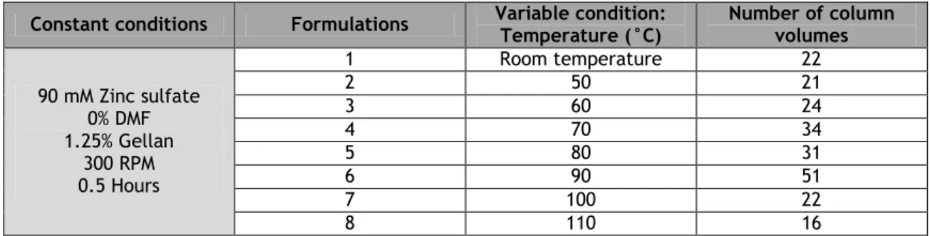
Table 3 – Number of column volumes obtained by varying the gellan concentration for the gellan gum gel formulation.
32 Table 4 – Number of column volumes obtained by varying the zinc sulfate
concentration for the gellan gum gel formulation.
33 Table 5 – Number of column volumes obtained by varying the reaction time for the
gellan gum gel formulation.
33 Table 6 – Number of column volumes obtained by varying the temperature time for
the gellan gum gel formulation.
34 Table 7 – Number of column volumes obtained by varying the DMF concentration for
the gellan gum gel formulation.
34 Table 8 – Dynamic binding capacity of cation-exchange resins and heparin for 0.5
mg/ml solution lysozyme.
x
AC Affinity chromatography ANN Artificial neural network ANX Diethylaminopropyl
BSA Bovine serum albumin CM Carboxymethyl CVs Column volumes DEAE Diethylaminoethyl
DMF N,N-Dimethylformamide FDA Food and Drug Administration
HIC Hydrophobic Interaction Chromatography IEC Ion Exchange Chromatography
IMAC Immobilized Metal Ion Affinity Chromatography MES 4-Morpholineethanesulfonic acid
NaCl Sodium chloride NiSO4 Nickel sulfate
PAGE Polyacrylamide Gel Electrophoresis PSA Ammonium persulfate
Q Quaternary ammonium
RPC Reversed Phase Chromatography S Methyl sulfonate
SDS Sodium dodecyl sulfate
SEC Size Exclusion Chromatography SP Sulfopropyl
TDP Thymine-5-diphosphate
TEMED N,N,N',N'-tetramethylethylenediamine TFA Trifluoroacetic acid
Tris Tris(hydroxymethyl)aminomethane UDP Uridine-5-diphosphate
USA United States of America ZnSO4 Zinc sulfate
1
Chapter I - Introduction
1. Chromatography
Protein purification has been performed for more than 200 years. Until the beginning of the 20th century, the only available separation technologies were filtration, precipitation, and crystallization methods. In 1906 the botanist Mikhail Tswett introduced the term chromatography, describing his work on separation of plant pigments on a column of calcium carbonate [1, 2]. In a few years, the purification of other biomolecules, such as proteins, nucleic acids, antibodies, was possible [3, 4].
Nowadays, most purification schemes involve some form of chromatography. As a result, chromatography has become an indispensable tool in the separation and purification of biological substances in every laboratory, due to its simplicity, robustness, versatility, and high reproducibility [5, 6].
The aim of a purification process is not only removal of unwanted contaminants, but also to obtain and maintain quality, stability and biological activity of biomolecules. The purity required depends on the science area in which the biomolecule will be applied. If the application consists on pharmaceuticals, the biomolecule must have a high purity degree, in contrast to biomolecules applied in laundry detergents where the purity degree is inferior [7, 8, 9].
Chromatography has become a preferential technique for separation, due to its high resolving power, the existence of several chromatographic methods with different selectivity and its applicability to an extensive spectrum of compounds [10, 11].
1.1 Chromatographic process
Chromatography is a method used to separate components of a sample, taking into account the physical and chemical properties of each biomolecule [10]. Charge, molecular size, hydrophobicity and polarity determine the type of interaction between the solute in a mobile phase with a stationary phase [7].
Solutes can stay in the mobile phase or be distributed in the stationary phase by promoting specific interactions. Stationary phase is packed into a vertical column and should be insoluble in the buffer. Mobile phase is pumped through the column and it can be liquid or gaseous [2].
2
Separation of the different sample solutes can occur when they interact with different intensities on the support, running the column with different times. This differential separation occurs due to continuous addition of mobile phase and, which is known as elution [10, 12]. Differential elution of several biomolecules causes a sequence of separated peaks. These peaks reflect the concentration of biomolecules versus time or volume of eluent at the column exit, whichis typically represented in a chromatogram [2].
The chromatographic process consists in five steps: equilibrium phase, loading of sample, washing and removal of unbound material, elution of the biomolecule and regeneration/cleaning matrix [12]. Equilibrium phase involves the adjustment of the mobile and stationary phase to optimal binding conditions of solutes. The sample loading corresponds to injection of a specific quantity protein solution in the column, depending of the sample origin and if the process has an analytical or preparative objective. In washing step, unbound material components that remained in the mobile phase are removed, because did not interact with the stationary phase [12, 13]. The elution of retained species corresponds to desorption of desired compound by stepwise or continuous change of mobile phase composition. Elution buffer decreases interactions between the matrix and target biomolecules [2, 12].
The target biomolecule that strongly adsorbed to the stationary phase elute slower than the weakly bound biomolecules, and the application of significant alterations in the mobile phase is necessary [14]. Alterations on ionic strength (type and salt concentration), pH and competitive agent addition are the most common means of eluting adsorbed proteins by linear gradient or stepwise gradient [5, 8, 15].
In linear gradient elution, the mobile phase composition is continuous changing for conditions that favor the dissociation of the protein and thus, the solute interaction with the support is variable. This elution form improves the separation and it is normally used when the sample is unknown. The eluent composition is changed step by step in stepwise gradient and various substances can be eluted with this strategy [15]. Stepwise elution is frequently favored for routine, large-scale purifications and additionally the buffer consumption is reduced [1, 16]. Alternatively, the composition of the eluent is selected to give weak or no existent interactions between sample components and chromatographic matrix, and these conditions are kept unchanged during binding and elution. The target biomolecule passes through the column slower or faster than impurities, resulting in different elution positions. This type of elution is known by isocratic elution where in composition of the mobile phase and the solute interaction with the support are constant [1, 16]. The gradient elution allows the separation of components with a wider range of properties when compared with isocratic elution [1]. The regeneration and cleaning matrix step is important, because it permits to reuse the matrix for new chromatographic assays [1]. In this step, sticky impurities such as endotoxins
3
and lipids are removed to maintain the native matrix characteristics such as selectivity and support life-time [9, 12]. A dirty medium may also induce decreased binding capacity and increased pressure [1]. Cleaning procedure depends on matrix type, but normally it is made with low or high-salt concentrated solutions, highly acid or basic conditions, organic solvents or some detergents. This process also avoids microbial contamination during storage [13]. The purification of the biomolecule of interest may happen by two different processes. The biomolecule itself can be separated from contaminants by binding to the stationary phase, followed by selective elution; or by binding impurities, allowing the target biomolecule to pass through the column without being retained (designated as negative chromatography) [1].
1.2 Preparative versus analytical chromatography
The product amount, purity degree and biomolecule stability are important requirements for choosing the type of chromatography [12].
Chromatography can be used for preparative or analytical scale. Liquid chromatography (mobile phase is a liquid) is the most used for preparative scale. Gas chromatography (mobile phase is a gas) is an indispensable tool to the analytical level [17–19].
In the last years, chromatographic stationary phase materials based on new concepts where developed to increase speed at both analytical and preparative scale [20].
Preparative liquid chromatography has become the primary tool for the purification of proteins from complex biological mixtures in biotechnological processes. In preparative chromatography, the aim is to separate very similar compounds, requiring high column efficiencies [16, 18]. Preparative separations require the introduction of larger amounts of sample onto the chromatographic column. Sample dissolution in the same solvents as used for elution is ideal to minimize peak broadening and distortion, because the higher the solubility, the higher the efficiency of the preparative productivities [21].
Preparative chromatography is the most tool used in the biotechnological industry, therefore it is used on a large scale, producing sometimes kg or even tons of the target biomolecule [20]. For this reason, large diameter columns and gels very porous to increase the binding capacity are necessary to use. Diffusion pathways for the proteins are relatively long, therefore low flow velocities are required to improve the resolution [22]. Preparative chromatography is becoming a well-established separation and purification method including chiral separations, extraction of recombinant proteins from fermentation broths and purification of basic compounds [17].
Whether the amount of purified compound isolated is going to be used of sale as a reagent or a pharmaceutical, among them the need to produce as concentrated a fraction as possible, to collect and transfer without impurities, and to do the separation as quickly and cheaply as
4
possible. As it is a large-scale process, the buffer should be cheap, because it will be used in large quantities [1, 17]. Separation costs account for 50-80% of total production costs and chromatography is often the most expensive unit process in a separation. Due to this fact it is important to develop more cost efficient chromatographic systems, to become the purification technique more economically viable and reproductive for to large-scale-up process [9].
Preparative chromatography is a powerful separation technique, but it is often associated with significant dilution of the product. Other problem is the gradient, for the typical large-diameter preparative columnslinear gradients are not a good choice. Thus, step gradients are more popular in preparative elution chromatography or displacement elution [2].
Separation power of preparative chromatography attracts the attention of those interested in producing pure chemicals in large amounts for a variety of purposes. Once produced, the identity, purity, potency, and safety of biopharmaceutical compounds must be demonstrated to the regulatory agencies before they can be used in humans, avoiding unwanted side effects. This aim may require the use of simple detectors (such as UV or FID) or coupling with the most complex detectors (such as multiple MS or high field strength NMR). From the moment required signals have been acquired for a detector, the chemical can be discarded [9, 24].
Analytical chromatography permits to identify and quantify the components of mixtures either simple or complex. In analytical chromatography, a small sample is applied to the column and maximum resolution is sought, by manipulating the characteristics of the different phases of the system [12, 17]. Preparative column loads more protein in three or four preparative cycles than an analytical column loads in two or three hundred cycles [9]. In analytical chromatography high flow velocities and cycle times of a few seconds are reported [24].
The goal of analytical chromatography remains to be the rapid determination of the structure of the component, through the direct acquisition of the proper information, and the calculation of its concentration, through calibration of the detector signal [17]. In this type of chromatography it is required a high selectivity and sensitivity, because the purification conditions may create very similar populations of proteins with structural variants (such as deamidated, amino acid residues oxidized and cleavage fragments of a single polypeptide) and their analysis becomes more difficult [24, 25]. Lately, development of complex instruments and the coupling two or several columns, have been applied to solve analytical or preparative separation problems [17].
5
1.3 Chromatographic equipment
Protein purification is always a race against time and there is a need to accelerate the procedure, because during the isolation step proteins are exposed to changes in environment, enzymatic degradation by proteases, or even time itself, leading to protein losses and reduction of biological activity. The separation process is fast to increase the yield of the target protein and helping to improve the cost effectiveness of the production process is essential [2, 22].
The combination of hardware, chemicals, operation mode, sample volume, purity, amount of purified protein required, presence of toxic components, number of samples to be processed, are determinant to choose of the purification format [1]. The principal purification format is based on chromatography in closed columns (chromatography system or an independent pump) or in open columns (gravity flow columns or spin columns) used in manual purification. A chromatographic system should be used when reproducible results are important and when the manual purification becomes more slow and inefficient [1].
The purification equipment has evolved into hardware and software that impart a high level of control over the process. These automated systems not only decrease labor costs, but perhaps more importantly contribute to the reproducibility of operation and recording [2]. Automatic systems provide more control than manual purification because it permits to control the flow rate, monitor the progress of the purification and allows automatic collection of narrow peaks as well as to make controlled gradients. These systems can perform simple step-gradient elution as well as high-resolution separations using accurately controlled linear-gradient elution. Although proteins may be separated using a variety of matrices, the basic components of a chromatographic system are similar [1, 15].
Usually the typical components of a chromatography system are pump, injection system, column, detector, recorder and collector, (Figure 1). The pump allows a constant flow during the process and variable pressure. Injection system is responsible for the injection of the sample into the column. The packaging is made of the stationary phase in the column where the separation occurs. Depending on the process in which it is employed, it can have different lengths, degree of chemical resistance and mechanical strength [2].
The detector allows the measurement of UV absorbance, fluorescence, conductivity, radioactivity or optical density of the output eluent. The recorder allows to record the chromatograms on paper or digitally. Finally the collector enables automatic collection of output eluent fractions according to the recorder [2, 14, 15]. This type of chromatographic systems designed for analysis contain narrow tubing in order to give optimum performance with high resolution columns [1].
6
Equipment and materials of construction should be nonreactive with mobile phase. System components should be able to pass a rigorous cleaning and validation test. In order to keep the equipment and consequently the chromatographic process in a controlled or validated state, regular preventive maintenance and calibration procedures must be made [2].
Figure 1 - Schematic representation of the low pressure chromatographic equipment adapted from [1].
1.4 Chromatographic methods
Depending on characteristics of target biomolecules several chromatographic methods can be used, which in turn explore different interactions between the support and these molecules [6]. Therefore, the interaction established with the target biomolecule is normally determined by the chemical composition of the chromatographic resins, being the objective to retain the biomolecule and to elute the contaminants [26, 27].
Basically, there are two mechanisms used for chromatographic separation of proteins: adsorption (separation proteins occurs according to some property that provides interaction to certain matrices) and molecular filter chromatography (retardation from proteins occurs without this property) [2, 5]. On the other hand, based on properties of the target biomolecule such as size, charge, hydrophobicity, polarity or affinity, different chromatographic methods can be applied in purification.
7
1.4.1 Size Exclusion Chromatography (SEC)
This type of chromatography is sometimes also referred as gel filtration, molecular sieve chromatography or gel-permeation chromatography [8]. SEC matrices consist on a variety of beads with a size pores similar to the target proteins. Thus, the pore size of the gel can be adjusted to delete or retain all molecules above a certain size [8, 28].
This technique is simple to use, because separation occurs according to molecular weight and conformation, and it is achieved by the differential exclusion or inclusion within porous particles [29]. The largest proteins cannot penetrate in the channels beads because the pores are too small, so they flow quickly around the external of the beads and elute out first. The smallest proteins are able to penetrate into the pores in the beads and thus get isolated temporarily after a while, they elute out. The gel beads have a range of pore size, so that intermediate sized proteins can spend some time inside in the beads, but not as much as the smallest proteins. Therefore, proteins are eluted in order of decreasing size and since proteins differ in size, gel filtration can be used to purify a target protein [2, 28, 30].
Gels may be formed from natural polymers (such as agarose or dextran) or synthetic polymers (such as polyacrylamide) [31]. A crosslinking process is applied to these polymers to form a three-dimensional network and the degree of crosslinking will define the pore size.Nowadays, many gels are commercially available in a broad range of porosities [8, 32].
Since molecules are not adsorbed but only retarded, proteins are isocratically eluted, which means a single buffer is used throughout the separation process. However, some proteins may exhibit ionic or hydrophobic interactions with the matrix resulting in slower elution or retention in the column [5]. In these cases, buffer conditions can be varied to suit the sample type, increase purification or analysis and maintain biological activity of proteins. Indeed, factors such as polarity, pH and ionic strength of mobile phase may have influence on their elution behavior [14, 30].
The main limitations of gel filtration are the low matrix capacity that allows only small sample volumes, viscosity effects and largest dilution of the sample. So, maximum resolution can be obtained with sample volumes of 0.5% to 2% of the total column volume, however, up to 5% may give acceptable separation [32, 33]. Concentrations above 70 mg/mL should be avoided, because viscosity effects may cause severe band broadening and consequently, reduces the resolution. To avoid these effects and increase the resolution, several parameters can be manipulated such as increase retention time, column length, decrease the flow rate and use beads of different sizes [1, 31]. When lower resolution is obtained, a pair of proteins with a molecular mass difference of 500 Da cannot be distinguished. However, identify the presence of aggregates, and investigating protein folding can be employed [28].
8
This technique is ideal for final polishing steps in purification when sample volumes have been reduced, for desalination, buffer exchange and it also allows to determinate molecular mass of proteins [5, 32].
1.4.2 Hydrophobic Interaction Chromatography (HIC)
Hydrophobic interactions are involved in the biological systems, namely they are responsible by structure stabilization, antibody-antigen reactions, enzyme-substrate reactions, protein folding and self-association of phospholipids and other lipids to form the bilayer of the cellular membrane [7].
HIC allows the biomolecule separation, under relatively soft conditions, according to differences in their hydrophobicity [7, 33]. The degree of hydrophobicity of a protein depends on the amount and position of hydrophobic amino acids. Isoleucine, valine, leucine and phenylalanine are examples of hydrophobic amino acids and they have side chains without active groups for formation of hydrogen-bonds with water [33]. Therefore, the separation occurs due to hydrophobic interactions between immobilized hydrophobic ligands and non-polar regions on the protein surface [7, 34]. These interactions are reversible and caused by Van del Waals forces, the most important hydrophobic interaction [35].
The adsorption of biomolecules to the matrix increases with high salt concentration in the mobile phase and the elution occurs by decreasing the salt concentration of the eluent [7, 34]. When proteins with nonpolar side chains are dissolved in water, the water molecules present an extremely orderly form. In this process there is a decrease in entropy, energetically unfavorable, consequently it does not occur spontaneously. In the presence of salt, there is an increase in entropy, due to displacement of the ordered water molecules around the hydrophobic groups and the interaction occurs. This process is energetically favorable, consequently occurs spontaneously. All this interactions are mainly determined by the change in entropy [2, 7].
Anions and cations have influence in hydrophobic interactions and they can be sorted according Hofmeister (lyotropic) series. Starting with anions that highly favor the interaction to those that will reduce hydrophobic forces are: PO43-, SO42-, CH3COO-, Cl-, Br-, NO3-,ClO4-, I -and SCN-. The cations are: NH
4+, Rb+, K+, Na+, Li+, Mg2+, Ca2+ and Ba2+. The ions at the beginning of this series are called cosmotropes or antichaotropic and they promote hydrophobic interactions. The ions at the final of this series are called chaotropic, because they decrease the strength of hydrophobic interactions [2, 7]. The salt increment can affect the surface tension of water, resulting on an increase in the strength of interaction between proteins and the matrix. But specific interactions between the protein and the salt may change the protein structure and the protein hydration and counteract these effects [7, 8].
9
The elution is usually performed by continuous decreasing of salt gradient. Sometimes, step-wise elution is preferred in large scale preparative applications since it is technically more simple and reproducible than gradient elution [36]. Proteins elute based on their different hydrophobicities, according to the increasing hydrophobicity [7, 14]. From theoretical calculations, ammonium sulfate is the best salt for the protein retention and it is applied in high-ionic-strength solution, but the used salt concentration must be kept below to the concentration that precipitates any protein in the sample [28]. In some cases the binding is too strong, therefore it is needed to add a decrease of the solvent polarity (such as ethylene glycol), detergents, organic solvents or chaotropic agents (such as urea, guanidine hydrochloride) [1].
Generally, the increase of temperature enhances protein retention and the decrease of temperature generally promotes the protein elution. The pH of buffers has a decisive influence on the protein adsorption. An increase in the pH value up to 9-10 decreases the hydrophobic interaction, due to the increased hydrophilicity promoted by the change in the charge of the protein. All these factors require a special attention because they can lead to protein denaturation [8, 28].
In general, HIC can be used for capture, intermediate to other techniques or polishing steps in a purification protocol [34]. This is a powerful bioseparation technique, since it allows the protein separation that differ in one amino acid residue, separating a native protein from incorrectly folded forms and the amount of recovered biomolecule is high. Therefore, at laboratorial and industrial scales, the biomolecule purification such as serum proteins, nuclear proteins, recombinant proteins, membrane proteins, enzymes and hormones is extremely used [35, 37].
1.4.3 Reversed Phase Chromatography (RPC)
Reversed Phase Chromatography is very similar to HIC, because both are based upon interactions between hydrophobic surfaces of biomolecules and the hydrophobic chromatographic matrix. However, the surface of a RPC medium is usually more hydrophobic than a HIC medium and the eluents are also different [34].
A typical biological sample contains a complex mixture of molecules. Some of these molecules can be sufficiently hydrophobic to bind strongly in the hydrophobic matrix, mainly proteins, peptides and oligonucleotides [35]. The sample is applied under conditions that favor binding, typically using an aqueous solution and a low concentration (3-5%) of organic solvent. Sometimes, an ion-pairing agent, such as trifluoroacetic acid (TFA), to enhance the hydrophobic interactions is used [8, 34].
As the binding between the biomolecule and the matrix is very strong, the application of organic solvents to elute the biomolecules is necessary [30, 39]. The organic solvent
10
decreases the polarity, causing elution. A large variety of organic solvents can be used, but the two most widely used are acetonitrile and methanol. The protein retention decreases according to following series of solvent modifiers: methanol, ethanol, acetonitrile and isopropanol. The use of isopropanol is limited due to its high viscosity that causes a decrease of column efficiency and an increase of pressure. Consequently, acetonitrile becomes the more used organic solvent. Other reason to use this organic solvent is the fact it promote much lower background absorbance at low wavelengths. This characteristic is important since the column elution is normally monitored by UV detectors [1, 8, 30].
However, the use of organic solvents has also disadvantages. They are explosive and flammable by nature, have an intense odor and its recycling has to be proper [28]. Normally, the addition of an organic solvent to the eluent leads to the protein denaturation. This phenomenon is a consequence of the disruption of the hydrophobic interactions between nonpolar side chains in the protein and disruption of the hydrogen bindings, destroying their three-dimensional structure. Thus, RPC is not recommended for protein purification if the recovery of activity and refolding to tertiary structure are required, but it is possible to use for to determination of protein primary structure [1, 34, 39]. In contrast, for proteins with molecular weight lower than 30 KDa, denaturation effects are often minimal or rapidly reversible, and they can be isolate in a biologically active form [8].
The elution is usually done by decreasing the polarity of the mobile phase and the separations frequently use elution gradients in order to minimize the run time. However, if there is a large difference in hydrophobicity betweenthe separating proteins a step elution can be used [30]. Samples are eluted in order of increasing hydrophobicity or in order of decreasing polarity [34]. The nature of the stationary phase most commonly used in RPC consists in porous silica beads with modified groups and synthetic organic polymers, such as beaded polystyrene [34, 38].
Due to the high resolving power, RPC is an extremely useful tool for final polishing of oligonucleotides and peptides. This method is also important for analytical separations requiring high selectivity and for the separation, purification and analysis of polypeptides and small proteolytic fragments such as peptide mapping [5, 34]. RPC has been applied in large-scale purification of recombinant proteins and synthetic peptides (insulin and growth hormone), which were obtained with a high chemical purity and biological activity [28].
1.4.4 Affinity Chromatography (AC)
Affinity Chromatography can include all types of interactions that occur in adsorption chromatographic techniques. However, interactions between biomolecules that interact in natural binding sites are much more significant [37]. The solute retention in this method is based on the same types of specific and reversible interactions established in biological
11
systems such as antibodies-antigens, hormone-receptor, enzyme-inhibitor or inhibitor-drug or other compounds with serum proteins [40, 41].
Ligand-protein interactions are often based on electrostatic interactions, hydrophobic and hydrogen bonds [2]. AC separates proteins on the basis of a specific and reversible interaction between the target protein and a specific ligand covalently attached to the chromatographic matrix [34, 41]. AC ligands can be of biological or non-biological origin such as metal ion complexes and synthetic dyes [37]. The sample of interest is applied in the affinity matrix under conditions that favor the specific binding to the ligand and unbounded biomolecules are washed away, staying only the target protein retained [38].
The choice of elution conditions depends on the nature and strength of the ligand-protein interaction. The elution is undertaken using a competitive ligand or by changing pH, ionic strength or polarity the buffer [1, 41]. Competitive ligand can bind either to the ligand or to the target molecule. The competitor binds to the matrix-bound ligand, by having a higher affinity or being in a higher concentration, and thereafter the competitor replaces the target molecule that is then eluted. On the other hand, if the target molecule forms a stronger binding with the free ligand, it will desorb from the matrix-bound ligand and elute together with the free ligand [8]. When the elution is due to pH change, a change in the state of ionization of ligand groups occurs and the interaction with target molecule weakens. If the interaction between ligand and biomolecule is constituted predominantly by electrostatic interactions, an increase on the ionic strength of the buffer is necessary for elution of the bounded proteins. Finally, in some cases it is necessary to reduce the polarity or include a chaotropic salt in the buffer. This elution type is typical when the binding is dominated by strong hydrophobic interactions. However, this elution method can lead to protein denaturation [5, 8, 41].
AC is ideal for a capture or intermediate step, because it offers high selectivity, resolution and usually high capacity for the protein of interest. So, this technique has been used in pharmaceutical science and biotechnology for purification of enzymes, immunoglobulins, glycoconjugates, nucleotides and cell fragments [5, 38]. Some advantages of this method are the reutilization the same ligand preparation for multiple experiments, and the relatively short periods of time required [37]. The nature of the chromatographic support is usually constituted by agarose, dextran, cellulose, silica, polystyrene and polyacrylamide [40, 41].
1.4.4.1 Immobilized metal ion affinity chromatography (IMAC)
IMAC is a specific type of affinity chromatography. This technique is based on the interaction between electron donor species present on the surface of biomolecules and metal ions immobilized via a chelating ligand [1, 42]. Generally, the most used ions are Fe3+, Co2+, Ni2+, Cu2+, Zn2+, Al3+ and Ca2+ due to their acidic character. These ions after being chelated form
12
reversible bonds with certain amino acid residues such as imidazole of histidine, thiol of cysteine and indole of tryptophan [42, 43].
However, many proteins do not have these electron donors. Nonetheless, due to recombinant DNA technology, it is possible to incorporate tails (tag) into proteins, which do not naturally contain electron donor species, making this separation process possible [42, 43]. Usually, the elution of bound proteins is performed using a competitive ligand, the imidazole [1, 42].
1.4.5 Ion exchange chromatography (IEC)
IEC separates biomolecules according to differences in their surface charge. The set of all charged side chains will give the protein surface charge [28]. The separation is based on the reversible interaction between protein surface charges and oppositely charged groups on the matrix [28]. Proteins vary considerably in their charge properties, depending on ionizable amino acid residues on their structures; proteins can have both positive and negative charges. Charged groups within a molecule possess different pKa values depending on the pH buffer [44, 45].
The protein isoelectric point corresponds to the pH at which the net charge is zero, and under this condition the protein will not interact with a charged medium. When the buffer pH is higher than the isoelectric point, the protein acquires negative net charge and will bind to a positively charged medium. However, if the buffer pH is lower than of the protein point isoelectric, the protein acquires positive net charge and will interact to a negatively charged medium [33, 45–47]. Therefore, if the matrix is negatively charged it is being applied cation exchange chromatography, whereas, when the matrix has a positive charge it corresponds to anion exchange chromatography [45].
Proteins with the same charge as the resin will pass through the column without adsorbing while proteins with the opposite charge will bind. Normally, in anion exchange chromatography, pH values above the isoelectric point of the protein of interest are used. On the contrary, cation exchange chromatography is carried out with pH values below the protein isoelectric point. Nonetheless, pH values to be used are limited by the pH range in which the protein is stable and optimal pH is the one that confers the largest charge difference as possible between the target protein and contaminants [5, 48].
Target protein elution occurs due to the change of the buffer pH or by increasing the ionic strength.When the ionic strength is increased, salt ions compete with bound biomolecules for charged groups on the matrix surface and they begin to elute.When the buffer pH is changed, the net charge of proteins is equal to the matrix charge and there is no longer interaction between both molecules. The elution normally takes place under mild conditions, so that the protein can maintain its native conformation during the chromatographic process [28, 44, 49].
13
IEC is a methodology that provides high resolution, high sample loading capacity and ability to separate molecular species that have only minor differences in their charge properties, for example two proteins differing by one charged amino acid [48, 49]. Nevertheless, this method has a incompatibility with mass spectrometry, especially in case of ionization mode, because the ionization of proteins and peptides are severely perturbed by ions [41]. This type of chromatography is adequate for capture, intermediate purification or polishing steps and it is used to microscale analysis or in large scale production of proteins [46]. Table 1 summarizes the main characteristics of the chromatographic methods referred above.
Table 1 - Characteristics and applications of several chromatographic methods, adapted from [1].
Method Protein property
Typical characteristics Purification phase Sample start Conditions Sample end conditions Re sol ut ion Ca pac it y Ca pt ur e In te rm ed ia te Pol is hin g AC Specific ligand recognition (biospecific or nonbiospecific) +++ or ++ +++ or ++ +++ ++ + Various binding conditions Specific elution conditions
IMAC Metal binding +++ ++ +++ ++ +
For purifying histidinetagged proteins using Ni Sepharose columns: 20-40 mM imidazole; pH > 7; 500 mM NaCl; no chelators Other proteins: low concentration of imidazole High concentration of imidazole, pH > 7, 500 mM NaCl GF Size ++ + + +++ Most conditions acceptable, limited sample volume Buffer exchange possible, diluted sample IEX Charge +++ +++ +++ +++ +++ Low ionic strength. pH depends on protein and IEX type High ionic strength or pH changed HIC Hydrophobicity +++ ++ ++ +++ +++ High ionic strength, addition of salt required Low ionic strength RPC +++ ++ + ++ Ion-pair reagents and organic modifiers may be required Organic solvents (risk for loss of biological activity)
14
1.5 Chromatographic applications
Chromatography is probably the most powerful technique to achieve high levels of biomolecule purification. Peptides, native proteins and recombinant proteins produced by bacteria, fungi, yeasts, animal cells, insect cells and plants, as well as nucleic acids (DNA and RNA) have been purified by different chromatographic methods which reflect the use of various chromatographic matrices [20, 42]. The ideal chromatographic technique for separating different proteins from a mixture depends on the most relevant properties and nature of proteins. The application of different chromatographic types within the same process is possible [1].
Nowadays, chromatography is established in the biotechnologic industry as a productive or an analytical tool. Thus, the protein purification is now performed in scales from micrograms and milligrams in research laboratories or kilograms and tonnes in industrial settings [9]. Requirements of a large-scale purification protocol are principally determined by the nature and quality of the desired final product and its intended use. Therefore, proteins for therapeutic use need to be extremely pure to minimize the risk of unwanted side effects or immunogenic response. Distinctly, materials to be used in industrial processes (for example laundry detergents) do not need always to be absolutely pure. Therefore, parameters such as purity, biological activity, necessary amount, costs and time frame for the work should be considered before this process implement [9].
According to Passarinha and coworkers, an example of protein purification through HIC is the recombinant human soluble catechol-O-methyltransferase purification [47]. This protein is involved in the biotransformation and detoxification of many endogenous and xenobiotic compounds [47]. Sousa and coworkers applied affinity chromatography as other purification methodology of the plasmid DNA for the development of gene therapy and DNA vaccine [4].
2. Matrices
2.1 Matrices and ligands used in chromatography
Over the years, it is increasingly relevant to increase the resolution, selectivity, efficiency and speed of the chromatographic process, because samples complexity and quantity have also increased. So, complex samples separation and to maintain satisfactory recovery yields is necessary to development new chromatographic supports [6, 23].
An ideal chromatographic support for successful application in chromatography should possess the following properties: solid, macroporous, inert, uniform, hydrophilic, nontoxic, incompressible, cheap, simple to use, chemical, physically stable and insoluble in the solvent. Furthermore, other characteristic should be considered such as to present high binding
15
capacity and mass transfer, keep good flow properties throughout the process, exhibit low nonspecific adsorption, promote stable immobilization of the ligand which should be reusable in various chromatographic runs and allow regeneration with extreme conditions [40, 51–53].
2.1.1 Chemical nature of the chromatographic supports
Normally, the chemical nature of proteins determines surface properties of the chromatographic medium, while the physical properties are determined by its size. Constitutive materials of the matrix can be classified as natural polymer, synthetic polymer, inorganic material and composite material [6, 54].
Natural polymers include agarose [52], cellulose [53], nitrocellulose [54] and dextran [55]. They are highly hydrophilic due to the high amount of hydroxyl groups in the polymer chain. In some cases, natural polymers have poor mass transfer properties and limited stabilities at high flow rates, because a high pressure causes the compression of the chromatographic medium. To counteract this effect and increase the stability, the materials have to be crosslinked with ion-exchange or hydrophobic groups [50].
Polyacrylamide derivatives, polymethacrylate and polystyrene are synthetic polymers. Due to the hydrophobic character of these polymers, their surface area should be coated with hydrophilic molecules in order to make possible a reversible interaction with target biomolecule [6, 54].
Hydroxyapatite, silica and glass are examples of inorganic materials used in chromatography. Unlike hydroxyapatite, the glass does not present good selectivity but has excellent flow and mass transfer properties due to its rigid and porous structure. In the case of silica, it is necessary inactivate the residual Si-OH groups, because they can interact with biomolecules. For this case, silica can be coated with natural or synthetic polymers also resulting in a good selectivity [55, 56].
One cryogel polymerized into a porous skeleton of methacrylate, silica or zirconium is considered a composite material and it can be operated at high speeds without losing its binding capacity. This material allows the direct capture of proteins from the supernatant of cell cultures, without additional clarification steps [57, 58].
2.1.2 Physical nature of the chromatographic supports
Packed particles can be non-porous or porous. Nonporous particles allow quick separation and analysis with high efficiency, because the mass transfer resistance and intraparticulate diffusion effects are eliminated. The absence of internal pore structure allows good recovery, avoiding biomolecules conformational changes, but the column loading capacity is relatively low due to small surface area of these particles [6,59]. Porous particles appeared to overcome some limitations of nonporous particles. These particles increase surface area and
16
consequently increase binding capacity. The pore size should be at least five times larger than the average size of the target biomolecule for its easy access. Porous particles allow faster mass transfer and higher flow rates by maintaining an efficient capture [53, 60]. Monoliths are considered the fourth generation of the chromatographic stationary phases, because they overcome some limitations of conventional columns [6]. The material can be polymerized into rigid disk and cylindrical or conical tubes as a solid block interlaced with branched channels, which avoid air bubble problems. The solute transport to their surfaces is done only by convection, in contrast to conventional supports that operate by diffusion. Therefore, this methodology allows fast separations with high mass transfer at low pressure [48, 61, 62]. Typically, monoliths applied for preparative separation are composed of polyacrylamide or polymethacrylate and for analytical separation are composed of polyacrylamide, polymethacrylate or polystyrene. One advantage of the monolithic column is the fact that the same column can be used on both small and industrial scales [6].
Membrane technology is often used to concentrate samples, to separate large molecules from small ones, in desalting processes and sometimes to remove cell debris. However, membrane absorbers arise as potential chromatography supports increasing resolution, efficiency and consequently productivity. In industrial applications they present a limitation, because they have higher costs [6].
Cryogels are also a type of chromatography support. They are prepared at low temperatures, forming ice crystals. The shape and size of the pores depend on the freezing process of cryopolymerization. Indeed, cryogels with macropores have the ability to process crude solutions that contain non-clarified cell or entire cell suspensions [6,62].
2.1.3 Role of ligand immobilization on chromatographic supports
Ligands play a very significant role in the success of the purification protocol, because they confer specific interactions to explore. The binding of adequate ligands allows high specificity for the target biomolecule and an efficient and directed purification [6,9].
The ligand immobilization procedure consists on activating the matrix to make it reactive with the ligand functional group. Then, the ligand is covalently coupled to the matrix through a chemical reaction. Finally, to ensure that all interactions are responsible by the ligand and the sample, unreacted groups are blocked by a large excess of a suitable low molecular weight substance such as ethanolamine [2]. When small ligands are immobilized, a spacer arm between the ligand and the matrix should be introduced to increase the ligand availability and reduce the steric hindrance [36].
Normally, several ligands such as nucleotides, lectins, protein A and protein G or non-biological such as dyes, metals and amino acids can be classified in non-biological ligands [9].
17
The ideal ligand would be a synthetic and stable molecule, safe, nontoxic, compatible with the solvents used during the procedure and inexpensive. Some dyes have been used in this way, but a great inconvenient has been a lack of specificity. However, protein ligands usually provide higher selectivity but are not ideal for production, since they are expensive, they can be irreversibly denatured and they must be pharmaceutically pure. Ligands can bind a single or a very small number of proteins with similar chemical characteristics and it should be compatible with the solvents used during the process [36–38].
2.2 Typical matrices and ligands in ion exchange chromatography
In general, ion exchangers are more densely substituted than other adsorbents used in protein chromatography and its capacity for protein binding is very high. Chromatographic matrices for ion exchange are made from porous or non-porous particles and they should trail several characteristics [44, 63] referred in the section 1.2. Matrices commonly used in ion exchange chromatography are summarized in Table 2.
The functional groups substituted into a chromatographic matrix determine the medium charge and they will also influence the separation. For example, the following functional groups Quaternary ammonium (Q), Diethylaminoethyl (DEAE) and Diethylaminopropyl (ANX) are anion exchangers. On the other hand, the functional groups Sulfopropyl (SP), Methyl sulfonate (S) and Carboxymethyl (CM) are cation exchangers. Ion exchangers are classified as weak (Q, DEAE, CM) or strong (Q, SP, S). This classification refers to the pKa values of their charged groups and does not reflect anything about the interaction strength. Strong ion exchangers are not affected by change of pH medium. On the contrary, weak ion exchangers have a limited pH range for their use [9,28,64].
18 Table 2 - Ion exchange chromatographic matrices, adapted from [41,42].
Stationary phase Base matrix Mean particle size (µm) SP Sepharose fast flow Cross-linked agarose 90
Sepharose XL Agarose 6%, dextran chains coupled to
agarose 90
Sepharose Big beads agarose 6% 200 Capto S Highly cross-linked agarose with flexible
dextran surface extender 90 UNOsphere rapid S Polyacrylamide network 80
S-HyperD M Ceramic shell filled with polyacryalamide
soft gel 80
Fractogel EMD SO3 _ M Polymethacrylate with Polyacrylamide
surface extender 65 POROS HS Polystyrene– divinylbenzene with through
pores 50
Source 30 S Polystyrene– divinylbenzene monobeads
with hydrophilic coating 30 SOURCE 15 Polystyrene with divinyl benzene 15 Toyopearl SP-650 M Polymethacrylate 65
GigaCap S Polymethacrylate with flexible polymeric
surface extender 65 MiniBeads Polystyrene with divinyl benzene 3 MonoBeads Polystyrene with divinyl benzene 10
2.3 Dynamic Binding Capacity
Over the last 30 years there have been significant improvements in the expressed levels of recombinant proteins, thus quantities increasing than µg/mL for mg/mL. The assessment of column binding capacity is a significant component for evaluate the efficiency of a purification processe [63, 65].
Dynamic binding capacity predicts the target protein amount that will bind to the matrix under adequate flow conditions, representing the adsorption capacity of the column for the target protein. The breakthrough curve can be also used to determine how much of the column capacity has been used, how much biomolecule has been wasted during the adsorption phase and the processing time or applied volume [66,67]. Dynamic binding capacity can be determined by different methods [69].
The binding capacity of the support is directly proportional to its surface area and consequently is affected by the amount of immobilized ligand. The larger is the surface area, the greater is the amount of immobilized ligands. On the other hand, surface area increase with decreasing porous size. All these parameters are important for evaluating the application of a new matrix in purification processes [6,9]. Dynamic binding capacity can forecast the mass transfer limitations value that may occur when the flow rate is increased [67].
19
3. Gellan gum
3.1 Physical and chemical properties of gellan gum
Gellan gum is a polysaccharide produced extracellularly by a non-pathogenic bacterial strain,
Sphingomonas paucimobilis of the Pseudomona elodea species [66, 67]. The gellan chemical
structure consists on a straight-chain constituted of repeating units of a tetrasaccharide (1,3-β-D-glucose; 1,4-β-D-glucuronic acid; 1,4 β-D-glucose and 1,4-α-L-rhamnose). The composition of this polysaccharide is approximately glucose 60%, rhamnose 20% and glucuronicacid 20% [68, 69].
Native polymer contains L-glyceryl group at C2 and acetyl group at C6 on glucose residues. In presence of these groups, high acyl gellan is obtained (Fig. 2) [66,70]. Immediately after the production by fermentation, native gellan gum acquires glyceryl and acetyl groups, but when exposed to strong alkali treatment at high temperatures they become hydrolyzed, and the low acyl gellan is obtained (Fig. 3). Commercially, the gellan gum is available in these two forms [68, 71].
Figure 2 - Representation of the repeating unit of chemical structure of native gellan gum, adapted from [76].
Figure 3 - Representation of the repeating unit of chemical structure of deacetylated gellan gum, adapted from [76].
Gellan gum is an anionic natural polymer with high molecular weight (500 kDa) and in solution at high temperature it acquires a random coiled structure. However, gellan gun conformation is changed to a double helix by decreasing temperature [77]. When dissolved in water and in presence of ions, gellan gum forms three dimensional double-helix networks. Low acyl gellan
20
gum forms a strong, firm and brittle gels. On the contrary, high acyl gellan gum produces soft and elastic gels [66,74].
Glycoside molecules have all their connections carbon 1 to carbon 4, with flat structure and similar to a loop. However, gellan gum has three of four glycosidic linkages, according other glycoside molecules, but it has one glycosidic linkages localized in carbon 1 and carbon 3 [79]. Gel rheological properties and melting point vary depending on the high acyl gellan to low acyl gellan proportion, gel concentration and temperature, as well as the type and concentration of cations added [72,76].
Previously, gellan gum had been reported by its S-60 and PS-60. Due to its unique properties, the gellan gum has been found to be applicable to a wide range of applications in the food, pharmaceutical and chemical industries [76].
3.2 Fermentative production of gellan gum
For the industrial production of gellan gum, the bacterium Sphingomonas paucimobilis ATCC 31461 is used. Sphingomonas is a group of gram-negative, chemoheterotrophic and strictly aerobic. These microorganisms are rod form and typically produce yellow-pigmented colonies [66, 77].
The growth media suitable for the production differs according to each polysaccharide nature. Accordingly, several factors can affect the gellan gum production such as media components, pH, agitation rate and temperature, in order to increase production yields [76]. The media used in the gellan gum production can be simple media, only containing carbon source, nitrogen source and inorganic salts or can be more complex with nutrient supplements, for example vitamins and amino acids [76]. Carbon source is the most important component of the media and the carbon amount varies between 2–4 % by mass. Carbohydrates such as glucose, fructose, maltose, sucrose and mannitol can be used either alone or in combination as carbon source [72,78].
According to Nampoothiri and coworkers (2003) and Bajaj and coworkers (2006), the soluble starch is the best carbon source for gellan gum production, when compared with glucose, lactose, maltose and sucrose [79, 80]. Following carbon source, nitrogen is the second more important medium component for gellan gum production. However, copious secretion of exopolysaccharide is usually most noticeable with an abundant carbon source and minimal nitrogen [85]. The choice of the nitrogen source has strong effect on broth characteristics. Several studies were made to find the best nitrogen source [85–87]. Among the organic and inorganic nitrogen sources used, tryptone supported the maximum gellan gum production according Nampoothiri and collaborators [84].
![Figure 1 - Schematic representation of the low pressure chromatographic equipment adapted from [1]](https://thumb-eu.123doks.com/thumbv2/123dok_br/18180046.874421/16.892.316.631.233.645/figure-schematic-representation-low-pressure-chromatographic-equipment-adapted.webp)
![Table 1 - Characteristics and applications of several chromatographic methods, adapted from [1]](https://thumb-eu.123doks.com/thumbv2/123dok_br/18180046.874421/23.892.142.793.373.1098/table-characteristics-applications-chromatographic-methods-adapted.webp)
![Figure 3 - Representation of the repeating unit of chemical structure of deacetylated gellan gum, adapted from [76]](https://thumb-eu.123doks.com/thumbv2/123dok_br/18180046.874421/29.892.182.704.526.726/figure-representation-repeating-chemical-structure-deacetylated-gellan-adapted.webp)
![Figure 4 - Schematic representation of the postulated pathway leading to the nucleotide sugar precursors, adapted from [95].](https://thumb-eu.123doks.com/thumbv2/123dok_br/18180046.874421/32.892.154.785.457.876/figure-schematic-representation-postulated-pathway-leading-nucleotide-precursors.webp)


![Figure 8 - Representation of the repeating unit of chemical structure of gellan gum methacrylate adapted from [103]](https://thumb-eu.123doks.com/thumbv2/123dok_br/18180046.874421/47.892.223.676.322.441/figure-representation-repeating-chemical-structure-gellan-methacrylate-adapted.webp)