Maria Fernanda Baeta Neves Alonso da Costa
I, Emilene Reisdorfer
II, Silvana Silveira Kempfer
I,
Gisele Cristina Manfrini Fernandes
I, André Luís Porporatti
I, Graziela De Luca Canto
II Universidade Federal de Santa Catarina. Florianópolis, Santa Catarina, Brazil.
II Centre for Addiction and Mental Health. Toronto, Ontario, Canada.
How to cite this article:
Costa MFBNA, Reisdorfer E, Kempfer SS, Fernandes GCM, Porporatti AL, Canto GDL. Diagnostic validity
of biomarkers in Parkinson’s Disease: systematic review and meta-analysis. Rev Bras Enferm [Internet]. 2018;71(6):3074-83. DOI: http://dx.doi.org/10.1590/0034-7167-2017-0822
Submission: 12-07-2017 Approval: 04-16-2018
ABSTRACT
Objective: To identify biomarkers for Parkinson’s disease, cerebrospinal fl uid, blood, saliva, and urine. Method: The studies were collected from the Cochrane, LILACS, PubMed, SCOPUS, WEB OF SCIENCE, OpenGrey, ProQuest and Google Scholar databases starting from May 3, 2016 and updated on March 20, 2017. Twenty-two studies were evaluated, by the Quality Assessment Tool for Diagnostic Accuracy Studies and Review Manager 5.3. Results: Evidence shows that serum antibodies can be used as highly specifi c and accurate biomarkers for the diagnosis of Parkinson’s disease at the outset. Biomarkers in the cerebrospinal fl uid are related to increased motor severity, postural instability, gait abnormality, and cognitive impairment. Conclusion: Serum and cerebrospinal antibodies can be used as diagnostic biomarkers at the onset of the disease.
Descriptors: Review; Parkinson’s Disease; Diagnostic; Biomarkers; Blood.
RESUMO
Objetivo: Identifi car os biomarcadores para a doença de Parkinson, no líquido cefalorraquidiano, sangue, saliva e urina. Método: Os estudos foram coletados nas bases de dados Cochrane, LILACS, PubMed, SCOPUS, WEB OF SCIENCE, OpenGrey, ProQuest
e Google Scholar, a partir de 3 de maio de 2016 e atualizados em 20 de março de 2017. Foram selecionados 22 estudos, avaliados pelo Quality Assessment Tool for Diagnostic Accuracy Studies e o Review Manager 5.3. Resultados: A evidência mostra que os anticorpos séricos podem ser usados como biomarcadores altamente específi cos e precisos para o diagnóstico da doença de Parkinson em seu início. Os biomarcadores no líquido cefalorraquidiano estão relacionados ao aumento da severidade motora, à instabilidade postural, ao distúrbio da marcha e ao declínio cognitivo. Conclusão: Os anticorpos séricos e cefalorraquidianos podem ser utilizados como biomarcadores de diagnóstico no início da doença.
Descritores: Revisão; Doença de Parkinson; Diagnóstico; Biomarcadores; Sangue.
RESUMEN
Objetivo: Identifi car los biomarcadores para la enfermedad de Parkinson, el líquido cefalorraquídeo, la sangre, la saliva y la orina. Método: Los estudios fueron recolectados en las bases de datos Cochrane, LILACS, PubMed, SCOPUS, WEB OF SCIENCE,
OpenGrey, ProQuest y Google Scholar, a partir del 3 de mayo de 2016 y actualizados el 20 de marzo de 2017. Se seleccionaron 22 estudios, evaluados por la Quality Assessment Tool for Diagnostic Accuracy Studies y el Review Manager 5.3. Resultados: La evidencia muestra que los anticuerpos séricos pueden ser utilizados como biomarcadores altamente específi cos y precisos para el diagnóstico de la enfermedad de Parkinson en su inicio. Los biomarcadores en el líquido cefalorraquídeo están relacionados con el aumento de la severidad motora, la inestabilidad postural, el disturbio de la marcha y la declinación cognitiva. Conclusión: Los anticuerpos séricos y cefalorraquídeos pueden utilizarse como biomarcadores de diagnóstico al inicio de la enfermedad.
Descriptores: Revisión; Enfermedad de Parkinson; Diagnóstico; Biomarcadores; Sangre.
Diagnostic validity of biomarkers in Parkinson’s
Disease: systematic review and meta-analysis
Validade diagnóstica de biomarcadores na doença de Parkinson: revisão sistemática e meta-análise
Validad diagnóstica de biomarcadores en la enfermedad de Parkinson: una revisión sistemática y metaanálisis
INTRODUCTION
Parkinson’s disease (PD) is a common neurodegenerative disease, characterized pathologically by the presence of alpha-synuclein (α-syn)(1)-rich Lewy bodies, and is considered a neurologi-cal disorder of movement that has affected more than six million people around the world(2). The onset of molecular and cellular neuropathology of PD probably occurs decades before the onset of the motor symptoms characteristic of the disease. Currently, the diagnosis of PD is clinical and depends on the manifestations of the Unified Parkinson’s Disease Rating Scale (UPDRS)(3) and the disease stage found on the Modified Hoehn and Yahr Scale(4). These criteria are subjective and can be applied only when motor characteristics appear. However, the clinical manifestations of PD do not appear until 50% and 70% of the dopaminergic neurons have been lost, so patients do not have the opportunity to perform early treatment. Therefore, the search for new biomarkers, objective and quantifiable, can contribute to the diagnosis of PD, especially in the early stages of the disease process(5).
There is an urgent need to develop biomarkers for early di-agnosis with the aim of intervening at the onset of the disease and monitoring the progress of therapeutic interventions that may delay or interrupt the course of the disease(6). Therefore, identifying biomarkers in Parkinson’s disease can contribute to reduce the person’s disability with the disease.
Studies that evidence biomarkers for the diagnosis of PD(7) are necessary. Biomarkers can be defined as features measured objectively as indicators of normal and pathogenic processes or organic reactions.
OBJECTIVE
To identify biomarkers for Parkinson’s disease, cerebrospinal fluid, blood, saliva, and urine.
METHOD
This systematic review was oriented according to the checklist Preferred Reporting Items for Systematic Reviews and Meta-Analyzes (PRISMA)(8), and in the selected studies quality and risk of bias were evaluated using the QUADAS-2 tool.
Inclusion criteria
The retained articles were only those studies whose objective was to evaluate the diagnostic validity of blood, salivary and cerebrospinal fluid biomarkers for PD, in comparison with the clinical examination.
Exclusion criteria
The following exclusion criteria were applied: 1. Compari-son of PD and other disorders; 2. Development of techniques/ methods for the diagnosis of PD; 3. Stages of PD progression; and 4. Non-diagnostic studies.
Information sources
Detailed individual search strategies were developed for each of the following bibliographic databases: Cochrane, LILACS,
PubMed, Scopus and Web of Science. A partial gray literature search was conducted using OpenGrey, ProQuest and Google Scholar. The search date was May 3, 2016 in all databases and updated on March 20, 2017. References cited in the selected articles have also been verified.
Research
The appropriate stemming and word combinations were selected and adapted for each database search. All references were managed by the appropriate reference software (EndNote® Web - Thomson Reuters, Philadelphia, PA) and the duplicates were removed.
Selection of studies
The selection was completed in 2 stages. In stage 1, two reviewers (M.F.B.N.A.C, E.R) independently reviewed the titles and abstracts of all citations from identified electronic databases. Articles that did not meet the inclusion criteria were discarded. In stage 2, the same reviewers applied the inclusion criteria in full text of the articles. The list of references of the selected studies was critically evaluated by both examiners (M.F.B.N.A.C, E.R). An agreement was reached between both authors.
Process of collecting data and data items
An author (M.F.B.N.A.C) collected the necessary information from selected articles, such as study characteristics (author, year of publication and country, study design and objective); characteristics of the population (total number, total sample, number of cases and control); characteristics of the intervention (biomarkers, collection and analysis) and results (main conclusions). Any disagreement was resolved through discussion and mutual agreement between both authors. When they did not reach consensus, the third author (S.S.K) intervened to make the final decision.
Risk of bias in individual studies
The methodology of selected studies was evaluated using the QUADAS-2 tool(9). This tool is designed to evaluate the quality of primary diagnostic accuracy studies and consists of 4 key domains: patient selection; index test; reference standard, and flow and time. Each domain is assessed in terms of risk of bias, and in terms of concerns regarding applicability. The articles were classified as ‘Yes”, “No”, “Unclear” and “Risk”, according to the analysis of each study.
Summary measure
The sensitivity and specificity of the diagnostic tests were the main outcomes assessed. The positive predictive value (PPV), the negative predictive value (NPV), the likelihood ratio of a positive test result (LR+), the likelihood ratio of a negative test result (LR-) and the diagnostic odds ratio (DOR) were secondary results.
Synthesis of results
data were not specified in the articles, so they were not calculated. The validity measures described in data items were transformed to design Receive Operationg Characteristic (ROC), curves and batch learning with the aid of Review Manager 5.3 (RevMan 5.3, The Nordic Cochrane Center, Copenhagen, Denmark). The heterogeneity within the stud-ies was evaluated considering clinical differ-ences (differdiffer-ences between participants, test of index and results), methodological (design and risk of bias) and statistical characteristics (effect of studies) or using inconsistency in-dices (I2), whereas a value greater than 50% was considered an indicator of substantial heterogeneity among studies. The level of significance was set at 5%(11).
RESULTS
Selection of studies
During initial research (stage 1) 557 differ-ent citations were iddiffer-entified in all electronic databases. Then, after a comprehensive evalua-tion of abstracts and removal of the duplicates, 175 were considered potentially useful and captured for the evaluation of stage 2.
In this stage, 22 were considered appropriate for reading the full text. After checking the refer-ence list, 48 potential studies were considered for inclusion, but were excluded. The gray literature citations were also considered, being 45 from Google Scholar, 2 from Open Gray and 1 from ProQuest, and none of these were selected to be included in the study. In the end, 22 studies integrated the review. A flow chart detailing the process of identifying, including and excluding studies is shown in Figure 1.
Characteristics of studies
The studies were published between 2008
and 2017, all in English. Furthermore, they were conducted in 14 different countries: Australia(12), Brazil(10,13), China(5,14-16), Germany(17), Israel(18), Italy(19-20), Korea(21), Mexico(22), Norway(23-24), Spain(25), Sweden(24), United Arab Emirates(26), the United King-dom(2,27), the USA(28-29). The studies used biomarkers in the cerebrospinal fluid, blood and saliva of 2,574 adults, with the objective of investigating the diagnosis of PD in early stages. The biomarkers analyzed in the cerebrospinal fluid were: α-synuclein; o-α-synuclein; t-α-synuclein; LRRK2; Cu/Zn-SOD; Lipid peroxi-dation assay; NOx; Ceruloplasmin ferroxidase; miR-1; miR-153; miR-409-3p; miR-19b-3p; miR-10ª-5p; miR153 / mir-409-3p; Aβ1-42 / t-tau ratio; Tau [T-tau]; [P-tau181]; α-synuclein; SKP1A; HSPA8; PSMC4; ALDH1A1; HIP2); in the blood (hsa-miR-195;
hsa-miR-15b; hsa-miR-221; hsa-miR-181a; hsa-miR-185;
phospho-α-synuclein; α-synuclein; oligo-α-synuclein;
oligo-phospho-α-synuclein; A. 57 Biomarkers; B. 21 Biomarkers; LRRK2;
α-synuclein; iPD; PSMA2; LAMB2; ALDH1A1; HIST1H3E; NM_001544.2; NM_024754.2; BC051695.1; PHR5001; NM_006790.1; NM_032855.1; BC005858.1; NM_003141.2; BC094687.1; BC027617.1; SNCA/PARK1/4; PRKN/PARK2; L1 (UCHL-1/PARK5); PINK1/PARK6; DJ-1 (DJ1/PARK7); LRRK2/ PARK8; VPS35/PARK17; EIF4G1/PARK18; miR-626; miR-505; miR-222; k-TSP1; k- TSP2; k- TSP3; k-TSP4 ; k-TSP5; k-TSP1–5; k-TSP1–4; k-TSP1–3 ; k-TSP1,3,4; 1; 22; 29a; miR-16-2; miR-26a2; miR-30a; SKP1A; HSPA8; PSMC4; ALDH1A1; HIP2, and in saliva (α-synuclein; o- α- synuclein; t-α- synuclein).
1 Adapted from PRISMA.
Identification
Scr
eening
Elegibility
Included
Cochrane n=10
Records identified through database searching n=557
Records after duplicates removed n=175
LILACS n=8
Records screened from databases n=44
Full-text articles assessed for eligibility n=44
Studies included in qualitative synthesis n=22
Records screened from ProQuest n=0
Reference lists n=0
Records screened from Google Scholar n=0
PUBMED n=347
SCOPUS n=1
OpenGrey n=2
Records screened from OpenGrey
n=0
Full articles excluded with reasons (n=22)
1. Comparison between Parkinson disease and other dementias; 2. Development of techniques/ methods to diagnosis of PD; 3. Stages of progression of PD; 4. Study not diagnosis.
ProQuest n=1
Google Scholar n=45 WEB OF SCIENCE
n=191
Risk of bias in studies
The studies were analyzed by the QUADAS-2 tool, from three domains: domain 1 (patient selection), domain 2 (index test), and domain 3 (reference standard). Four of them were found with moderate risk of bias:Goldknopf et al, 2009(28); Grünblatt et al, 2010(17);Kang et al, 2013(12); Khoo et al, 2012(30), and eigh-teen with low-risk of bias: Aasly et al, 2014(23), Aguiar et al,(13), Boll et al, 2008(22), Cao et al,(16), Ding et al, 2016(5), Foulds et al, 2011(27), Foulds et al, 2013(2);Gorostidi et al, 2012(25), Gui et al 2015(14), Han et al 2012(29), Karlsson et al 2013(24), Margis et al 2011(10), Molochnikov et al, 2016(18), Park et al, 2011(21), Parnetti et al, 2014(19), Tokuda et al, 2010(26), Vivacqua et al, 2016(20), Wang, et al, 2015(15).
Results of individual studies
Eight studies are of the cerebrospinal fluid(12-14,18-19,21-23), and 14 of the blood(2,5,10,15-19,21,24-25,27,30). Salivary biomarkers, α-synuclein and oligo/total α-synuclein were found in one study only(20).
Synthesis of results
The results demonstrate that serum antibodies can be used as highly specific and accurate biomarkers for the diagnosis of PD at the onset of disease(5). In another study, ten different antibod-ies were found in the blood (NM_001544.2; NM_024754.2; BC051695.1; PHR5001; NM_006790.1; NM_032855.1; BC005858.1; NM_003141.2; BC094687.1; BC027617.1), which presented sensitivity of 93.1% and specificity of 100%(29). A combination of a set of serum antibodies produced a panel of biomarkers for PD (K-TSP1: miR-1826/miR-450-3p; miR-626 and miR-505) with high predictive value, 91% sensitivity, 100% specificity, 100% positive predictive value, and 88% negative predictive value(30). On the other hand, miR-1; MiR-22 and miR-29a showed low expression in blood samples in PD. This analysis of miRNA expression showed a significant difference between the group of individuals with untreated PD and the control group. The miR-16-2, miR-26a2 and miR-30a allowed to distinguish treated patients from those not treated(10). Only one study was found with antibodies from the cerebrospinal fluid and showed that miR-1 and miR-19b-3p were significantly reduced in PD and miR-10a-5p; miR153/ mir-409-3p mais expressivas(14).
Identifying autoantibodies through blood has a low cost and can be incorporated into routine health care. Blood testing is reliable for PD and can cause great clinical impact not only for patients but also for pharmaceutical companies attempting to evaluate the efficacy of disease modifying drugs in clinical trials(29).
Lower levels of Aβ1-42 and P-tau181; CSF T-tau and α-synuclein were associated with increased motor severity; postural instabil-ity and dominant phenotype of gait abnormalinstabil-ity(12).
The plasma level of phosphorylated α-synuclein has potential value as a diagnostic tool, while the level of total α-synuclein may act as a surrogate marker for PD progression. Longitudi-nal aLongitudi-nalysis shows that the level of “Ser-129α-phosphorylated synuclein” in blood plasma remains high and does not change during the disease, whereas the level of total α-synuclein (which includes both phosphorylated and unphosphorylated) tends to increase over time for up to 20 years after appearance(2).
Measurements in the cerebrospinal fluid of Aβ1-42, T-tau, P-tau181 and α-synuclein have the potential to diagnose PD at its baseline. The combination of o/t-α-syn and Aβ42/tau in cerebrospinal fluid improves the diagnosis of PD. Patients with PD who have low levels of Lβ42 at baseline are more likely to develop cognitive decline(19).
In only one study the biomarkers were found, in the cere-brospinal fluid, SKP1A, ALDH1A, PSMC4 and HSPA8 altered at the early stage of the disease and unaffected by disease progression. However, the selective elevation of HIP2 was only found in individuals with PD at an advanced stage and may reflect an evolution of the disease(18). Total oligo/α-synuclein in cerebrospinal fluid is significantly lower in patients with PD than in healthy individuals, and they can be useful for early diagnosis and detection of PD(26).
Α-Synuclein is a classical cytoplasmic protein that exists unfolded in its native form. However, numerous transient structures can be found from monomers to oligomers and filamentous forms, depending on the environment. Although the functional role of α-synuclein has not yet been established, its membrane-binding affinity and the presynaptic location of α-synuclein indicate a role in synaptic transmission. Clas-sically, it is in the cytoplasmic field, and the pathological implications have been amplified with recent studies that have produced evidence of an extracellular localization for
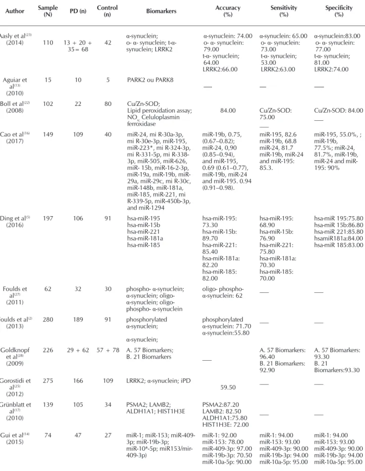
α-synuclein in the body fluids of patients with PD(21). Studies have shown that people with Parkinson’s disease have some altered biomarkers in CSF: LRRK25; A-synuclein, o-a-synuclein and t-a-synuclein(12,19,21,23,26); Cu/Zn-SOD, lipid peroxidation assay, NOx and ceruloplasmin ferroxidase(22); Hsa-miR-195, hsa-miR-15b, hsa-miR-221, hsa-miR-181a, hsa-miR-185(14); Aβ1-42, Tau [T-tau] and [P-tau181](12, 19); SKP1A, HSPA8, PSMC4, ALDH1A1 and HIP2(18). And others in blood, such as hsa-miR-195, hsa-miR-15b, hsa-miR-221, hsa-miR-181a and hsa-miR-185(5,10,27,30); Phospho-a-synuclein; Α-synuclein, oligo-α-synuclein and oligo-phospho-α-synuclein(2,15,21,25,27); A. 57 and B. 21(28); LRRK2 and α-synuclein(24-25); PSMA2, LAMB2, ALDH1A1 and HIST1H3E(17-18); NM_001544.2, NM_024754.2, BC051695.1, PHR5001, NM_006790.1, NM_032855.1, BC005858.1, NM_003141.2, BC094687.1 and BC027617.1(29). Information is shown in Table 1.
Table 1 – Accuracy Test
Author Sample (N) PD (n) Control (n) Biomarkers Accuracy (%) Sensitivity (%) Specificity (%)
Aasly et al(23)
(2014) 110 13 + 20 + 35= 68 42
α-synuclein; o- α- synuclein; t-α- synuclein; LRRK2
α-synuclein: 74.00 o- α- synuclein: 79.00
t-α- synuclein; 64.00 LRRK2:66.00
α-synuclein: 65.00 o- α- synuclein: 73.00
t-α- synuclein; 53.00 LRRK2:63.00
α-synuclein:83.00 o- α- synuclein: 77.00
t-α- synuclein; 81.00 LRRK2:74.00 Aguiar et
al(13)
(2010)
15 10 5 PARK2 ou PARK8
--- ----
---Boll et al(22)
(2008) 102 22 80 Cu/Zn-SOD;Lipid peroxidation assay; NOx; Celuloplasmin ferroxidase 84.00 Cu/Zn-SOD: 75.00 ___ Cu/Zn-SOD: 84.00 ___
Cao et al(16)
(2017) 149 109 40 miR-24, mi R-30a-3p, mi R-30e-3p, miR-195, miR-223*, mi R-324-3p, mi R-331-5p, mi R-338-3p, miR-505, miR-626, miR- 15b, miR-16-2-3p, 19a, 19b, miR-29a, miR-29c, mi R-30c, miR-148b, miR-181a, miR-185, miR-221, mi R-339-5p, miR-450b-3p, and miR-1294 miR-19b, 0.75, (0.67–0.82); miR-24, 0,90 (0.85–0.94), and miR-195, 0.69 (0.61–0.77), miR-19b, miR-24 and miR-195. 0.94 (0.91–0.98). miR-195, 82.6 miR-19b, 68.8 miR-24, 81.7 miR-19b, miR-24 and miR-195: 85.3.
miR-195, 55.0%, ; miR-19b,
77.5%; miR-24, 81.7%, miR-19b, 24 and miR-195: 90%
Ding et al(5)
(2016) 197 106 91 hsa-miR-195hsa-miR-15b hsa-miR-221 hsa-miR-181a hsa-miR-185 hsa-miR-195: 73.30 hsa-miR-15b: 89.70 hsa-miR-221: 85.40 hsa-miR-181a: 82.20 hsa-miR-185: 82.00 hsa-miR-195: 68.90 hsa-miR-15b: 76.90 hsa-miR-221: 75.80 hsa-miR-181a: 70.30 hsa-miR-185: 70.00 hsa-miR 195:75.80 hsa-miR 15b:86.80 hsa-miR 221:85.80 hsamiR181a:84.00 hsa-miR 185:83.00 Foulds et al(27) (2011)
62 32 30 phospho- α-synuclein;
α-synuclein; oligo-
α-synuclein; oligo- phospho- α-synuclein
oligo- phospho-
α-synuclein: 62 ___ ___
Foulds et al(2)
(2013) 280 189 91 phosphorylated α-synuclein;
α-synuclein;
phosphorylated
α-synuclein: 71.70
α-synuclein:55.80
___ ___
Goldknopf et al(28)
(2009)
226 29 + 62 57 + 78 A. 57 Biomarkers;
B. 21 Biomarkers ___
A. 57 Biomarkers: 96.40
B. 21 Biomarkers: 92.90
A. 57 Biomarkers: 93.30 B. 21 Biomarkers:93.30 Gorostidi et al(25) (2012)
275 166 109 LRRK2; α-synuclein; iPD
59.50 ___ ___
Grünblatt et al(17)
(2010)
139 105 34 PSMA2; LAMB2; ALDH1A1; HIST1H3E PSMA2:87.20 LAMB2: 82.50 ALDH1A1:75.80 HIST1H3E: 72.00 ___ ___
Gui et al(14)
(2015) 74 47 27 miR-1; miR-153; miR-409-3p; miR-19b-3p; miR-10ª-5p; miR153/mir-409-3p) miR-1: 92.00 miR-153: 78.00 miR-409-3p: 97.00 miR-19b-3p: 70.50 miR-10a-5p: 90.00 miR-1: 94.00 miR-153: 93.00 miR-409-3p: 90.00 miR-19b-3p: 94.00 miR-10a-5p: 95.00 miR-1: 94.00 miR-153: 93.00 miR-409-3p: 90.00 miR-19b-3p: 94.00 miR-10a-5p: 95.00
Author Sample (N) PD (n) Control (n) Biomarkers Accuracy (%) Sensitivity (%) Specificity (%)
Han et al (29)
(2012)
69 29 40 Autoantibody (10): NM_001544.2 NM_024754.2 BC051695.1 PHR5001 NM_006790.1 NM_032855.1 BC005858.1 NM_003141.2 BC094687.1 BC027617.1
___
93.10 100
Kang et al (12)
(2013)
102 63 39 Aβ1–42; Tau [T-tau];
[P-tau181]; α-synuclein ___ ___
Karlsson et al (24)
(2013)
154 79 75 α-synuclein (SNCA/ PARK1/4); (PRKN/PARK2); L1 (UCHL-1/PARK5); (PINK1/PARK6); DJ-1 (DJ1/PARK7); (LRRK2/ PARK8); (VPS35/PARK17); (EIF4G1/PARK18)
94.00 79.00 75.00
Khoo et al(30)
(2012)
64 32 32 626; 505; miR-222; k-TSP1; k- TSP2; k- TSP3; k-TSP4; k-TSP5; k-TSP1–5; k-TSP1–4; k-TSP1–3; k-TSP1,3,4
___
miR-626: 83.00 miR-505: 72.00 miR-222: 78.00 k-TSP1: 100.00 k- TSP2: 32.00 k- TSP3: 96.00 k-TSP4: 0.00 k-TSP5:0.00 k-TSP1–5: 96.00 k-TSP1–4: 96.00 k-TSP1–3: 96.00 k-TSP1,3,4: 96.00
miR-626: 100.00 miR-505: 97.00 miR-222: 73.00 k-TSP1: 56.00 k- TSP2: 00.00 k- TSP3: 9.00 k-TSP4: 100.00 k-TSP5:20.00 k-TSP1–5: 27.00 k-TSP1–4: 55.00 k-TSP1–3: 55.00 k-TSP1,3,4: 64.00
Margis et al(10)
(2011)
16 8 8 miR-1; miR-22; miR 29a; miR-16-2; miR-26a2; miR-30a
___ ___ ___
Molochnikov et al(18)
(2012)
156 92 64 SKP1A; HSPA8; PSMC4;
ALDH1A1; HIP2 95.00 90.30 89.10
Park et al(21)
(2011)
52 23 29 α- synuclein oligomer;Total α- synuclein
__ __ __
Parnetti et al(19)
(2014)
69 44 25 Aβ142/t-tau ratio; T-α- synuclein; O-α-synuclein; O/t-α-synuclein
Aβ42/t-tau ratio: 64.00
T-α-syn: 68.00 O-α-syn: 72.00 O/t-α-syn ratio: 78.00
Aβ42/t-tau ratio: 82.00
T-α-syn: 59.00 O-α-syn: 89.00 O/t-α-syn ratio: 82.00
Aβ42/t-tau ratio:56.00 T-α-syn: 80.00 O-α-syn: 48.00 O/t-α-syn ratio: 64.00
Tokuda et al(26)
(2010)
60 32 28 α-synuclein; oligo/total α-
synuclein α
-synuclein: 85.90 oligo/total α- synuclein: 94,80
α-synuclein: 75.00 oligo/total α- synuclein: 89.30
α-synuclein: 87.50 oligo/total α- synuclein:90.60
Vivacqua et(20)
(2016)
100 60 40 α-synuclein; oligo/total α-
synuclein __ __ __
Wang et al(15)
(2015)
202 100 102 α-synuclein; oligo/total α- synuclein
76.00 79.00 64.70
Risk of bias between studies
The main concern about the studies was the representativeness of the sample. The limitations of methodology were related to poor reporting of domain 2 (Index Test) from the QUADAS-2 tool.
DISCUSSION
Currently, the diagnosis of PD is based mainly on clinical symptoms, with incidence in the population over 65 years, from 1 to 2% worldwide and prevalence in Brazil of 3%(31).
To date, no laboratory biomarker is available to detect indi-viduals at risk for developing PD before most of their dopami-nergic neurons have been lost. Numerous studies suggest that neuronal cell death may result from the formation of oligomers (o-α-synuclein) in the brain(5,8-9,11,22,25-27).
In this study, we confirmed that the expressions of 10 antibod-ies (NM_001544.2, NM_024754.2, BC051695.1, PHR5001, NM_006790.1, NM_032855.1, BC005858.1, NM_003141.2, BC094687.1, BC027617.1) may be used as biomarkers to detect PD, exhibiting great sensitivity and specificity, using a small blood sample. Although the function of a number of antibod-ies is unknown, it has been found that the presence of some specific disorders in its profile is useful for the detection and diagnosis of various diseases. The present study demonstrates that PD is also linked to changes in serum antibody expression profiles. These changes allow the unbiased identification and selection of specific antibodies that can function effectively as diagnostic biomarkers. It is shown here that, with only 10 antibody biomarkers, the serum samples from the PD patients were easily distinguished in sera from the control group with a sensitivity of 93.1% and a specificity of 100%(1,29).
Several studies have begun to analyze antibody (miRNA) levels in body fluids (including serum, plasma and cerebrospinal fluid) or circulating cells in the fluids of patients with PD. The first study reports mentioned above exam-ined the miRNAs in blood cells. The following studies were then performed with plasma or serum samples from patients with PD to check serum miRNA levels in patients with idiopathic PD and LRRK2 mutations. Other studies have found that antibodies (miR-133b(5,10,14,29-30); 195, miR-185, miR-221 and miR-181a) are decreased in serum. All 5 miRNAs appear to be closely related to the nervous system and to the neurodegenerative process, which may lead to the alterna-tion of these levels in the brain and, ultimately, to the change in the serum concentrations of miRNAs(5,10,14,30).
In another study, 9 pairs of predictors for PD were identified using k-TSP classification analysis and 13 miRNAs. A combination of both sets of data produced a panel of predictor biomarkers: k-TSP1 (miR-1826 / miR-450b-3p), miR-626, miR-505, obtain-ing 91% sensitivity, 100% specificity, 100% positive predictive value, and 88% negative predictive value in the replicate set(30).
A set of 6 microRNAs formed two groups: miR-1, miR-22 and miR-29 allowed to distinguish PD in untreated individuals from healthy individuals; the miR-16-2, miR-26a2 and miR30a allowed to distinguish treated patients from untreated patients. This study is innovative in contributing to the development of effective biomark-ers for PD(10). Another blood study found 57 specific proteins with abnormal levels in PD. Of these, 21 were associated with patients with mild PD symptom, and 14 with moderate to severe PD symptom. When the same study was applied in the second place of research, the 21 proteins showed sensitivity of 93.3% and specificity of 92.9%.
A pilot study in peripheral blood found four genes: pro-teasome (prosome, macropain) alpha subunit type 2 (PSMA2, p=0.0002, OR=1.15, 95% CI 1.07-1.24), laminin, beta-2 (laminin S) (LAMB2, P=0.0078, OR=2.26, 95% CI 1.24-4.14), A1 family member aldehyde dehydrogenase (ALDH1A1, p=0.016, OR=1,5 CI 95% 1.01-1.1) and histone cluster-1 H3e (HIST1H3E, p=0.03, OR=0.975, 95% CI 0.953-0.998) altered in PD. These four biomarkers are also used in the diagnosis of PD, with sensitivity and specificity for more than 80%(17-18). Other studies have evaluated that, in blood and cerebrospinal fluid, o-α-synuclein levels are elevated in symptomatic and asymptomatic LRRK2 mutation carriers in PD and begin several years before experiencing any motor symptoms(2,23-25,27-28).
(t-tau) and phosphorylated tau (p-tau) are known markers of PD disease, because they faithfully reflect the condition. Α-synuclein induces aggregation and polymerization of tau, which promotes the formation of abundant intracellular tau-amyloid inclusions. In addition, the presence of α-synuclein was detected in the neurons of patients with PD, and the measurement in the cerebrospinal fluid of DJ1/PARK7 (multifunctional protein due to oxidative stress) was decreased in PD. However, the sensitivity and specificity ap-pear to be only moderate and no correlation was observed with the severity or progression of PD(12-14,19). The present study is the first report on biomarkers in cerebrospinal fluid (Aβ1-42, T-tau, P-tau181 and α-synuclein). Several biomarkers characteristics related to the clinical aspects of PD were found. The levels of Aβ1-42, T-tau, Ptau181, T-tau/Aβ1-42 and α-synuclein were sig-nificantly lower, and the concentrations of T-tau and α-synuclein associated with severity of motor dysfunction in PD. Α-Synuclein levels were found to have a strong correlation with levels of tau proteins (T-tau and P-tau181) in patients with PD. The L-Aβ1-42 and P-tau181 motor phenotypes presented lower concentrations when associated with the phenotype of postural instability-gait abnormality. Finally, we found a significant correlation between
α-synuclein levels and T-tau and P-tau levels(12-14,19).
Recent studies have demonstrated that α-synuclein and the DJ-1 protein in cerebrospinal fluid are biomarkers used for early diagnosis and detection of PD. Α-synuclein and DJ-1 have a strong predictive value for the diagnosis of PD and may possibly help identify individuals at pre-symptomatic stages (patients with depression, sleep disorders) and neuroprotective treatment(18).
The levels of α-synuclein and the ratio of α-synuclein to total (t-α-synuclein) are higher in cerebrospinal fluid in patients with PD, unlike saliva. Previous studies have shown a small t-α-synuclein concentration in the saliva of patients with PD, and no study previously examined salivary o-α-synuclein con-centration or assessed the correlation between o-α-synuclein and t-α-synuclein concentration(2,15,20-21,23,25-27).
In the cerebrospinal fluid, increased free copper, ferroxidase, lipid peroxidation and NOx (nitrites and nitrates) is present in PD. The decrease in ferroxidase activity was a common feature related to the increase of iron deposition in degenerated areas. In PD, free copper is also significantly related to the clinical stage and duration of clinical manifestations(22).
In the present study, it was found that total salivary α-synuclein is significantly lower in PD patients than in healthy subjects. On the other hand, salivary oligo-α-synuclein levels are higher in PD patients than in healthy subjects. Consequently, the α-synuclein/ oligo-synuclein ratio is significantly higher in patients with PD than in healthy subjects. Neuropathological studies have shown that in the early stages of PD the intracellular aggregation of α-synuclein occurs in several brainstem nuclei, including the dorsal motor vagus nucleus and probably the superior and inferior salivatory nuclei and the parasympathetic salivary ganglia. It suggests that
α-synuclein can spread from neuronal cell bodies of salivary neurons, along axons, to the synaptic terminals around the epi-thelial cells of the salivary glands, where it also accumulates in the saliva. Therefore, it is possible that the reduced concentration of total α-synuclein detected in the saliva of PD patients is due to intracellular and axonal α-synuclein aggregation in salivatory nuclei or salivary gland neurons(15).
Study limitations
In this study, only blood, cerebrospinal fluid and salivary biomarkers were used in PD. The full range of body fluids (bile, cerumen, peritoneal fluid, tears, breast milk, amniotic fluid, mucus, sebum, semen, gastric juice, sweat, vomit) were not considered. Unfortunately, studies were excluded because they had insufficient information and gaps for the predictive values of sensitivity and specificity and could cause bias in the findings.
Contributions to the Nursing, Health or Public Policy Areas Care to people with PD should be directed with the objective of improving their quality of life. Pharmacological treatment of PD is extremely complex and involves the intervention of a multidisci-plinary team, and should be associated with non-pharmacological treatment, such as Physical Therapy, Speech Therapy, Nutrition, and others to attenuate symptoms and promote quality of life.
Nurses are the professionals trained to understand the particulari-ties of each individual, to care for and educate the person with PD and their relatives. Nurses’ action occurs, mainly in Primary Health Care, as a point of entry into the care network. It is in this context that nurses develop health education groups and perform care, in addition to pharmacological and multiprofessional treatment. Nurs-ing care includes the reception, clinical evaluation and guidance on disease prevention and health promotion, both for the person with the disease, as well as for their relatives and/or caregivers.
CONCLUSION
We observed that the countries that developed the majority of studies were China (3) and the USA (2), followed by Italy (2) with groups of antibodies and proteins present in the blood and cerebrospinal fluid. Most had low risk of bias and were performed in blood. Evidence shows that serum antibodies can be used as highly specific and accurate biomarkers for the diagnosis of PD at the onset of disease. The biomarkers identified in the cerebrospinal fluid also have the potential to diagnose PD at baseline and are related to increased motor severity, postural instability, gait abnormality, and cognitive decline, and are not affected by disease progression. It is hoped that early, even pre-symptomatic screening methods can be established. Serum antibodies are considered as a new class of pathologically rel-evant molecules that can be explored for a better understanding of disease mechanisms and potential therapies.
REFERENCES
2. Foulds PG, Diggle P, Mitchell JD, Parker A, Hasegawa M, Masuda-Suzukake M, et al. A longitudinal study on alpha-synuclein in blood plasma as a biomarker for Parkinson’s disease. Sci Rep[Internet]. 2013[cited 2017 May 28];3:2540. Available from: https:// www.ncbi.nlm.nih.gov/pmc/articles/PMC3756331/pdf/srep02540.pdf
3. Martignoni E, Franchignoni F, Pasetti C, Ferriero G, Picco D. Psychometric properties of the Unified Parkinson’s Disease Rating Scale and of the Short Parkinson’s Evaluation Scale. Neurol Sci[Internet]. 2003[cited 2017 May 23];24(3):190-1. Available from: https://link.springer.com/article/10.1007/s10072-003-0124-0
4. Schenkman ML, Clark K, Xie T, Kuchibhatla M, Shinberg M, Ray L. Spinal movement and performance of a standing reach task in participants with and without Parkinson disease. Phys Ther[Internet]. 2001[cited 2017 May 28];81(8):1400-11. Available from: https://www.ncbi.nlm.nih.gov/pubmed/11509070
5. Ding H, Huang Z, Chen M, Wang C, Chen X, Chen J, et al. Identification of a panel of five serum miRNAs as a biomarker for Parkinson’s disease. Parkinsonism Relat Disord[Internet]. 2016[cited 2017 May 23];22:68-73. Available from: http://www.prd-journal.com/article/S1353-8020 (15)30038-9/pdf
6. Miller DB, O’Callaghan JP. Biomarkers of Parkinson’s disease: present and future. Metabolism[Internet]. 2015[cited 2017 May 28];64(3Suppl1):S40-6. Available from: https://www.ncbi.nlm.nih.gov/pmc/articles/PMC4721253/pdf/nihms-747493.pdf
7. Santos-Garcia D, Mir P, Cubo E, Vela L, Rodriguez-Oroz MC, Marti MJ, et al. COPPADIS-2015 (Cohort of Patients with Parkinson’s Disease in Spain, 2015), a global--clinical evaluations, serum biomarkers, genetic studies and neuroimaging--prospective, multicenter, non-interventional, long-term study on Parkinson’s disease progression. BMC Neurol[Internet]. 2016[cited 2017 May 28];16:26. Available from: https://www.ncbi.nlm.nih.gov/pmc/articles/PMC4766717/pdf/12883_2016_Article_548.pdf
8. Moher D, Liberati A, Tetzlaff J, Altman DG. Preferred reporting items for systematic reviews and meta-analyses: the PRISMA statement. Int J Surg[Internet]. 2010[cited 2017 May 23];8(5):336-41. Available from: http://www.journal-surgery.net/article/ S1743-9191(10)00040-3/pdf
9. Whiting PF, Rutjes AW, Westwood ME, Mallett S, Deeks JJ, Reitsma JB, et al. QUADAS-2: a revised tool for the quality assessment of diagnostic accuracy studies. Ann Intern Med[Internet]. 2011[cited 2015 Jul 23];155(8):529-36. Available from: http://annals. org/aim/fullarticle/474994/quadas-2-revised-tool-quality-assessment-diagnostic-accuracy-studies
10. Margis R, Rieder CR. Identification of blood microRNAs associated to Parkinsonis disease. J Biotechnol[Internet]. 2011[cited 2017 May 28];152(3):96-101. Available from: http://www.sciencedirect.com/science/article/pii/S0168165611000678?via%3Dihub
11. Leeflang MM, Deeks JJ, Takwoingi Y, Macaskill P. Cochrane diagnostic test accuracy reviews. Syst Rev[Internet]. 2013[cited 2017 May 28];2:82. Available from: https://www.ncbi.nlm.nih.gov/pmc/articles/PMC3851548/pdf/2046-4053-2-82.pdf
12. Kang JH, Irwin DJ, Chen-Plotkin AS, Siderowf A, Caspell C, Coffey CS, et al. Association of cerebrospinal fluid beta-amyloid 1-42, T-tau, P-tau181, and alpha-synuclein levels with clinical features of drug-naive patients with early Parkinson disease. JAMA Neurol[Internet]. 2013[cited 2015 Jul 23];70(10):1277-87. Available from: https://www.ncbi.nlm.nih.gov/pmc/articles/PMC4034348/ pdf/nihms573438.pdf
13. Aguiar PMC, Severino P. Biomarkers in Parkinson Disease: global gene expression analysis in peripheral blood from patients with and without mutations in PARK2 and PARK8. Einstein[Internet]. 2010[cited 2017 May 23];8(3 Pt 1):291-7. Available from: http:// apps.einstein.br/revista/arquivos/PDF/1674-Einsteinv8n3_pg291-7_eng.pdf
14. Gui Y, Liu H, Zhang L, Lv W, Hu X. Altered microRNA profiles in cerebrospinal fluid exosome in Parkinson disease and Alzheimer disease. Oncotarget[Internet]. 2015[cited 2017 May 28];6(35):37043-53. Available from: https://www.ncbi.nlm.nih.gov/pmc/articles/ PMC4741914/pdf/oncotarget-06-37043.pdf
15. Wang X, Yu S, Li F, Feng T. Detection of alpha-synuclein oligomers in red blood cells as a potential biomarker of Parkinson’s disease. Neurosci Lett[Internet]. 2015[cited 2017 May 23];599:115-9. Available from: http://www.sciencedirect.com/science/ article/pii/S0304394015003870?via%3Dihub
16. Cao XY, Lu JM, Zhao ZQ, Li MC, Lu T, An XS, Xue LJ. MicroRNA biomarkers of Parkinson’s disease in serum exosome-like microvesicles. Neurosci Lett[Internet]. 2017[cited 2017 May 23];644:94–9. Available from: http://europepmc.org/abstract/med/28223160
17. Grunblatt E, Zehetmayer S, Jacob CP, Muller T, Jost WH, Riederer P. Pilot study: peripheral biomarkers for diagnosing sporadic Parkinson’s disease. J Neural Transm[Internet]. 2010[cited 2017 May 28];117(12):1387-93. Available from: https://link.springer. com/article/10.1007%2Fs00702-010-0509-1
18. Molochnikov L, Rabey JM, Dobronevsky E, Bonucelli U, Ceravolo R, Frosini D, et al. A molecular signature in blood identifies early Parkinson’s disease. Mol Neurodegener[Internet]. 2012[cited 2017 May 28];7:26. Available from: https://www.ncbi.nlm.nih. gov/pmc/articles/PMC3424147/pdf/1750-1326-7-26.pdf
19. Parnetti L, Farotti L, Eusebi P, Chiasserini D, De Carlo C, Giannandrea D, et al. Differential role of CSF alpha-synuclein species, tau, and A beta 42 in Parkinson’s Disease. Frontiers Aging Neurosci[Internet]. 2014[cited 2017 May 28];6. Available from: https:// www.ncbi.nlm.nih.gov/pmc/articles/PMC3978246/pdf/fnagi-06-00053.pdf
20. Vivacqua G, Latorre A, Suppa A, Nardi M, Pietracupa S, Mancinelli R, et al. Abnormal Salivary Total and Oligomeric Alpha-Synuclein in Parkinson’s Disease. Plos One[Internet]. 2016[cited 2017 May 23];11(3). Available from: http://journals.plos.org/plosone/article/ file?id=10.1371/journal.pone.0151156&type=printable
Patients with Parkinson’s Disease. J Clin Neurol[Internet]. 2011[cited 2017 May 23];7(4):215-22. Available from: https://www.ncbi. nlm.nih.gov/pmc/articles/PMC3259496/pdf/jcn-7-215.pdf
22. Boll MC, Alcaraz-Zubeldia M, Montes S, Rios C. Free copper, ferroxidase and SOD1 activities, lipid peroxidation and NO(x) content in the CSF: a different marker profile in four neurodegenerative diseases. Neurochem Res[Internet]. 2008[cited 2017 May 28];33(9):1717-23. Available from: https://link.springer.com/article/10.1007%2Fs11064-008-9610-3
23. Aasly JO, Johansen KK, Bronstad G, Waro BJ, Majbour NK, Varghese S, et al. Elevated levels of cerebrospinal fluid alpha-synuclein oligomers in healthy asymptomatic LRRK2 mutation carriers. Frontiers Aging Neurosci[Internet]. 2014[cited 2017 May 23];6:248. Available from: https://www.ncbi.nlm.nih.gov/pmc/articles/PMC4174885/pdf/fnagi-06-00248.pdf
24. Karlsson MK, Sharma P, Aasly J, Toft M, Skogar O, Saebo S, et al. Found in transcription: accurate Parkinson’s disease classification in peripheral blood. J Parkinsons Dis[Internet]. 2013[cited 2017 May 28];3(1):19-29. Available from: https://content.iospress.com/ download/journal-of-parkinsons-disease/jpd120159?id=journal-of-parkinsons-disease%2Fjpd120159
25. Gorostidi A, Bergareche A, Ruiz-Martinez J, Marti-Masso JF, Cruz M, Varghese S, et al. α-synuclein levels in blood plasma from LRRK2 mutation carriers. PLoS One[Internet]. 2012[cited 2017 May 28];7(12):e52312. Available from: http://journals.plos.org/ plosone/article/file?id=10.1371/journal.pone.0052312&type=printable
26. Tokuda T, Qureshi M, Ardah M, Varghese S, Shehab S, Kasai T, et al. Detection of elevated levels of α-synuclein oligomers in CSF from patients with Parkinson disease. Neurol[Internet]. 2010[cited 2017 May 28];75(20):1766-72. Available from:http://onlinelibrary. wiley.com/o/cochrane/clcentral/articles/665/CN-00772665/frame.html.
27. Foulds PG, Mitchell JD, Parker A, Turner R, Green G, Diggle P, et al. Phosphorylated alpha-synuclein can be detected in blood plasma and is potentially a useful biomarker for Parkinson’s disease. Faseb J[Internet]. 2011[cited 2017 May 23];25(12):4127-37. Available from: http://www.fasebj.org/content/25/12/4127.long
28. Goldknopf IL, Bryson JK, Strelets I, Quintero S, Sheta EA, Mosqueda M, et al. Abnormal serum concentrations of proteins in Parkinson’s disease. Biochem Biophys Res Commun[Internet]. 2009[cited 2017 May 28];389(2):321-7. Available from: http://www. sciencedirect.com/science/article/pii/S0006291X09017392?via%3Dihub
29. Han M, Nagele E, DeMarshall C, Acharya N, Nagele R. Diagnosis of Parkinson’s disease based on disease-specific autoantibody profiles in human sera. PLoS One[Internet]. 2012[cited 2017 May 23];7(2):e32383. Available from: http://journals.plos.org/plosone/ article?id=10.1371/journal.pone.0032383
30. Khoo SK, Petillo D, Kang UJ, Resau JH, Berryhill B, Linder J, et al. Plasma-based circulating MicroRNA biomarkers for Parkinson’s disease. J Parkinsons Dis[Internet];2012[cited 2017 May 23];2(4):321-31. Available from: https://content.iospress.com/articles/ journal-of-parkinsons-disease/jpd012144