Cognitive Performance and Cerebrospinal
Fluid Biomarkers of Neurodegeneration: A
Study of Patients with Bipolar Disorder and
Healthy Controls
Sindre Rolstad1
*, Joel Jakobsson1¤a, Carl Sellgren2¤b, Carl-Johan Ekman3¤c,
Kaj Blennow1, Henrik Zetterberg1,4¤d, Erik Pålsson1, Mikael Landén1,2,3
1Institute of neuroscience and physiology, the Sahlgrenska Academy at the Gothenburg University, Gothenburg, Sweden,2Department of Medical Epidemiology and Biostatistics, Karolinska Institutet, Stockholm, Sweden,3Department of Clinical Neuroscience, Karolinska Institutet, Stockholm, Sweden, 4UCL Institute of Neurology, Queen Square, University College London, London, United Kingdom
¤a Current address: The Sahlgrenska Academy at Gothenburg University, Institute for neuroscience and
physiology, Gothenburg, Sweden
¤b Current address: Department of Medical Epidemiology and Biostatistics, Karolinska Institutet, Stockholm,
Sweden
¤c Current address: Karolinska Institutet, Department of Clinical Neuroscience, Stockholm, Sweden ¤d Current address: University College London, London, United Kingdom
*sindre.rolstad@neuro.gu.se
Abstract
The purpose of the present study was to investigate if cerebrospinal fluid (CSF) biomarkers of neurodegeneration are associated with cognition in bipolar disorder and healthy controls, respectively. CSF concentrations of total and phosphorylated tau, amyloid beta (Aβ)1-42, ratios of Aβ42/40 and Aβ42/38, soluble amyloid precursor proteinαandβ, and neurofila-ment light chain protein were analyzed in relation to neuropsychological performance in 82 euthymic bipolar disorder patients and 71 healthy controls. Linear regression models were applied to account for performance in five cognitive domains using the CSF biomarkers. In patients, the CSF biomarkers explained a significant proportion of the variance (15–36%,
p=.002 -<.0005) in all cognitive domains independently of age, medication, disease status,
and bipolar subtype I or II. However, the CSF biomarkers specifically mirroring Alzheimer-type brain changes, i.e., P-tau and Aβ1-42, did not contribute significantly. In healthy con-trols, CSF biomarkers did not explain the variance in cognitive performance. Selected CSF biomarkers of neurodegenerative processes accounted for cognitive performance in per-sons with bipolar disorder, but not for healthy controls. Specifically, the ratios of Aβ42/40 and Aβ42/38 were consistently associated with altered cognitive performance.
OPEN ACCESS
Citation:Rolstad S, Jakobsson J, Sellgren C, Ekman C-J, Blennow K, Zetterberg H, et al. (2015) Cognitive Performance and Cerebrospinal Fluid Biomarkers of Neurodegeneration: A Study of Patients with Bipolar Disorder and Healthy Controls. PLoS ONE 10(5): e0127100. doi:10.1371/journal.pone.0127100
Academic Editor:Jing A. Zhang, UW Medicine Neuropathology, UNITED STATES
Received:November 25, 2014
Accepted:April 11, 2015
Published:May 8, 2015
Copyright:© 2015 Rolstad et al. This is an open access article distributed under the terms of the Creative Commons Attribution License, which permits unrestricted use, distribution, and reproduction in any medium, provided the original author and source are credited.
Data Availability Statement:Due to ethical restrictions, data are available upon request. Interested researchers may contact the
corresponding author oranne.snellman@vgregion.
sefor sensitive data.
Funding:This research study was supported by grants from the Swedish Medical Research Council (http://www.vr.se/inenglish.4.
Introduction
The hallmark of bipolar disorder is recurrent episodes of depression and mania or hypomania [1]. The worldwide prevalence of the main subtypes of bipolar disorder, type I and II, is esti-mated to about 1–3% [2]. The disorder is associated with high societal costs, of which indirect costs due to sick leave and early retirement are the main drivers [3]. It has been increasingly recognized that cognitive dysfunction is an important predictor of functional outcomes in bi-polar disorder [4].
Meta-analyses suggest that attention/speed, memory, and executive functions are impaired in euthymic bipolar disorder [5,6]. An independent individual patient data meta-analysis pro-vided further evidence of significant cognitive impairment in bipolar disorder, albeit less sub-stantial than previous reports suggested [7]. Another meta-analysis found that differences between bipolar type I and II are negligible with the exception of memory and semantic fluency [8]. Whereas there is some evidence of cognitive deterioration during the course of illness [7,
9], most cognitive functions appear to remain persistently impaired over time [10].
No biological correlates of cognitive impairment in bipolar disorder have been established. Meta-analyses of structural magnetic resonance imaging (MRI) studies report morphologi-cal differences between persons with bipolar disorder and controls [11,12], but these structural abnormalities have not been linked to cognitive deficits [13]. It is hence undecided whether the neuroimaging findings in bipolar disorder indicate a neurodegenerative process, a premorbid condition, effects of alcohol intake, altered hormone levels, or medication effects [14].
In two recent studies, we investigated the applicability of cerebrospinal fluid (CSF) biomark-ers to study neurodegenerative processes in bipolar disorder [15,16]. We found decreased con-centrations of the soluble forms amyloid precursor protein (APP)—sAPPαand sAPPβ- and
higher ratios of amyloidβ(Aβ) 42/40 and Aβ42/38 in persons with bipolar disorder compared with healthy controls [16]. The physiological role of APP is not fully understood, but it has been linked to synaptic formation and repair as well as axonal regeneration [17]. APP has also been suggested to be important for neural connectivity, plasticity, and activity, as well as for memory functions. We found no group difference between bipolar disorder patients and con-trols with respect to total or phosphorylated tau (T-tau/P-tau) that reflect axonal damage and neurofibrillary degeneration [18], or Aβ1–42 that indicate plaque deposition [19]. In the subse-quent study, however, we found higher mean CSF concentrations of neurofilament light chain protein (NFL) in persons with bipolar disorder compared to controls [15]. NFL is a cytoskeletal constituent of intermediate filaments. Increased CSF NFL is considered to reflect neuronal and axonal degeneration and loss [20]. Taken together, these previous studies suggested that altered APP metabolism and axonal injury might occur in bipolar disorder.
CSF biomarkers of neurodegeneration have been linked to cognitive impairment in other disorders [18,21]. It is hence not farfetched to suggest that they might also be associated with cognitive dysfunction in bipolar disorder. Clarifying this issue is important for at least two rea-sons. First, it might yield insights as to the biological underpinnings of cognitive impairment in bipolar disorder, which in turn is important for identifying treatment targets to alleviate cogni-tive impairment. Second, biomarkers of neurodegeneration might prove useful to predict wors-ening of cognitive function during the course of illness. If biomarkers could help identifying vulnerable individuals, targeted intervention programs to prevent cognitive decline could be developed.
The aim of this study was to evaluate potential associations between CSF biomarkers of de-generation (T-tau, P-tau, Aβ1–42, Aβ42/40 and Aβ40/38 ratios, sAPPαand sAPPβ, and NFL) and cognitive function in patients with bipolar disorder. Regression models with five aggregat-ed cognitive domains were appliaggregat-ed using CSF biomarkers as praggregat-edictors and covariates as
foundation (http://swedishbrainpower.se/en/), and the Swedish Federal Government under the LUA/ALF agreement (ALF 20130032, ALFGBG-142041). The funders had no role in study design, data collection and analysis, decision to publish, or preparation of the manuscript.
appropriate. The models were repeated in healthy age- and sex-matched controls to determine if observed associations were disease dependent.
Methods
Patients
The St. Göran Bipolar Project is a clinical longitudinal study of persons with bipolar disorder. The procedures in this project have been described in detail elsewhere [22]. In brief, patients were enrolled at the bipolar outpatient unit at the Northern Stockholm Psychiatric Clinic (Stockholm, Sweden). The inclusion criteria for the St. Göran Bipolar Project are18 years of age, fulfilling the DSM-IV criteria for bipolar disorder type I or II. Exclusion criteria were in-ability to complete the standard clinical assessment or incapin-ability of providing informed consent.
The key clinical assessment instrument was a Swedish version of the Affective Disorder Evaluation ADE), which is a standardized interview protocol developed for the Systematic treatment Enhancement Program of Bipolar Disorder (STEP-BD) [23]. The ADE directs the interviewer through a systematic assessment of the patient’s current mental state, psychiatric
history, and diagnosis according to DSM-IV criteria as per the Structured Clinical Interview for DSM-IV (SCID) [24]. The ADE includes a social anamnesis, and a medical history. The lifetime severity of bipolar disorder is rated using the 7-point Likert scale Clinical Global Im-pression (CGI), which ranges from healthy to extremely ill. In addition to the ADE, the Mini International Neuropsychiatric Interview (M.I.N.I.) [25] was completed to screen for other psychiatric diagnoses than bipolar disorder. Alcohol Use Disorders Identification Test (AUDIT) and the Drug Use Disorders Identification Test (DUDIT) were used to screen for substance and alcohol abuse, as well as serum levels of carbohydrate-deficient transferrin [26]. The ADE and M.I.N.I. interviews were conducted by board-certified psychiatrists working at the tertiary bipolar outpatient unit, or residents in psychiatry completing their training at this unit. To minimize risk of inter-rater bias, a best-estimate diagnostic decision was made based on all information available at admission by a consensus panel of experienced board certified psychiatrists specialized in bipolar disorder. All available sources of information, encompassing patient interview, case records and, if available, interview with the next of kin, were utilized in the diagnostic assessment.
Both the CSF sampling and the cognitive examination procedures were carried out when patients were in a euthymic mood. Euthymia was defined as MADRS (Montgomery–Åsberg Depression Rating Scale) and YMRS (Young Mania Rating Scale) scores<14.
Controls
occurred at separate occasions for patients. Controls presenting potentially pathological find-ings were discussed between examining clinician, primary investigator, and study coordinator at case conferences. Exclusion criteria were: any current psychiatric disorder, a family history of schizophrenia or bipolar disorder in first-degree relatives, drug or alcohol abuse (based on DUDIT and AUDIT as well as serum levels of carbohydrate-deficient transferrin), neurological conditions except mild migraines, pregnancy, untreated endocrine disorders, dementia, and se-vere personality disorder.
Ethics
The study was approved by the Regional Ethics Committee in Stockholm (case no. 2005/ 554-31/3) and conducted in accordance with the latest Helsinki Protocol. After complete de-scription of the study, all enrolled patients and controls consented orally and in writing to par-ticipate in the study.
CSF sampling and biomarker analyses
To reduce the risk of diurnal fluctuations, lumbar puncture was performed at 0900–1000 AM after night fasting. The spinal needle was inserted into the L3/L4 or L4/L5 interspace, and a total volume of 12 ml of CSF was drawn, gently inverted to avoid gradient effects, and divided into 1.0–1.6 ml aliquots that were stored at -80°C pending analysis. For ethical reasons, patients
were not taken off their prescribed medication at the time of the sampling. All samples were thawed and refrozen once before analysis. All biochemical analyses were performed at the Clin-ical Neurochemistry Laboratory in Mölndal, Sweden, by board-certified laboratory technicians blinded with respect to clinical information. The CSF concentrations of sAPPαandβwere de-termined using the MSD sAPPα/sAPPβMultiplex Assay, while CSF Aβ38, Aβ40, and Aβ42
were analyzed using the MSD Human/Rodent (4G8) Assay (Meso Scale Discovery, Gaithers-burg, MD, USA), as described previously [27]. CSF concentrations of hyperphosphorylated Tau (P-tau), total tau (T-tau), and Aβ1–42 were measured simultaneously by the Luminex
xMAP technology using the Inno-Bia AlzBio3 kit (Innogenetics, Zwijndrecht, Belgium), as de-scribed preciously [28]. NFL was analyzed as previously described with a commercial ELISA assay (NF-light, UmanDiagnostics AB, Umeå, Sweden). The intra-assay coefficient of variabili-ty was<10% for all biomarkers and the inter-assay coefficient of variability varied from 2% (Aβ38) to 20% (sAPPβ).
Neuropsychological examination
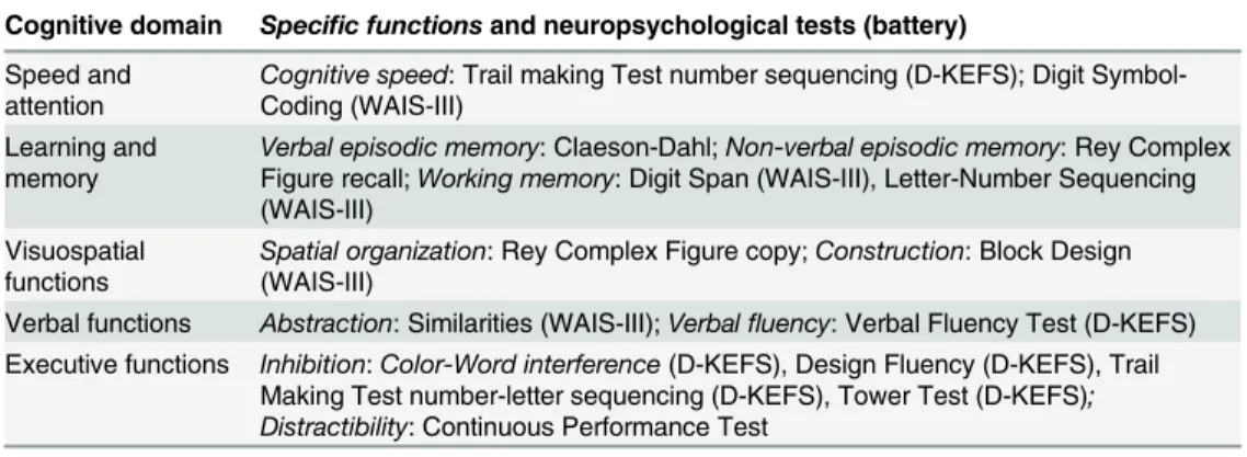
controls. Specific neuropsychological tests and functions measured are displayed inTable 1. The purpose of analyzing cognitive domains rather than individual cognitive tests is to suc-cinctly communicate the underlying measuring entities [32], decrease the test-specific associa-tions, and to reduce the potential alpha inflation resulting from a larger battery of tests.
Statistical analysis
The primary outcome of this study was the ability of the CSF biomarkers to explain cognitive performance in bipolar disorder. The applicability of the biomarkers to account for cognitive performance was further tested in the healthy age- and sex-matched controls.
Preliminary analyses were performed to ensure no violation of the following prerequisites for regression: linearity, normality, absence of multicollinearity, and homoscedasticity. Vari-ables that violated requirements for linearity were transformed as appropriate and variVari-ables that were highly inter-correlated were excluded for the model of interest. Linear regression was performed to assess the degree to which CSF biomarkers can explain the variance in the aggre-gated cognitive domain scores. CSF biomarkers were entered as independent variables in all models. The following variables were included as covariates for patients: age, sex, bipolar sub-type, CGI, MADRS, YMRS, treatment with any mood stabilizer, lithium, anticonvulsants, depressants, antipsychotics, benzodiazepines, and anxiolytics (non-benzodiazepine anti-anxiety medication). For healthy controls, age and sex were the only applicable covariate.
We report the adjustedr2of the model, the standardized beta values, and thep-value (two-tailed tests) of the individual variables. Analysis of variance and chi-square, where applicable, were used for group comparisons. P-values<.05 were considered significant. Alpha correction was not applied. To test if the covariates accounted for the influence of the CSF biomarkers on cognition, mediation effects were assessed using bootstrapping for continuous mediators and logistic mediation analysis for dichotomous mediators (medication). Analyses were performed using SPSS version 21 (Armonk, NY: IBM Corp.) and SAS JMP version 10 (Cary, NC: SAS inst.).
Results
The current study included 82 cases of bipolar disorder type I and II that had completed sam-pling of CSF and cognitive examination (Table 2). The patient sample was predominantly fe-male (58.5%) with a mean (SD) age of 38.4 (±12.6) and an average of 12.9 (±2.8) years of
Table 1. Cognitive domains and functions assessed in the St Göran bipolar project.
Cognitive domain Specific functionsand neuropsychological tests (battery)
Speed and attention
Cognitive speed: Trail making Test number sequencing (D-KEFS); Digit Symbol-Coding (WAIS-III)
Learning and memory
Verbal episodic memory: Claeson-Dahl;Non-verbal episodic memory: Rey Complex Figure recall;Working memory: Digit Span (WAIS-III), Letter-Number Sequencing (WAIS-III)
Visuospatial functions
Spatial organization: Rey Complex Figure copy;Construction: Block Design (WAIS-III)
Verbal functions Abstraction: Similarities (WAIS-III);Verbalfluency: Verbal Fluency Test (D-KEFS) Executive functions Inhibition:Color-Word interference(D-KEFS), Design Fluency (D-KEFS), Trail
Making Test number-letter sequencing (D-KEFS), Tower Test (D-KEFS); Distractibility: Continuous Performance Test
WAIS-III = Wechsler’s Adult Intelligence Scale version III, D-KEFS = Delis-Kaplan executive function system.
education. The mean (SD) CGI score was 4.47 (±. 98). Most patients were bipolar type I and a majority were prescribed mood stabilizers such as lithium, valproate, or lamotrigine (86.6%, 61% lithium in total) at the time of examination. To investigate if the associations between CSF biomarkers and cognitive performance were specific to bipolar disorder, this study also includ-ed healthy controls (Table 2).
Data from the St. Göran cohort on test specific cognitive performance [33] as well as CSF biomarkers concentrations have been presented previously [15,16]. However, in the present study, only subjects that had completed both cognitive testing and lumbar puncture were in-cluded. In the present study, controls did not differ significantly from patients with respect to age and gender distribution, but had significantly more years of education (F(1,151) = 8.7,p=. 004). Patients’sAPPα(F(1,151) = 8.1,p=. 005) and sAPPβ(F(1,151) = 4.7,p=. 03) were lower whereas their Aβ42/38 ratio (F(1,151) = 4.4,p=. 03) and NFL concentrations (F(1,151) = 5.4,
p=. 02) were higher. Patients performed worse than healthy controls in the domains of memory (F(1,151) = 13.6,p<.0005) and verbal functions (F(1,151) = 7.1,p=. 009) (Table 3).
Table 2. Demographics and clinical characteristics of patients with bipolar disorder type I and II and healthy controls.
Patients (N = 82) Controls (N = 71)
Mean/frequency SD/% Mean/frequency SD/%
Females 48 58.5 44 61.9
Age 38.3 12.5 37.8 14.6
Education, years 12.9 2.8 14.1 1.7
Bipolar type I/II 53/29 64.6/35.4 -
-Clinical Global Impression 4.4 .9 -
-Mood stabilizers 71 86.6 -
-Antidepressants 34 41.5 -
-Lithium 50 61 -
-Anxiolytics 20 24.4 -
-Benzodiazepines 23 28 -
-Anticonvulsants 36 43.9 -
-Antipsychotics 17 20.7 -
-sAPPαa 723.26 (307.81) 859.58 (282.80)
sAPPβb 300.29 (156.87) 354.93 (152.88)
Aβ42/40c .1152 (.0199) .1107 (.0177)
Aβ42/38d .7729 (.1133) .7345 (.1103)
Aβ1–42e 253.88 (62.33) 254.38 (55.42)
T-tauf 34.32 (12.48) 37.17 (14.09)
P-taug 26.72 (6.79) 28.27 (6.89)
NFLh 485.73 (425.62) 254.38 (55.42)
Frequencies and percentages are italicized.
a
sAPPα/bsAPPβ(ng/ml) = secreted form of beta-amyloid precursor proteinα/β;
c
Aβ42/40 = CSF amyloid beta 42/40 ratio (pg/ml);
dAβ42/38 = CSF amyloid beta 42/38 ratio (pg/ml); e
Aβ1–42 = CSF amyloid beta 1–42 (pg/ml);
f
T-tau = CSF total tau (pg/ml);
gp-tau = phosphorylated tau (pg/ml); h
NFL = Neurofilament light subunit (pg/ml)
Memory/learning domain
The CSF biomarkers explained a significant proportion of the variance in the memory domain; the model as a whole accounted for 15% (adj. r2=. 15, F(4, 82) = 5.0.p. 001) of the observed variance (Table 4). Higher concentrations of NFL and a higher ratio of Aβ42/40 were
associat-ed with worse memory performance, whereas a higher ratio of Aβ42/38 was associated with better memory performance. NFL was the most influential predictor in this model (beta = -.37) indicating that an increase of 425 pg/ml NFL (1 SD) reduced memory performance by-.37 SD.
As working memory can be conceived as a separate memory system [34], the association be-tween CSF biomarkers and memory/learning performance was repeated without the working memory tests (Digit Span and Letter-Number Sequencing). Whereas a smaller percentage was accounted for when working memory was removed from the domain (adj. r2=. 12, F(4, 82) = 4.4.p=. 006), the contribution of the CSF biomarkers NFL, Aβ42/40, and Aβ42/38 on memory
remained essentially unchanged (data not shown). We also performed a separate analysis of the association between working memory and CSF biomarkers and found that the CSF bio-markers explained a significant proportion of the variance in working memory performance (adj. r2=. 27, F(4, 82) = 8.0.<.0001). Low working memory performance was associated with
an elevated concentration of NFL (beta = -.43,p<.0001) and a higher Aβ42/38 ratio (beta = -.28,p=. 03). Use of lithium was associated with increased working memory performance (beta =. 27,p =. 001).
Attention/speed domain
In total, the model explained 24% (adj. r2=. 24, F(8, 75) = 4.01.p<.0005) of the variance in speed and attention performance. Low performance in the attention/speed domain was associ-ated with lower concentrations of sAPPαand a lower Aβ42/40 ratio, whereas a higher Aβ42/38 ratio again was associated with higher performance. Lower age and use of anticonvulsants and antipsychotics were positively associated with speed and attention, whereas use of benzodiaze-pines had a negative impact. sAPPαwas the only CSF biomarker with a marked impact on
at-tention/speed (beta = -.27, lower concentration was associated with worse performance). Use of benzodiazepines was negatively associated with attention (beta = -.31).
Executive domain
The model accounted for a significant proportion (29%, adj. r2 =. 29, F(9, 75) = 4.3. p<.0005) of the executive domain scores. Higher concentrations of T-tau explained reduced executive performance (beta = -.35). Use of anxiolytics (except benzodiazepines) was associated with in-creased performance, whereas benzodiazepine use was associated with reduced performance.
Table 3. Cognitive performance for patients with bipolar disorder and healthy controls.
Patients Controls
Mean (SD) Mean (SD)
Memory functions -.26 (.46) .00 (.39)
Executive functions -.62 (.69) -.42 (.66)
Visuospatial functions -.23 (1.01) .03 (.73)
Speed/attention .09 (.75) -.09 (.50)
Verbal functions -.27 (.76) .02 (.59)
Verbal domain
In total, CSF biomarkers and covariates accounted for 21% (adj. r2=. 21, F(10, 76) = 3.08.p=.
002) of the variance in performance in this domain. Better verbal performance was associated with higher T-tau concentrations and a lower Aβ42/40 ratio. Higher concentrations of NFL
was most strongly negatively associated with verbal performance (beta = -.39). Use of anticon-vulsants was negatively associated with verbal functioning.
Visuospatial domain
The total variance explained in visuospatial performance was 36% (adj. r2=. 36, F(11, 82) =
5.37.p=<.0005) (Fig 1A). Higher concentrations of T-tau, higher Aβ42/38 ratio, and lower Aβ42/40 ratio were associated with increased visuospatial performance. Use of lithium and
lower age was positively associated with visuospatial functions. Age (beta = -.60), Aβ 42/40-(beta = -.52) and Aβ42/38 (beta =. 48) ratios were the most influential predictors in explaining
visuospatial performance.
Table 4. Cerebrospinal fluid biomarkers associated with cognitive performance in bipolar disorder.
Dependent variable Predictor Beta T P
Memory NFLa -.37 -3.64 <.0005
Aβ42/40b -.30 -2.58 .01
Aβ42/38c .27 2.60 .01
Attention/speed Antipsychotics .35 -2.19 .03
Benzodiazepines -.31 -2.15 .03
sAPPαd -.27 -2.22 .03
Age -.23 -2.40 .01
Anticonvulsants .21 2.09 .04
Aβ42/40 -.07 -2.47 .01
Aβ42/38 .06 2.34 .02
Executive functions Benzodiazepines -.69 -2.63 .01
Anxiolytics .61 2.72 .008
T-tau -.35 3.73 <.005
Verbal functions NFL -.39 -3.76 <.005
Aβ42/40 -.18 -2.07 .04
Anticonvulsants .14 3.11 .002
T-tau .14 2.11 .003
Visuospatial functions Age -.60 -6.09 <.005
Aβ42/40 -.52 3.30 <.005
Aβ42/38 .48 2.27 .04
T-tau .33 2.63 .01
Lithium .17 2.10 .03
Non-significant results are not displayed.
aNFL = Neuro
filament light subunit (pg/ml);
bAβ42/40 = CSF amyloid beta 42/40 ratio (pg/ml); cAβ42/38 = CSF amyloid beta 42/38 ratio (pg/ml);
dsAPPα(ng/ml) = secreted form of beta-amyloid precursor protein; eT-tau = CSF total tau (pg/ml)
Mediation effects
Mediation effects were not found for any of the covariates included in the regression models (results not displayed); CSF biomarkers and covariates contributed independently to the vari-ance observed in the cognitive domains.
Controls
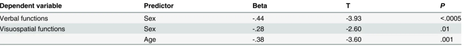
CSF biomarkers did not account for any variance in cognitive performance for the controls. The only significant predictors of cognitive performance in healthy controls were sex and age for visuospatial functions (Fig 1B) and sex for verbal functions (Table 5).
Discussion
This is the first study investigating the relationship between CSF biomarkers of neurodegenera-tion and cognineurodegenera-tion in bipolar disorder. The biomarkers independently accounted for a signifi-cant part of the individual variability of cognitive performance in the bipolar disorder group. Intriguingly, the findings could not be reproduced in the control sample. This suggests that an association with CSF neurodegeneration biomarkers and cognitive performance is not a gener-al phenomenon. It remains to be elucidated if these associations are specific to persons with bi-polar disorder, or if CSF biomarkers of neurodegeneration might also be associated with cognitive performance in other psychiatric disorders such as schizophrenia.
Biomarkers and covariates best explained the variance in visuospatial functions, which is somewhat surprising as visuospatial functions are typically relatively well preserved in bipolar disorder [7]. As the individual tests constituting the visuospatial domain—Rey Complex Figure Test and Block Design—also load on executive functions, there is a possibility that some of the variance in this domain may be accounted for by other cognitive functions [35]. The rel-atively large proportion of variance accounted for by CSF biomarkers in the verbal domain was also unexpected as most studies have reported intact verbal functions in bipolar disorder [6]. By contrast, attention and processing speed was only weakly accounted for by the biomarkers; covariates had a stronger influence on performance.
In the group with bipolar disorder, higher concentration of NFL, as well as higher Aβ42/40
ratio and lower Aβ42/38 ratio, was consistently associated with decreased cognitive perfor-mance. Altered ratios of Aβwere the CSF biomarkers that accounted for most of the variance
in verbal performance. Again, this finding could be related to cross-loadings on executive func-tions. NFL was the most important predictor of memory performance. However, the model
and healthy controls (1B). In patients with bipolar disorder cerebrospinal fluid (CSF) Amyloidβ(Aβ) 42/40 ratio, CSF Aβ42/38 ratio, CSF total tau, age, and lithium contributed significantly to the model (adj. r2=. 36, p=<.0005). In patients with bipolar disorder only sex and age were associated with visuospatial functions (adj. r2=. 23,p=<.0005).
doi:10.1371/journal.pone.0127100.g001
Table 5. Variables associated with cognitive performance in healthy controls.
Dependent variable Predictor Beta T P
Verbal functions Sex -.44 -3.93 <.0005
Visuospatial functions Sex -.28 -2.60 .01
Age -.38 -3.60 .001
Non-significant results are not displayed.
only accounted for 15 percent of the variance in the memory domain, which dropped to 12 per-cent when working memory tests were removed from the memory/learning domain. Working memory alone was more strongly associated with CSF biomarkers (27%) and NFL was the CSF biomarker that contributed the most to the model. Nonetheless, this suggests that neuronal and axonal dysfunction or degeneration might contribute to reduced memory performance in bipolar disorder. Aβ40 is more adhesive than the Aβ38 isoform and thus considered more harmful [36]. A decrement in both ratios (42/40 and 42/38) would normally be associated with reduced neuropsychological performance [37]. Our finding that ahigherAβ42/40 ratio was as-sociated with decreased cognitive performance is therefore somewhat puzzling. However, the Aβ42/38 and Aβ42/40 ratios have also been found to increase prior to deposition of plaque in Alzheimer’s disease [38]. The association between increased Aβ42/40 ratio and cognitive
im-pairment could thus be interpreted as a response to neurotoxicity. Alternatively, elevated Aβ
ratios can indicate aγ-secretase dysfunction.γ-secretase cleaves amino acids 37–40 and 42 of the Aβdomain, and the Aβratios are expected to increase if this enzyme is functioning less well. Indeed, impairedγ-secretase has been associated with impaired brain plasticity in animal
models [39].
T-tau was associated with cognitive performance in three domains, but inconsistently so; higher T-tau concentrations was positively associated with verbal and visuospatial functions, but negatively associated with executive functions. Moreover, there was no difference in CSF T-tau between patients and controls. Hence, the value of T-tau as a predictor of cognitive per-formance might be limited. Also, sAPPβwas only associated with performance in one cognitive
domain; lower sAPPβconcentrations correlated with decreased speed/attention performance. Furthermore, the more AD specific biomarkers P-tau and Aβ1–42 were not associated with cognitive performance in any cognitive domain. As P-tau and Aβ1–42 are established biomark-ers for current and future cognitive performance in mild cognitive impairment and AD [21], findings from the current study accords with the study by Jakobsson and colleagues who found no evidence of an Alzheimer like CSF pattern in patients with bipolar disorder [16]. Although there is some evidence that psychiatric disorders may increase the risk of dementia [40], a neuropathological study found no increase of amyloid plaques or neurofibrillary tangles in post-mortem brain tissues in a sample of psychiatric patients [41]. Hence, it is more likely that the associations between CSF biomarkers and cognitive domains in patients reflect a neurotox-ic state rather than a neurodegenerative process. As the mean age of this sample was relatively low (38 years) we cannot rule out the possibility that neurodegenerative processes are present in an older cohort.
all covariates of interest. For example, the potential influence of education on cognitive func-tioning was not analyzed in the current study. The included patients were euthymic at the time of the investigation, which precludes any speculation as to how CSF biomarkers affect cogni-tion in non-euthymic states. As episodes of mania, hypomania, and depression were not re-corded during the investigation it cannot be ruled out that the associations seen in this study are sequelae following mania/depression. There were, however, no significant associations be-tween MADRS/YMRS scores and cognition. Finally, whereas the effect of medication was con-trolled for, this cross-sectional study cannot rule out that medication influences
CSF concentrations.
In conclusion, we found that CSF biomarkers of neurodegeneration were associated with cognitive performance in euthymic bipolar disorder, but not in healthy controls, in all cognitive domains independently of age, medication, disease status, and bipolar subtype. Notably, ratios of Aβ42/40 and Aβ42/38 were consistently associated with altered cognitive performance. It
re-mains to be studied whether CSF biomarkers can also be utilized to predict changes in cogni-tive functioning during the course of illness.
Acknowledgments
We wish to thank the staff at the St. Göran bipolar affective disorder unit, including coordina-tor Martina Wennberg, study nurses Agneta Carlswärd-Kjellin and Benita Gezelius, and data manager Haydeh Olofsson. Yngve Hallström is acknowledged for performing lumbar punc-tures. Erik Joas, Kristoffer Bäckman and Mathias Kardell are acknowledged for statistical and database support. We are also indebted to Björn Hultman, Pascal Borgström, and Josefin Östlind for designing the cognitive test battery. We also thank the patients and controls partici-pating in this study. We finally wish to thank the BBMRI.se and KI Biobank at Karolinska Institutet for professional biobank service.
Author Contributions
Conceived and designed the experiments: SR ML. Performed the experiments: SR. Analyzed the data: SR JJ ML EP. Wrote the paper: SR JJ CS CJE KB HZ EP ML.
References
1. Phillips ML, Kupfer DJ. Bipolar disorder diagnosis: challenges and future directions. Lancet. 2013; 381 (9878):1663–71. doi:10.1016/S0140-6736(13)60989-7PMID:23663952.
2. Merikangas KR, Jin R, He JP, Kessler RC, Lee S, Sampson NA, et al. Prevalence and correlates of bi-polar spectrum disorder in the world mental health survey initiative. Archives of general psychiatry. 2011; 68(3):241–51. doi:10.1001/archgenpsychiatry.2011.12PMID:21383262; PubMed Central PMCID: PMC3486639.
3. Ekman M, Granstrom O, Omerov S, Jacob J, Landen M. The societal cost of bipolar disorder in Swe-den. Social psychiatry and psychiatric epidemiology. 2013. doi:10.1007/s00127-013-0724-9PMID:
23754681.
4. Andreou C, Roesch-Ely D, Veckenstedt R, Bohn F, Aghotor J, Kother U, et al. Predictors of early stable symptomatic remission after an exacerbation of schizophrenia: The significance of symptoms, neuro-psychological performance and cognitive biases. Psychiatry research. 2013. doi:10.1016/j.psychres. 2013.08.019PMID:23998362.
5. Arts B, Jabben N, Krabbendam L, van Os J. Meta-analyses of cognitive functioning in euthymic bipolar patients and their first-degree relatives. Psychological medicine. 2008; 38(6):771–85. doi:10.1017/ S0033291707001675PMID:17922938.
7. Bourne C, Aydemir O, Balanza-Martinez V, Bora E, Brissos S, Cavanagh JT, et al. Neuropsychological testing of cognitive impairment in euthymic bipolar disorder: an individual patient data meta-analysis. Acta psychiatrica Scandinavica. 2013; 128(3):149–62. doi:10.1111/acps.12133PMID:23617548. 8. Bora E, Yucel M, Pantelis C. Cognitive endophenotypes of bipolar disorder: a meta-analysis of
neuro-psychological deficits in euthymic patients and their first-degree relatives. Journal of affective disorders. 2009; 113(1–2):1–20. doi:10.1016/j.jad.2008.06.009PMID:18684514.
9. Torrent C, Martinez-Aran A, del Mar Bonnin C, Reinares M, Daban C, Sole B, et al. Long-term outcome of cognitive impairment in bipolar disorder. The Journal of clinical psychiatry. 2012; 73(7):e899–905. doi:10.4088/JCP.11m07471PMID:22901360.
10. Mora E, Portella MJ, Forcada I, Vieta E, Mur M. Persistence of cognitive impairment and its negative im-pact on psychosocial functioning in lithium-treated, euthymic bipolar patients: a 6-year follow-up study. Psychological medicine. 2013; 43(6):1187–96. doi:10.1017/S0033291712001948PMID:22935452. 11. Kempton MJ, Geddes JR, Ettinger U, Williams SC, Grasby PM. Meta-analysis, database, and
meta-re-gression of 98 structural imaging studies in bipolar disorder. Archives of general psychiatry. 2008; 65 (9):1017–32. doi:10.1001/archpsyc.65.9.1017PMID:18762588.
12. Lim CS, Baldessarini RJ, Vieta E, Yucel M, Bora E, Sim K. Longitudinal neuroimaging and neuropsy-chological changes in bipolar disorder patients: review of the evidence. Neuroscience and biobehavior-al reviews. 2013; 37(3):418–35. doi:10.1016/j.neubiorev.2013.01.003PMID:23318228.
13. Savitz J, Solms M, Ramesar R. Neuropsychological dysfunction in bipolar affective disorder: a critical opinion. Bipolar disorders. 2005; 7(3):216–35. doi:10.1111/j.1399-5618.2005.00203.xPMID:
15898960.
14. Weinberger DR, McClure RK. Neurotoxicity, neuroplasticity, and magnetic resonance imaging mor-phometry: what is happening in the schizophrenic brain? Archives of general psychiatry. 2002; 59 (6):553–8. PMID:12044198.
15. Jakobsson J, Bjerke M, Ekman CJ, Sellgren C, Johansson AG, Zetterberg H, et al. Elevated Concentra-tions of Neurofilament Light Chain in the Cerebrospinal Fluid of Bipolar Disorder Patients. Neuropsy-chopharmacology. 2014. doi:10.1038/npp.2014.81PMID:24694925.
16. Jakobsson J, Zetterberg H, Blennow K, Johan Ekman C, Johansson A, Landén M. Altered concentra-tions of amyloid precursor protein metabolites in the cerebrospinal fluid of patients with bipolar disorder. Neuropsychopharmacology: official publication of the American College of Neuropsychopharmacology. 2013; 38(4):664–72. doi:10.1038/npp.2012.231PMID:23212456
17. Turner PR, O'Connor K, Tate WP, Abraham WC. Roles of amyloid precursor protein and its fragments in regulating neural activity, plasticity and memory. Progress in neurobiology. 2003; 70(1):1–32. PMID:
12927332.
18. Blennow K, Hampel H, Weiner M, Zetterberg H. Cerebrospinal fluid and plasma biomarkers in Alzhei-mer disease. Nature reviews Neurology. 2010; 6(3):131–44. doi:10.1038/nrneurol.2010.4PMID:
20157306.
19. Fagan AM, Mintun MA, Mach RH, Lee SY, Dence CS, Shah AR, et al. Inverse relation between in vivo amyloid imaging load and cerebrospinal fluid Abeta42 in humans. Annals of neurology. 2006; 59 (3):512–9. doi:10.1002/ana.20730PMID:16372280.
20. Rosengren LE, Karlsson JE, Karlsson JO, Persson LI, Wikkelso C. Patients with amyotrophic lateral sclerosis and other neurodegenerative diseases have increased levels of neurofilament protein in CSF. J Neurochem. 1996; 67(5):2013–8. PMID:8863508.
21. Rolstad S, Berg AI, Bjerke M, Johansson B, Zetterberg H, Wallin A. Cerebrospinal fluid biomarkers mir-ror rate of cognitive decline. Journal of Alzheimer's disease: JAD. 2013; 34(4):949–56. doi:10.3233/ JAD-121960PMID:23313924.
22. Ryden E, Thase ME, Straht D, Aberg-Wistedt A, Bejerot S, Landen M. A history of childhood attention-deficit hyperactivity disorder (ADHD) impacts clinical outcome in adult bipolar patients regardless of current ADHD. Acta psychiatrica Scandinavica. 2009; 120(3):239–46. doi:10.1111/j.1600-0447.2009. 01399.xPMID:19426162.
23. Sachs GS, Thase ME, Otto MW, Bauer M, Miklowitz D, Wisniewski SR, et al. Rationale, design, and methods of the systematic treatment enhancement program for bipolar disorder (STEP-BD). Biological psychiatry. 2003; 53(11):1028–42. PMID:12788248.
24. Association AP. Diagnostic and Statistical Manual of Mental Disorders, Fourth Edition: DSM-IV-TR: American Psychiatric Association; 2000.
26. Saunders JB, Aasland OG, Babor TF, de la Fuente JR, Grant M. Development of the Alcohol Use Dis-orders Identification Test (AUDIT): WHO Collaborative Project on Early Detection of Persons with Harmful Alcohol Consumption—II. Addiction. 1993; 88(6):791–804. PMID:8329970.
27. Zetterberg H, Andreasson U, Hansson O, Wu G, Sankaranarayanan S, Andersson ME, et al. Elevated cerebrospinal fluid BACE1 activity in incipient Alzheimer disease. Archives of neurology. 2008; 65 (8):1102–7. doi:10.1001/archneur.65.8.1102PMID:18695061.
28. Olsson A, Vanderstichele H, Andreasen N, De Meyer G, Wallin A, Holmberg B, et al. Simultaneous measurement of beta-amyloid(1–42), total tau, and phosphorylated tau (Thr181) in cerebrospinal fluid by the xMAP technology. Clinical chemistry. 2005; 51(2):336–45. doi:10.1373/clinchem.2004.039347
PMID:15563479.
29. Yatham LN, Torres IJ, Malhi GS, Frangou S, Glahn DC, Bearden CE, et al. The International Society for Bipolar Disorders-Battery for Assessment of Neurocognition (ISBD-BANC). Bipolar disorders. 2010; 12 (4):351–63. doi:10.1111/j.1399-5618.2010.00830.xPMID:20636632.
30. Lezak M, Howieson D, Bigler E, Tranel D. Neuropsychological Assessment: USA: Oxford University Press; 2012.
31. Strauss E, Sherman EMS, Spreen O. A Compendium of Neuropsychological Tests: Administration, Norms, and Commentary: Oxford University Press; 2006. PMID:17067774
32. Bearden CE, Shih VH, Green MF, Gitlin M, Sokolski KN, Levander E, et al. The impact of neurocogni-tive impairment on occupational recovery of clinically stable patients with bipolar disorder: a prospecneurocogni-tive study. Bipolar disorders. 2011; 13(4):323–33. doi:10.1111/j.1399-5618.2011.00928.xPMID:
21843272; PubMed Central PMCID: PMC3157039.
33. Palsson E, Figueras C, Johansson AG, Ekman CJ, Hultman B, Ostlind J, et al. Neurocognitive function in bipolar disorder: a comparison between bipolar I and II disorder and matched controls. BMC psychia-try. 2013; 13:165. doi:10.1186/1471-244X-13-165PMID:23758923; PubMed Central PMCID: PMC3691847.
34. Smith EE, Jonides J. Storage and executive processes in the frontal lobes. Science. 1999; 283 (5408):1657–61. PMID:10073923.
35. Schwarz L, Penna S, Novack T. Factors contributing to performance on the Rey Complex Figure Test in individuals with traumatic brain injury. The Clinical neuropsychologist. 2009; 23(2):255–67. doi:10. 1080/13854040802220034PMID:19172529.
36. Page RM, Baumann K, Tomioka M, Perez-Revuelta BI, Fukumori A, Jacobsen H, et al. Generation of Abeta38 and Abeta42 is independently and differentially affected by familial Alzheimer disease-associ-ated presenilin mutations and gamma-secretase modulation. The Journal of biological chemistry. 2008; 283(2):677–83. doi:10.1074/jbc.M708754200PMID:17962197.
37. Schoonenboom NS, Mulder C, Van Kamp GJ, Mehta SP, Scheltens P, Blankenstein MA, et al. Amyloid beta 38, 40, and 42 species in cerebrospinal fluid: more of the same? Annals of neurology. 2005; 58 (1):139–42. doi:10.1002/ana.20508PMID:15984010.
38. Kuperstein I, Broersen K, Benilova I, Rozenski J, Jonckheere W, Debulpaep M, et al. Neurotoxicity of Alzheimer's disease Abeta peptides is induced by small changes in the Abeta42 to Abeta40 ratio. The EMBO journal. 2010; 29(19):3408–20. doi:10.1038/emboj.2010.211PMID:20818335; PubMed Cen-tral PMCID: PMC2957213.
39. Bittner T, Fuhrmann M, Burgold S, Jung CK, Volbracht C, Steiner H, et al. Gamma-secretase inhibition reduces spine density in vivo via an amyloid precursor protein-dependent pathway. The Journal of neu-roscience: the official journal of the Society for Neuroscience. 2009; 29(33):10405–9. doi:10.1523/ JNEUROSCI.2288-09.2009PMID:19692615.
40. Kessing LV, Andersen PK. Does the risk of developing dementia increase with the number of episodes in patients with depressive disorder and in patients with bipolar disorder? Journal of neurology, neuro-surgery, and psychiatry. 2004; 75(12):1662–6. doi:10.1136/jnnp.2003.031773PMID:15548477; PubMed Central PMCID: PMC1738846.
41. Damadzic R, Shuangshoti S, Giblen G, Herman MM. Neuritic pathology is lacking in the entorhinal cor-tex, subiculum and hippocampus in middle-aged adults with schizophrenia, bipolar disorder or unipolar depression. Acta neuropathologica. 2002; 103(5):488–94. doi:10.1007/s00401-001-0496-2PMID:
11935265.