Introduction
The clam R. decussatus is a suspension-feeding bivalve mol-lusc widely distributed in European and Mediterranean
coastal waters. Due to its economic importance it is exten-sively cultured in many countries, and particularly in Portugal where its production represents > 80 % of total shellfish production. The selection of the clam R. decussa-tus as a bioindicator was a consequence of its economic importance and the need to use a filter-feeding bivalve to evaluate environmental changes of metal concentrations [1]. Like many other bivalves, this species resides in sedi-ments and accumulates metal concentrations reflecting gra-dients of metal contamination in the surrounding environ-ment [2,3,4]. This suggests that its tissues have mechanisms, related to its filter-feeding habit, that inhibit the toxic effects of these contaminants.
Like many other molluscs R. decussatus has evolved a number of subcellular systems for accumulation, regulation and immobilization of metals. These include, among others, the binding of essential and pollutant metals to soluble lig-ands such as MT. These two-domain molecules are low mol-ecular, heat-stable proteins of non-enzymatic nature that occur mainly in the cytoplasm. They have strong affinity to class B metal cations which enable them to be differentiated from most of the other proteins [5]. The induction of these proteins has, therefore, been proposed as a specific indica-tor and possible “early warning marker” for the detection of detrimental effects caused by exposure to excess of essen-tial and pollutant metals.
MT induction has been detected in the whole soft tissues, gills, digestive gland and remaining tissues of the clam R. decussatus after exposure to essential and toxic metals [4,6] and this paper represents an overview of MT function in this bivalve species.
MT induction in laboratory experiments
Several laboratory experiments have been carried out to assess MT induction in R. decussatus exposed to sublethal concentrations of cadmium (100 and 400 µ g/l), copper (75 µg/l) and lindane (34.5 µg/l) [4,7,6,8].
Chromatographic elution profiles of the heat-treated cytosol of the whole soft tissues of unexposed clams revealed that cadmium in this pool (> 80 % of total Cd) was distributed between two cadmium-binding proteins of a mol-ecular weight of around 10,000 and 20,000 Da, respectively, that increased with Cd accumulation. Similarly an increase in the –SH containing proteins (by 70 % compared with unexposed clams), based on differential pulse polarography
386 ANALUSIS, 2000, 28, N° 5
© EDP Sciences, Wiley-VCH 2000
■
Metallothioneins in the clam Ruditapes
decussatus: an overview
M.J. Bebianno
*, M.A. Serafim and D. Simes
University of Algarve, Campus de Gambelas, 8000 Faro, Portugal
The clam Ruditapes decussatus is a suspension-feeding bivalve mollusc widely distributed in European waters and in the Mediterranean. Due to its economic importance it is heavily harves-ted in many countries, and particularly in Portugal. Its ability to accumulate high metal concentrations along with its economic impor-tance was the main reason for its selection as a bioindicator.
Metallothionein (MT) concentrations in the clams R. decussatus followed by gel filtration chroma-tography, differential pulse polarography and SDS-PAGE, after Cd exposure, revealed that MT is induced in different tissues (whole soft sues, gills, digestive gland and remaining tis-sues) but the level of MT induction is tissue dependent. MT from the gills and the digestive gland give a more sensitive response to assess the effects of metal exposure directly from the water or from the food than in the whole soft tis-sues.
MT levels were also measured in the gills, diges-tive gland and remaining tissues of R. decussa-tus collected in the Ria Formosa lagoon (Portugal) from areas of different metal load and during the period of sexual differentiation of the clam. Data revealed that there were significant differences of MT concentrations among sites and season but not among sex.
Purification of MT from the digestive gland of R. decussatus revealed four MT isoforms. The mole-cular weight of one of these isoforms, determi-ned by SDS-PAGE, was of the same order of magnitude as that of MT from other bivalve spe-cies. Similarly the amino acid sequence of the βdomain of the MT of the digestive gland of the clam also shows some degree of similarity with the similar MT sequence from mussels and oys-ters. It is, therefore suggested that there is some degree of similarity in the MT structure among these species.
[9], was related to the increase in Cd levels. Therefore Cd in the heat-treated cytosol of the whole soft tissues of R. decussatus is principally bound to MT [4].
MT concentrations, determined by differential pulse polarography [9] in the heat-treated cytosol of unexposed clams (2.05 ± 0.41 mg.g–1dw, Tab. I) are similar to the basal
levels of MT proposed for marine bivalves (2 mg.g–1dw)
[5]. However, de novo MT production in the whole soft tis-sues of Cd-exposed clams, although significant at times, was relatively low, in contrast with a 3- to 4-fold MT induction observed in the mussels Mytilus edulis and Mytilus gallo-provincialis exposed to the same Cd concentrations [10,11]. Therefore the whole soft tissues were considered unsuitable for following the effects of sublethal Cd contamination in the environment [4].
The gills function both as a site of metal uptake and as an important reservoir for metal storage and for Cd in R. decussatus are the major site of accumulation, at least ini-tially, though subsequent transport to and from the digestive gland seems likely [4,7]. As in whole soft tissues, MT in the gills is the major Cd-binding component (binding > 80 % of total body Cd in contaminated species and 45 % unex-posed clams). Characteristically, 84 % of the –SH groups (major Cd-binding sites) in the heat-treated cytosol were associated with MT compared with 65 % in the gills of unexposed clams.
MT concentrations determined in selected tissues of unex-posed clams (Tab. I) revealed that MT concentrations ranked from digestive gland>remaining tissues>gills>. In unexposed R. decussatus under-saturation with Cd in the MT pool was evident for all tissues. After exposure to Cd (100 and 400 µg/l for 40 days) there was a significant net increase in MT concentrations in all tissues but the sequence of MT concentrations among tissues remained unchanged [4,7].
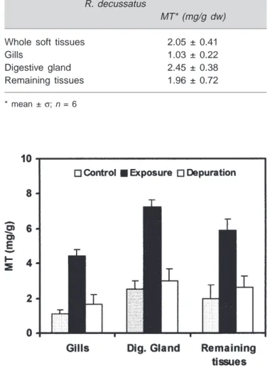
MT concentrations in the gills of unexposed clams (1.03 ± 0.22 mg.g–1dw) are of the same order of magnitude
as the whole soft tissues. After 40 days of Cd (100 µg/l) exposure, MT was synthesised in the gills of Cd exposed clams at net MT rate of 0.04 mg.g–1d–1and increased
four-fold during that time (Fig. 1). However this rate is four-four-fold less than that in the gills of the mussel M. galloprovincialis exposed to the same Cd concentration over the same period of time [1].
After depuration MT concentrations in the different tis-sues of the clams decreased to levels slightly higher than those of unexposed clams suggesting that protein turnover although different in the three tissues was faster than Cd [7]. However, MT levels in the gills were still 2-fold higher than basal levels at the end of the depuration period suggesting that MT are involved in the accumulation and elimination of Cd from the other tissues [7].
Roméo and Gnassia-Barelli [6], using a fluorometric method, observed that MT concentrations in the gills of R. decussatus exposed to 250 µ g Cd.l–1 for 7 days
(45 ± 1 µ g.g–1ww) were not different from control gills
(42 ± 1 µ g.g–1ww). This negligible increase in MT
concentrations in the gills of the clams was possibly due to limited period of exposure and indicates that at the begin-ning of the treatment, Cd might be associated with existing soluble proteins, such as MT. Subsequently Cd was dis-placed to other tissues or to the particulate fraction or accu-mulated in intracellular granules [6]. However MT induction in the gills of R. decussatus was observed after exposure to 75 µg Cu.l–1 for 7 days (58 ± 6 µg.g–1ww) confirming the
monitoring potential of R. decussatus in the gills for the evaluation of the effect of dissolved and particulate metals in the environment [6].
The greater induction of MT in the gills, when compared with the other tissues suggests that MT in this tissue is involved in detoxification and elimination of Cd. The deter-mination of MT concentrations in the gills of the clams seems, therefore, preferable [4,7].
Table I. MT concentrations determined by DPP in diffe-rent tissues of the clam.
R. decussatus
MT* (mg/g dw) Whole soft tissues 2.05 ± 0.41 Gills 1.03 ± 0.22 Digestive gland 2.45 ± 0.38 Remaining tissues 1.96 ± 0.72
* mean ±σ;n = 6
Figure 1.Ruditapes decussatus. Variation of MT concentrations in the gills, digestive gland and remaining tissues of clams exposed to Cd (100 µg/l) for 40 days and depurated for 50 days (data represents mean ±σ,n = 6).
Analysis of other body components of the clams revealed that metals are predominantly localised in the digestive gland [12,4,7,6]. MT concentrations in this tissue of unex-posed clams (2.45 ± 0.38 mg.g–1dw) was two-fold higher
than that of the gills. In Cd exposed clams, however, MT binds only 70 % of the Cd in the digestive gland compared with 84 % in the digestive gland of unexposed clams. Nevertheless an increase was observed in MT in the diges-tive gland of Cd exposed clams reaching 3-fold after 40 days of Cd (100 µg/l) exposure (Fig. 1).
When the source of contamination stopped MT levels in the digestive gland also decreased, as in the gills, but MT levels after 50 days although significantly different from MT in the digestive gland of unexposed clams, were much lower than that of the gills (Fig. 1).
When R. decussatus were exposed to Cu (75 µg/l) or to lindane (34.5 µg/l), separately or in combination, MT levels in the clam digestive gland (0.52 ± 0.17 mg.g-1 ww),
deter-mined by differential pulse polarography, did not change sig-nificantly (0.62 ± 0.13 -Cu, 0.53 ± 0.16 -lindane, 0.42 ± 0.07 Cu+lindane mg.g-1 ww). Therefore it was
observed that the increase in Cu in the soluble fraction was
not correlated with MT concentrations [8]. Cu was also shown to increase the lipid peroxidation of membranes in the digestive gland of R. decussatus. This toxic phenome-non may facilitate Cu precipitation so that less Cu is avail-able for MT induction. Lindane does not appear to have any effect on MT induction [13].
MT concentrations in clams from the field
MT concentrations were measured, by differential pulse polarography, in the heat-treated cytosol of the gills, diges-tive gland and remaining tissues of R. decussatus sampled over a period of one year (1994). Clams were collected, every two months, from two zones with different metal loads in the Ria Formosa lagoon in the south coast of Portugal (Fig. 2). The variation of MT concentrations in the gills, digestive gland and remaining tissues from clams from these two sites are presented in figure 2. MT distribution among the different tissues was similar to that observed in the lab-oratory experiments, with the highest MT levels occuring in the digestive gland, followed by the gills and remaining tis-sues. MT concentrations in the three tissues were signifi-cantly different between sites reflecting the different metal
load (highest at Faro) [14]. Similarly a seasonal variation was observed in MT concentrations in the three tissues with higher MT levels found in summer and winter (p < 0.05).
Once the highest MT levels in this species was estab-lished to be those of the digestive gland, MT variation in this tissue of R. decussatus was followed in a natural pop-ulation during a period of 4 months (from June to September 1997) to evaluate the effect of sex on MT levels. This period corresponded, in this species, to the period of sexual differ-entiation. MT levels were determined in the digestive gland from the site A in the Ria Formosa lagoon (Portugal) between June and September in males and females and data is presented in figure 3. As can be seen there is no signifi-cant difference of MT levels between males and females during the period of sexual differentiation. However, MT levels in both sexes showed seasonal variation with MT lev-els in August (14.4 ± 0.9 nmol.g–1and 17.5 ± 3.8 mol.g–1for
males and females, respectively) that were significantly dif-ferent from the other months (p < 0.05). Similarly, high Cd and Cu levels were found in August while Zn did not show any seasonal variation [21]. Data revealed that sex did not affect MT concentrations in this species and in this marine ecosystem.
To evaluate the efficiency of MT as a biomarker a simi-lar study was carried out along the Tunisian coast using the same species. MT and metal levels in the subcellular frac-tions of R. decussatus digestive gland were determined in a natural population of the golf of Gabès (Tunisia) during the same four month period (June to September) to evaluate metal pollution and the effects of biotic (sex, size and repro-ductive state) parameters on MT levels. Mean MT concen-trations in the digestive gland of R. decussatus from the Tunisian population, determined by differential pulse polarography (3.4 mg.g–1ww) was of the same order of
magnitude as that of the same tissue of the Portuguese clam population [22] (Fig. 1). The effect of size and reproductive effects on MT levels were less perceptible in males than in females. Data suggested that other factors than metal cont-amination affect MT synthesis in this species. Therefore Hamza-Chaffai et al. [22] proposed that MT levels in the digestive gland of males should be used as suitable bio-marker for detecting metal contamination due to its high condition index and low MT variability linked with size and spawning period. Uniformely-sized organisms should be used in order to avoid, as far as possible, size effects.
MT characterization in the digestive gland of R. decussatus
MT in the digestive gland of Cd exposed (100 µ g/l) R. decussatus was further characterised, after purification, by gel-permeation and ion-exchange chromatography. A molecular weight of 13,700 Da was obtained for MT by gel filtration chromatography, similar to the MT molecular weight determined for other mollusc species with the same
method [5]. The molecular weight determined on calibrated gel SDS/Page electrophoresis was 7328 Da that is also sim-ilar to the molecular weight of MT in the mussel Mytilus edulis (7,740 [15]) and Crassostrea virginica (7,318 [16]).
Four MT isoforms were separated by ion-exchange chro-matography followed by HPLC in the digestive gland of R. decussatus. They all had high Cd content and UV spec-tra indicative of the presence of characteristic Cd-thiolate complexes and low aromatic content. Two of these MT iso-forms were purified, characterised and the amino acid of the NH2-terminal sequenced (Tab. II). The sequence of the NH2
-terminal, that corresponded to the β domain of the protein, revealed the absence of methionine which is consistent with
Table II. Comparison of the sequence of NH2-terminal of MT in marine bivalves.
R. decussatus MT-1a GDPCNVAETGCQVCAQCCK
R. decussatus MT -2a GEPCN
C. virginicab MSDPCNCIETGTCACSDSCPAT
M. edulis (10-IV)c PAPCNCIETNVCICGTGCSGH
a) adapted from Simes et al. (submitted); b) Unger et al., 1991; c) Mackay
et al., 1993
Figure 3.Ruditapes decussatus. Variation of MT concentrations between males and females (adapted from Serafim & Bebianno, in press) (data represents mean ±σ, n=6).
other non-mammalian MTs. From the sequence of the NH2
-terminal and its chemical characteristics of the two isoforms the MTs in the clam digestive gland can be classified as a class I MT. For the other two isoforms isolated, one was sligtly contaminated and the other had the NH2-terminal
blocked. [17]. These isoforms are due to polimorphism within this clam population and revealed a similarity with other MT mollusc species, the mussel Mytilus edulis where at least nine isoforms were detected [18] and the oyster Crassostrea virginica [19,16]. A comparison between the sequence of R. decussatus MT NH2-terminal from the
diges-tive gland with the sequence of MT from other mollusc species reported in the literature showed a higher degree of similarity with the oyster Crassostrea virginica MT NH2
-ter-minal (52 % identity) [16].
However, despite partial sequencing the function of MT in the digestive gland remains unclear. Suggested possibili-ties for the various isoforms include different specificity of the promotor region of the gene [20], different metal-bind-ing affinities [23] or simply the need for a large number of copies to facilitate a rapid response when necessary.
References
1. Bebianno, M.J.; Serafim, M.A. The Science of Total
Environment 1998, 214, 123-131.
2. Henry, M.; Huang, G.; Cistiani, C.; Belluau, M.; Durbec, J.P. Toxicité et bioaccumulation du cadmium chez la palourde
Ruditapes decussatus, en fonction des facteurs temps,
concen-tration et température; Vies Journées d’Études Pollution
Marines en Méditerranée, Cannes: CIESM, 1982, pp 737-742.
3. Henry, M.; Huang,W.; Cornet, C.; Belluau, M.; Durbec, J.P.
Oceanologica Acta 1984, 7, 329-335.
4. Bebianno, M.J.; Nott, J.A.; Langston, W.J. Aquatic Toxicology
1993, 27, 315-334,
5. Langston, W.J.; Bebianno, M. J.; Burt, G. Metabolic pathways in marine invertebrates. In: Metal Metabolism in Aquatic
Environments; W. J. Langston & M. J. Bebianno (Eds),
London: Chapmann & Hall, 1998, pp 219-284.
6. Roméo, M.; Gnassia-Barelli, M. Comp. Biochem. Physiol.
1995, 111C, 457-463.
7. Bebianno, M.J.; Serafim, M.A.; Rita, M.F. Bull. Environ.
Contam. Toxicol. 1994, 53, 726-732.
8. Hamza-Chaffai, A.; Roméo, M.; Gnassia-Barelli, M.; El Abed, A. Bull. Environ. Contam. Toxicol. 1998, 61, 397-404. 9. Bebianno, M.J.; Langston, W.J. Portugaliæ Electrochimica
Acta 1989, 7, 59-64.
10. Bebianno, M.J.; Langston, W.J. Marine Biology 1991, 108, 91-96.
11. Bebianno, M.J.; Langston, W.J. Comp. Biochem. Physiol.
1992, 103C, 79-85.
12. Vicente, N.; Baghdiguian, S.; Henry, M.; Riva, A. Oceanis
1988, 14, 125-131.
13. Roméo, M.; Gnassia-Barelli, M. Comp. Biochem. Physiol.
1997, 118C, 33-37.
14. Bebianno, M.J. Sci. Total. Environ. 1995, 171, 107-115. 15. Viarengo, A.; Pertica, M.; Canesi, L.; Mazzucotelli, A.;
Orunesu, M.; Bouquegneau, J. M. Comp. Biochem. Physiol.
1989, 93C(2), 389- 395.
16. Unger, M.E.; Chen, T.T.; Murphy, C.M.; Vestling, M.M.; Fenselau, C.; Roesijadi, G. Biochimica Biophysica Acta 1991,
1074, 371-377.
17. Simes, D.C.; Bebianno, M.J.; Moura, I.; Moura J.J.G. Biochem
Biophys. Res. Com. (submitted).
18. Mackay, E.A.; Overnell, J.; Dunbar, B.; Davidson, I.; Hunziker, P.E.; Kagi, J.H.R.; Fothergill, J.E. Journal of
Biochemistry 1993, 218, 183-194.
19. Roesijadi, G.; Kielland, S.L.; Klerks, P. Archives Biochemical
Biophysics 1989, 273, 403-413.
20. Schmidt, C. J.; Jubier, M.–F.; Hamer, D.H. J. Biol. Chem.
1985, 260, 7731-7737.
21. Serafim, M.A.; Bebianno, M.J. Environ. Toxicol. & Chem. (in press).
22. Hamza-Chaffai, A.; Amiard, J. C.; Cosson, R. Comp. Biochem.
Physiol. 1999, 123C, 153-163.
23. Winge, D.R.; Miklossy, K.A. J. Biol. Chem. 1982, 257, 3471-3476.