Uncovering the shell game with barcodes: diversity
of meiofaunal Caecidae snails (Truncatelloidea,
Caenogastropoda) from Central America
Christina Egger1,2, Timea P. Neusser3, Jon Norenburg4,Francesca Leasi5, Barbara Buge6, Angelo Vannozzi7, Regina L. Cunha2, Cymon J. Cox2, Katharina M. Jörger1
1 SNSB-Zoologische Staatssammlung München, Münchhausenstr. 21, 81247 Munich, Germany 2 CCMAR,
Campus de Gambelas, Universidade do Algarve, 8005-139 Faro, Portugal 3 LMU Munich, Biocenter, Dept. II, Großhaderner Str. 2, 82152 Planegg-Martinsried, Germany 4 Department of Invertebrate Zoology, Na-tional Museum of Natural History, Smithsonian Institution, Washington, DC 20560, USA 5 Department of Biology, Geology and Environmental Science. University of Tennessee at Chattanooga. 615 McCallie Ave. Chattanooga, TN 37403, USA 6 Muséum national d’Histoire naturelle, 55 Rue Buffon, 75231 Paris, France
7 Independent researcher, Via M.L. Longo 8, Rome, Italy Corresponding author: Christina Egger (christinaegger@gmx.de)
Academic editor: Eike Neubert | Received 11 April 2020 | Accepted 5 August 2020 | Published 16 September 2020 http://zoobank.org/4296306E-51B9-4873-AB6F-4B475194CA98
Citation: Egger C, Neusser TP, Norenburg J, Leasi F, Buge B, Vannozzi A, Cunha RL, Cox CJ, Jörger KM (2020) Uncovering the shell game with barcodes: diversity of meiofaunal Caecidae snails (Truncatelloidea, Caenogastropoda) from Central America. ZooKeys 968: 1–42. https://doi.org/10.3897/zookeys.968.52986
Abstract
Caecidae is a species-rich family of microsnails with a worldwide distribution. Typical for many groups of gastropods, caecid taxonomy is largely based on overt shell characters. However, identification of spe-cies using shell characteristics is problematic due to their rather uniform, tubular shells, the presence of different growth stages, and a high degree of intraspecific variability. In the present study, a first integra-tive approach to caecid taxonomy is provided using light-microscopic investigation with microsculptural analyses and multi-marker barcoding, in conjunction with molecular species delineation analyses (ABGD, haplotype networks, GMYC, and bPTP). In total 132 specimens of Caecum and Meioceras collected dur-ing several sampldur-ing trips to Central America were analyzed and delineated into a minimum of 19 species to discuss putative synonyms, and supplement the original descriptions. Molecular phylogenetic analyses suggest Meioceras nitidum and M. cubitatum should be reclassified as Caecum, and the genus Meioceras might present a junior synonym of Caecum. Meiofaunal caecids morphologically resembling C. glabrum from the Northeast Atlantic are a complex of cryptic species with independent evolutionary origins, https://zookeys.pensoft.net
Copyright Christina Egger et al. This is an open access article distributed under the terms of the Creative Commons Attribution License (CC BY 4.0), which permits unrestricted use, distribution, and reproduction in any medium, provided the original author and source are credited.
likely associated with multiple habitat shifts to the mesopsammic environment. Caecum invisibile Egger & Jörger, sp. nov. is formally described based on molecular diagnostic characters. This first integrative approach towards the taxonomy of Caecidae increases the known diversity, reveals the need for a reclas-sification of the genus Caecum and serves as a starting point for a barcoding library of the family, thereby enabling further reliable identifications of these taxonomically challenging microsnails in future studies. Keywords
DNA taxonomy, marine biodiversity, meiofauna, molecular species delineation, Mollusca
Introduction
In the past fifteen years molecular barcoding and molecular species delineation have revolutionized the assessment of species diversity and traditional taxonomy, allowing for fast and reproducible species identification and delimitation, and adding to ob-jectivity and reliability in species diagnoses (Leasi et al. 2013; Fontaneto et al. 2015; Scarpa et al. 2016; Martínez-Arce et al. 2020). Molecular data enables testing for mor-phologically cryptic species as well as phenotypic plasticity, and the evaluation of intra- versus inter-specific variability (Jörger et al. 2012; Leasi et al. 2013, 2016). Given the number of described species and 250 years of taxonomic practice that delimit species based largely on distinct morphologies, it is unsurprising that despite the success of modern molecular approaches, many clades of Metazoa have yet to have their morpho-logical classification tested against molecular markers.
Traditionally, the taxonomy of Gastropoda, one of the most species-rich and better-known clades of invertebrates in the marine environment, is largely based on shell char-acteristics (Bouchet and Strong 2010). However, this approach is generally problematic as several studies have revealed species exhibiting phenotypic plasticity in shell form due to environmental factors or predation (Trussell 2000; Weigand et al. 2011), and un-covered cryptic species with the aid of molecular data (Haase et al. 2007; Puillandre et al. 2010; Jörger et al. 2012). Consequently, these studies question evolutionary hypoth-eses based on species delimited by shell characteristics alone and point to the need for an integrative approach using both molecular and morphological data in future research.
Members of the family Caecidae Gray, 1850 can be found in different marine hab-itats (e.g., among algae or corals) including the marine mesopsammon (i.e., the aque-ous interstitial pore spaces of marine sediments). As adults they have uncoiled tubular shells that are likely an adaptation to their infaunal lifestyle (Swedmark 1968). In early descriptions zoologists associated Caecidae snails with tusk-shells (nowadays known as scaphopod molluscs) (see e.g., Montague 1803) or classified them among annelid tube worms (Brown 1827; see Pizzini et al. 2013 for a classificatory history). Even after Caecidae were settled among gastropods (Clark 1849), with current phylogenetic hy-potheses placing them among caenogastropod Truncatelloidea (Criscione and Ponder 2013), their unusual tubular shells still posed challenge to taxonomists. Caecid lar-val shells (protoconch) are usually planspirally coiled with two whorls (Bandel 1996)
and closely resemble related gastropod veliger shells. After settlement of the larvae the adult shell (teleoconch) is formed through differing degrees of uncoiling, with the protoconch either remaining attached (Parastrophia de Folin, 1869, Ctiloceras R. B. Watson, 1866, Enigmerces Iredale & Laseron, 1957, Jayella Iredale & Laseron, 1957, Ponderoceras Bandel, 1996, Strebloceras Carpenter, 1859) or being shed (Caecum J. Fleming, 1813, Meioceras Carpenter, 1859, Pizzinia Vannozzi, 2017, and Mauroceras Vannozzi, 2019). In the latter case, the growing teleoconch is closed by a septum (Ban-del 1996). The snails continue to shed part of the teleoconch until the fully developed adult shell is formed (Draper 1974). The number of repetitions of shedding likely is variable between species, but unknown for the majority of caecids (Pizzini et al. 1998). This complex shell ontogeny results in highly variable shell morphologies during on-togeny (with a minimum of three different shell morphologies: the larval shell-form, the juvenile shell form(s) and the adult shell form), which hampers species identifica-tion and delineaidentifica-tion based on single shells if no comparative data is available for the entire morpho-series (i.e., all developmental stages). Moreover, the tubular shells have few taxonomic characters, thus the current taxonomy is largely based on conchological characters such as size, shell shape, ornamentation, construction of the aperture, sep-tum and mucro (i.e., an evagination of the sepsep-tum, see Fig. 1 for terminology) (Light-foot 1992a, b, 1993a, b; Pizzini and Raines 2011; Pizzini et al. 2013; Vannozzi et al. 2015; Vannozzi 2017). However, these characters can change for an individual during its lifetime, for instance, young specimens can be entirely smooth and express shell ornamentation only later during maturation, and also shell shape may change as they continue to add shell material at their aperture (i.e., shell opening, see Fig. 1) (Draper 1974; Pizzini 1998b; Lima et al. 2013). Additional difficulties arise in determining whether the septum and mucro are temporary or final (Pizzini et al. 1998).
While the phylogenetic position of the family among truncatelloid gastropods is supported by molecular and morphological data, the taxonomy within the family still is based largely only on shell morphology alone. Indeed, anatomical data is scarce (e.g., Götze 1938; Draper 1974) and thought to offer few diagnostic characters, while molec-ular barcoding approaches are lacking entirely. Currently, the family Caecidae contains approx. 260 described species in ten genera (MolluscaBase 2019). Most genera (i.e., Strebloceras, Ctiloceras, Jayella, Enigmerces, Ponderoceras, Pizzinia, and Mauroceras) are species-poor and limited in distribution to the Indo-Pacific (Iredale and Laseron 1957; Bandel 1996; Pizzini et al. 2013; Vannozzi 2016, 2017). Only Caecum, currently with 210 valid species (according to MolluscaBase 2019), shows a circumglobal distribution in temperate and tropical zones. Their abundance is particularly high in tropical waters such as the Indo-Pacific and Central America (Vokes 1983; De Jong and Coomans 1988; Lightfoot 1992a, b, 1993a, b; Díaz Merlano and Puyana Hegedus 1994; Pizzini 1998a; Pizzini and Bonfitto 2008; Discover Life 2020). Meioceras, which differs from Caecum in the general shape of the shell (i.e., with the widest part towards the middle of the shell), was erected by Carpenter (1858–1859) due to the slightly coiled shape of their juveniles. The genus was recently split into Indo-West Pacific Mauroceras and Western Atlantic Meioceras (Vannozzi 2019). While recent taxonomic works have
de-scribed the caecid fauna in the Indo-Pacific based on microsculptural investigations of the shell (Pizzini et al. 2013; Vannozzi 2017, 2019), knowledge of caecid diversity in Central American waters is still limited to light-microscopic identification of shells for a large majority of described species.
In this study we present data on caecid diversity based on several recent collecting trips to Central America. We identified the collected Caecidae specimens based on traditional taxonomy and used additional microsculptural observations and molecular barcodes to reliably assign different growth stages to taxa. We applied an integrative experimental ap-proach including multi-marker barcoding and molecular species delineation analyses to test our morphology-based taxonomy, and to identify putative cryptic species.
Materials and methods
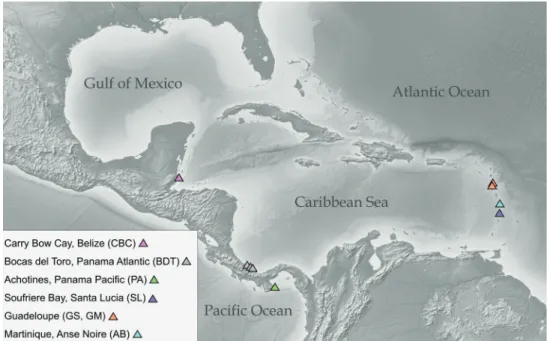
We collected and microscopically investigated a total of 132 individuals of meiofaunal caecid snails from five different sites in tropical Central America. Of 132 specimens, 67 were selected for further analyses (see Fig. 2 for sampling sites and Tables 1, 2 for details on material and sampling sites). Specimens were extracted from samples of coarse subtidal sands by resting them in buckets for at least 1–2 days to deplete oxygen and ac-cumulate the meiofauna in the surface layer. The surface layer was skimmed off, and the snails extracted by a decantation technique after anesthetization with MgCl2-seawater solution using a sieve with a mesh size of 100 µm (Jörger et al. 2014). All specimens were documented alive and grouped into preliminary morphotypes based on light mi-croscopic (LM) examination of shell characters in the field and fixed in 75–96% etha-nol. Specimens provided by the Muséum national d’Histoire naturelle (MNHN) Paris had previously been removed from their shells in the field by the use of a microwave oven (Galindo et al. 2014), this method is advantageous and recommended over the destructive sampling described below, applied in the beginning of our survey.
Shell characteristics and microsculptural analyses
We documented the main taxonomic characters of the tubular shells (Fig. 1), such as the morphology of aperture, septum, and mucro, and measured size and diameter of the shells. Initial species identification in the field was carefully revised in the laboratory, and specimens were assigned to species according to these shell characteristics. The microscu-lpture of the shell of one representative of each putative morphospecies was investigated via scanning electron microscopy (SEM), whenever a voucher was available (Table 1).
Microscopic debris on the shell was manually removed using an eyelash, and the shell rinsed in 96% ethanol. Specimens were dried by evaporation of the ethanol and transferred onto SEM stubs covered with self-adhesive carbon stickers. We used a sput-ter coasput-ter Polaron SC510 to coat the samples with gold in argon atmosphere. The shells were analyzed with a LEO 1430 VP SEM at a voltage of 15 kV.
All light microscopic images and SEM-micrographs are available through FigShare (https://figshare.com/projects/Central_American_Caecidae/84929).
DNA extraction, amplification, and sequencing
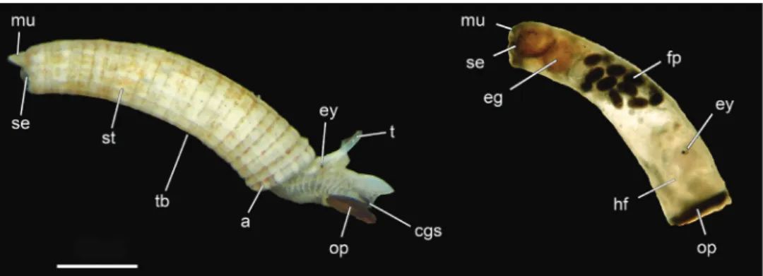
DNA was extracted from 121 of the 132 investigated specimens. The sputter-coated individuals previously investigated by SEM were crushed mechanically using pestles (Bergmeier et al. 2016); specimens investigated only by LM were also crushed if tis-sue was not already separated. Subsequently, DNA was extracted by the procedure of Knebelsberger and Stöger (2012) combining lysis with 2-mercaptoethanol in CTAB buffer, chloroform-isoamyl precipitation, and recovery using columns with silica-membrane (Nucleo Spin, Macherey-Nagel GmbH & Co. KG, Düren, Germany). The DNA was eluted twice with 25 µl aliquots of pre-heated elution buffer to gain high yield. DNA of specimens deposited to the USMN-Smithsonian Institution were extracted in the Laboratories for Analytical Biology, SI using the standard protocols of the Autogen Prep 956 Extractor (eluting with 100 µl Autogen R9 buffer). Three different markers were partially amplified by PCR: mitochondrial cytochrome oxidase subunit I (COI) and 16S rRNA gene, and nuclear 28S rRNA gene, using the standard PCR primers for gastropods (see Klussmann-Kolb et al. 2008). We either used the Phire polymerase (Thermo Fisher Scientific Inc., Waltham, USA) with the following protocol for PCRs on 16S/ COI resp. 28S at the LMU: 98 °C 1 min (98 °C 30 sec; 46–50 °C 20 sec; 72 °C 20 sec) × 36–38 cycles 72 °C 1 min resp.98 °C 30 sec (98 °C 15 sec; 55–60 °C 5 sec; 72 °C 20 sec) × 35 cycles 72 °C 1 min or the KlenTaq poly-merase (AB Peptides, Inc.) with the following program for sequences generated at the SI: 95 °C 3 min, (95 °C 30–45 sec; 48–52 °C 30–45 sec; 72 °C 45–90 sec) × 35–40, 72 °C 7 min. PCR products were either cleaned using a spin column purification kit (Zymo Research, Irvine, California, USA) or were purified with QIAquick (Qiagen Inc.). Samples at the LMU were cycle sequenced on an ABI 3730 48 capillary sequenc-er (Applied Biosystems, Fostsequenc-er City, USA) using Big Dye 3.1 (Thsequenc-ermo Fishsequenc-er Scientific Inc.) at the sequencing service of the Ludwig-Maximilians-Universität (LMU) Bio-center, Munich, Germany. At the SI, cycle sequencing was also conducted with BigDye Figure 1. Morphology of Caecum (Caecidae) including important shell features used for morphological species identification. USNM 1618850 Caecum imbricatum – specimens to the left. Abbreviations: a, aperture; cgs, ciliated gliding sole; eg, egg; ey, eye; fp, fecal pellets; hf, retracted head and foot; mu, mucro; op, operculum; se, septum; st, shell structure (ornamentation); t, tentacle; tb, tube. Scale bar: 500 µm.
Table 1. List of investigated Caecidae specimens, museums numbers (ZSM: SNSB-Bavarian State Col-lection, MNHN: Muséum National d’Histoire Naturelle, USMN: Smithsonian Institution), and NCBI GenBank accession numbers of sequenced genes and the type of voucher of the material of Caecidae analyzed in the present study. An asterisk (*) marks individuals used for SEM scans.
Species Field-code Locality
code
Specimen catalog number
Voucher GenBank number
COI 16S rRNA 28S rRNA
Caecum imbricatum CBC_26 CBC3 USNM 1618850 DNA MT727051 MT704281
Caecum imbricatum BDT_04 BRS101 USNM 1618852 DNA MT704261
Caecum imbricatum BDT_07 BRS103 USNM 1618854 DNA MT727047
Caecum imbricatum BDT_08 BRS103 USNM 1618855 DNA MT727048
Caecum striatum CBC_8 CBC24 USNM 1618845 DNA MT727061 MT704275
Caecum invisibile
sp. nov. CBC_1bB CBC1b ZSM-Mol-20200109 DNA, paratype MT727054 MT704267 MT731696
Caecum invisibile
sp. nov. CBC_1bC CBC1b ZSM-Mol-20100320 DNA*, holotype MT727055 MT704268 MT731697
Caecum invisibile
sp. nov. CBC_3a CBC1b USNM 1618839 DNA MT727056 MT704269 MT731698
Caecum invisibile
sp. nov. CBC_3c CBC1b USNM 1618840 DNA MT727057 MT704270 MT731699
Caecum invisibile
sp. nov. CBC_3d CBC1b USNM 1618841 DNA MT727058 MT704271 MT731700
Caecum invisibile
sp. nov. CBC_3e CBC1b USNM 1618842 DNA MT727059 MT704272 MT731701
Caecum invisibile
sp. nov. CBC_3f CBC1b USNM 1618843 DNA MT727060 MT704273 MT731702
Caecum invisibile
sp. nov. CBC_13a CBC1b USNM 1618846 DNA MT727062 MT704276 MT731704
Caecum invisibile
sp. nov. CBC_13b CBC1b USNM 1618847 DNA MT727063 MT704277 MT731705
Caecum invisibile
sp. nov. CBC_13c CBC1b USNM 1618848 DNA MT727064 MT704278 MT731706
Caecum invisibile
sp. nov. CBC_13d CBC1b USNM 1618849 DNA MT727065 MT704279 MT731707
Caecum invisibile
sp. nov. BDT_20 BRS104 USNM 1618856 DNA MT727049 MT704264 MT731689
Caecum invisibile
sp. nov. BDT_48 BRS200 USNM 1618859 DNA MT727052 MT731694
Caecum regulare BDT_22 BRS108 USNM 1618883 DNA MT727050 MT731690
Caecum regulare CBC_22B CBC22 ZSM-Mol-20100321 DNA* MT704280 MT731708
Caecum donmoorei BDT_23 BRS108 USNM 1618857 DNA MT704265 MT731691
Caecum donmoorei BDT_25 BRS108 USNM 1618858 DNA MT704266 MT731692
Caecum donmoorei CBC_6 CBC1b USNM 1618844 DNA MT704274 MT731703
MOTU I BDT_17 ZSM-Mol-20200039 DNA* MT704263 MT731688
MOTU II BDT_06 BRS101 USNM 1618853 DNA MT727046 MT704262 MT731687
MOTU II BDT_46 BRS110 USNM 1618852 DNA MT727051 MT731693
MOTU II BDT_49 BRS200 USNM 1618860 DNA MT727053 MT731695
Caecum cf.
corrugulatum PA_C04 PA14 USNM 1618861 DNA MT727069 MT731722 Caecum heptagonum PA_28A PA23a ZSM-Mol-20200030 DNA* MT704283 MT731717
Caecum heptagonum PA_G10 PA23a USNM 1618866 DNA MT704291 MT731726
Caecum cf. teres PA_E10 PA23a USNM 1618865 DNA MT727070 MT704289 MT731724
Caecum cf. teres PA_30B PA23a ZSM-Mol-20200033 DNA* MT704284 MT731718
Caecum cf. teres PA_30G PA23a ZSM-Mol-20200037 DNA* MT704286 MT731720
Caecum cf.
chemistry (PerkinElmer) and standard cycles (4 min denaturation at 96 °C, followed by 25 cycles of 10 sec at 96 °C, 5 sec at 50 °C and 4 min at 60 °C), and sequenced on an ABI 3730xl 96-well capillary sequencer. In total, 34%, 43% and 50% of the partial COI, 16S rRNA, and 28S rRNA gene sequences, respectively, were successfully am-plified and sequenced. All sequences were edited in Geneious Prime (vers. 11.02011, Biomatters, Ltd., Auckland, New Zealand). Primer sequences were removed and base calls checked for misreads against their chromatogram. The sequences were then com-pared to sequences in the public database NCBI GenBank (http://ncbi.nlm.nih.gov/ genbank) by using the BLAST online web service to check for putative contamination. In total 29 COI, 40 16S rRNA and 43 28S rRNA gene sequences were deposited in NCBI GenBank (see Table 1 for accession and voucher numbers).
Phylogenetic analyses
Multiple sequence alignments of the 28S rRNA and COI genes were constructed us-ing Mafft (vers. 7.419; Katoh et al. 2002; Nakamura et al. 2018) with default param-eter settings. The mitochondrial 16S rRNA sequences were aligned using the program Muscle (vers. 3.8.31; Edgar 2004) with default parameter settings. Alignments were visualized using Seaview (vers. 3.2; Gouy et al. 2009). COI sequences were translated into amino acids. The program Gblocks (vers. 0.91b; Castresana 2000; Talavera and Castresana 2007) was applied to the 16S and 28S rRNA gene alignments to check
Species Field-code Locality
code
Specimen catalog number
Voucher GenBank number
COI 16S rRNA 28S rRNA
Caecum cf.
strangulatum PA_A07 PA14 USNM 1618864 DNA MT727068 MT704287 MT731721 Caecum cf.
semilaeve PA_11B PA14 ZSM-Mol-20200028 DNA* MT704282 MT731716 Caecum cf.
semilaeve PA_30C PA23a ZSM-Mol-20200034 DNA* MT704285 MT731719
Caecum sp. PA_H05 PA15 USNM 1618862 DNA MT704292 MT731728
Caecum sp. PA_E06 PA14 USNM 1618885 DNA MT704288 MT731723
Caecum sp. PA_F06 PA14 USNM 1618886 DNA MT727071 MT704290 MT731725
Caecum sp. PA_H06 PA14 USNM 1618863 DNA MT727073 MT704293
Caecum pulchellum SL_01 SL1 ZSM-Mol-20090485 DNA* MT727074 MT704300 MT731729
Caecum cooperi Gu12_20 GS32
MNHN-IM-2019-32 DNA, shell* MT704297 MT731713
Caecum cf.
clathratum Gu12_06 GM01 IM-2019-17MNHN- DNA, shell* MT704294 MT731710
Caecum debile Gu12_15 GS32
MNHN-IM-2019-27a DNA, shell* MT704295 MT731711
Caecum debile Gu12_16 GS32
MNHN-IM-2019-27b DNA, shell MT704296 MT731712
Meioceras nitidum Ma16_01 AB102
MNHN-IM-2013-2087a DNA, shell* MT704298 MT731714
Meioceras nitidum Ma16_02 AB102
MNHN-IM-2013-2087b DNA, shell MT704299 MT731715
Table 2. Details on sampling localities and habitat of the investigated specimens.
Locality code Region Station Latitude, Longitude Depth Date Habitat
CBC1b Carrie Bow Cay,
Belize House reef 16.8015, -88.0790 10 m 14/01/2010 open plain CBC3 Carrie Bow Cay,
Belize House reef 16.8037, -88.0769 31 m 15/01/2010 trough inside ridge CBC15 Carrie Bow Cay,
Belize House reef 16.8021, -88.0768 31 m 22/01/2010 trough inside ridge CBC22 Carrie Bow Cay,
Belize Curlew Reef 16.7911, -88.0761 15 m 24/01/2010 protected sand in patches CBC24 Carrie Bow Cay,
Belize House reef 16.8024, -88.0776 19 m 25/01/2010 small sand patches on ridge BRS101 Bocas del Toro,
Panama Atlantic of Punta South Cauro
9.3609, -82.3467 3 m 08/06/2010 small sandy patches, silty, medium coarse
sand BRS103 Bocas del Toro,
Panama Atlantic GardenSolarte 9.3222, -82.2215 4.5 m 09/06/2010 patches, silty, fineexposed, sandy BRS104 Bocas del Toro,
Panama Atlantic Wild Cane Rock 9.3503, -82.1723 14 m 10/06/2010 deep, sand plain, long ripples, medium coarse sand BRS108 Bocas del Toro,
Panama Atlantic Near Tiger Rock 9.2141, -81.9318 8.5 m 10/06/2010 n/a BRS110 Bocas del Toro,
Panama Atlantic Wild Cane Reef 9.3507, -82.1724 15 m 12/06/2010 sand plain, medium coarse sand BRS200 Bocas del Toro,
Panama Atlantic Wild Cane Reef 9.3507, -82.1724 3 m 12/06/2010 coarse sand 200 µm
PA4 Achotines,
Panama Pacific Achotines Bay 7.4145, -80.1765 2–4 m 25/02/2016 sand pits between corals, coarse sand PA12 Achotines,
Panama Pacific Achotines Back of Laboratory
7.4119, -80.1735
intertidal-subtidal 28/02/2016 action, scoarse sandtide pools, wave PA14 Achotines,
Panama Pacific Isla Iguana south 7.6207, -80.0013 12 m 29/02/2016 sandy plain around rocks, lots of organic matter, coarse to fine PA15 Achotines,
Panama Pacific Isla Iguana west 7.6301, -80.0022 11–16 m 29/02/2016 rubble, coarse to fineslope with coral PA23a Achotines,
Panama Pacific Isla Iguana north 7.6349, -79.9968 10 m 06/03/2016 sand plain, partially with organic matter, gravel and coarse PA23b Achotines,
Panama Pacific Isla Iguana north 7.6346, -79.9965 10 m 06/03/2016 rocky coral, gravel, patches next to course SL1 Santa Lucia Soufriere
Bay 13.8494, -61.0675 8–9m 19/02/2009
GS32 Guadeloupe west Fajou 16.3558, -61.5965 2 24/05/2012 lagoon terrace with sandy bottom
GM01 Guadeloupe small
marine dead end
16.2235, -61.5305 1 02/05/2012 AB102 Martinique Anse Noire 14.5283, -61.0883 6 06/09/2016
for unambiguously aligned sites. Proposed exclusion sites were reviewed, adjusted, and subsequently removed (alignments before and after editing are deposited at https://doi. org/10.5281/zenodo.3613958). Sequences available from NCBI GenBank for in-group
taxa (C. glabrum (Montagu, 1803) and C. glabellum (A. Adams, 1868)), as well as for out-group taxa were added (Table 3). Outgroups were assigned based on the recent mo-lecular phylogenetic analyses by Golding (2014a, b) and Criscione and Ponder (2013). Two combined data sets were generated: (1) a concatenated alignment of all three marker genes and (2) a concatenated alignment comprising only the mitochondrial 16S rRNA gene, and COI. The data were combined into single matrices using P4 (Foster 2004). The combined data sets were then partitioned by gene and COI codon position.
Maximum likelihood (ML) and Bayesian inference (BI) were used to construct the phylogenetic tree from single genes and from combined and partitioned alignments. For each alignment jModelTest2 (vers. 2.1.10; Darriba et al. 2012) was run and the calculated likelihood scores weighted under the Akaike Information criterion (AICc) (Hurvich and Tsai 1989) which suggested GTR+I+G as the best fitting model. ML was performed using IQ-TREE (multicore vers. 1.6.7.1 for Linux 64-bit; Nguyen et al. 2014) with the GTR+G4+FO model (equivalent to GTR+G in RAxML vers. 8.2; Stamatakis 2014) with 300 bootstrap replicates. Bayesian MCMC analyses were per-formed using the program MrBayes (vers. v.3.2.6; Huelsenbeck and Ronquist 2001) with the same model. The Bayesian analyses were run in duplicates by default, with each run having four parallel Markov chains (MCMC) to estimate posterior prob-ability support. Each chain was run for 5 million generations, sampling trees every 1000th generation. Sampled trees were combined into a consensus tree after the first 1000 sampled trees (1000000 generations), considered as ‘burn-in’, were discarded. A general time-reversible model of nucleotide substitutions with a gamma-distribution of among-site rates (GTR+G) was used for the ML analyses. All trees were visualized Figure 2. Map of color-coded sampling sites (triangles) for Caecidae in Central American waters.
Table 3. List of included Caecidae and outgroup taxa for phylogenetic analyses downloaded from NCBI GenBank (including accession numbers).
Genus Species Author GenBank number
28S rRNA 16S rRNA COI
Caecum glabrum (Montagu, 1803) FN820514
Caecum glabellum (A. Adams, 1868) AB930352 AB930481
Elachorbis subtatei (Suter, 1907) KC110005 KC109953 KC439807
Aenigmula criscionei Golding, 2014 KC439956 KC439911 KC439788
Pseudomerelina mahimensis (Melvill, 1893) KC439943 KC439894 KC439772
Auricorona queenslandica Golding, 2014 KC439953 KC439907 KC439786
Nozeba topaziaca (Hedley, 1908) KC439952 KC439906 KC439784
Clenchiella minutissima (Wattebled, 1884) KC439803 KC109947 KC109999
Calopia imitata Ponder, 1999 KC439790 KC439912 KC439957
Calopia laseroni Ponder, 1999 KC439792 KC439914 KC439959
and annotated using Figtree (vers. 1.4.4; Rambaut 2007). Boostrap support values (BS) > 85% and posterior probabilities > 0.95 were considered statistically significant.
Species delimitation and characterization based on molecular data
Four different methods of species delineation were used with both the COI and 16S rRNA gene mitochondrial data sets. The Automatic Barcode Gap Discovery (ABGD) webserver was used to partition the data set into putative species based on the calculated gap between intra- and interspecific genetic differences (https://bioinfo.mnhn.fr/abi/ public/abgd/abgdweb.html; Puillandre et al. 2012). J ModelTest2 (vers. 2.1.10; Dar-riba et al. 2012) was applied to the uncorrected COI and 16S rRNA gene alignments and the parameters were weighted under the corrected Akaike Information criterion (AICc) (Hurvich and Tsai 1989). For both alignments the Jukes-Cantor (JC69) as well as Kimura (K80) model showed to be within the 100% confidence interval however, K80 had slightly higher likelihood scores. Both models were applied with the default settings (TS/TV = 2.0, relative gap width = 1.5, Pmin = 0.001, and Pmax = 0.10) on the uncorrected COI and 16S rRNA gene alignments.
To evaluate haplotype connectivity, we generated haplotype networks based on the COI as well as the 16S rRNA gene sequence alignment using the software TCS (vers. 1.21; Clement et al. 2000) using the standard 95% parsimony setting. Ambiguous sites in both alignments were removed to prevent the creation of artificial haplotypes.
The bPTP web server (https://species.h-its.org/) was used to conduct the Bayesian implementation of the PTP model for species delimitation (Zhang et al. 2013) on the optimal ML trees of the individual and the combined COI and 16S rDNA datasets. We applied the default settings with 100000 generations, thinning for each 100th sample with a burn-in of 10% and checked for convergence of the MCMC chains of each run. Posterior probability (PP) support values above 0.95 were considered as strong support.
For the General Mixed Yule-Coalescent model (GMYC) (Pons et al. 2006), ul-trametric trees from the COI, 16S rRNA gene, and combined COI and 16S rRNA
gene data were obtained using a time calibrated Bayesian evolutionary analysis in Beast (vers. 1.7.4; Drummond and Rambaut 2007). For the tree prior, we used a Yule pro-cess and two fossil records, Caecum cooperi and Caecum imbricatum [2.58–1.80 myr] (Mansfield 1930; Cooke 1936; Ward and Blackwelder 1987) and the in-group Caecum [50–55 myr] (Goedert and Raines 2016) with a lognormal distribution (logL). The analysis was run with the GTR substitution model and under a strict clock assump-tion. The analysis was started from a random tree and two Markov chains run for 10 000 000 generations with a sampling frequency of 1000. Convergence of the chains was checked in Tracer (vers. 1.7.4.; Rambaut et al. 2018) and effective sampling sizes (ESS) were confirmed as > 200 for all values (Rambaut et al. 2018). The first 10% of sampled trees were removed as burn-in and the trees were combined in TreeAnnota-tor (vers. 1.7.4.; Drummond et al. 2012) using the maximum clade credibility option and mean node height. The ultrametric trees were uploaded to the web server (https:// species.h-its.org/gmyc) for single, as well as, multiple threshold GMYC analyses.
The software QUIDDICH (vers. 1.0.0; Kühn and Haase 2019) was used to identi-fy the diagnostic molecular characters of morphologically cryptic species. We extracted diagnostic characters of type 1 (i.e., characters, which distinguish each individual of the investigated species from other caecids with a fixed character state in the investi-gated species) and of type 2 (i.e., characters, which distinguish each individual of the investigated species from all other caecids, but vary also within the investigated species) from the COI, 16S rRNA gene and 28S rRNA gene alignments of the same dataset also used for the species delineation and phylogenetic analyses.
Results
Molecular phylogeny and primary species hypothesis
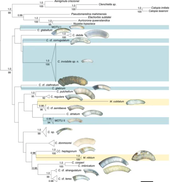
In our phylogenetic analyses Caecidae form a well-supported clade (1.0 PP, 99% BS; Fig. 3). The two established genera Caecum and Meioceras, however, are not recovered as reciprocally monophyletic but instead species of Meioceras group among Caecum species in different parts of the tree (Fig. 3, taxa highlighted in yellow): M. nitidum sis-ter to C. heptagonum (0.96 PP), and M. cubitatum sissis-ter to C. cf. semilaeve (no statisti-cal support). The phylogeny groups the Caecidae into 21 clades which show moderate to high support values ranging from 0.95 PP/85% BS to full support (Fig. 3). Other clades are only statistically supported by one analysis (C. regulare, 92% BS) or do not have statistical support (C. donmoorei). The sister group relationships of C. pulchellum and C. regulare (1.0 PP, 95% BS), and C. cooperi and C. imbricatum (1.0 PP, 100% BS) are well supported; otherwise, deeper nodes and higher-level relationships among clades are not supported. In agreement with the molecular data, C. pulchellum and C. regulare as well as C. cooperi and C. imbricatum show morphological similarities in shell ornamentation and microsculpture. Inconspicuous specimens with smooth shells and few characters that were morphologically ascribable to C. glabrum or the American
Pa-cific look-alikes like C. glabriforme are polyphyletic and form four lineages separated by branches of comparable length to morphologically distinct species (Fig. 3, highlighted in blue). These lineages are distinct from C. glabrum from the North Atlantic (Table 3) included in the analyses, indicating the presence of morphologically cryptic species in this ‘C. glabrum species complex’.
Figure 3. Optimal ML tree of the concatenated 28S rRNA, 16S rRNA and COI genes partitioned by genes and COI codon positions. Bootstrap values (below nodes) of the ML analysis are shown for values > 80% and posterior probability support (above nodes) of the BI analysis are shown for values > 0.95. Specimens previously classified as Meioceras are indicated in yellow color. Smooth, translucent specimens, lacking diagnostic features and summarized in the ´Caecum glabrum-like complex` are indicated in blue.
C. = Caecum, M. = Meioceras, MOTU I/ MOTU II = molecular operational taxonomic unit within the
Molecular species delineation
The methods that were used for species delineation are largely congruent with regard to the assignment of taxa to molecular operational taxonomic units (MOTUs), how-ever individual analyses deviate and evidently differences occur due to incomplete sampling of one of the markers (Fig. 4, Table 1). Both PTP/ bPTP and GMYC (single threshold) delimit 21 MOTUs for the concatenated dataset of COI and 16S rRNA genes (excluding the species whose sequences were retrieved from NCBI GenBank, i.e. North Atlantic C. glabrum and Japanese C. glabellum). These results are in concord-ance with the preliminary species hypotheses based on morphological investigation and the molecular phylogenetic tree (Fig. 3) with the exception of additional splits of C. debile and C. regulare into two distinct MOTUs each. Caecum cf. teres resulted in a single species for 16S rDNA alone, and the Bayesian implementation of bPTP split M. nitidum into two separate species based on the 16S rRNA genes (however, support value for the split is 0.501%). The multiple threshold analyses in GMYC additionally splits C. invisibile sp. nov. of the ‘C. glabrum complex’ into two MOTUs, as does TCS but into differing entities. In analyses of individual datasets (numbers not directly comparable due to missing data) ABGD identified 15 MOTUs in our 16S rRNA gene dataset (Fig. 4), while the COI dataset resulted in a hypothesis of 10 MOTUs independent of the application of the JC69 or the K80 model. In comparison to the other methods, TCS appears to oversplit MOTUs (see e.g., TCS analyses of the COI of C. donmoorei in Fig. 4). The algorithm of this haplotype-network software splits the 16S rDNA dataset into 19 independent haplotype networks, while it recovered 13 networks for the COI dataset (Fig. 4). Additionally, TCS also splits MOTU II of the ‘C. glabrum-like complex’ into two networks and C. donmoorei into three independ-ent networks based on 16S rRNA sequence data. Haplotype networks divided C. cf teres and C. cf. strangulatum into two unconnected networks. However, the split is not congruent with the two monophyletic sister populations of the species tree (Fig. 4). In summary, we consider only splits relevant, which are supported by at least two dif-ferent analyses or markers, singular deviating signal might either resemble errors in analyses or might be informative in population analyses (for more details see remarks in Systematics section).
Taxonomy of Central American Caecidae
Systematics
Class Gastropoda Cuvier, 1797 Family Caecidae Gray, 1850 Genus Meioceras Carpenter, 1859
Type species. Caecum nitidum Stimpson, 1851 from Florida by subsequent designa-tion, Carpenter (1859): 438.
Based on the molecular phylogeny, specimens identified as Meioceras nitidum and M. cubitatum both group among Caecum species and should therefore be transferred to this genus. However, considering that only one M. nitidium is statistically supported, in the interest of taxonomic stability this finding is pending further molecular studies, once additional material is available, preferably including material from the type localities.
Meioceras nitidum (Stimpson, 1851)
Caecum nitidum Stimpson, 1851 in Stimpson (1851a): 112. Type locality: Florida. Caecum lermondi Dall, 1924: 7; Caecum rotundum de Folin, 1868: 49, pl. 5, fig. 2;
Meioceras bitumidum de Folin, 1869: 9, fig. 4; Meioceras carpenteri de Folin, 1869: 8, 9, fig. 3; Meioceras cingulatum Dall, 1892: 302, pl. 16, figs 6, 7; Meioceras con-tractum de Folin, 1874: 213, t. 2, pl. 4, fig. 7; Meioceras coxi de Folin, 1869: 13, fig. 9; Meioceras crossei de Folin, 1869: 11, 12, fig. 7; Meioceras deshayesi de Folin, 1869: 11, fig. 6; Meioceras elongatum de Folin, 1881: 17, pl. 1, fig. 9; Meioceras fischeri de Folin, 1870: 188, pl. 26, figs 3, 4; Meioceras imiklis de Folin, 1870: 189, pl. 26, figs 5, 6; Meioceras leoni Bérillon, 1874: 251, pl. 5, fig. 3; Meioceras moreleti de Folin, 1869: 10, fig. 5; Meioceras subinflexum de Folin, 1869: 165, pl. 23, fig. 8; Meioceras undulosum de Folin, 1869: 12, fig. 8.
Material examined. French Antilles • 1 (Fig. 5A–D); Martinique, Anse Noir; 14.528, -61.088; depth 6 m; 6 Sep 2016; MNHN Madibenthos exped.; Stat. AB102; GenBank: MT704298, MT731714; MNHN-IM-2013-72087a • 1; same collection data as for preceding; GenBank: MT704299, MT731715; MNHN-IM-2013-72087b.
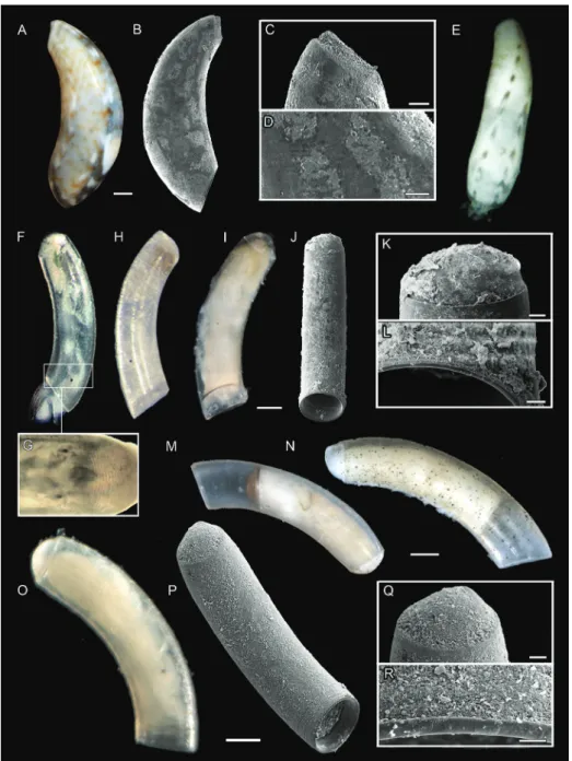
Shell morphology. Shell translucent, glossy. Light brown zig-zag pattern covering entire shell in rings with irregular white dorsal patches (Fig. 5A). Bulbous tube, taper-ing towards aperture and posterior end. Maximum width at about one third of shell length. Slightly more bowed towards aperture. Septum flat, with triangular, pointed mucro (Fig. 5C). No sculpture or microsculpture diagnostic features (Fig. 5D).
Remarks. Meioceras and in particular “M. nitidum” has a complex taxonomic his-tory involving at present 16 synonyms and several reallocations between Meioceras and Caecum (MolluscaBase 2019). Vannozzi (2017) highlighted the problems with the ambiguous type specimen of M. nitidum (Stimpson 1851a), which encouraged multiple novel descriptions (e.g., Carpenter 1858–1859; De Folin and Périer 1867), nowadays recognized as synonyms. Our two investigated specimens from Martinique are consistent with the meagre original description by Stimpson (1851a) based on specimens from Florida and several redescriptions based on material from the Carib-bean and southern America, now all accepted as M. nitidum (M. nitidum see Bandel 1996: 99, pl. 5, figs 1–7; M. contractum see de Folin and Périer 1875: pl. 9, fig. 7). Our specimens differ morphologically from several “M. nitidum” specimens from Central American waters all of which were described as a different species in the past but later synonymized with M. nitidum acknowledging intraspecific variability (i.e., M. elon-gatum, holotype accessible through the online catalogue of the MNHN
(MNHN-IM-2000-32923) and M. subinflexum (see de Folin and Périer 1867: pl. 23, fig. 8). Molecular comparison of specimens spanning the morphological and geographical range is needed to clarify the species status and distribution of the species.
Meioceras cubitatum de Folin, 1868
Meioceras cubitatum de Folin, 1868 in De Folin and Périer (1867–1871): 50, pl. 5, fig. 4. Type locality: Baie de Bahia [Bahia Bay, Brazil].
Figure 4. Molecular based species delimitation of Central American Caecidae. Guide tree used for PTP and bPTP based on the optimal likelihood tree of the concatenated three-marker dataset. Color codes indicate our preliminary species hypothesis derived from the phylogenetic tree. Color bars reflect the species delimitation suggested by the four consulted species delimitation programs (including ML and Bayesian implementation for PTP and single and multiple threshold for GMYC). Bars are missing where no sequence data obtained.
Figure 5. A–D Meioceras nitidum, specimen MNHN-IM-2013-72087 A light microscopic picture B SEM scan C SEM close-up of mucro and D microsculpture E M. cubitatum, specimen USNM 1618851 F, G C. cf. corrugulatum, specimen USNM 1618861 F light microscopic picture G light mi-croscopic close-up of microstructure H MOTU II, specimen USNM 1618853 I–L MOTU I, specimen ZSM-Mol-20200039 I light microscopic picture J SEM scan K SEM close-up of mucro and L microscu-lpture M C. glabrum, specimen Mol-20200096 N C. glabellum auctt. non Adams, specimen ZSM-Mol-20200074 O–R C. invisibile sp. nov., holotype ZSM-Mol-20100320 O light microscopic picture P SEM scan Q SEM close-up of mucro and R microsculpture. Scale bars: 10 µm (R); 20 µm (C, K, L, Q); 50 µm (D, E) 100 µm (I, J, O, P); 200 µm (A, B, M, N).
Caecum cubitatum (de Folin, 1868): 19; Meioceras tenerum de Folin, 1869: 24.
Material examined. Belize • 1 (Fig. 5E); Carrie Bow Cay; 16.8021, -88.0767; depth 31 m; 22 Jan 2010; USNM Belize 2010 exped.; Stat. CBC15; DNA voucher; Gen-Bank: MT727067, MT731709; USNM 1618851.
Shell morphology. Shell opaque white and solid. Mottled grayish pattern over whole shell, two rows of distinct brown dashes along dorsal side (Fig. 5E). Specimen approx. 2 mm. Tube not evenly curved but appears bulbous and is rounded strongly towards aperture, decreasing towards mucro. Mucro thin and sharp.
Remarks. Our molecular phylogenetic results delimited M. cubitatum as a separate species, despite similarities to M. nitidum in its bulbous shell shape and pattern. Sur-prisingly, our molecular analyses do not retrieve these morphologically similar Meioceras species as a monophyletic entity but suggest independent origin within Caecum. Mor-phological differences towards M. nitidum (characterized above) are a more slender shell with more pronounced curvature towards the anterior end and the opaque color of the present individual. We assigned the specimen to Meioceras cubitatum sensu de Folin, 1869 from Bahia, Brazil (dos Santos Gomes and Absalão 1996: 523, figs 12–15; De Folin and Périer 1867: pl. 5, fig. 4; Redfern 2001: fig. 178A, B) = Meioceras cor-nucopiae Carpenter, 1859 (from the West Indies, exact type locality unknown) sensu Lima et al. (2015: 3, fig. 7). Nevertheless, this Meioceras species likely should also be reallocated to the genus Caecum based on the results of our phylogenetic analyses.
Genus Caecum J. Fleming, 1813
Type species. Dentalium trachea Montagu, 1803 from England by subsequent desig-nation, Gray 1847: 203.
Cryptic lineages revealed in molecular analyses
Twenty-four specimens from Central American waters are smooth and glossy without ornamentation except for occasional growth lines (i.e., possess few shell characteristics), but vary in adult shell length between 0.7 and 2.5 mm (Figs 3, 5F–L, O–R). Morphologically, these specimens all closely resemble Caecum glabrum (Montagu, 1803) which is one of the best-known species of caecids, and abundant in the northern Atlantic (Montagu 1803; Wood and Harmer 1848; Götze 1938; Chambers 2009). Caecum glabrum was originally described from Biddlesford Bay and Barnstable, Devon, England, the included sequences from GenBank (see Table 3) originates from specimens collected in Norway, but own unpublished data from Roscoff, northern France, supports the wide distribution range of C. glabrum along European coastlines based on molecular data. We refer to cryptic species with simple shells lacking characteristic features as ‘Caecum glabrum-like’ species complex. In previous works, specimens similar to C. glabrum have also been described from the Pacific (C. glabellum as Brochina glabella A. Adams, 1868 from Akashi, Japan, C. glabriforme Carpenter,
1857 and C. corrugulatum Carpenter, 1857, both from Mazatlán, Mexico and C. parvulum de Folin, 1867 from Panama Bay, Brazil), have been reported and described from Japan, Hawaii, and central America (Carpenter 1855–1857; Adams 1868; Lightfoot 1993b; Pizzini et al. 2007, Takano and Kano 2014). Our study clearly shows an independent evolutionary origin of C. glabrum from the northeast Atlantic and C. glabellum from Japan, and the cryptic C. glabrum-like MOTUs from central America (see Fig. 3, ‘C. glabrum-like-complex’ highlighted in blue color). Our molecular species delineation revealed a minimum of four cryptic MOTUs (see above, Fig. 4).
Caecum cf. corrugulatum Carpenter, 1857
Caecum corrugulatum Carpenter, 1857: 327, pl. 37, figs 375, 1547. Type locality: Mazatlán, 1 sp. off Chama [Mexico].
Material examined. Panama • 1 (Fig. 5F, G); Achotines; 7.6207, -80.0013; depth 12 m; 29 Feb 2016; USNM Achotines2016 exped.; Stat. PA14; DNA voucher; Gen-Bank: MT727069, MT731722; USNM 1618861.
Shell morphology. Shell color whitish translucent. Tube regularly curved, shape equal in width but bears prominent edge at transition to septum (Fig. 5F). Septum round and blistered lacking a mucro. Aperture equally wide as tube with straight edge. Sculpture ap-pears completely smooth but shows fine concentric ribs at higher magnification (Fig. 5G).
Remarks. We assigned the specimen collected in the Pacific coast of Panama to C. cor-rugulatum based on the description of Carpenter (1858–1859) who already highlighted its similarity with another inconspicuous species (C. glabriforme). Both species are de-scribed from the same geographic area (Mazatlán, Pacific coast of Mexico) and resemble the C. glabrum-like type: translucent, blistered septum without mucro, smooth, however slightly bigger than the eponymous C. glabrum from European waters. Caecum corrugu-latum, can be distinguished by microsculptural concentric wrinkles, which could be ob-served with higher magnification in our specimen. So far, only C. glabriforme was recorded in Pacific Panama (Lightfoot 1993b) and recollection at the type locality is needed to 1) confirm the validity of both co-occurring species and reject conspecificity and 2) to con-firm their putative distribution range from Mexico to Panama and exclude the possibility of further cryptic species among C. glabriforme and C. corrugulatum species along the Pacific Coast of Central America (as discovered herein for the Atlantic Coast, see below).
Caecum invisibile Egger & Jörger, sp. nov.
http://zoobank.org/4183679F-44F4-4817-A2E1-7325687E5F0A
Material examined. Holotype Belize • 1 (Fig. 5O–R); Carrie Bow Cay; 16.8015, -88.0790; depth 10 m; 14 Jan 2010; USNM Belize2010 exped.; Stat. CBC1b; DNA voucher; DNA bank: r462p15f2t91; GenBank: MT727055, MT704268, MT731697; ZSM-Mol-20100320. Paratypes Belize • 1; same data as for holotype; DNA voucher;
DNA bank: r462p14f2t91; GenBank: MT727054, MT704267, MT731696; Mol-20200109. Belize • 2; same data as for holotype; Mol-20200111, ZSM-Mol-20200112. Other material Belize • 10; same data as for holotype; DNA voucher; GenBank: MT727056–MT727065, MT704269–MT704279, MT731698–
MT731707; USNM 1618839, USNM 1618840, USNM 1618841, USNM
1618842, USNM 1618843, USNM 1618846, USNM 1618847, USNM 1618848, USNM 1618849. Panama • 1; Bocas del Toro; 9.2140, -81.9318; depth 8.5 m; 5 Jun 2010; USNM BRS2010 exped.; Stat. BRS104; DNA voucher; GenBank: MT727049,
MT704264, MT731689; USNM 1618856. • 1; Bocas del Toro; 9.3507, -82.1724; depth 3 m; 13 Jun 2010; USNM BRS2010 exped.; Stat. BRS200; DNA voucher; GenBank: MT727052, MT731694; USNM 1618859.
Molecular diagnostic characters. see Table 4.
Morphological description. All investigated specimens were very similar in ap-pearance, with little or no variation in shell morphology. Shell completely translucent. Length 0.8 mm long, width 0.2 mm (holotype, Fig. 5O). Tube regularly curved, shape equal in width but bears prominent edge at transition to septum, edge with smaller diameter. Septum round and blistered lacking a distinct mucro. Septum slightly inclin-ing towards the left, dorsal side in holotype with slight variation between the speci-mens. Aperture equally wide as tube with straight edge. Sculpture appears smooth, only with faint growth lines (Fig. 5R). Whitish translucent body visible through trans-lucent shell. Operculum transtrans-lucent, slightly tinted yellowish. Radula formula shows taenioglossate pattern 2.1.1.1.2. with very small central rhachidian tooth. Large lateral teeth oriented towards the rhachidian tooth. Marginal teeth finer, outer marginal teeth are scoop-like curved. All the specimens investigated are adults based on the cylindrical shape of the tube and the shape of the aperture showing a reflected lip without cutting edge, which is normally present in immature specimens.
Etymology. The Latin adjective invisibile (invisible, unable to be seen) refers to the minute size of specimens, the translucent color of its shell, its hidden lifestyle between sand grains, and its taxonomic crypsis.
Table 4. Type 1 characters and type 2 characters (Kühn and Haase 2019) for COI, 16S rRNA and 28S rRNA sequence data (no type 2 characters for 16S rRNA and 28S rRNA present) of C. invisibile sp. nov.
COI 16S rRNA 28S rRNA
Type 1 Type 2 Type 1 Type 2 Type 1 Type 2
Position States Position States Position States Position States Position States Position States
15 A 501 T 7 C 378 G 595 CA 171 G 450 G 9 C 392 A 267 G 32 T 414 T 279 T 93 G 426 C 300 G 97 T 515 T 104 T 598 A 191 T 612 G 223 G 649 A 227 G 664 T 247 C
Distribution. Type locality: Carrie Bow Cay, Belize. (16.8015°N, -88.0790°W, -10 m). Distributed in Central American Atlantic from Carrie Bow Cay, Belize to Bocas del Toro, Panama. Interstitial in coarse biogenic sediments (calcareous sand and shell hash), shallow subtidal at ten meters’ depth.
Remarks. Caecum invisibile sp. nov. is described as a new species based on mo-lecular diagnostic characters, which show it as distinct from the European C. glabrum (Fig. 5M), as well as the morphologically similar C. corrugulatum (Fig. 5F, G) from the Central American Pacific and C. glabellum from Japan (Fig. 5N).
MOTU I
Material examined. Panama • 1 (Fig. 5I–L); Bocas del Toro; 2010; USNM BRS2010 exped.; DNA voucher; DNA bank: r462p13f2t91; GenBank: MT704263, MT731688; ZSM-Mol-20200039.
Morphological characterization. Shell size 1.3 mm long, 0.3 mm wide. Translu-cent, with whitish body. Tube regularly curved and equal width. Septum hemispherical (Fig. 5K). Aperture straight, with lip indicating an adult specimen. Operculum brown-ish. No sculpture or microsculpture diagnostic features (compare Fig. 5L).
Remarks. MOTU I is highly similar to the European C. glabrum (Fig. 5M) and Caecum invisibile sp. nov. (Fig. 5O–R). However, MOTU I shows some small mor-phological differences such as a bigger shell size and a tiny rim at the aperture (Fig. 5L) which is absent in C. glabrum. The septum is further completely round and blistered (Fig. 5K), whereas the one of Caecum invisibile sp. nov. slightly inclines (Fig. 5Q). MOTU I is based on a singleton and an incomplete molecular dataset, lacking COI sequence data. Additional material from the same locality is necessary to justify proper species description in future research.
MOTU II
Material examined. Panama • 1 (Fig. 5H); Bocas del Toro; 9.4333, -82.347; depth 3 m; 5 Jun 2010; USNM BRS2010 exped.; Stat. BRS101; DNA voucher; GenBank:
MT727046, MT704262, MT731687; USNM 1618853. • 1; Bocas del Toro; 9.3507, -82.1724; depth 15 m; 13 Jun 2010; USNM BRS2010 exped.; Stat. BRS110; DNA voucher; GenBank: MT727051, MT731693; USNM 1618852. • 1; Bocas del Toro; 9.3507, -82.1724; depth 3 m; 13 Jun 2010; USNM BRS2010 exped.; Stat. BRS200; DNA voucher; GenBank: MT727053, MT731695; USNM 1618860.
Morphological characterization. Shell size unknown. Translucent, with trans-lucent body. Tube regularly curved, slightly increasing in diameter towards aperture. Septum round, slightly flattened (Fig. 5H). Aperture straight. No sculpture visible, microsculptural data missing.
Remarks. MOTU II is based on the molecular data of three specimens; however, we unfortunately lack SEM scans and thus microsculptural data of the shell and light
microscopic images are only available for one specimen (Fig. 5H). This specimen is a juvenile, and due to uncertainty with regards to adult ornamentation of the shell, and its possible identity with an already described species, we refrain from providing a formal description based on the available material only.
Adding barcodes to known Central American Caecidae
Caecum heptagonum Carpenter, 1857
Caecum heptagonum Carpenter, 1857: 319, t. 1524. Type locality: Mazatlán [Mexico].
Material examined. Panama • 1 juv. (Fig. 6B–E); Achotines; 7.6349, -79.9968; depth 10 m; 6 Mar 2016; USNM Achotines2016 exped.; Stat. PA23a; DNA voucher; DNA bank: r462p4f2t91, GenBank: MT704283, MT731717; ZSM-Mol-20200030. • 1 juv. (Fig. 6A); same collection data as for preceding; DNA voucher; GenBank:
MT704291, MT731726; USNM 1618866. • 1 juv.; same collection data as for pre-ceding; Mol-20200116. • 1 juv.; same collection data as for prepre-ceding; ZSM-Mol-20200117. • 1 juv.; Achotines; 7.6346, -79.9965; depth 10 m; 6 Mar 2016; USNM Achotines2016 exped.; Stat. PA23b; ZSM-Mol-20200115.
Shell morphology. In juvenile specimens, shell fragile, translucent brownish color. Tube doubles diameter towards aperture, with a moderate curvature in anterior half, increasing distally in curvature. Septum level beneath cutting plane, slightly rising to-wards mucro (Fig. 6D). Mucro slender finger-like shape (Fig. 6D). Aperture fragile and partly broken. Shell sculptured by seven longitudinal ridges with transverse ribs cross-ing, knobs at intersections, ridges less prominent towards posterior. Microsculpture of fine rugose longitudinal stripes, noticeably increasing in width on transversal rings in comparison to interspaces (Fig. 6E).
Remarks. Due to the characteristics of the heptagonal tube with the transversal rings, considered unique among caecids (Lightfoot 1993a), the investigated specimen could be unambiguously assigned to C. heptagonum. However, illustrations of C. hep-tagonum indicate a very thick shell with distinct differentiated aperture with inner bulge forming a round opening instead of the outer polygonal shape (Keen 1974; Pizzini et al. 1998: 142, figs 1–13) including an inner bulge in the aperture, forming a round opening instead of the outer polygonal shape which is absent in the rather thin and fragile investigated specimens. As our samples only comprised juvenile specimen, however, we can attribute this variation to the unfinished shell state.
Caecum imbricatum Carpenter, 1858
Caecum imbricatum Carpenter, 1858: 422, pl. 69, fig. 10. Type locality: “W. Indies [Carribean].
Caecum coronatum de Folin, 1867: 50–52, pl. 2, fig. 5; Caecum formulosum de Folin, 1869: 24–125, pl. 11, figs 9, 10 (with three varieties paucicostata, simplex and
sulcate); Caecum insigne de Folin, 1867: 52, 53, pl. 2, fig. 4; Caecum sculptum de Folin, 1881: 15, pl. I, figs 1, 2.
Material examined. Belize • 1 (Fig. 1, specimen to the left); Carrie Bow Cay; 16.8037, -88.0769; depth 31 m; 15 Jan 2010; USNM Belize2010 exped.; DNA voucher; GenBank: MT727051, MT704281; USNM 1618850. Panama • 1; Bo-cas del Toro; 9.4333, -82.3467; depth 3 m; 5 Jun 2010; USNM BRS2010 exped.; DNA voucher; GenBank: MT704261; USNM 1618852. • 1; Bocas del Toro; 9.3222, -82.2215; depth 4.5 m; 5 Jun 2010; USNM BRS2010 exped.; DNA voucher; Gen-Bank: MT727047; USNM 1618854. • 1; same collection data as for preceding; Gen-Bank: MT727048; USNM 1618855.
Shell morphology. Shell opaque, yellowish. Lighter color in interspaces, darker colored ridges of prominent rhombic pattern (Fig. 1, first specimen from left). Tube evenly narrows towards posterior end, which is approximately half as wide in diameter as aperture. Septum flat, triangular shaped, strongly pointed mucro (Fig. 1, first speci-men from left). Rhombic pattern consisting of distinct longitudinal ridges crossed by axial ridges, pattern more distinct towards aperture, last row forms bumps at intersec-tions ((Fig. 1, specimen to the left).
Remarks. See remarks on C. cooperi. “ after paragraph on shell morphology of C. imbricatum.
Caecum cooperi S. Smith, 1860
Caecum cooperi S. Smith, 1860: 154–155. Type locality: northern part of Gardiner’s Bay, four or five fathoms [United States].
Caecum costatum A. E. Verrill, 1872: 283, pl. 6, fig. 6, Caecum smithi Cooper, 1872.
Material examined. French Antilles • 1 (Fig. 6F–I); Guadeloupe; 16.3558, -61.5965; depth 2 m; 24 May 2012; MNHN KARUBENTHOS exped.; Stat. GS32; GenBank: MT704297, MT731713; MNHN-IM-2019-32.
Shell morphology. Shell opaque, with whitish diffuse patterns. Size > 2 mm, tube narrow and elongated, curvature increasing towards posterior end (Fig. 6F, G). Septum flat, triangular shaped, strongly pointed mucro (Fig. 6H) similar to mucro in C. imbricatum. Prominent and conspicuous shell ornamentation, minimum of 20 longitudinal strings of beads, more pronounced towards aperture (Fig. 6I).
Remarks. Our molecular species delimitation separates C. cooperi and C. imbricatum into two independent evolving sister species (see Figs 3, 4). This is supported by (minor) morphological differences such as a finer but more pronounced bead-like ornamentation in C. cooperi in comparison to flattened squarish and less frequent longitudinal pattern. The included barcodes and distinguishing diagnostic features should help to overcome previous taxonomic uncertainty suggestive of synonymy (see C. imbricatum sensu Moore (1972: 888, fig. 6)). The putative synonymy of
Figure 6. A–E Caecum heptagonum A specimen USNM 1618866 juvenile specimen with larval shell still attached B–E specimen ZSM-Mol-20200030, juvenile specimen already resembling closely the adult form B light microscopic picture C SEM scan D close-up of mucro and E microsculpture F–I C.
coop-eri, specimen MNHN-IM-2019-32 F light microscopic picture G SEM scan H close-up of mucro and
I microsculpture whole specimen and close-up of mucro and microsculpture J–M C. debile, specimen MNHN-IM-2019-27 J light microscopic picture K SEM scan L close-up of mucro and M microsculpture N–Q C. striatum, specimen ZSM-Mol-20100322 N light microscopic picture O SEM scan P close-up of mucro and Q microsculpture R–U C. clathratum, specimen MNHN-IM-2019-17 R light microscopic picture S SEM scan T close-up of mucro and U microsculpture. Scale bars: 10 µm (D, E, I, Q); 20 µm (H, M, P, U); 100 µm (A, B, C, L, N, O, T); 200 µm (F, G, J, K whole specimen); 300 µm (R, S).
C. insularum Moore, 1969 and C. imbricatum (compare C. imbricatum sensu Tunnell et al. (2010: 144) = C. insularum sensu Moore (1970: 370, fig. 1A, B) needs to be tested by molecular data, ideally based on material recollected from the type localities.
Caecum debile Verrill & Bush, 1900
Caecum debile Verrill & Bush, 1900: 538. Type locality: Bermuda, Ship Channel and Bailey Bay, in 12 to 40 feet.
Material examined. French Antilles • 1 (Fig. 6J–M); Guadeloupe; 16.3558, -61.5965; depth 2 m; 24 May 2012; MNHN KARUBENTHOS exped.; Stat. GS32 GenBank: MT704295, MT731711; MNHN-IM-2019-27a. • 1; same collection data as for preceding; GenBank: MT704296, MT731712; MNHN-IM-2019-27b.
Shell morphology. Color whitish, slightly translucent. Specimens about 2.0 mm long, 0.5 mm wide. Tube of adult specimen 2.1 mm long, 0.5 mm wide. Tube evenly curved and evenly wide over entire length (Fig. 6J, K). Septum separated by sharp rim from tube and hemispherical without protruding mucro (Fig. 6L). Aperture with protruding ring. Shell structured by longitudinal striae clearly observable via LM. Mi-crosculpture of fine wavy striation (Fig. 6M).
Remarks. The present specimens were assigned to C. debile based on the characteristic microsculpture (see Absalão and Gomes 2001). Morphologically, C. debile might present a synonym of C. infimum, de Folin, 1867 (de Folin and Périer 1867: 26, pl .3, fig. 2) but, in awareness of cryptic species we refrain from synonymizing until C. infimum from the type locality is available for molecular analyses. Some species delineation analyses, separate C. debile into two independent species (Fig. 4), which might indicate a putative speciation, but more data on the genetic variability is needed to exclude the presence of an artefact in analyses.
Caecum striatum de Folin, 1868
Caecum striatum de Folin, 1868 in De Folin and Périer (1867–1871): 49, pl. 5, fig. 3 (with variety obsoleta de Folin, 1874). Type locality: Baie de Bahia [Bahia Bay, Brazil].
Material examined. Belize • 1; Carrie Bow Cay; 16.8024, -88.0776; depth 19 m; 25 Jan 2010; USNM Belize2010 exped.; Stat. CBC24; DNA voucher; GenBank:
MT727061, MT704275; USNM 1618845. • 1 (Fig. 6N–Q); same collection data as for preceding; ZSM-Mol-20100322.
Shell morphology. Shell translucent with mottled ochre and white marbling (Fig. 6N). Size 1.5 mm in length and 0.3 mm in width with thick shell (= 10 µm at aperture). Tube curved regularly with equal width at posterior and anterior end. Blistered, dome-shaped septum with prominent ring separating tube from septum
(Fig. 6P). Mucro central, flat and rounded, pointing slightly dorsal, hardly separated from septum. Aperture simple, straight. Sculpture not visible under the light micro-scope, i.e., shell appears rather smooth despite two faint transversal rings slightly no-ticeable close to aperture. SEM examination reveals longitudinal and slightly wavy structure (Fig. 6Q).
Remarks. Caecum striatum was identified based on a comparison with the material collected in the sampling region and dedicated as lectotypes by Absalão and Gomes (2001). Their microsculptural description of the type material does correspond to the longitudinal striation of our investigated specimen. Furthermore, shape, mucro, and the noticeably sharp aperture are identical to our specimen. A comparison with a speci-men of C. striatum pictured by Pastorino and Chiesa (2014: figs 10–16), also shows the same fine-lined microsculpture as our specimen. Type specimens of three highly similar species, namely C. johnsoni Winkley, 1809, C. antillarum Carpenter, 1858 and C. strigosum de Folin, 1867, are described from Central American waters. Differences can be compared in the reinvestigation of Absalão and Gomes (2001: 20, figs 39–41, 12, figs 11, 12 and figs 7, 8 respectively).
Caecum cf. clathratum Carpenter, 1857
Caecum clathratum Carpenter, 1857 in Carpenter (1855–1857): 322, pl. 34, figs 269, 1528. Type locality: Mazatlán [Mexico].
Material examined. French Antilles • 1 (Fig. 6R–U); Guadeloupe; 16.2235, -61.5305; depth 1 m; 2 May 2012; MNHN KARUBENTHOS exped.; Stat. GM01; GenBank: MT704294, MT731710; MNHN-IM-2019-17.
Shell morphology. Large, thick shell (3.0 mm length and 0.8 mm width) with and even curvature (Fig. 6R). Color opaque yellow brownish, entire specimen covered in dense dark periostracum. Septum triangular merged with pointed mucro (Fig. 6T). Aperture oblique and constricted. Shell with 21 strong and protruding sharp ribs and deep interspaces narrowing at aperture (Fig. 6R, S). Ribs and interspaces smooth without microsculptural diagnostic features (Fig. 6U).
Remarks. The specimen corresponds to C. clathratum, which differs from other ribbed Caecum species by its exceptional size, golden color and lack of microsculpture (compare with Lightfoot 1993a: 15, fig. 1 and a syntype collected by Carpenter available through the online catalogue of the Natural History Museum London catalogue number 1857.6.4.1528). However, the specimen is described and known only from the Eastern Pacific. Our herein investigated specimen from the Atlantic might thus present a (morphologically cryptic) sister species new to science, which potentially originated when populations were separated via the formation of the Isthmus of Panama. But molecular data of specimens collected from the Eastern Pacific is required to confirm the molecular identity or justify the description of a new species.
Caecum pulchellum Stimpson, 1851
Caecum pulchellum Stimpson, 1851 in Stimpson (1851b): 36, pl. 2, fig. 3. Type local-ity: New England, Buzzard’s Bay [New Bedford Harbor, Massachusetts].
Caecum capitanum de Folin, 1874: 227, 228, pl. 9, fig. 8; Caecum conjunctum de Folin, 1867: 46, pl. 4, figs 5, 6, Caecum curtatum de Folin, 1867: 20, pl. 2, figs 4, 5
Material examined. Saint Lucia • 1 (Fig. 7A, C, E, F); Soufriere Bay; 13.8494, -61.0675; depth 8–9 m; 19 Feb 2009; ZSM stuff leg.; DNA voucher; DNA bank: r462p-19f2t91; GenBank: MT727074, MT704300, MT731729; ZSM-Mol-20090485. • 1, juv. (Fig. 7B, D); same collection data as for preceding; DNA voucher; DNA bank: r462p20f2t91; ZSM-Mol-20200118.
Shell description. Color opaque whitish, slightly translucent, shell thick (Fig. 7A). Adult specimen 2.1 mm long, 0.5 mm wide. Tube slightly tapering, constricted at ap-erture, with thickened lip (Figs 7C). Septum slightly lower than posterior end of tube, rising, small and peaked mucro (Fig. 7E). Shell bears 27 squares transverse ribs of even width and equal interspaces, except two or three ribs which meld close to the aperture. Topmost ring sloped towards septum and smaller than others. Fine inconspicuous longitudinal microstriae cover ribs (Fig. 7F). Interspaces covered with organic material, therefore no microsculptural pattern visible.
Remarks. Our investigated specimens agree well with recent descriptions and geo-graphical records of C. pulchellum (e.g., Bandel (1996): 112, 113, pl. 6, figs 1–4, 6, pl. 7, figs 1, 2 from Columbia and Curaçao; Lightfoot (1992a): 143, fig. 2). The type specimen, however, is originally described from Buzzard’s Bay in Massachusetts, USA. A molecular comparison of specimen from the Caribbean and northeast America is needed to confirm the distribution range based on morphology.
Caecum regulare Carpenter, 1858
Caecum regulare Carpenter, 1858 in Carpenter (1858–1859): 428–429, pl. 69. Type locality: W. Indies (Woodward) [Caribbean].
Material examined. Panama • 1, juv.; Bocas del Toro; 9.2141, -81.9318; depth 8.5 m; 5 Jun 2010; USNM BRS2010 exped.; Stat. BRS108; DNA voucher; GenBank:
MT727050, MT731690; USNM 1618883. Belize • 1, juv. (Fig. 7H–K); Carrie Bow Cay; 16.7911, -88.0761; depth 15 m; 24 Jan 2010; USNM Belize2010 exped.; Stat. CBC22; DNA voucher; DNA bank: r462p17f2t91; GenBank: MT704280, MT731708; ZSM-Mol-20100321. • 1, juv.; same collection data as for preceding; ZSM-Mol-20200114.
Shell morphology. Shell translucent, color white to yellowish (Fig. 7H). Size 1.0 mm long, 0.4 mm wide. Tube curved regularly in moderate angle, tapering to-wards posterior, widening at aperture, thick lip (10 µm) (Fig. 7H, I). Septum slightly domed. Mucro slender and sharply pointed (Fig. 7J, but tip broken in specimen
ZSM-Mol-20100321). Sculpture consists of 32 marked and rounded transverse ribs, nar-rowing towards septum, with narrow deep interspaces (Fig. 7I). Microsculpture shows longitudinal fusiform lobes covering the ribs (Fig. 7K).
Remarks. We identified the specimens as C. regulare by referring to the drawings of Carpenter’s original description (Carpenter 1857; 1858–1859), the syntype ma-terial from the Natural History Museum, London, UK accessed through the online catalogue (catalogue numbers 1858.12.9.19, 1858.12.9.20, 1858.12.9.21) and the re-description of Moore (1972): 888, fig. 7. The conspicuous widening of the shell very close to the aperture in our specimen investigated can be interpreted as a character of a sub-adult growth stage (Bandel 1996), thus not contradicting the assignment to C. reg-ulare. A literature survey suggests at least one putative synonym of C. regulare, resp. Caecum planum, de Folin, 1874 (de Folin and Périer 1875: 277, t. 2, pl.10, figs 8, 9). However, the geographical distribution differs (C. regulare was originally described from the Caribbean and C. planum from Brazil), and there are no evident diagnostic differences to C. regulare. Hence, molecular data is needed for clarification. Further data also is needed for the complementary gene sequences (16S rRNA and COI) for the two investigated specimens. We consequently attribute the split of our two investi-gated specimens into distinct molecular species to missing data in our analyses.
Caecum donmoorei Mitchell-Tapping, 1979
Caecum donmoorei Mitchell-Tapping, 1979: 104, 105, figs 21, 22, 31, 32 Type locality: In 5 m of water in Sprat Baz, Water Island, USVI.
Material examined. Panama • 1, juv.; Bocas del Toro; 9.2141, -81.9318; depth 8.5 m; 5 Jun 2010; USNM BRS 2010 exped.; Stat. BRS108; DNA voucher; GenBank: MT704265,
MT731691; USNM 1618857. • 1; same collection data as for preceding; DNA voucher; GenBank: MT704266, MT731692; USNM 1618858. Belize • 1 (Fig. 7G); Carrie Bow Cay; 16.8015, -88.0790; depth 10 m; 14 Jan 2010; USNM Belize2010 exped.; Stat. CB-C1b; DNA voucher; GenBank: MT704274, MT731703; USNM 1618844.
Shell morphology. Shell opaque white and solid. Size large > 2.0 mm. Tube mod-erately curved and curvature stronger towards aperture (Fig. 7G). Septum blistered with pointed, sharp mucro (Fig. 7G). Aperture surrounded by three narrow thick rings. Sculpture consists of 25 distinct squarish ribs with wide, deep interspaces. No microsculpture available.
Remarks. The collected specimen USNM 1618844 closely resembles the descrip-tion of C. donmoorei from the Virgin Islands (Mitchell-Tapping 1979), however mi-crosculptural data for comparison is missing. Our molecular data suggests the spe-cies identity among specimens with less separated, strongly flattened rings and almost vanishing interspaces (e.g., in juvenile USNM 1618857). The same has been observed for the confusingly similar species C. quadratum, Carpenter (1855–1857: 322, pl. 34, fig. 370). It can also exhibit a considerable variety of shell morphologies (Lightfoot