UNIVERSIDADE DO
ALGARVE
RECOMBINANT PROTEINS IN DIFFERENTIATION OF
STEM CELLS
Ana Rita Fragoso Nascimento
Dissertação de Mestrado Mestrado em Ciências Biomédicas
Trabalho efetuado sob a orientação de Professor Doutor José Bragança
e co-orientação de
Professor Doutor Guilherme Ferreira
UNIVERSIDADE DO
ALGARVE
RECOMBINANT PROTEINS IN DIFFERENTIATION OF
STEM CELLS
Ana Rita Fragoso Nascimento
Dissertação de Mestrado Mestrado em Ciências Biomédicas
Trabalho efetuado sob a orientação de Professor Doutor José Bragança
e co-orientação de
Professor Doutor Guilherme Ferreira
Declaração de autoria de trabalho:
Declaro ser a autora deste trabalho, que é original e inédito. Autores e trabalhos consultados estão devidamente citados no texto e constam da listagem de referências incluída.
Ana Rita Fragoso Nascimento
Copyright ®
A Universidade do Algarve tem o direito, perpétuo e sem limites geográficos, de arquivar e publicitar este trabalho através de exemplares impressos reproduzidos em papel ou de forma digital, ou por qualquer outro meio conhecido ou que venha a ser inventado, de o divulgar através de repositórios científicos e de admitir a sua cópia e distribuição com objetivos educacionais ou de investigação, não comerciais, desde que seja dado crédito ao autor e editor.
I am using this opportunity to express my gratitude to everyone who supported me throughout the course of this Master Thesis.
I would like to express my special thanks to my supervisor Professor José Bragança, who has given me the opportunity to work in his laboratory. I would like to thank you for encouraging my research and for allowing me to grow as a research scientist. Your advice and support have been priceless.
To Dr. Guilherme Ferreira, I appreciate the support given during the orientation of this dissertation. The opportunity and knowledge acquired express my gratitude.
I would also like to thank to Professor Rui Martinho, Professor Eduardo Melo and Carlos Lopes, who were essential at some developmental stages of this work.
To my laboratory colleagues and friends from 1.12 and from 3.30, Gisela Oliveira, Ana Jesus, João Charneca, Vanessa Afonso, Eduarda Guerreiro and Carina Silva, for all the support given as well as the good and fun moments through this year. I would like to specially thank to João Santos for all his help and patience, his invaluably constructive criticism and friendly advice during the project work. I’m also very grateful to Catarina Águas, for all her help and incentive in the development of this thesis.
To all my friends for being present and giving me support through all this year, including the entertaining moments which allowed me to put aside the laboratory concerns and overcome them, but also for the patience in certain times along this dissertation year.
Last but not the least, I would like to express my gratitude to my family for believing in me and giving me the opportunity to get my MsC degree. A special word of thanks for the love, encouragement, and tolerance of Rúben, who has made all the difference in my life. Without his patience and sacrifice, I could not have completed this thesis.
Cardiomyocytes derived from embryonic stem cells (ESCs) offer a great alternative to generate a large number of cells with potential application in biomedical research and drug or toxicology screening. Although the differentiation efficiency can be improved by the genetic manipulation of ESCs to over-express cardiac-specific transcription factors, current protocols for differentiation are time consuming, have low yield, and lack of reproducibility. Protein transduction has been demonstrated as an alternative approach for increasing the efficiency of ESCs differentiation toward cardiomyocytes.
The objective of this work was to produce a recombinant chimeric protein with the intrinsic ability to transduce into cells. In this work, we successfully produced and purified a novel recombinant protein from bacteria, which is capable of overcome the cellular membrane barrier. This protein is composed by two domains: the human protein CITED2 fused at its N-terminal domain to a Protein Transduction Domain (PTD), rich in arginines, which confers biologically active proteins the capability to translocate across the membrane and deliver them inside the cell.
We showed that CITED2 recombinant protein, added to the culture medium of the cells, was successfully internalized, localized in the nucleus and functional. In mouse embryonic stem cells (mESC), the knockout of Cited2 gene impairs these cells to differentiate into cardiomyocytes. We showed that the supplementation, on the second day of differentiation, of CITED2 recombinant protein to the culture medium of Cited2 depleted cells, rescued the cardiogenic defects of these cells. On the other hand, the supplementation at the onset of differentiation, while the cells are still in a pluripotent state, suggested a delay in the overall differentiation process.
Although further studies are still required, the direct application of the developed recombinant protein suggests a capability to replace the endogenous protein effects, further suggesting that CITED2 protein has a relevant role in cardiogenesis and pluripotency.
Keywords: Cardiac differentiation, Cited2, Mouse Embryonic Stem Cells, Protein Transduction Domain, Recombinant protein.
A utilização de cardiomiócitos derivados de células estaminais embrionárias (ESC) permite-nos acesso a um grande número de células para investigação biomédica e uso de fármacos ou ensaios toxicológicos. Apesar de ser possível aumentar a eficiência de diferenciação através de manipulação genética sobre expressando fatores cardíacos específicos, os protocolos atuais são bastante morosos, têm baixo rendimento e são dificilmente reprodutíveis. A transdução proteica é um método alternativo para aumentar a eficácia de diferenciação de células estaminais embrionárias em cardiomiócitos.
O objetivo consistiu na construção de uma proteína quimérica com a capacidade intrínseca de transdução celular. Produzimos e purificámos esta proteína recombinante, com a capacidade de atravessar a membrana celular, sendo composta por dois domínios: a proteína humana CITED2, ligada através do domínio terminal-N a um Domínio de Transdução Proteica (PTD), rico em argininas, que confere a capacidade de transduzir proteínas biologicamente ativas e entregá-las no interior da célula.
Com este trabalho, conseguimos demonstrar que esta proteína recombinante CITED2, quando adicionada ao meio de cultura celular, atravessa a membrana celular, alcança o núcleo, e é funcional.
Em células embrionárias de ratinho (mESC), o knockout do gene Cited2 leva à diferenciação destas células em cardiomiócitos. A suplementação com a proteína recombinante CITED2, no segundo dia de diferenciação, no meio de cultura em que as células tinham sofrido a depleção de Cited2, tem a capacidade de compensar os defeitos na diferenciação cardíaca. Por outro lado, se o meio de cultura for suplementado na fase inicial de diferenciação (dia 0), enquanto as células ainda se encontram num estado de pluripotência, sugere um atraso no processo de diferenciação geral.
Apesar de serem necessários estudos mais aprofundados, a aplicação direta da proteína desenvolvida parece ter a capacidade de substituição dos efeitos causados pela proteína endógena, sugerindo assim que a proteína CITED2 desempenha um papel importante tanto em cardiogénese como em pluripotência.
Palavras-chave: Diferenciação cardíaca, Cited2, Células estaminais embrionárias de ratinho, Domínio de Transdução Proteica, Proteína recombinante.
As doenças cardiovasculares são a principal causa de morte nos países desenvolvidos, constituindo um problema de saúde pública que urge minorar. No entanto, o coração tem uma capacidade limitada de regeneração e quando uma lesão ocorre, este não consegue substituir o tecido cardíaco danificado por novo músculo cardíaco, pois os cardiomiócitos (células altamente diferenciadas do coração com propriedades contráteis) morrem quer por apoptose ou necrose do tecido. Assim, essas células são substituídas por fibroblastos que contribuem para a formação de tecido cicatricial, tornando o coração mais frágil, com as suas propriedades contráteis comprometidas.
Um dos principais objetivos da investigação biomédica tem sido o desenvolvimento de métodos para a geração de cardiomiócitos de novo, tanto in vitro como in vivo, tal como novas ferramentas terapêuticas e métodos para regenerar o músculo cardíaco. Tendo em conta a informação anterior, um modo de tentar investigar os mecanismos moleculares que contribuem para o desenvolvimento de doenças do miocárdio, é essencialmente gerar cardiomiócitos funcionais, uma vez que a sua utilização na descoberta de medicamentos e estudos de toxicidade seria altamente benéfico, pois iria permitir testar novas moléculas farmacológicas para o tratamento deste tipo de doença. A solução ideal seria obter novas células, com propriedades contráteis de modo a reparar o tecido danificado.
As células estaminais são consideradas uma das mais promissoras fontes de células para regeneração, sendo que vários tecidos humanos já foram propostos como uma fonte de células estaminais com potencial cardiogénico (e portanto, capaz de gerar novos cardiomiócitos). O transplante autólogo de células estaminais dos próprios pacientes constitui uma das melhores hipóteses propostas para a reparação eficiente de corações fragilizados por doenças cardíacas, principalmente devido ao potencial de populações de células estaminais (como as células progenitoras cardíacas (CPCs)) que podem contribuir para promover a regeneração cardíaca.
Relativamente à diferenciação celular em cardiomiócitos, para gerar células seguras para aplicações terapêuticas é essencial evitar integração viral, e portanto é fundamental reduzir e evitar a manipulação genética, tornando a produção e aplicação de proteínas recombinantes uma alternativa mais viável para a expressão do gene em estudo.
Cited2, um gene que codifica uma proteína nuclear, liga-se diretamente e com elevada
afinidade, à primeira região enriquecida com cisteína e histidinas (CH1) do p300 e CBP, estando presente em todos vertebrados. Vários estudos demonstraram que as variações/mutações no gene Cited2 está relacionada com defeitos cardíacos congénitos.
Foi observado que Cited2 é essencial na manutenção da pluripotência uma vez que regula diretamente a expressão de fatores de transcrição tal como Oct4, Sox2, Nanog, Klf4 e
Tbx3, que fazem parte da rede nuclear dos genes responsáveis pelo estado de pluripotência
nas ESC. Uma variação dos níveis de expressão destes reguladores de pluripotência dos níveis de expressão ótimos, levam à diferenciação das ESC. Vários estudos suportam esta hipótese, uma vez que a sobrexpressão de CITED2 em mESC suporta a autorrenovação destas células, mesmo na ausência de leukemia inhibitory factor (LIF). Pelo contrário, a ausência de CITED2 durante a diferenciação de mESC leva a um atraso em genes de autorrenovação, ativando vias de diferenciação hematopoiéticas, neuronais e cardíacas.
A entrega intracelular de macromoléculas biológicas (como por exemplo fármacos) constituem um campo importante da medicina, uma vez que muitas macromoléculas biológicas, como as proteínas, péptidos, e ácidos nucleicos têm demonstrado ser úteis para o tratamento de vários problemas de saúde. No entanto, a entrega de macromoléculas nos seus locais-alvo não é fácil, principalmente devido à estrutura da membrana celular eucariótica. Um dos sistemas que podem ser utilizados para superar este problema, é a aplicação de domínios de transdução proteicos (PTDs), que são capazes de transportar a moléculas associadas através da membrana e desta forma permitir a entrada das proteínas biologicamente ativas no interior da célula.
Assim sendo, o objetivo desta tese foi a produção de uma proteína recombinante com a capacidade de transdução celular. Neste trabalho, clonámos o gene Cited2 num plasmídeo, de modo a produzir uma nova proteína recombinante. Esta proteína consiste na fusão de CITED2 com um péptido rico em argininas, denominado PTD, através do domínio terminal-N. Este PTD tem a capacidade de atravessar a membrana celular e desta forma, seria capaz de transportar a proteína CITED2 para o interior da célula.
Assim, este projeto de investigação pode ser dividido em três etapas gerais: • Clonagem molecular dos fragmentos de DNA no plasmídeo pGEX-6P-1; • Produção e purificação das proteínas recombinantes;
• Realização de ensaios celulares para análise da entrada e efeitos na pluripotência e diferenciação cardíaca em células estaminais embrionárias de ratinho.
Tendo em conta o objetivo proposto, começámos por clonar os fragmentos de DNA desejados num plasmídeo que permitiria a posterior produção destas proteínas. Após a produção das proteínas recombinantes TAT-CITED2 e 8R-CITED2, estas foram purificadas através de FPLC: TAT-CITED2 e 8R-CITED2. No entanto, a validação funcional a nível celular foi testada apenas com a proteína 8R-CITED2 por falta de tempo.
Quando suplementada no meio de cultura, a proteína recombinante 8R-CITED2 demonstrou ter sido internalizada nas células. A atividade da mesma foi comprovada através de um ensaio de BiFC, onde se demonstrou a competitividade contra a proteína CITED2 endógena pelo domínio p300. Por último, em ensaios de diferenciação celular, a proteína recombinante apresentou um padrão semelhante à proteína endógena quando esta última é suprimida. Ao estudar o papel desta proteína recombinante, podemos concluir que esta tem a capacidade de restauração dos defeitos causado pelo knockout de Cited2 endógeno em mESC quando aplicada ao meio de cultura no segundo dia de diferenciação, e parece ter alguma influência no atraso da diferenciação cardíaca quando o meio é suplementado no dia 0.
Usando esta abordagem, esta proteína pode ser utilizada para expandir células estaminais embrionárias de ratinho num ambiente controlado e auxiliar no controlo da diferenciação destas células. Este método oferece melhores alternativas do que os métodos utilizados anteriormente, visto que envolvem transfeções que geralmente resultam em sistemas artificiais e níveis subótimos de CITED2. No entanto, pesquisas e ensaios adicionais são necessários para aumentar a eficiência e segurança desta proteína recombinante.
Acknowledgments ... iv
Abstract ... v
Resumo ... vi
Resumo Alargado ... viii
List of Contents ... xi
List of Figures ... xiii
List of Tables ... xv
Abbreviations and Acronyms ... xvi
I. Introduction ... 1
I.1. Cardiovascular Disease ... 1
I.2. Stem Cells ... 2
I.2.1. Differentiation ... 3
I.2.2. Cardiac differentiation ... 5
I.3. CITED2 ... 8
I.4. Recombinant Proteins ... 10
II. Aims of the Project ... 14
III. Materials and Methods ... 15
III.1. Molecular cloning ... 15
III.1.1. Restriction enzyme digest ... 16
III.1.2. Agarose gel electrophoresis ... 18
III.1.3. Gel purification ... 19
III.1.4. Dephosphorilation ... 20
III.1.5. Ligation reaction ... 20
III.2. Preparation of Chemically Competent cells... 21
III.3. Transformation of competent cells ... 22
III.4. Extraction of DNA ... 23
III.4.1. Miniprep... 23
III.4.2. Midiprep... 24
III.5. Recombinant Protein Production ... 25
III.5.1. Production of GST-8R-C2 and GST-TAT-C2 proteins ... 25
III.6. SDS-PAGE ... 26
III.7. Protein Purification ... 29
III.7.1. GST-3C Protease ... 29
III.7.2. GST-8R-C2 and GST-TAT-C2 ... 30
III.8. Protein Quantification: Bradford Assay ... 31
III.9. Western Blot ... 31
III.10. Cell Assays ... 32
III.10.2. Cellular entrance of Recombinant Protein ... 33
III.10.3. Fluorescence Immunocytochemistry ... 34
III.10.4. Bimolecular Fluorescence Complementation (BIFC) ... 36
III.10.5. Cardiac Differentiation - Hanging Drop Method ... 37
III.11. Statistical Analysis ... 38
IV. Results and Discussion... 39
IV.1. Construction of Expression Vectors ... 39
IV.2. Recombinant Protein Production ... 44
IV.2.1. Production and Purification of 3C Protease ... 44
IV.2.2. Production and Purification of GST-TAT-CITED2 and GST-8R-CITED2 protein ………47
IV.3. Cellular Entrance of Recombinant Protein ... 53
IV.4. Fluorescence Immunocytochemistry ... 54
IV.5. Competition essay between 8R-CITED2 and Endogenous Cited2 for p300 interaction….. ... 55
IV.6. 8R-CITED2 Rescues Cardiac Differentiation defects caused by Cited2-knockout… ... 57
V. Conclusion ... 61
VI. Future Prospects ... 63
VII. Bibliography ... 65
VIII.Appendices ... 71
Appendix A ... 71
Appendix B ... 72
Figure I.1 – Schematic overview of activation of endogenous multipotent cardiac stem cells by various means for myocardial repair. ... 4 Figure I.2 – Diagram illustrating sequential steps in differentiation of pluripotent stem cells to cardiomyocytes. ... 6 Figure I.3 – Schematic representation of different stem cell sources for cardiac regeneration. ... 7 Figure I.4 – Schematic representation of variety of cargo covalently linked to arginine-dependent protein transduction domains (PTDs).. ... 11 Figure III.1 – Schematic representation of the work developed in course of this thesis. ... 15 Figure IV.1 – Visualization of pGEX-6P-1 digestion product by BamHI in a 1% agarose gel. ... 39 Figure IV.2 – Visualization of the plasmid DNA isolated from positive colonies to pGEX-6P-1+TAT, digested with SacII and PstI in a 1% agarose gel.. ... 40 Figure IV.3 – Visualization of the plasmid DNA isolated from positive colonies to pGEX-6P-1+8R, digested with SacII and PstI in a 1% agarose gel. ... 41 Figure IV.4 – Visualization of pSB54, pGEXT and pGEXR double digested with BamHI and XhoI, in a 1% agarose gel in order to isolate and purify inserts and vectors ... 42 Figure IV.5 – Double digestion with BamHI and XhoI of pGEXTC2 and pGEXRC2 in a 1% agarose gel in 1X TAE, stained with GreenSafe, to confirm the insertion of CITED2 gene. . 43 Figure IV.6 – SDS gel stained with Coomassie Blue to verify the expression of the recombinant protein: GST-3C. ... 45 Figure IV.7 – Purification chromatogram of the recombinant protein GST-3C. ... 46 Figure IV.8 – SDS-Page analysis of sample purification steps stained with Coomassie Blue. ... 47 Figure IV.9 – SDS-Page gels stained with Coomassie Blue to verify the expression of the recombinant proteins (A) GST-TAT-CITED2 and (B) GST-8R-CITED2. ... 48 Figure IV.10 – Purification chromatogram of the recombinant protein 8R-CITED2 with on-column cleavage. ... 49 Figure IV.11 – Flow chart of the affinity purification procedure and GST-3C Protease cleavage of GST fusion proteins. ... 50 Figure IV.12 – SDS-PAGE gel stained with Coomassie Blue to monitor the purification process of TAT-CITED2. ... 51 Figure IV.13 – SDS-Page gel stained with Coomassie Blue to monitor the purification process of 8R-CITED2. ... 51 Figure IV.14 – Western Blots to confirm the identity of the purified proteins: (A) TAT-CITED2 and (B) 8R-TAT-CITED2. ... 53 Figure IV.15 – Western Blot to evaluate the penetration of the recombinant protein 8R-CITED2. ... 54 Figure IV.16 – Fluorescence microscopy of MG5 cells treated with 20 µg/mL of 8R-CITED2 detected by immunocytochemical reaction against CITED2, 24h after supplementation, at 100X magnification. ... 55
Figure IV.17 – Representative fields of E14/T ESC co-transfected cells with plasmids expressing VEN-CH1 and VEC-CITED2 and quantification of the fluorescence detected with different concentrations of 8R-CITED2. ... 56 Figure IV.18 – Stages of mESC development. ... 57 Figure IV.19 – Comparison between C2fl/fl[Cre] treated with EtOH, 4HT and 4HT+8R-CITED2. ... 58 Figure IV.20 – Daily qRT-PCR analysis in aggregate differentiation of mESCs.. ... 59 Figure IV.21 – Comparison between C2fl/fl[Cre] treated with EtOH, 4HT and 4HT+8R-CITED2. ... 60
Table 1 – Reaction mix used for the usual restriction digestion of the pDNA. ... 17
Table 2 – List of restriction enzymes used for digestions, with their recognition site and optimal conditions. ... 18
Table 3 – Composition of 1% TAE Agarose gel. ... 19
Table 4 – Sample preparation prior agarose gel electrophoresis. ... 19
Table 5 – Ligation mix components. ... 21
Table 6 – Gene primers used in to prepare TAT and 8R inserts. ... 21
Table 7 – Sample preparation prior SDS-Page electrophoresis. ... 27
Table 8 – Composition of each layer of a 12% acrylamide SDS-Page gel. ... 28
α-MHC α-Myosin Heavy Chain Amp Ampicillin 16R Arginine hexadecamer 4HT 4-hydroxytamoxifen 4R Arginine tetramer 8R Arginine octamer
AR-CPPs Arginine-rich Cell Penetrating Peptides
BIFC Bifluorescence Complementation Method
bp Base pairs
BSA Bovine Serum Albumine
CBP cAMP-responsive element-biding protein
CH1 Cysteine-histidine rich
CIP Calf Intestine Phosphatase
Cited2 CBP/p300-Interating transactivators with ED rich tail 2
CPCs Cardiac progenitor cells
CPPs Cell-penetrating peptides
CSC Cancer Stem Cells
cTnT Cardiac isoform of Troponin-T
D Aspartic acid
DAPI 4’,6-diamidino-2-phenylindole
DNA Desoxirribonucleic acid
DTT Dithiothreitol
E Glutamic acid
E. coli Escherichia coli
EB Embryoid Body
ECM Extracellular matrix
EDTA Ethylenediamine tetraacetic acid
ESC Embryonic stem cells
FBS Fetal Bovine Serum
FHF First Heart Field
GMEM Glasgow Minimum Essential Medium
GSH Reduced Glutathione
GST Glutathione S-Transferase
hESCs Human Embryonic Stem Cells
HIF-1 Hypoxia-inducible factor
HRP Horseradish Peroxidase
InsF Insoluble Fraction
iPSCs Induced Pluripotent Stem Cells
IPTG Isopropyl β-D-1-thiogalactopyranoside
Isl1 Islet1
KO Knockout
LB Lysogeny broth
LIF Leukaemia Inhibitory Factor
MEFs Mouse Embryonic Fibroblasts
mESCs Mouse Embryonic Stem Cells
NSCLC Non-Small Cell Lung Cancer
O.D. Optical density
O.N. Overnight
PBS Phosphate Buffered Saline
pDNA Plasmid DNA
PMSF Phenylmethylsulfonyl fluoride
PTDs Protein Transduction Domains
qRT-PCR quantitative Reverse Transcriptase-Polymerase Chain Reaction
R Arginine
RF Reprogramming factors
RNA Ribonucleic acid
RT Room temperature
SDS-PAGE Sodium Sodecyl Sulphate-Polyacrylamide Gel Electrophoresis
SF Fraction/injected Sample
SHF Second Heart Field
TAE Tris-acetate
TAT-RFs Reprogramming factors fused with TAT
I.1. CARDIOVASCULAR DISEASE
Heart disease is the leading cause of mortality worldwide, resulting in 17.3 million deaths (31.5%) in 2013 up from 12.3 million (25.8%) in 1990, with insufficient therapeutic options and poor prognosis.1,2 Cardiovascular disease affects older adults, amongst which coronary artery disease and stroke account for 80% of deaths in males and 75% of deaths in females with cardiovascular disease.3
Despite significant advances in therapeutic modalities and prevention strategies, the high mortality rates associated with heart disease is fearsome. This has driven research into new therapeutic strategies including cardiac regenerative therapy as a new approach for severe cardiac diseases resistant to the conventional treatments.1
To investigate the molecular mechanisms leading to myocardial diseases in humans, it is essential to generate functional cardiomyocytes. Their use in drug discovery and toxicology studies would be highly beneficial, allowing that new pharmacological molecules for the treatment of cardiac disorders to be validated pre-clinically on cells of human origin.4
Recent studies from several laboratories have demonstrated that cardiomyocyte turnover occurs throughout life in mammals. However, most of the reports find a remarkably low annual post-natal cardiomyocyte renewal rate of approximately 1%, which increases modestly after injury but declines with age.2 The regenerative potential of the adult mammalian heart is very limited and when a lesion occurs it cannot replace the damaged cardiac tissue with new function muscle, since the cardiomyocytes die either by apoptosis or necrosis of the tissue. Then these cells are replaced by fibroblasts for scar tissue formation, turning the heart fragile, without good contractile properties.2,5,6
An optimal solution is to obtain new cells, with contractile properties to replace the injured tissue. However the access to the human heart tissue is very limited, and although human cardiomyocytes could be isolated from heart through biopsies, the procedure is complicated, invasive, unpractical to obtain viable cell preparations in large quantities (since they lack of proliferation capacity), and the majority of the cells acquired do not beat spontaneously.6,7
To overcome this problem, biomedical research fields have been developing methods for the generation of de novo cardiomyocytes, both in vitro and in vivo, together with novel therapeutic tools and approaches to regenerate cardiac muscle for diseased hearts.2,5,6
The development of new approaches has resulted in the isolation of cardiomyocytes from various new-born animals or the production of genetically engineered cell lines. Nevertheless, these models have significant limitations since they present basic physiological differences in comparison to human cardiomyocytes as well as high costs and ethical questions.8,9
Regarding cardiac cell-based treatments, stem cells are a promising cell source, which are being prioritized by scientists for basic research and clinical trials.10,11 Several different human tissues have already been proposed as a source of stem cells with cardiogenic potential (therefore capable to generate new cardiomyocytes) (e.g., fetal cardiomyocytes, adult cardiac progenitor cells, skeletal myoblasts, bone marrow-derived stem cells, adipose-derived stem cells, umbilical cord-derived stem cells, and pluripotent stem cells), and some methods to isolate and expand these cells have been developed aiming cardiac regenerative therapy.7,10–12 In this field, stem cell-derived cardiomyocytes would facilitate the discovery of small molecules promoting cardiomyocyte differentiation that could be used for the activation of endogenous cardiac stem cells in clinical settings.12
Stem cell autologous transplantation in patients is one of the candidates approach for efficient repair of impaired heart, due to the potential of stem cell populations (like cardiac progenitor cells (CPCs)) to promote cardiac regeneration and repair in experimental models and in patients with heart disease.2 Even so, there are complex and challenging pathobiological apprehensions. To make this possible, cell enhancement strategies need to be improved.13 For instance, patient derived cells undergoing a pre-treatment with small molecules or genetic modification which may contribute to an augmented recruitment of important factors which could enhance differentiation or other beneficial functions.11
I.2. STEM CELLS
Stem cells are often described by their capabilities of self-renewal, to be progenitors of tissue or organ specific cells and their ability to functionally reconstitute a given tissue in
These cells are present throughout the different developmental stages of an organism into and during the adult life and can be divided into two groups: embryonic and adult stem cells. Embryonic stem cells (ESC) are found only in the embryo and are classified as pluripotent, as they can give rise to any cell type of the three germ layers. On the other hand, adult stem cells are found in adult organisms, within fully differentiated tissues, and are classified as multipotent because they can only self-renew and differentiate into the different cells of the specific tissue where they reside or from which they were isolated.
Pluripotent ESC are derived from the inner cell mass of the blastocyst. Unlike adult stem cells, ESC have a very high degree of self-renewal capability, at least in part due to the fact that they express high levels of telomerase.14 This property makes them hold great potential as cell-based therapies to promote vascularization and tissue regeneration.15
At day 4 and day 7 to 10 of mouse and human development, respectively, cells of the blastocyst become lineage restricted, which means that they become committed to a germline and subsequently to a specialization within certain tissues. So, when working in vitro with ESC cells, we must to have in mind the need to maintain their pluripotency (preventing differentiation) and their self-renewal capacity using specific culture conditions.14,16
In the field of cardiac regenerative therapy, it was shown that skeletal myoblasts extracted from patients themselves, have the potential to improve the function of a failing heart when they are transplanted into the heart as myoblast sheets17. However, these cells are
not able to differentiate into cardiomyocytes, and therefore cannot replace the defective cardiomyocytes in a failing heart. A more likely efficient cell source in cardiac regenerative therapy could be if the transplantable cardiomyocytes could be prepared from ESCs.17,18
I.2.1. DIFFERENTIATION
Several strategies have been established to regulate the differentiation of ESC into all the three germ layers: the mesoderm, endoderm, and ectoderm.
There are several methods that can be used to differentiate ESC towards a specific fate (Figure I.1). One of these methods is genetic manipulation, which force the expression of some transcription factors and can lead to direct differentiation of ES cells toward specific lineages.
Figure I.1 – Schematic overview of activation of endogenous multipotent cardiac stem cells by various means for myocardial repair. (A) Molecules (e.g. growth factors, cytokines), (B)
noncardiac stem cells (e.g. bone marrow), (C) or gene therapy (e.g. micro-RNAs, gene transfer). Upon activation, resident endogenous cardiac stem cells can proliferate and mature into newly formed cardiac myocytes (yellow cardiac myocytes).
For example, the overexpression of HOXB4, a gene involved in hematopoietic lineage, significantly enhances the hematopoietic potential of mouse ESC (mESC) differentiation in vitro. Expression of GATA-6 and GATA-4 genes, involved in myocardial differentiation and function, induces mESC differentiation into the extra-embryonic endoderm, while GATA-4 overexpression alone enhances cardiogenesis and markedly increases the number of terminally differentiated beating cardiomyocytes.19
Another way to genetically control the differentiation of these cells is the use of micro-RNAs, since a single miRNA can target multiple pathways simultaneously, being the effects on gene expression very powerful.20–22 Recently, a combination of microRNAs (miRNAs: 1,
133, 208 and 499) has been identified as capable to reprogram murine fibroblasts to cardiomyocyte-like cells even though the efficiency rate being very low (about 1.5-7.7%).22
The most recent study combined a subset of four transcription factors (Gata4, Hand1, Tbx5 and myocardin) with two miRNAs (1, 133) to successfully reprogram human fibroblasts into cardiomyocyte-like cells. 23
The control of cell proliferation by growth factors, being often added to a medium to promote differentiation, is another approach that could affect survival of specific cell types. For instance, this direct differentiation can be implemented to overcome the heterogeneous cell mixture derived from differentiation cultures, and could be achieved by controlling the nutrient intake, the extracellular matrix (ECM), co-culturing with inductive cell types or adding a signaling molecule that have an impact on gene expression and cell proliferation.23
However, the formation of ESC aggregates is the most widely used, consisting in the formation of suspended spherical aggregates called Embryoid Bodies (EBs).18 This structure facilitates multicellular interactions, in which cell-cell contact exists and gap junctions may be established. EB formation is stimulated in the absence of leukemia induced factor (LIF) from the culture media or mouse embryonic fibroblasts (MEFs) feeder layer, changing from two-dimensional monolayer cell cultures to three-two-dimensional cell based structures in suspension.19,24 In the case of mESC, spherical ESC aggregates with morula-like structures formed in 2–6 days in suspension culture, and the removal of molecules which promote self-renewal from the culture medium, combined with cellular aggregation is an efficient way to differentiate them.19,25
I.2.2. CARDIAC DIFFERENTIATION
The ability of ESC to differentiate into spontaneously contracting cardiomyocyte-like cells has attracted substantial interest from the scientific community. Initially it was a process considered difficult to control, but now cardiomyogenesis in vitro is a process which, to a certain extent, could be effectively manipulated and directed in the future.12
Heart development involves a series of highly complex morphogenetic processes that are chronologically regulated by multiple phase-specific signals. The heart is the first organ to form in the embryo where its early function is essential to the circulation of nutrients and removal of waste. The construction of the heart is a complex process and involves the integration of different cell populations at distinct site as development proceeds.
Using chicken and mice embryos as models it has been demonstrated that the heart tissue is composed of three major mesoderm-derived cell lineages: the cardiac myocyte, the vascular smooth muscle, and the endothelial cell lineages. Few days after fertilization, the
three embryonic layers form, the endoderm, the ectoderm, and the mesoderm. The primitive streak is formed from primitive endoderm and is the origin of many tissues, being cardiac progenitors formed in the posterior primitive streak.
The cardiac mesoderm gives rise to the endocardium, the first heart field (FHF, which forms the atria, left ventricle, and the nodal conduction system), the secondary heart field (SHF, which forms the right ventricle, outflow tract, and part of the atria), and the proepicardial mesenchyme.26
The generation of cardiomyocytes from pluripotent stem cells can be divided into four phases: formation of mesoderm, driving of mesoderm toward anterior mesoderm or cardiogenic mesoderm, formation of cardiac mesoderm, and finally the maturation of early cardiomyocytes. All of these steps are conducted by the expression of transcription factors, primarly T/Brachyury is expressed for primitive streak mesoderm, followed by Mesp-1 for cardiogenic mesoderm, and Nkx2.5, Tbx5/20, Gata-4, Mef2c, and Hand1/2 for cardiac mesoderm.12,27
Figure I.2 – Diagram illustrating sequential steps in differentiation of pluripotent stem cells to cardiomyocytes. Pluripotent stem cells differentiate into early mesoderm cells, which further
differentiate to cardiac mesoderm. Then, cells become committed to cardiac progenitors and differentiate to functional beating cardiomyocytes. The typical markers for each step are indicated.12
Further identification of maturing cardiomyocytes could be done by the expression of cardiac structural proteins such as α-actinin, α-myosin heavy chain (α-MHC), and/or the cardiac isoform of Troponin-T (cTnT). Figure I.2 shows the sequential steps in the differentiation of pluripotent stem cells to cardiomyocytes.9,12
There are several possible approaches for generating and expanding cardiomyocytes from major sources of starting cells like: induced pluripotent stem cells (iPSCs), adult heart-derived cardiac progenitor cells (CPCs), and reprogrammed fibroblasts (Figure I.3).5,12,28
Besides their potential in clinical applications, these cells can offer more physiologically and clinically relevant reproducible human cell models than the ones presently available. Since they can offer the cardiac phenotype and the functional proper
ties of the pluripotent stem cell-derived cardiomyocytes, they can be used as models to study early events of human cardiogenesis and have the potential to be used in pharmaceutical drug discovery and safety toxicology.12
A genetic manipulation approach, like reprogramming somatic cells into cardiac lineage cells, bypassing the pluripotent state, is another possibility. Human embryonic stem cells (hESCs), and the recent discovery of iPSCs generation, have attracted the curiosity of many investigators regarding the potential of these cells, trying to develop strategies to efficiently and reliably direct stem cell differentiation into the cardiovascular lineage. 5
Differentiation of cardiomyocytes from hESCs has progressed rapidly through a growth factor-mediated approach of Srivastava group, who showed successful direct conversion of fibroblasts into cardiomyocyte-like cells in vitro and in vivo by a specific combination of cardiac transcriptional factors (Gata4, Mef2c, and Tbx5). 2,25
Figure I.3 – Schematic representation of different stem cell sources for cardiac regeneration.
ESCs/iPSCs can be generated by reprogramming fibroblasts and then differentiated into cardiac lineages. Another of the approaches rely on the direct conversion of mature somatic cells (like fibroblasts) into other mature cell types like cardiomyocytes or to an intermediate stage between full pluripotency and total maturity: a cardiac progenitor stage.21
Also, Itskovitz-Eldor et al. (2000)29 have demonstrated that contracting
cardiomyocytes can be generated from of hPSCs opening the possibility of producing an unlimited number of human cardiomyocytes to rebuild the heart. These advances in embryology and hPSC differentiation have offered significant insights into the mechanisms of cardiopoiesis, building a promising future regarding the repair of injured hearts through clinical applications of these cells. 5,29
Although the efficiency of differentiation protocols has increased over time, there are some disadvantages using this type of method. One of them relies on the optimization of the methods used, to obtain a better efficiency. Another concern is the fact that a possible risk of viral transduction-mediated tumorigenesis could occur, making this method not the safest one.2,25
Another alternative source of de novo cardiomyocytes is the direct reprogramming of cardiac fibroblasts and other adult cell types into cardiomyocytes using cardiac-specific transcription factors: Gata4, Mef2c, and Tbx5.30 Using this approach, reprogramming cardiac fibroblasts in vivo for heart regeneration becomes a possibility, with direct application of these transcription factors.5
Ultimately, the derivation of cardiomyocytes without viral integration is essential for the generation of safe cells for therapeutic applications, and to avoid genetic manipulation. Therefore the production and application of recombinant proteins constitute an alternative for the over-expression of a desired gene.
I.3. CITED2
CITED2 is a CREB-binding protein (CBP)/p300-interacting transactivator with glutamic acid (E) and aspartic acid (D) enriched tail, previously named melanocyte-specific gene-related gene (MRG)1/p35srj. This gene locates within the 6q23 region in humans and it encodes a nuclear protein which is ubiquitously expressed, and binds directly, and with high affinity, to the first cysteine–histidine-rich (CH1) region of p300 and CBP, being present in all vertebrates.31–33
As a CBP/p300-dependent transcription factor, CITED2 regulates gene transcription by interacting with other transcription factors acting as a co-activator or a co-repressor.
Several studies have shown that variations/mutations or abnormal methylation of the CITED2 gene are related with congenital heart defects.34,35
In mouse, CITED2 is essential for normal development, since disruption of the gene is embryonic lethal. When CITED2 is not expressed in mice, the embryos die in utero with cardiac and aortic arch malformations, adrenal gland agenesis, small cranial and dorsal root ganglia, exencephaly, neural crest and left-right patterning defects.32,36 The cardiac malformations are various, such as atrial and ventricular septal defects, double outlet right ventricle, common arterial trunk, transposition of the great arteries, interrupted and aberrant aortic arches and others. 35,37–39
CITED2 is essential at two different times of mESC fate, first maintaining ESC in pluripotency state via direct regulation of a number of activated signaling pathways that control the expression of the transcription factors Oct4, Sox2 and Nanog, and then leading to differentiation towards cardiomyocytes. 35,40
Interestingly, a shift of the expression levels of these core pluripotency master regulators from their optimal expression prompt ESC to differentiate. Therefore, the maintenance of pluripotency depends on the stringent control of their expression by forward and feedback regulation loops.35 Studies supported this hypothesis, since the overexpression
of CITED2 in mESC sustains self-renewal of these cells, even in the absence of LIF.36,41 One
of the possible explanations is the fact that CITED2 is a direct target of FoxP1 which mediates pluripotency in mESC, and could be involved in the maintenance of fetal and adult hematopoietic stem cells. In contrast, the absence of CITED2 during mESC differentiation leads to a delay in self-renewal genes activating hematopoietic, neuronal and cardiac differentiation pathways. 40,42
Recent work developed in our group showed that CITED2-depletion at the onset of differentiation significantly impairs the generation of cardiomyocytes and decreases the expression of early mesoderm markers (Brachyury, Mesp1), pro-cardiogenic transcription factors (Isl1, Gata4, Tbx5) and secreted molecules (Wnt5a and Fgf10). In opposition, CITED2 overexpression is sufficient to stimulate the expression of these genes in undifferentiated ESC and to promote cardiac differentiation. Finally it was shown that CITED2 expression is highly associated with Cardiac Progenitor Cells (CPC) populations, particularly cardiac progenitors of the SHF marked by Isl1 expression (Manuscript in preparation: Cited2 synergizes with Isl1
and promotes cardiac differentiation of mouse embryonic stem cells; Ivette Pacheco-Leyva, A. Matias, D. Oliveira, Rita Nascimento, V. Afonso, JMA Santos, E. Guerreiro, Anna C. Michell, Annebel M. van De Vrugt, G. Oliveira, K. Kranc, G. Ferreira, I. and J. Bragança).
I.4. RECOMBINANT PROTEINS
Intracellular delivery of biological macromolecules is a major topic in the field of drug delivery, since many biological macromolecules, like proteins, peptides, and nucleic acids, have proven to be useful for the treatment of various health problems.43
However, the delivery of macromolecules to their target sites is not easy, mostly due to the structure of the eukaryotic cell membrane structure. The plasma membrane consists of a lipid bilayer in which proteins and glycoproteins are inserted, and their hydrophobic nature makes the delivery of proteins less efficient, since it prevents the hydrophilic compounds to cross the membrane. 25,44
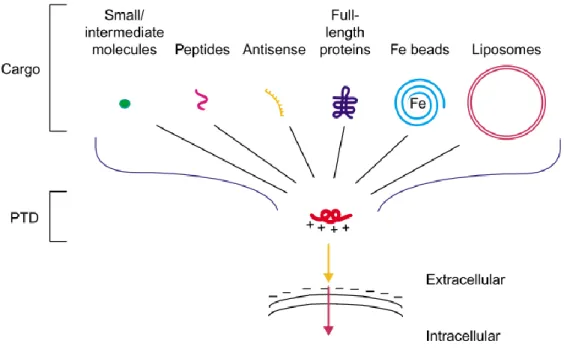
One of the systems that can be used to overcome the problem of the entry into the cells, is the application of protein transduction domains (PTDs), also known as cell-penetrating peptides (CPPs). These kind of peptides are capable of transporting cargo across the membrane and delivering biologically active proteins inside the cell (Figure I.4). Besides, they can be linked to another cargos such as peptides, proteins, oligonucleotides, pDNA, or liposomes.25,43,45
Figure I.4 – Schematic representation of cargo variety covalently linked to arginine-dependent protein transduction domains (PTDs). Adapted from 46.
The initial discovery of PTDs was originated from the independent observation by Green and Frankel in 1988, when a special aminoacid sequence derived from HIV-1 TAT protein, the TAT PTD, showed that could penetrate cells in a receptor-independent, concentration-dependent and activate HIV-1-specific target genes. That peptide could translocate across the plasma membrane by its 11 basic aminoacids (residues 47–57) and to deliver heterogeneous proteins into cells. 25,47–50 This PTD has a higher efficiency for protein delivery into the cells when compared to other PTD signals.25
Once added to the culture media, TAT-mediated transduction occurs through a rapid, temperature and energy-independent process, suggesting direct penetration across the lipid bilayer because of the strong binding of the PTD to the cell surface. 50
One of the hypothesis for the capacity of translocation of these kind of molecules is that their positive charges allow the protein to interact with lipid rafts in a membrane which is negatively charged. That way it can overcome the cell membrane barrier by different mechanisms, like macropinocytosis.25,47,49 Endocytosis was later suggested as an alternative internalization pathway. It is now thought that endocytosis and direct translocation are two coexisting pathways.51 In all cases, after endocytic uptake, the internalized CPPs (either alone
or linked to cargos) should escape from the endocytic vesicles to the cytosol to avoid degradation.43 Within cells, the TAT-fusion proteins are either degraded or refolded by the
cellular machinery into functional proteins.49
One of the characteristics of the TAT PTD sequence which seems to be responsible for the transduction of these proteins is the enrichment by arginine (R) residues and that the sequences with 6-12 consecutive R residues are functional PTDs.47 These kind of arginine-rich cell-penetrating peptides (AR-CPPs) are the most widely studied.43
These oligoarginine peptides present differential manners of internalization and cellular localization depending on the number of arginine residues in the molecules. For example, a tetramer of arginines (4R) did not show significant internalization, while an octamer (8R) showed efficient internalization and nuclear localization, very similar to that of TAT. Conversely, a larger number of residues of arginine did not result in an increased internalization capability, as an hexadecamer of arginines (16R) showed less efficient internalization compared to TAT, without showing significant nuclear localization.52
The transduction of these PTD-based proteins showed to be very efficient working for virtually all types of cells tested. The localization of transduced proteins within the cells depends on the nature of imported proteins, the cell type used and delivery approach.47
In 2009, Zhou et al. reported the successful reprogramming of mouse somatic cells to pluripotency using recombinant proteins of 11R-Sox2, 11R-Oct4, 11R-Klf4 and 11R-c-Myc. Kim et al.(2009) also reported the successful induction of pluripotency in human fibroblast cells using 293T cell extracts that contained the same four reprogramming factors (RF) with the application of a polyarginine peptide C-terminal 9R PTD. 47,53–55
Following the same point of view, Zhang et al. (2012) used reprogramming factors fused with TAT (TAT-RFs) or 11R to induce human foreskin fibroblast and reprogrammed these cells to iPSCs with success. The RFs used were the same as the mentioned by previous groups and one more, Nanog, and all of them were added to the culture medium. Comparing the efficiency of transduction between TAT and 11R, it was found that they were almost the same. However, regarding the reprogramming efficiency, TAT-RFs presented better results than 11R-RFs.47
Concerning the use of recombinant proteins in cardiac differentiation, Fonoudi and collaborators (2013) developed a recombinant protein which was added to hESCs cultures, and efficiently penetrated into the cells, enhancing the differentiation into cardiac cells. To do this, they developed a transduction system based on the fusion of TAT with Islet1 (Isl1). Isl1 is a marker of myocardial lineage during mammalian cardiogenesis and marks a common population of progenitors in the heart that can differentiate into cardiomyocytes, smooth muscle and endothelial cells. The intent was to improve the cardiomyocyte differentiation rate under a suspension culture condition and they have successfully demonstrated that the application of TAT-ISL1 increased the differentiation of cardiomyocytes (2–3 folds) without genetic modification.25
Since this method lacks of genetic manipulation, these kind of molecules can be easily applied in drug discovery or cell therapy since the risk for the application is diminished.
In this thesis, it was proposed to combine TAT and an arginine-rich peptide (composed by 8 arginines) with a human protein – CITED2 – which is involved in cardiac differentiation, to evaluate the transduction into mESC.
The specific aims of this study were the following:
- Cloning of two different PTDs into a pGEX vector and sequential cloning of the CITED2 gene
- Production and purification of GST-3C Protease cleavage protein - Overexpression of recombinant proteins in E.Coli
- Purification and cleavage of the recombinant proteins
- Assessment of proteins functionality through their capacity to rescue Cited2 knockout mESCs cardiogenic defects
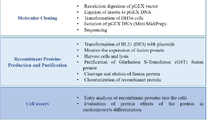
This practical work aimed the synthesis and application of recombinant proteins to mESC. The process is not simple and involved many phases, since the DNA cloning to the direct application of the protein to the stem cells.
Figure III.1 – Schematic representation of the work developed in course of this thesis.
III.1. MOLECULAR CLONING
Gene cloning is a complex process that requires several DNA techniques, in which a desired fragment of DNA is inserted in a cloning vector, such as a plasmid, in order to increase the number of copies of the fragment of interest. This is usually achieved by inserting the vector in a host, being the most commonly used bacteria.
This can be done with enzymes which are used to excise the fragments of interest and the same enzymes to open up the plasmid. After obtaining the DNA from the vector and the desired fragment, another enzyme will be needed to integrate the fragment in the host genome (ligation).
The aim for our final cloning was to obtain CITED2 linked to TAT and 8R in a pGEX vector which will permit the further purification of the proteins, so we needed to do four subclonings into the pGEX-6P-1 vector (Amersham Biosciences):
- Insertion of TAT - Insertion of 8R
- Insertion of CITED2 into each one of the created vectors
Figure III. 1 - Schematic representation of sequential fragment cloning into pGEX-6P-1 vector.
The pGEX-6P-1 vector (Appendix 1) was the expression vector chosen and is designed to express a Glutathione S-Transferase (GST) tag fused to the N-terminal of the protein of interest through a short peptide chain containing a specific endoprotease cleavage site. GST tag will allow the binding of the fusion protein to glutathione affinity columns for subsequent purification. This vector has a T7 promoter system, which is similar to the lac operon in E. coli and, being Isopropyl β-D-1-thiogalactopyranoside (IPTG) sensitive, so when IPTG is added to the culture medium, the transcription of protein linked to GST is induced. For bacterial selection, it has a Ampicillin (Amp) resistance gene.
III.1.1. RESTRICTION ENZYME DIGEST
A restriction enzyme is a naturally occurring bacterial endonuclease that allows to cut a DNA sequence, recognizing a specific nucleotide sequence also known as restriction site. This method have turn out to be the most widely used method to selectively transfer a specific
gene or a DNA sequence from one plasmid to another when the size of the plasmid insert is known. Another application is when the size of the plasmid insert and vector backbone are known, this technique can be used to verify and confirm the construct (diagnostic digest).
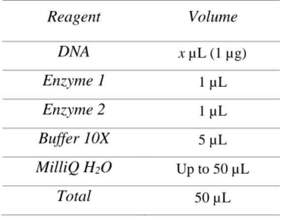
To diagnostic digests, the samples were prepared accordingly with Table 1, at 37ºC for 1h. To isolate inserts, samples were also prepared in the same way but the digestion was performed O.N. (overnight) at 37ºC.
Table 1 – Reaction mix used for the usual restriction digestion of the pDNA.
Reagent Volume DNA x µL (1 µg) Enzyme 1 1 µL Enzyme 2 1 µL Buffer 10X 5 µL MilliQ H2O Up to 50 µL Total 50 µL
To perform the restriction enzyme digest the double stranded DNA has to be incubated with the appropriate restriction enzymes in a suitable buffer recommended by the supplier.
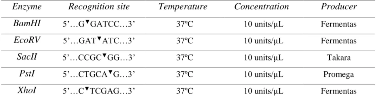
For this work, we performed several digestions to accomplish different goals:
- Double digestion of pGEX-6P-1 with BamHI and EcoRV to check the quality and purity of the plasmid
- Digestion of pGEX-6P-1 with BamHI to insert TAT and 8R
- Double digestion of pGEX-6P-1+TAT (pGEXT) with SacII and PstI to check if cloning of TAT was successful
- Double Digestion of pGEX-6P-1+8R (pGEXR) with BamHI and PstI to check if cloning of 8R was successful
- Double Digestion of pGEXT with BamHI and XhoI to insert Cited2 - Double Digestion of pGEXR with BamHI and XhoI to insert Cited2
- Double Digestion of pGEXTC2 and pGEXRC2 with BamHI and XhoI to check if cloning of Cited2 was successful.
Table 2 – List of restriction enzymes used for digestions, with their recognition site and optimal conditions.
Enzyme Recognition site Temperature Concentration Producer BamHI 5’…G▼GATCC…3’ 37ºC 10 units/μL Fermentas
EcoRV 5’…GAT▼ATC…3’ 37ºC 10 units/μL Fermentas
SacII 5’…CCGC▼GG…3’ 37ºC 10 units/μL Takara
PstI 5’…CTGCA▼G…3’ 37ºC 10 units/μL Promega
XhoI 5’…C▼TCGAG…3’ 37ºC 10 units/μL Fermentas
Buffers were selected considering the enzymes used. In the case of double digestion, the buffer was selected considering the best activity for both enzymes chosen.
III.1.2. AGAROSE GEL ELECTROPHORESIS
Gel electrophoresis is a standard method in molecular biology used to separate molecules accordingly with their size (in this case by length in base pairs) for visualization and purification. Applying an electric charge, negatively charged DNA move through an agarose gel matrix towards a positive electrode. This way, smaller molecules move faster than larger molecules, because they migrate more easily through the matrix.
This process was made to check the DNA from the vector, to purify the desired fragments or to know if the clonings were successful.
For each run was prepared a fresh 1% TAE-gel by dissolving the agarose powder into TAE buffer in a microwave for about 1 to 3 min (see composition at Table 3). After it cooled down for 5 min, GreenSafe (NYZTech, Portugal) was added, and the molten agarose was poured into the flat mold for hardening. A comb was placed to form wells for running the samples and let the agarose gel solidify for at least 30 min, removing carefully the comb before the run.
Table 3 – Composition of 1% TAE agarose gel.
Agarose Gel Agarose 0.5 g
TAE 1X 50 mL
GreenSafe 2 µL
The gel was then transferred into an electrophoresis unit filled with TAE-buffer. Samples were previously prepared with loading buffer (Orange Loading dye 6X) accordingly with Table 4, and carefully loaded into the wells along with a ladder mix (GeneRuler DNA Ladder Mix, Thermo Scientific), under a constant electric potential of 120 V for 40 min.
The posterior visualization of DNA bands was performed in ChemiDoc XRS Molecular Imager (Bio-Rad).
Table 4 – Sample preparation prior agarose gel electrophoresis.
DNA Sample
DNA 20 µL
Orange Loading Dye 6X 3 µL
III.1.3. GEL PURIFICATION
Gel purification is the method that allows to isolate and purify DNA fragments after a standard agarose gel electrophoresis, cutting out the DNA bands which had the expected size of the agarose gel and executing several steps to purify the DNA samples.
In this work, we performed this method for several occasions: to isolate the plasmid vector pGEX-6P-1 DNA digested with BamHI, the pGEX-6P1+TAT and the pGEX-6P1+8R digested with BamHI and XhoI, and also CITED2 DNA insert from a preexisting plasmid (pSB54), digested with BamHI and XhoI. All the purifications were performed using GeneJETTM Gel Extraction Kit from Fermentas.
Purification was carried out according to the manufacturer’s instructions. In summary, the excised fragments were placed in centrifuge tubes and weighted. For each 100 mg of
agarose gel, 100 µL of binding buffer was added, then the mix was incubated at 50-60ºC for 10min, inverting the tube a few times. The binding of DNA was performed by transferring the solution to a column, which was placed in a collection tube (provided by the kit). The sample was centrifuged for 1 min at 12000g and the flow-through discarded. Then, 700 µL of Wash Buffer were added to the column and centrifuged in the same conditions, being the flow-through also discarded. The centrifugation was repeated but with the empty column. Afterwards, the column was placed in a fresh 1.5 mL centrifuge tube and 50 µL of Elution Buffer was added to the column, which allowed the sample to be obtained after a centrifugation for 1 min at 12000g.
III.1.4. DEPHOSPHORILATION
One of the steps prior to the ligation of fragments to the vector, isdephosphorilation. This process will avoid self-closure after the restriction digestion since the pDNA become linearized.
To achieve that, 1 µL of Calf Intestine Phosphatase (CIP) (Finnzymes, Thermo Scientific) was added to the DNA obtained, mixed and incubated for 30 min at 37 ºC in a dry bath (CH-100, Heating/Cooling Dry Block, Biosan). After this time, the tubes needed to be submitted to heat inactivation of the enzyme, leaving it at 65ºC for 30min, to ensure that the enzyme would not interfere in the ligation reaction.
III.1.5. LIGATION REACTION
After restriction of the inserts and the vector, and subsequently dephosphorilation of the last, the ligation reactions were performed in order to the inserts to be integrated into the host vector (Table 5). For the ligation to happen, both DNAs needed to be incubated with an enzyme – T4 DNA ligase (Fermentas) – and its buffer (10X T4 Ligase Buffer, Fermentas), for at least 2 hours (sometimes it was necessary to extend the time to 4 hours) at Room Temperature (RT).
TAT and 8R DNA fragments were already prepared by professor J.Bragança (primers used are listed in Table 6). Both set of oligonucleotides encoding TAT or 8R were chemically
synthetized to harbor, after annealing, a BglII hemi-site at their 5’ end and a BamHI site at their 3’ end flanking the TAT or 8R sequence. The BglII hemi-site can hybridize a BamHI hemi-site, however the resulting BglII/BamHI composite site can no longer be digested by neither of these enzymes. Since these oligonucleotides were synthetic, their 5’ extremities were phosphorylated with a T4 polynucleotide kinase in the presence of ATP before the annealing.
Table 5 – Ligation mix components.
Table 6 – Gene primers used in to prepare TAT and 8R inserts.
Gene Forward 5’-3’ Reverse 5’-3’
TAT GATCTGGCTACGGCCGCAAGAAACGCC GCCAGCGCCGCCGCGGTG GATCCACCGCGGCGGCGCTGGCGGCGTT TCTTGCGGCCGTAGCCA 8R GATCTGGCCGCCGCCGCCGCCGCCGCCG CCGCCGCCGCCGCGGTG GATCCACCGCGGCGGCGGCGGCGGCGG CGGCGGCGGCGGCGGCCA
After ligation procedure, 5 µL of cloned DNA was used to transform chemically competent E. coli cells.
III.2. PREPARATION OF CHEMICALLY COMPETENT CELLS
Competent cells are bacterial cells that possess more easily altered cell walls by which foreign DNA can be passed through easily. The majority of cells cannot take up DNA efficiently unless they have been exposed to special chemical or electrical treatments to make them competent. Reagent Volume 10X T4 Ligase Buffer 1 µL pGEX-6P-1 3 µL Insert 5 µL T4 DNA ligase 1 µL MilliQ H2O Up to 10 µL Total 10 µL
In this work E. coli cells were subjected to chemical treatment, where they were treated with high cation concentration (in this case MgCl2) and then exposed to a heat shock
which makes the cell membrane to become permeable for plasmid DNA.
Cultures were grown in 25 mL of Lysogeny Broth (LB; 10g/L Tryptone, 5g/L Yeast Extract, 10g/L NaCl, pH 7.0) medium at 37ºC in an orbital shaker (Agitorb 200 IC, Aralab®)
until they reach an O.D.600nm of 0.47. Cells were harvested by centrifugation at 1000 g for 10
min (Centrifuge 5810R, Eppendorf®), discarding supernatant. Accordingly with culture volume, cells were resuspended in 1/10 (v/v) of ice cold TSS buffer, previously prepared (10% Polyethylene glycol 8000, 5% Dimethyl Sulfoxide, 20 mM MgCl2 and 85% autoclaved
LB, pH 7.0), and incubated on ice for 10 min. Cells were either used immediately or frozen in 100 µL aliquots.
III.3. TRANSFORMATION OF COMPETENT CELLS
Transformation is the process where foreign DNA (like plasmids) is introduced into a bacterial cell. This process is very common and useful since it allows the replication of a specific plasmid DNA. The majority of the plasmids carry a gene for antibiotic resistance which can be used as a selection tool.
However, not all bacteria strains can be transformed, so they need to enroll a process to become competent, which makes their cell walls more likely to uptake DNA (see section III.2).
The bacterial cells used in this study were from the strain E.coli DH5α (for cloning) and BL21(DE3) (for protein expression), previously made competent. All the transformations were performed using the heat-shock method.
In cloning, bacteria was transformed by adding 5 µL of the ligation mix to 50 µL of DH5α and incubated on ice for 30 min. Further the sample was incubated at 42ºC for 1 min and placed on ice for 2 min. To make the cells recover from the heat shock, 250 µL of Super Optimal broth with Catabolite repression (SOC) were added to the cell suspension and incubated at 180 rpm, at 37ºC for 1 hour in an orbital shaker (Agitorb 200 IC, Aralab®). Bacteria were plated in LB agar plates with Amp (100µg/mL) and placed inverted on the
incubator at 37ºC O.N. For the usual transformation with purified and amplified DNA (miniprep, midiprep or protein expression), only 1 µL was needed to make the transformation.
When the colonies were the result from a ligation reaction transformation, we checked if the plasmid was well constructed, by purifying pDNA, double digesting it and analyzing the result by electrophoresis.
III.4. EXTRACTION OF DNA
III.4.1. MINIPREP
In order to extract the pDNA from the recombinant bacteria, to check by electrophoresis if the inserts were cloned in the right way to the cloning vector, the GeneJET™ Plasmid Miniprep (Thermo Scientific) was used.
Several colonies were isolated and transferred from the plate into a 2 mL LB medium, supplemented with Amp (100 µg/mL) and incubated O.N. at 37ºC, with agitation (200 rpm).
To perform the extraction, cultures were placed in a clean and sterile microcentrifuge tube, and spun down at 6800 g for 2 min being the supernatant discarded. To the pelleted cells 250 µL of Resuspension Solution (which contained RNase A) was added and a micropipette was used to resuspend the cells. Then 250 µL of Lysis Solution was used to disrupt the bacterial walls, being the solution mixed by inversion, making the solution clear and viscous. A volume of 350 µL of Neutralization solution was added to the mix, and the tube was inverted 4 to 6 times. The mixture was centrifuged at 14000 g for 10 min (Centrifuge 5810R, Eppendorf®).
The supernatant was transferred carefully to a fresh tube (avoiding the white precipitate) using a micropipette and applied to the Thermo Scientific GeneJET Spin Column. The columns were then centrifuged for 1 min and the flow-through was discarded, since the plasmid DNA binds to the column in this step. Then 500 µL of Wash Solution were added to the column to wash it and the column was centrifuged again for 1 min, being the flow-through discarded. This step was repeated one more time. The empty column was centrifuged for an additional 1 min to remove residual wash buffer.
Finally, the column was placed in a clean 1.5 mL microcentrifuge tube and the DNA was eluted by incubation in 50 μl Elution Buffer for 2 min and centrifugation for 2 min.
Once resuspended, the samples were quantified by spectrophotometry with NanoDrop 2000 spectrophotometer (Thermo Fisher Scientific Inc, USA) and stored at -20ºC until further use.
Clonings which seemed to be successful were sent to StabVida for sequencing (plasmid maps and sequencing results at Appendix 2 and 3). Once the identity of the cloned fragments was confirmed, a larger scale preparation of plasmid DNA was required. To achieve this goal, midiprep was performed.
III.4.2. MIDIPREP
In the various stages of this experimental work several DNA constructs needed to be purified such as pGEX-6P-1 vector for the preparation of the ligation reactions, and/or extraction of various products of ligation reactions for the sequencing.
To obtain the plasmid DNA, the QIAGEN® Plasmid Midi Kit was used according to the manufacturer’s instructions.
Previously, E. coli was transformed with the desired DNA and the day after, a colony was picked and inoculated in 100 mL of LB medium supplemented with Amp (100 μg/mL), allowing growth to occur O.N. at 37ºC in an incubator with agitation (200 rpm).
Cell suspension was then transferred to 50 mL conical centrifugal tubes and centrifuged for 15 min at 6000 g, at 4ºC. Supernatant was discarded and the pellet resuspended with 4 mL of Buffer P1 (which contained RNase) until no cell clumps were visible. After, 4 mL of Buffer P2 was added and all components were mixed thoroughly by inversion and incubated for 5 min at RT. Buffer P3 was prechilled and 4 mL were added to the suspension, mixed by inversion and incubated on ice for about 15 min. The sample was centrifuged for 30 min at 4ºC at 18000 g (Centrifuge 5810R, Eppendorf®).
Meanwhile, the resin column (Qiagen-tip 100) was assembled and needed to be equilibrated in a suitable buffer which was provided by the kit. A volume of 4 ml of Buffer QBT was added to the column and let through the column, being the flow through discarded.