DOI: 10.5935/2359-4802.20180076
Mailing Adress: Delcio Gonçalves da Silva Junior
Rua Praia de Piatã, 51, Postal Code: 79022-490, Autonomista, Campo Grande, MS - Brazil. E-mail: [email protected]
Cardiovascular Disease in Patients with Ankylosing Spondylitis from the Rheumatology
Outpatient Clinic of the UFMS-affiliated Hospital
Delcio Gonçalves da Silva Junior and Izaias Pereira da Costa
Universidade Federal de Mato Grosso do Sul, Mato Grosso do Sul, MS - Brazil
Manuscript received November 27, 2017, revised manuscript April 23, 2018, accepted June 19, 2018.
Abstract
Background: Cardiovascular diseases are a major cause of morbidity and mortality today. Despite its wide
distribution, it presents particularly prevalent in certain groups of individuals, particularly when exposed to a higher degree of inflammation, giving increased cardiovascular risk. Rheumatic diseases expose their holders to this increased cardiovascular risk condition; however only recently have been associated with spondyloarthritis, particularly ankylosing spondylitis (AS). For being a classically autoimmune disease related to HLA histocompatibility system, AS may present phenotypic variations in different ethnic groups with possible diverse cardiovascular consequences.
Objectives: To estimate the prevalence of cardiovascular disease (CVD) and the cardiovascular risk profile,
correlating the time since diagnosis and activity of ankylosing spondylitis (AS) in patients from the rheumatology outpatient clinic of the UFMS-affiliated hospital.
Methods: Of 55 patients with AS, 42 were selected consecutively and compared to a control group (CG) in a
cross-sectional study. Patients with diabetes, indigenous background and pregnant women were excluded. Quantitative variables were assessed by use of Student t test, while qualitative variables, by chi-square test. The patients underwent electrocardiography, echocardiography and carotid Doppler examination, measurement of serum lipid levels and inflammatory markers, and were stratified according to global cardiovascular risk. The AS activity and impairment were evaluated by use of the BASMI, BASDAI, BASFI and ASDAS.
Results: Mean age, 42.87 ± 12.37 years; time since AS diagnosis, 10.76 ± 8.74 years. There was no difference in
cardiovascular risk stratification between the groups, most of the patients being at high or moderate risk (AS: 64.3%, and CG: 52%, p = 0.134). The prevalence of manifest CVD (2%) showed no difference between the groups, except for right bundle-branch block (AS: 14%, and CG: 2%, p = 0.027). The prevalence of subclinical CVD showed no difference between the groups, except for higher carotid medial-intimal thickness (CIMT) in the AS group (AS: 1.82 ± 2.63, and CG: 0.67 ± 0.16, p = 0.018). There was no correlation between AS activity or inflammatory markers and CVD, but with time since AS diagnosis and CIMT (p = 0.039, r = 0.328).
Conclusions: Prevalence of CVD and risk factors was similar in the groups. Subclinical atherosclerosis degree was
higher in the AS group, related to the time since diagnosis, but was independent of the cardiovascular risk factors or inflammation. Most patients with AS are at high cardiovascular risk. (Int J Cardiovasc Sci. 2019;32(1)10-18)
Keywords: Cardiovascular Diseases; Risk Factors; Cross-Sectional Studies; Spondylitis, Ankylosing/complications;
Spondylitis, Ankylosing/ diagnosis.
Introduction
Cardiovascular diseases (CVD) are currently a major cause of morbidity and mortality. Recent data from
the World Health Organization have shown that 30% of the total of deaths worldwide in past decades result
from CVD, approximately 17 million individuals.1,2
metabolic syndrome, dyslipidemia, hypertension, smoking and family history, have been well established as predictors of cardiovascular events, and measures for their control and monitoring are recommended to reduce
and fight CVD.3
However, in certain clinical conditions, such as rheumatic diseases, the traditional risk factors do not
seem to contribute to the total incidence of CVD.4 Despite
the well described higher incidence of CVD in rheumatic diseases, especially rheumatoid arthritis, systemic sclerosis, systemic vasculitis, antiphospholipid syndrome and systemic lupus erythematosus, it was only recently that the association of CVD and ankylosing spondylitis
(AS) gained attention.5,6
The greater mortality of individuals with AS compared to that of the general population seems to
be related to CVD.7,8 Early atherosclerosis has been
reported in association with chronic inflammation and
with AS.9 Some studies have shown that CVD and their
traditional risk factors are more prevalent in patients
with AS,10,11 which is still controversial when interpreting
inflammation as the major responsible for the higher cardiovascular impairment.
Despite the international publications, Brazilian studies correlating CVD and AS are scarce and more recent, especially those assessing their various presentation forms
(clinically manifest or subclinical CVD).12-14
The characteristic racial miscegenation of the Brazilian population increases the interest for such studies, because it can lead to differences in the prevalence of CVD in that population, considering the association between AS and the HLA system.
This study was aimed at estimating the prevalence of CVD in individuals with AS from the Spondyloarthritis Outpatient Clinic of the Rheumatology Service of the Mato Grosso do Sul Federal University hospital (HU-UFMS), in addition to correlating the traditional risk factors and the inflammatory process of CVD.
Patients and methods
This is a quantitative descriptive study carried out from March to October 2015 with individuals with AS selected by convenience and consecutively from the patients regularly cared for at the Spondyloarthritis Outpatient Clinic of the Rheumatology Service of the HU-UFMS, which has records of 170 patients diagnosed with spondyloarthritis (psoriatic arthritis, AS, reactive arthritis, and enteropathic arthritis). Of those 170 patients,
55 were diagnosed with AS. A control group (CG) was formed by convenience sampling with employees of the UFMS without rheumatic disease and was similar to the AS group regarding age and sex. The inclusion and exclusion criteria allowed the selection of 42 individuals for the AS group and 50 for the CG.
Before data collection, all individuals provided written informed consent, duly registered in the Ethics Committee in Research of the UFMS (CAAE: 34043614.8.0000.0021).
Individuals with the following characteristics were excluded: diabetes, indigenous background, hypothyroidism or neoplasia, illiteracy, and pregnant women. All participants were screened for CVD and cardiovascular risk factors.
All individuals underwent cardiology clinical examination, laboratory tests, electrocardiography, echocardiography and carotid doppler examination on the same day, and within 20 days from the initial interview, which collected data from medical history and AS. The laboratory tests, performed after a 10-12-hour fasting, comprised the following measurements: total cholesterol and fractions, glycemia, glycated hemoglobin, uric acid, microalbuminuria, erythrocyte sedimentation rate (ESR), high-sensitivity C-reactive protein (hs-CRP) and interleukin 6 (IL-6).
The criteria to assess cardiovascular risk according to the V Brazilian Guideline on Dyslipidemia and Atherosclerosis Prevention comprised the presence of dyslipidemia, metabolic syndrome, arterial hypertension, current smoking, family history of early CVD, cardiac hypertrophy, and elevated levels of glycated hemoglobin,
uric acid and hs-CRP.15
Cardiovascular disease was classified as clinically manifest or subclinical. Individuals with manifest CVD were those with history of myocardial infarction, stroke, coronary artery bypass graft surgery (CABG), peripheral revascularization surgery or procedure (PRV), or peripheral obstructive arterial disease (POAD), echocardiographic evidence of segmental, systolic, diastolic (over grade I) or hypertrophic cardiomyopathy, arrhythmias or bundle-branch and atrioventricular blocks on the electrocardiogram, and presence of carotid plaque obstruction > 50% of the lumen on Doppler.
Subclinical CVD was identified by an ankle-brachial index (ABI) < 0.9 or > 1.3, a carotid intima-media thickness (CIMT) >1 mm, but without significant plaque
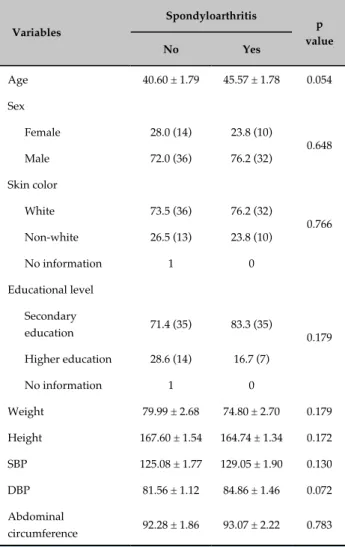
Table 1 - Sociodemographic and physical data of the individuals with and without spondyloarthritis assessed in this study
Variables
Spondyloarthritis
p value
No Yes
Age 40.60 ± 1.79 45.57 ± 1.78 0.054
Sex
Female 28.0 (14) 23.8 (10)
0.648 Male 72.0 (36) 76.2 (32)
Skin color
White 73.5 (36) 76.2 (32)
0.766 Non-white 26.5 (13) 23.8 (10)
No information 1 0
Educational level
Secondary
education 71.4 (35) 83.3 (35) 0.179
Higher education 28.6 (14) 16.7 (7)
No information 1 0
Weight 79.99 ± 2.68 74.80 ± 2.70 0.179
Height 167.60 ± 1.54 164.74 ± 1.34 0.172
SBP 125.08 ± 1.77 129.05 ± 1.90 0.130
DBP 81.56 ± 1.12 84.86 ± 1.46 0.072
Abdominal
circumference 92.28 ± 1.86 93.07 ± 2.22 0.783
SBP: systolic blood pressure; DBP: diastolic blood pressure. The results are expressed as mean ± standard error of the mean or relative frequency (absolute frequency). P value in the Student t test (quantitative variables) or the chi-square test (qualitative variables).
Disease activity was assessed by using the Bath Ankylosing Spondylitis Disease Activity Index (BASDAI) and Ankylosing Spondylitis Disease Activity Score (ASDAS). The ASDAS-CRP and ASDAS-ESR indices16 were calculated after blood collection, using the information obtained when assessing the Bath Ankylosing Spondylitis Metrology Index (BASMI) and the Bath Ankylosing Spondylitis Functional Index (BASFI). In addition, the disease activity indices were correlated with the serum levels of IL-6, hs-CRP and uric acid, and compared to the occurrence of clinically manifest or subclinical CVD.
The AS group individuals were also assessed regarding the occurrence of CVD up to the age of 40 years and after 40 years.
Statistical analysis17
The results referring to the quantitative variables are presented as mean ± standard deviation, while the results of the categorical variables are presented as relative frequency followed by absolute frequency. The quantitative variables were compared between individuals with and without spondyloarthritis by use of parametric Student t test for independent samples (nonpaired) because most of the samples were normally distributed (Shapiro-Wilk test; p > 0.05). Student t test was used to compare quantitative variables between age groups. The association between the qualitative variables and the presence or absence of spondyloarthritis was assessed by use of chi-square test, which was also used to assess the association between the qualitative variables and the patients’ age groups. The linear correlation between some quantitative variables was assessed by use of Pearson’s linear correlation test. The results of the other variables were presented as descriptive statistics or tables and graphs. The SPSS statistical program, version 22.0, was used for statistical analysis, considering a 5% significance level.
Results
General characteristics
There was no significant difference between individuals with or without AS regarding age, sex, skin color, educational level, weight, height, blood pressure and body mass index (Table 1).
Risk factors for cardiovascular disease
Table 2 shows the results regarding the cardiovascular risk factors in the two groups. There was no significant
difference between the groups regarding the variables family history, smoking habit, alcoholism, abdominal circumference, systemic arterial hypertension, total cholesterol, HDL-cholesterol, triglycerides, glycated hemoglobin, hs-CRP, microalbuminuria, metabolic syndrome, and IL-6. The LDL-cholesterol levels in the AS group, however, were higher than those in the CG (p = 0.012). The uric acid levels in the AS group were lower than those in the CG (p = 0.019).
Table 2 - Cardiovascular risk factors of the individuals with and without spondyloarthritis assessed in this study
Variables
Spondyloarthritis
p value
No Yes
Family history
No 76.0 (38) 73.2 (30)
0.757 Yes 24.0 (12) 26.8 (11)
No information 0 1
Smoking
No 90.0 (45) 92.9 (39)
0.628
Yes 10.0 (5) 7.1 (3)
Alcoholism
No 60.0 (30) 71.4 (30)
0.252 Yes 40.0 (20) 28.6 (12)
BMI 27.99 ± 0.76 27.33 ± 0.90 0.575
SAH
No 64.0 (32) 57.1 (24)
0.502 Yes 36.0 (18) 42.9 (18)
Total cholesterol 170.60 ± 5.36 180.67 ± 6.66 0.237
HDL 43.13 ± 2,45 45.81 ± 2.18 0.420
LDL 88.87 ± 6.36 110.79 ± 5.65 0.012
Triglycerides 137.58 ± 14.19 123.40 ± 11.76 0.455
Glycated
hemoglobin 6.65 ± 1.41 5.59 ± 0.08 0.484
Uric acid 6.02 ± 0.26 5.22 ± 0.20 0.019
hs-CRP 2.35 ± 0.31 16.10 ± 8.88 0.130
Microalbuminuria 3.87 ± 0.95 8.31 ± 2.04 0.053
Metabolic syndrome
No 62.0 (31) 69.0 (29)
0.480 Yes 38.0 (19) 31.0 (13)
Interleukin-6 4.62 ± 0.48 4.84 ± 0.80 0.806
BMI: body mass index; SAH: systemic arterial hypertension; hs-CRP: high-sensitivity C-reactive protein. The results are expressed as mean ± standard error of the mean or relative frequency (absolute frequency). P value in the Student t test (quantitative variables) or the chi-square test (qualitative variables).
To assess the cardiovascular risk of well-controlled patients, and thus with lower inflammation level, the global cardiovascular risk was calculated separately in patients with ASDAS-CRP < 2 and low disease activity level, but no significant statistical difference was found between the CG and the AS group patients with that characteristic.
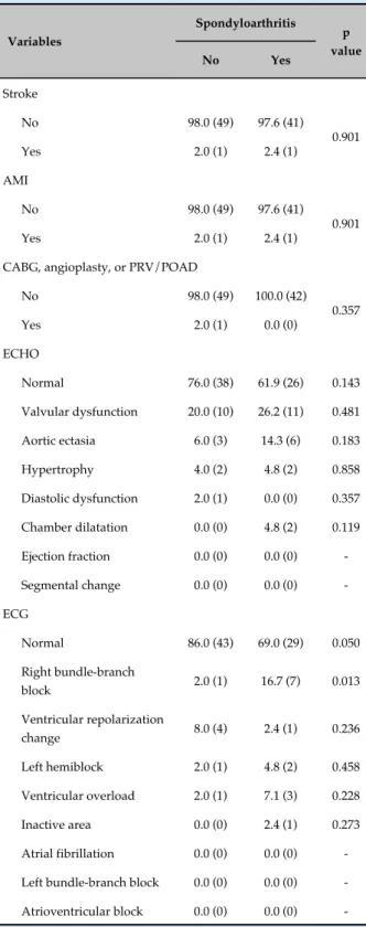
Manifest cardiovascular disease
Most individuals assessed in this study had no clinically manifest CVD, with prevalence between 2% and 26%, and valvular dysfunctions were the most frequent change, although all of them were mild. The percentage of individuals with right bundle-branch block was higher in the AS group than in the CG. The following variables showed no association with the presence or absence of AS: stroke, acute myocardial infarction, CABG, angioplasty, PRV/POAD, presence of carotid plaques and other findings on echocardiogram and electrocardiogram. None of the individuals in the CG and AS group showed significant carotid obstruction
(plaque obstruction ≥ 50% of the lumen) (Table 3).
Subclinical cardiovascular disease
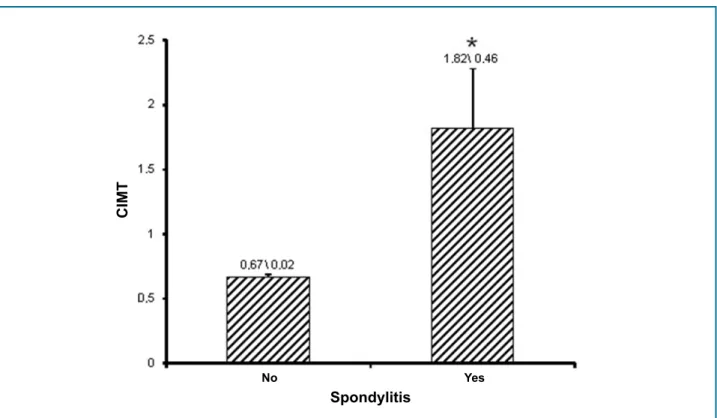
None of the individuals achieved the cutoff values for the occurrence of microalbuminuria (> 30 mg/g) or altered ABI (< 0.9 and > 1.3); in addition, the mean values of ABI and microalbuminuria were similar in both groups. Carotid plaques without significant obstruction (< 50% of the lumen) were slightly more frequent in the AS group [CG: 12.2% (n = 6); AS: 21.4% (n = 9)], but with no significant statistical difference (p = 0.239). However, as shown in Figure 1, CIMT was higher in the AS group (p = 0.018), and the cutoff value to determine altered CIMT (> 1 mm) was more often observed in the AS group [CG: 2.3 (n = 1); AS: 24.2 (n = 8)] (p = 0.003). This difference was observed when individuals with carotid plaques were excluded. Considering only the individuals with carotid plaques, no significant difference regarding CIMT was observed between the groups (CG: 0.78 ± 0.13; AS: 1.45 ± 2.12) (p = 0.464).
Ankylosing spondylitis activity and specific data
Most individuals were HLA positive (64.3%, n = 27) and had exclusive (33.3%, n = 14) or predominantly (61.9%, n = 26) axial clinical manifestations and extra-association between the presence or absence of AS and
Table 3 - Results regarding manifest cardiovascular disease of the individuals with and without spondyloarthritis assessed in this study
Variables
Spondyloarthritis
p value
No Yes
Stroke
No 98.0 (49) 97.6 (41)
0.901
Yes 2.0 (1) 2.4 (1)
AMI
No 98.0 (49) 97.6 (41)
0.901
Yes 2.0 (1) 2.4 (1)
CABG, angioplasty, or PRV/POAD
No 98.0 (49) 100.0 (42)
0.357
Yes 2.0 (1) 0.0 (0)
ECHO
Normal 76.0 (38) 61.9 (26) 0.143
Valvular dysfunction 20.0 (10) 26.2 (11) 0.481
Aortic ectasia 6.0 (3) 14.3 (6) 0.183
Hypertrophy 4.0 (2) 4.8 (2) 0.858
Diastolic dysfunction 2.0 (1) 0.0 (0) 0.357
Chamber dilatation 0.0 (0) 4.8 (2) 0.119
Ejection fraction 0.0 (0) 0.0 (0)
-Segmental change 0.0 (0) 0.0 (0)
-ECG
Normal 86.0 (43) 69.0 (29) 0.050
Right bundle-branch
block 2.0 (1) 16.7 (7) 0.013
Ventricular repolarization
change 8.0 (4) 2.4 (1) 0.236
Left hemiblock 2.0 (1) 4.8 (2) 0.458
Ventricular overload 2.0 (1) 7.1 (3) 0.228
Inactive area 0.0 (0) 2.4 (1) 0.273
Atrial fibrillation 0.0 (0) 0.0 (0)
-Left bundle-branch block 0.0 (0) 0.0 (0)
-Atrioventricular block 0.0 (0) 0.0 (0)
-AMI: acute myocardial infarction; CABG: coronary artery bypass graft surgery; PRV: peripheral revascularization surgery or procedure; POAD: peripheral obstructive arterial disease; ECHO: echocardiography; ECG: electrocardiography. The results are expressed as relative frequency (absolute frequency). P value in the chi-square test.
articular symptoms (54.8%, n = 23), enthesitis being the most frequent (69.6%, n = 16).
Time since diagnosis ranged from 2 to 34 years (mean of 10.76 ± 8.74 years). The time since symptom onset ranged from 3 to 47 years (mean of 19.36 ± 11.02 years). The AS activity grade and impairment were: BASDAI = 2.84 ± 2.04, BASFI = 3.77 ± 2.82, BASMI = 4.59 ± 2.17, ASDAS-CRP = 2.17 ± 1.10 and ASDAS-ESR = 1.77 ± 0.81.
The hs-CRP and IL-6 levels were analyzed in both groups as one of the inflammation markers. The IL-6 showed no statistically significant difference (CG: 4.62 ± 3.41, AS: 4.84 ± 5.20; p = 0.806). Although more elevated in the AS group, the hs-CRP levels showed no significant difference as compared to those in the CG (CG: 2.35 ± 2.21, AS: 16.10 ± 56.85; p = 0.130).
There was moderate and positive significant linear correlation between the hs-CRP values obtained and those of ASDAS-CRP (Pearson, p < 0.001; r = 0.657), in addition to a positive significant, although weak, linear correlation between the hs-CRP values obtained and those of ASDAS-ESR (p = 0.007; r = 0.418). On the other hand, there was no significant linear correlation between the other inflammation markers and the disease activity indices assessed in this study (p-value between 0.177 and 0.875).
There was no linear correlation between the values obtained in ASDAS-CRP and the CIMT measures (p = 0.932, r = -0.014). In addition, there was no linear correlation between the values of hs-CRP and CIMT (p = 0.625, r = -0.079) or between time since diagnosis and CIMT (p = 0.152, r = -0.225). The lack of statistical significance in the linear correlation between CIMT and ASDAS-CRP, hs-CRP and time since diagnosis persisted even when individuals with and without plaque were separated.
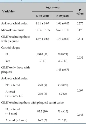
In addition, the AS group patients were assessed according to age group (up to 40 years and over 40 years). There was no significant association between age group and manifest CVD. Regarding subclinical CVD, however, the percentage of patients older than 40 years with carotid plaques was significantly higher than that of those under 40 years. Microalbuminuria, ABI and CIMT showed no statistically significant difference according to age group (Table 4).
Figure 1 - Graph showing the carotid intima-media thickness (CIMT) of individuals with and without ankylosing spondylitis. Each column represents the mean, and the bar represents the standard error of the mean.
* Significant difference as compared to individuals without spondyloarthritis (Student t test, p = 0.018).
No Yes
Spondylitis
CIMT
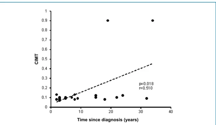
r = -0.362). Among individuals with AS older than 40 years and no carotid plaque, the correlation between time since diagnosis and CIMT was also positive (Figure 2).
Medications
In the AS group, 34 patients (81%) used immune biologics, 26 (61.9%) used nonsteroidal anti-inflammatory drugs, 13 (31%) were on immunosuppressant drugs (methotrexate/sulfasalazine), and only 4 (9.5%) were using corticosteroids. There was no significant statistical difference between the CG and the AS group regarding the use of statin, angiotensin-converting-enzyme inhibitor/angiotensin-receptor-blocker, beta-blocker, calcium-channel blockers, and acetylsalicylic acid.
Discussion
The higher frequency of cardiovascular impairment in rheumatic diseases, whose major background is chronic inflammation, explains the higher cardiovascular
morbidity and mortality of those patients.18 However, the
role of inflammation in the occurrence of CVD remains controversial in AS.
Although the prevalence of cardiovascular impairment
in AS is not high,12 there is consensus that the mortality
rate of those patients is higher than that of the general population, mainly due to higher cardiovascular
mortality.7,8 In addition, the more accurate definition
of the weight of each factor involved, such as chronic inflammation, traditional cardiovascular risk factors and genetics, still lacks. The cyclic characteristic of AS throughout life, the time of exposure to disease, the effect of treatment, and the variety of exposure to cardiovascular risk factors can interfere with the CVD occurrence in that population, hindering the better understanding of the correlation between AS and heart diseases.
Unlike rheumatoid arthritis, whose cardiovascular
risk score is higher,9 publications on AS are controversial.
Some authors have explained the higher prevalence of subclinical atherosclerotic disease, such as a higher CIMT, in patients with AS because of the higher exposure to
cardiovascular risk factors,7 while others, have reported
that the cardiovascular impairment was independent
of the presence of the traditional risk factors for CVD.19
Table 4 - Results regarding subclinical cardiovascular disease of the individuals up to 40 years of age and those older than 40 years assessed in this study
Variables
Age group
p value ≤ 40 years > 40 years
Ankle-brachial index 1.11 ± 0.05 1.06 ± 0.02 0.375
Microalbuminuria 15.04 ± 6.35 5.62 ± 1.10 0.170
CIMT (excluding those
with plaques) 1.97 ± 0.88 1.73 ± 0.53 0.811
Carotid plaque
No 100.0 (12) 70.0 (21)
0.032
Yes 0.0 (0) 30.0 (9)
CIMT (only those with
plaques) - 1.45 ± 0.71
-Ankle-brachial index
Not altered 75.0 (9) 93.3 (28)
0.097 Altered
(< 0.9 or > 1.3) 25.0 (3) 6.7 (2)
CIMT (excluding those with plaques) cutoff value
Not altered
(< 1 mm) 83.3 (10) 71.4 (15) 0.443
Altered (> 1 mm) 16.7 (2) 28.6 (6)
CIMT: carotid intima-media thickness. The results are expressed as mean ± standard error of the mean or relative frequency (absolute frequency). P value in the Student t test (quantitative variables) or the chi-square test (qualitative variables).
population with traditional risk factors for CVD similar to those of the general population, being an independent marker of risk. The presence of carotid plaques and the positive correlation between CIMT and the time since diagnosis of AS in individuals older than 40 years reinforces that hypothesis.
In addition, it is worth noting the considerably higher mean value of hs-CRP of the AS group individuals, indicating their higher cardiovascular risk. Furthermore, a good part of those with well-controlled rheumatic disease and low inflammation (ASDAS < 2) were at high cardiovascular risk and out of the recommended LDL-cholesterol target (< 70 mg/dL).
We do not know any Brazilian study using this cardiovascular risk stratification approach for those patients, but it is worth noting that some researchers have sought cardiovascular risk markers specific for
AS,23 according to the recommendations established for
rheumatoid arthritis and systemic lupus erythematosus.9
The lack of correlation between markers of inflammation, activity and disease impairment in the AS group can be partially explained by the low mean age of the patients or the use of immune biologics in 80% of them. By being a cross-sectional study might have contributed to that, although other authors have found no correlation between inflammation and early atherosclerosis when
cardiovascular risk factors were excluded.13,21
Conclusions
There is no difference in the prevalence of manifest CVD in patients with AS as compared to that of the CG. However, subclinical CVD is more prevalent in AS patients when assessed by use of CIMT, has no relation to the AS activity, but to the time since AS diagnosis and to more advanced ages. Most patients with AS are not only at higher cardiovascular risk, even when clinically controlled, but their CVD prevention is inadequate as well.
Ethics approval and consent to participate
This study was approved by the Ethics Committee of the Universidade Federal do Mato Grosso do Sul under the protocol number CAAE: 34043614.8.0000.0021. All the procedures in this study were in accordance with the 1975 Helsinki Declaration, updated in 2013. Informed consent was obtained from all participants included in the study. LDL-cholesterol, higher in the AS group, which can be
related to the use of immune biologics20,21 or only to a
more cautious diet in the CG.
No difference in the prevalence of manifest and subclinical CVD was found between the groups; however, most individuals were classified as intermediate and high risk. Considering that most individuals in both groups were young, the prevalence of risk factors for CVD at an early age is clear, which is especially relevant in patients with rheumatic diseases.
Excluding the limitations of a non-blinded study, the higher mean values of CIMT in the AS group indicate higher subclinical atherosclerosis. Despite the lack of
consensus in the literature,14,22 we believe that such
Figure 2 - Dispersion graph showing the moderate positive linear correlation between time since diagnosis and carotid intima-media thickness (CIMT), considering only patients with ankylosing spondylitis, without plaque, aged ≥ 40 years. Each symbol represents the value for both variables for a single individual. The dashed line represents the line of linear regression between those variables. The p value and r were obtained with Pearson’s linear correlation test.
Time since diagnosis (years)
CIMT
1. Beaglehole R, Bonita R, Global public health: a scorecard. Lancet. 2008;372(9654):1988-96.
2. Buttler D, UN Targets top killers. Nature. 2011;477(7364):260-1.
3. Simão AF, Precoma DB, Andrade JP, Correa Filho H, Saraiva JFK, Oliveira
GMM et al; Sociedade Brasileira de Cardiologia. [I Brazilian Guidelines for cardiovascular prevention]. Arq Bras Cardiol. 2013;101(6 Suppl 2):1-63. Erratum in: Arq Bras Cardiol. 2014;102(4):415.
4. del Rincón ID, Williams K, Stern M, Freeman GL, Escalante A High incidence of cardiovascular events in a rheumatoid arthritis cohort not explained by traditional cardiac risk factors. Arthritis Rheum. 2001;44(12):2737-45.
5. Haroon NN, Michael Paterson JM, Robert Inman RD, Haroon N. Patients with ankylosing spondylitis have increased cardiovascular and cerebrovascular mortality: a population-based study. Ann Intern Med. 2015;163(6):409-16.
References
Author contributions
Conception and design of the research: Silva Junior DG. Acquisition of data: Silva Junior DG. Analysis and interpretation of the data: Silva Junior DG. Statistical analysis: Silva Junior DG. Writing of the manuscript: Silva Junior DG. Critical revision of the manuscript for intellectual content: Silva Junior DG, Costa IP.
Potential Conflict of Interest
No potential conflict of interest relevant to this article was reported.
Sources of Funding
There were no external funding sources for this study.
Study Association
6. Horreau C, Pouplard C, Brenaut E, Barnetche T, Misery L, Cribier B, et al. Cardiovascular morbidity and mortality in psoriasis and psoriatic arthritis: a systematic literature review. J Eur Acad Dermatol Venereol. 2013;27 Suppl 3:12-29.
7. Papagoras C, Markatselia TE, Saougoub I, Alamanos Y, Zikou AK, Voulgari PV, et al. Cardiovascular risk profile in patients with spondyloarthritis. Joint Bone Spine. 2014;81(1):57-63.
8. Prati C, Claudepierre P, Pham T, Wendling D. Mortality in spondylarthritis. Joint Bone Spine. 2011;78(5):466-70.
9. Peters MJ, Symmons DP, Mccarey D, Dijkmans BA, Nicola P, Kvien TK,
et al. EULAR evidence-based recommendations for cardiovascular risk management in patients with rheumatoid arthritis and other forms of inflammatory arthritis. Ann Rheum Dis. 2010;69(2):325-31.
10. Mathieu S, Gossec L, Dougados M, Soubrier M. Cardiovascular profile in ankylosing spondylitis: a systematic review and meta-analysis. Arthritis Care Res (Hoboken). 2011;63(4):557-63.
11. Han C, Robinson DW Jr, Hackett MV, Paramore LC, Fraeman KH, Bala MV. Cardiovascular disease and risk factors in patients with rheumatoid arthritis, psoriatic arthritis, and ankylosing spondylitis. J Rheumatol. 2006;33(11):2167-72.
12. Rodrigues CM, Vieira WP, Bortoluzzo AB, Gonçalves CR, Da Silva JA, Ximenes AC, et al. Low prevalence of renal, cardiac, pulmonary, and neurological extra-articular clinical manifestations in spondyloarthritis: analysis of the Brazilian Registry of Spondyloarthritis. Rev Bras Reumatol. 2012;52(3):375-83.
13. Skare TL, Verceze GC, Oliveira AA, Perreto S. Carotid intima-media thickness in spondyloarthritis patients. Sao Paulo Med J. 2013;131(2):100-5.
14. Valente RL, Valente JM, De Castro RR, Zimmermann AF, Fialho SC, Pereira IA. Subclinical atherosclerosis in ankylosing spondylitis: is there a role for inflammation? Rev Bras Reumatol. 2013;53(5):377-81.
15. Xavier HT, Izar MC, Faria Neto JR, Assad MH, Rocha VZ, Sposito AC, et al; Sociedade Brasileira de Cardiologia. [V Brazilian Guidelines on dyslipidemias and prevention of atherosclerosis]. Arq Bras Cardiol. 2013;101(4 Suppl 1):1-20.
16. Machado P, Landewé R, Lie E, Kvien TK, Braun J, Baker D, et al; Assessment of SpondyloArthritis international Society. Activity states and improvement scores (ASDAS): defining cut-off values for disease Ankylosing Spondylitis Disease Activity Score. Ann Rheum Dis. 2011;70(1):47-53.
17. Shott S. Statistics for health professionals. Philadelphia: WB Saunders Company; 1990.
18. Mason JC, Libby P. Cardiovascular disease in patients with chronic inflammation: mechanisms underlying premature cardiovascular events in rheumatologic conditions. Eur Heart J. 2015;36(8):482-9c.
19. Üstün N, Kurt M, Atci N, Yağiz E, Güler H, Turhanoğlu A. Increased
epicardial fat tissue is a marker of subclinic atherosclerosis in ankylosing spondylitis. Arch Rheumatol. 2014;29(4):267-72.
20. Mathieu S, Jean-Jacques Dubost JJ, Tournadre A, Malochet-Guinamand S, Ristori JM, Soubrier M. Effects of 14 weeks of TNF alpha blockade treatment on lipid profile in ankylosing spondylitis Joint Bone Spine. 2010;77(1):50-2.
21. Tam L, Tomlinson B, Chu T, Li M, Leung Y, Kwok L, et al. Cardiovascular risk profile of patients with psoriatic arthritis compared to controls--the role of inflammation. Rheumatology (Oxford). 2008;47(5):718-23.
22. Gupta N, Saigal R, Goyal L, Agrawal A, Bhargava R, Agrawal A. Carotid intima media thickness as a marker of atherosclerosis in ankylosing spondylitis. Int J Rheumatol. 2014;2014:839135.
23. Cece H, Yazgan P, Karakas P, Karakas O, Demirkol A, Toru I, et al. Carotid intima-media thickness and paraoxonase activity in patients with ankylosing spondylitis. Clin Invest Med. 2011;34(4):E225.