Spine surgery in patients with ankylosing
spondylitis
Natália Maria Fernandes Britto, M.D.1 Beatriz Souza Renor, M.D.1 Enrico Ghizoni, M.D., Ph.D.2 Helder Tedeschi, M.D., Ph.D.2 Andrei Fernandes Joaquim, M.D., Ph.D.3
1. Neurosurgery resident - Universidade Estadual de Campinas, Unicamp, Campinas-SP, Brasil. 2. Neurosurgery Professor - Universidade Estadual de Campinas, Unicamp, Campinas-SP, Brasil. 3. Neurosurgeon – Universidade Estadual de Campinas, Unicamp, Campinas-SP, Brasil.
http://dx.doi.org/10.1590/1806-9282.64.04.379
SUMMARY
INTRODUCTION: Ankylosing spondylitis (AS) is an idiopathic seronegative spondyloartropathy that involves mainly the axial skeleton and the sacroiliac joints. AS promotes biomechanical changes in the spine that predispose to fractures, spinal deformity and spondylo-discitis. The aim of this article is to report the clinical and laboratorial characteristics of patients with AS who underwent spinal surgery at our Institution.
METHODS: Retrospective review of medical charts of patients who had AS and underwent spinal interventions.
RESULTS: Nine patients were found and eight were included in the present study. There were three men and six women and the patients’ mean age was 57 years old. All patients had pain at the involved spinal level and one patient had tetraparesis due to cervical myelop-athy. Acute-phase proteins were positive in six patients (75%), and HLA-B27 was found in two patients (25%). Four patients had the radiological diagnosis of spondylodiscitis (50%) and underwent a spinal disc biopsy. They were all characterized as having aseptic spondylodiscitis. Three patients were free of pain with analgesics in their last follow-up and one patient had only partial solution of his pain. Three additional patients had spinal fractures surgically treated (37.5%) and one patient was operated because of a cervical kyphotic deformity (12.5%). There were no deaths or surgical complications in this series.
CONCLUSIONS: the majority of our clinical and laboratories findings were discrepant with the medical literature. These differences may be secondary to regional characteristics or by the fact that our population included only those patients who underwent spinal surgery.
KEYWORDS: Spondylitis, ankylosing. Fractures, bone. Discitis.
ABBREVIATIONS: MRI = magnetic resonance imaging; CT = computed tomography, AS= Ankylosing spondylitis, HLA=Human leuko-cyte antigen, ASAS= Assessment of Spondyloarthritis international society, SpA= Spondyloarthitis, NSAIDSs= Nonsteroidal anti-in-flammatory drugs.
INTRODUCTION
Ankylosing spondylitis (AS) is an idiopathic se-ronegative spondyloartropathy that involves mainly the axial skeleton and the sacroiliac joints. The dis-ease has an estimated prevalence of 0.1 to 1.4% of the adult population, typically affecting men and with clinical symptoms starting at 20-30 years of age.1
DATE OF SUBMISSION: 28-Jun-2017
DATE OF ACCEPTANCE: 15-Jul-2017
CORRESPONDING AUTHOR: Andrei F. Joaquim
Departamento de Neurologia - Hospital das Clínicas da Faculdade de Medicina de Campinas Cidade Universitária Zeferino Vaz
Rua Padre Anchieta, 2770 – CEP 13083-888 – Campinas-SP E-mail: [email protected]
[email protected] [email protected] [email protected] [email protected]
There is a rough correlation between the preva-lence of HLA B27 and the incidence and prevapreva-lence of AS.2 Although 6-10% of the world population is
pos-itive for HLA B27, only 5% of them will develop AS. However, almost 90% of the AS patients are positive to HLA B27.1,3,4 There are no laboratorial findings that
inflam-matory diseases, acute-phase proteins are frequently in the normal range.
The diagnosis was first defined using the modi-fied New York criteria.4, 5 These criteria presented
high sensibility and specificity; however they were not useful for early diagnosis or prevention. Because of that the most recent ASAS classification criteria for axial spondyloarthritis were developed for early and established cases and include the MRI technique (to depict active inflammation) as an important tool for early diagnosis (Table 1).6
AS has a caudo-rostral progression, altering the biomechanical properties of the spine and diminish-ing its resistance through a process of remodeldiminish-ing, which involves ligamentary ossification, vertebral joint fusion, osteoporosis and finally, spinal defor-mity. The ankylosed spine is prone to fractures even after minor traumas; additionally, fractures are often unstable and require proper treatment to avoid pri-mary and secondary neurological injury, disability, and progressive deformity.7,8,8,10
Other possible complication is the occurrence of spondylodiscitis, especially for those patients who receive medicaments such as biologic agents or tu-mor necrosis factor antagonists, who may increase the chances of infections. It is radiologically suspect-ed by the combination of narrowing of the disc space and erosions at the terminal plates summed to pe-ripheral sclerosis, most of the times with gadolini-um enhancement at the MR. Of note, it may also be aseptic, secondary to AS disease itself.9, 10,11,12,13
The aim of the present manuscript is to
ana-lyze the patients with AS who underwent spinal sur-geries at our tertiary University Hospital.
METHODS
A retrospective study at the University Hospital of the State University of Campinas, São Paulo, Bra-sil, was performed. The medical charts of all patients with the diagnosis of AS that had spine surgery, were reviewed. Institutional Review Board approval was obtained under the number 17337313.7.0000.5404.
The inclusion criteria were: patients with diagno-sis of AS established by the internal medical depart-ment of our institution that underwent a spinal pro-cedure by any reasons in our hospital. All propro-cedures were performed by the same spine surgeon (AFJ).
The exclusion criteria were: lack of medical or ra-diological data or lack of postoperative follow-up.
The following variables evaluated were:
Clinical information: gender, age, time of disease progression, clinical manifestation, neurological sta-tus at diagnosis, description of the surgical proce-dure and clinical evolution, conventional criteria to evaluation of AS (peripheral arthritis, enthesopathy, uveitis), comorbidities, and history of trauma.
Laboratorial information: acute-phase proteins re-sults, presence of antigen HLA B27, results of hemo-cultures and biopsy results
Radiological findings: reported on spine MRI and CT scans.
RESULTS
Nine patients were found (three males and six fe-males), but one patient was excluded due to the lack of data in the medical charts. Eight patients were in-cluded in the present study and were fully evaluated.
The male/ female ratio was 1:3; the mean age was 57 years, ranging from 30 to 73 years; the mean time of disease progression was 9.5 years, ranging from five to 20 years. The most common clinical presen-tation of spinal disease was pain in the affected seg-ment, present in all cases.
Considering the neurological status, only one pa-tient had incomplete neurological deficits (Frankel C) (12.5%). This patient was diagnosed with spondylo-discitis at C45 and C67 disc and had a concomitant myelophaty and tetraparesis. He underwent an open anterior cervical disc biopsy, and the tetraparesis improved spontaneously after a few months. No bac-teriological origin was confirmed. All the remaining patients were neurologically intact.
Peripheral arthritis was documented in 50% of the cases, entesopathy in 25% of them and uveitis
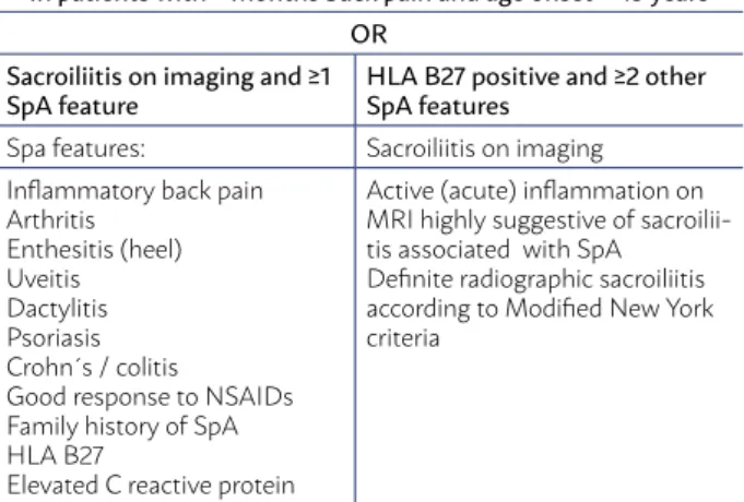
TABLE 1 - ASAS CLASSIFICATION CRITERIA FOR AXIAL
SPONDYLOARTHRITIS (SPA)
In patients with ≥ months back pain and age onset < 45 years OR
Sacroiliitis on imaging and ≥1
SpA feature HLA B27 positive and ≥2 other SpA features
Spa features: Sacroiliitis on imaging Inflammatory back pain
Arthritis Enthesitis (heel) Uveitis Dactylitis Psoriasis Crohn´s / colitis Good response to NSAIDs Family history of SpA HLA B27
Elevated C reactive protein
Active (acute) inflammation on MRI highly suggestive of sacroilii-tis associated with SpA
was not reported in the included patients. The most common comorbidities were arterial hypertension and diabetes that presented concomitantly in three patients. Two patients were smokers.
The laboratorial findings showed that acute
-phase proteins were positive in six patients (75%), and HLA-B27 was found in two patients (25%).
Four patients had the diagnosis of spondylodisci-tis (50%). One case at L5S1, one case at C45 and C67 (with myelopathy, as mentioned above), one case at T12L1 disc, and the last case at T1112. The clinical pre-sentation was limiting pain in the affected segment in all cases, and the cervical case had some degree of cervical myelopathy in the affected segment. All of them presented negative hemocultures and uro-cultures, but two of them had elevated acute phase proteins. After that, a percutaneous needle spine bi-opsy was performed in three cases and one had an open spinal biopsy. The results revealed unspecific chronic inflammatory process in all cases, without positive identification of the etiological agent, char-acterizing aseptic spondylodiscitis. Treatment con-sisted in a short-term empiric antibiotics treatment,
pain medication and physical therapy. The outcome was complete pain resolution in three patients, and one patient had partial resolution of the pain in the following months.
Three patients had spinal fractures (37.5%), as-sociated with minor trauma and one woman had a cervical kyphotic deformity that precluded her from looking horizontally when walking (12.5%). The cer-vical kyphotic deformity was treated by posterior instrumented cervical fusion (C2T3) with a pedicle subtraction osteotomy of c7.
The three patients with thoracic fractures were treated by a posterior instrumented fusion and had an AO type B3 injury with anterior distraction (T1011; T910 and T89 fractures). All of them had a posterior instrumented fusion two segments above and two segments below the involved level with a reasonable clinical improvement of their pain.
DISCUSSION
Our spine surgery division performs around 150 procedures each year. However, in the period be-tween 2011 and 2016, only nine patients had the di-agnosis of AS, with an estimated incidence of one per 100 patients who underwent a spinal procedure at our institution.
Regarding gender, our study found a predomi-nance of the female population, with a male/female ratio of 1/3. The literature estimates about two men for each women affected by AS.8,13 Some authors
sug-gested an overestimation of male prevalence due to lighter forms of presentation in women, leading to sub notification of those cases.1, 5
The laboratory findings in our patients also di-verged from the reported in the literature.1,14,15 In our
series, most patients (62.5%) presented positive acute phase proteins, and the minority (25%) presented positive antigens HLA B27. These differences may be secondary to regional characteristics of our patients or by the fact that our population included only those patients who underwent a spinal procedure.
The majority (50%) of our evaluated patients was diagnosed with an aseptic spondylodiscitis also known as Andersson’s lesions (described for AS). This data diverges from the literature which suggests that discitis is a rare presentation in the setting of AS. However, its incidence may be underestimated due to the asymptomatic presentation of some cases of aseptic spondylodiscitis.11 Of note, the biopsy of
patients with spondylodiscitis may not identify any infectious agent, leading to false negative results. This should be taken into account as a potential bias of our high rate of aseptic spondylodiscitis.
The incidence of surgically treated fractures was low, corresponding to only three cases (37.5%). The only segment fractured in our small sample was the lower thoracic spine. Both data disagree with the lit-erature in occurrence and location, which presents the majority of the cases in the cervical spine.11 The
progressive cervical kyphotic deformity present in these patients results in major susceptibility to cer-vical fractures in comparison to the other segments. Other factors involved in the vulnerability of the cer-vical segment are: small vertebral bodies, oblique ar-ticular facets and the mobility of the heavy skull on the cervical spine.11,12 Therefore, the most common
level of fractures is the cervical level. Westerveld et al.11 reviewed 93 articles and found 280 (81.2 %) cases
of cervical fractures in a total of 345 patients with AS and diagnosis of spinal fractures However, our series does not include conservatively managed patients, which may also change the epidemiological presen-tation of spinal fractures.
Although limited by the small case number, and retrospective nature, our retrospective study pro-vides unique information about AS patients treated for spinal diseases in a Brazilian university hospital.
CONCLUSIONS
This series displays epidemiological differences in clinical and laboratorial findings, with a high in-cidence of aseptic spondylodiscitis, when compared to the literature. The regional features of surgical-ly managed patients with diagnosis of AS, could be used as future reference for the management of pa-tients in our country.
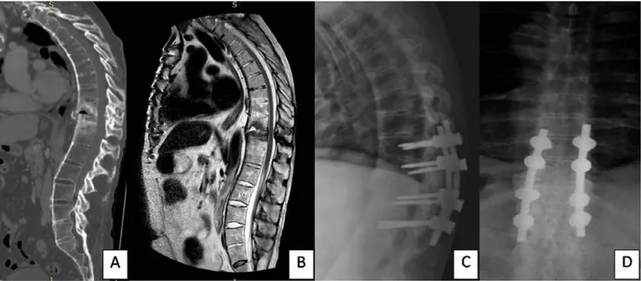
FIGURE 2 – This 54 patient had a fall from the height 2 months before, developing chronic low back pain. Figure 2A - A sagittal
T2 sequence thoracic spine MRI shows a T1011 fracture with anterior vertebral elements distraction. Figure 2B - The CT scan documented the fracture and also gas inside the intervertebral disc. A T9101112 pedicle screw fixation was performed with total pain relief (Figure 2C with the plain radiographs after surgery).
RESUMO
INTRODUÇÃO: A espondilite anquilosante (EA) é uma espondiloartropatia soronegativa, caracterizada principalmente pelo envolvi-mento do esqueleto axial e das articulações sacroilíacas. A EA promove alterações biomecânicas que predispõem a coluna a fraturas, deformidades e à espondilodiscite. O objetivo do presente estudo é reportar as características clínicas e laboratoriais dos pacientes com EA que foram submetidos a procedimentos cirúrgicos na coluna vertebral em nossa instituição.
MÉTODOS: Estudo retrospectivo com revisão de dados médicos dos pacientes com EA que foram submetidos a intervenções na coluna vertebral.
REFERENCES
1. Braun J, Sieper J. Ankylosing spondylitis. Lancet. 2007;369(9570):1379-90.
2. Feldtkeller E, Khan MA, van der Heijde D, van der Linden S, Braun J. Age
at disease onset and diagnosis delay in HLA-B27 negative vs. positive pa-tients with ankylosing spondylitis. Rheumatol Int. 2003;23(2):61-6.
3. Brewerton DA, Hart FD, Nicholls A, Caffrey M, James DC, Sturrock RD. Ankylosing spondylitis and HL-A 27. Lancet. 1973;1(7809):904-7.
4. van der Linden S, Valkenburg HA, Cats A. Evaluation of diagnostic criteria for ankylosing spondylitis. A proposal for modification of the New York criteria. Arthritis Rheum. 1984;27(4):361-8.
5. Vender JR, McDonnell DE. Ankylosing spondylitis. Contempor Neurosurg.
2000;22:1-8.
6. Sieper J, Rudwaleit M, Baraliakos X, Brandt J, Braun J, Burgos-Vargas
R, et al. The Assessment of SpondyloArthritis International Society (ASAS) handbook: a guide to assess spondyloarthritis. Ann Rheum Dis. 2009;68(Suppl. 2):ii1-44.
7. Goei The HS, Steven MM, van der Linden SM, Cats A. Evaluation of
di-agnostic criteria for ankylosing spondylitis: a comparison of the Rome, New York and modified New York criteria in patients with a positive clin-ical history screening test for ankylosing spondylitis. Brit J Rheumatol. 1985;24(3):242-9.
e dois eram HLA-B27 positivos. Em quatro pacientes houve o diagnóstico radiológico presumido de espondilodiscite e estes foram submetidos à biópsia de disco (três por via percutânea e um com biópsia aberta) – em nenhum deles houve identificação de agente infeccioso. Desses, três pacientes tiveram melhora total da dor durante o seguimento, enquanto um deles mantinha dores leves. Houve três casos de fraturas tratadas cirurgicamente (37,5%) e um caso de deformidade cervical cifótica grave (12,5%). Não houve mortes ou complicações relacionadas às cirurgias nessa série.
CONCLUSÕES: A maioria dos dados clínicos e laboratoriais de nosso estudo divergiu da literatura. Essas diferenças podem ser atribuídas às características regionais de nossa população ou pelo fato de incluirmos apenas pacientes que foram submetidos à intervenção cirúrgica.
PALAVRAS-CHAVE: Espondilite anquilosante. Fraturas ósseas. Discite.
8. Jacobs WB, Fehlings MG. Ankylosing spondylitis and spinal cord
inju-ry: origin, incidence, management, and avoidance. Neurosurg Focus. 2008;24(1):E12.
9. Bouvier M, Tebib J, Colson F. Évolution des spondylodiscopathies de la spondylarthrite ankylosante. Rev Rhum. 1987;54:229-33.
10. Bron JL, Vries MK, Snieders MN, van der Horst-Bruinsma IE, van Royen
BJ. Discovertebral (Andersson) lesions of the spine in ankylosing spondy-litis revisited. Clin Rheumatol. 2009;28(8):883-92.
11. Westerveld LA, Verlaan JJ, Oner FC. Spinal fractures in patients with anky-losing spinal disorders: a systematic review of the literature on treatment, neurological status and complications. Eur Spine J. 2009;18(2):145-56.
12. Winkelstein BA, Myers BS. The biomechanics of cervical spine injury and implications for injury prevention. Med Sci Sports Exerc. 1997;29(7 Sup-pl.):S246-55.
13. Hunter T. The spinal complications of ankylosing spondylitis. Semin
Ar-thritis Rheum. 1989;19(3):172-82.
14. Braun J, Bollow M, Remlinger G, Eggens U, Rudwaleit M, Distler A, et al. Prevalence of spondylarthropathies in HLA-B27 positive and negative blood donors. Arthritis Rheum. 1998;41(1):58-67.
15. Gran JT, Husby G. The epidemiology of ankylosing spondylitis. In: