João Pedro Nunes Paulo da Silva Martins
Licenciado em Biologia Celular e Molecular
Nuclear movement to the periphery of
skeletal muscle cells
Dissertação para obtenção do Grau de Mestre em Genética Molecular e Biomedicina
Orientador: Edgar Rodrigues Almeida Gomes, PhD, Instituto
de Medicina Molecular
Júri:
Presidente: Prof. Doutora Margarida Casal Ribeiro Castro Caldas Braga Arguente: Prof. Doutor Sérgio Jerónimo Rodrigues Dias
L
O
M
B
A
D
A
Nuclear movement to the periphery of skeletal muscle cells
João Pedro Martins
João Pedro Nunes Paulo da Silva Martins
Licenciado em Biologia Celular e Molecular
Nuclear movement to the periphery of
skeletal muscle cells
Dissertação para obtenção do Grau de Mestre em Genética Molecular e Biomedicina
Orientador: Edgar Rodrigues Almeida Gomes, PhD, Instituto
de Medicina Molecular
Júri:
Presidente: Prof. Doutora Margarida Casal Ribeiro Castro Caldas Braga Arguente: Prof. Doutor Sérgio Jerónimo Rodrigues Dias
Nuclear movement to the periphery of skeletal muscle cells
Copyright João Pedro Martins, FCT/UNL, UNL
A Faculdade de Ciências e Tecnologia e a Universidade Nova de Lisboa têm o direito, perpétuo e sem
limites geográficos, de arquivar e publicar esta dissertação através de exemplares impressos
reproduzidos em papel ou de forma digital, ou por qualquer outro meio conhecido ou que venha a ser
inventado, e de a divulgar através de repositórios científicos e de admitir a sua cópia e distribuição
com objectivos educacionais ou de investigação, não comerciais, desde que seja dado crédito ao autor
I Agradecimentos
Muito obrigado Edgar pela oportunidade que me deste para trabalhar no laboratório, foi um privilégio.
Obrigado pela disponibilidade e pelas palavras de apoio e incentivo ao longo do ano. Mas
principalmente obrigado por me teres proporcionado as ferramentas necessárias para que eu me
tornasse um cientista melhor e uma pessoa melhor.
Obrigado William, por toda a dedicação, disponibilidade e paciência para me ensinares, foste um
excelente professor. Obrigado por me teres acompanhado ao longo deste ano, incentivando-me a ser
independente mas sempre presente para qualquer dúvida ou problema que pudesse ter. Permitiste que
aprendesse com os meus próprios erros o que me ajudou bastante.
Obrigado Mafalda, pela tua disponibilidade para me ajudares quando o William não podia. Em
particular, muito obrigado por todas as discussões científicas em que me fizeste pensar e despertando o
meu espírito crítico.
A todos os elementos dos grupos do Edgar Gomes e do Cláudio Franco que ao longo do ano estiveram
sempre dispostos a ajudar, obrigado. Todos eles contribuíram, de uma forma ou de outra, para a minha
aprendizagem e para que fosse capaz de levar este projecto a bom porto. Mais do que colegas,
tornaram-se amigos e por tudo isto gostaria de agradecer à Judite, à Vânia, à Patrícia, à Cátia, à Mini
Cátia, ao Graciano, ao Francisco, à Telma, à Sara, à Cheila, à Anna, ao Pedro, à Catarina, à Joana, à
Aida, à Ana, à Isabela e ao Cláudio.
Um grande obrigado a toda a minha família pelo apoio que me têm dado ao longo dos anos. Em
especial, aos meus pais e à minha irmã que tanta paciência e compreensão demonstraram ao longo dos
anos que me permitiram chegar onde cheguei.
Aos meus amigos, que sempre estiveram lá para mim nos momentos bons e nos momentos mais
III Resumo
Os movimentos do núcleo das células são conduzidos por forças polarizadas exercidas por proteínas
motoras e pelo citoesqueleto, sendo importantes para uma multiplicidade de funções celulares.
Durante o desenvolvimento e regeneração do músculo esquelético os núcleos movem-se do centro
para a periferia da miofibra. Este movimento tem início com a formação de uma dobra nuclear entre
miofibrilas, que aumenta gradualmente de tamanho originando um hérnia nuclear. Durante o
movimento para a periferia, o núcleo sofre deformações dramáticas devido à pressão das miofibrilas,
até atingir a periferia da miofibra. O posicionamento dos núcleos na periferia das miofibras é crucial
para uma funções muscular ideal, visto que núcleos localizados no centro de miofibras estão
relacionados com várias patologias musculares. Com este trabalho conseguimos demonstrar que o
mecanismo de movimento nuclear para a periferia depende de alterações locais da rigidez nuclear
regulada por Lamina A/C. Descobrimos também que este movimento é controlado por reticulação de
miofibrilas dependente de Desmina que as aproxima num processo semelhante a um fecho éclair. Por
outro lado a Plectina, Arpc5L e γ actina são responsáveis pela organização desta rede de Desmina nas
linhas-Z. As Nesprinas são componentes principais do complexo LINC e também estão envolvidas
neste mecanismo. Por fim, a depleção de Nesprina1 originou uma redução considerável de núcleos à
periferia o que sugere que estas proteínas também estão envolvidas no movimento nuclear para a
periferia, possivelmente através de mecanotransdução
Palavras-chave: Miofibra-Movimento nuclear para a periferia-Rigidez nuclear-Reticulação de
V Abstract
Nuclear movements are important for multiple cellular functions and are driven by polarized forces
generated by motor proteins and cytoskeleton. During skeletal muscle development and regeneration,
nuclei move from the center to the periphery of the myofiber. Moreover, nuclear movement to the
periphery begins with the emergence of a nuclear wrinkle between myofibrils that gradually increases
forming a bud. The nucleus undergoes severe deformations while being squeezed by myofibrils until it
is finally expelled to the periphery. Nuclear positioning at the periphery of myofibers is crucial for
proper muscle function, with centrally located nuclei being linked to several muscle disorders. Here
we demonstrate that nuclear movement to the periphery of myofibers is dependent on local changes in
nuclear stiffness regulated by Lamin A/C. Furthermore, we found that this movement is mediated by
Desmin dependent myofibril crosslinking and zipping, while Plectin, Arpc5L and γ actin are necessary
for proper Desmin organization at the z-lines. Finally, Nesprin1 depletion resulted in a severe decrease
of peripheral nuclei which suggests that it might play a role in nuclear movement to the periphery
possibly associated with mechanotransduction.
Keywords: Myofiber-Nuclear movement to the periphery-Nuclear stiffness-Myofibril
VII Index
Agradecimentos ... I
Resumo ... III
Abstract ... V
Indexes of Figures ... IX
Indexes of Tables ... XI
List of abbreviations, acronyms and symbols ... XIII
1. Introduction ... 1
1.1. Skeletal muscle cell ... 1
1.2. Lamins ... 7
1.3. LINC complex ... 9
1.4. Desmin ... 11
1.5. Plectin ... 12
1.6. Centronuclear Myopathies ... 13
1.6.1. MTM1 related CNM ... 14
1.6.2. BIN1 related CNM ... 15
1.6.3. DNM2 related CNM ... 15
1.6.4. RYR1 related CNM ... 16
1.6.5. TTN related CNM ... 16
1.7. Laminopathies ... 17
1.8. Desminopathies ... 17
1.9. Plectinopathies ... 18
1.10. Objectives ... 18
VIII
2.1. Myoblast Isolation ... 19
2.2. Myoblast Differentiation ... 20
2.3. Transfections ... 20
2.5. Immunofluorescence ... 21
2.6. Microscopy and Image analysis ... 23
3. Results ... 25
3.1. Lamins ... 26
3.1.1. Nuclear stiffness in nuclear movement to the periphery ... 26
3.1.2. Lamin distribution throughout the nuclear envelope during nuclear movement to the periphery ... 28
3.1.3. Lamin A/C dynamics during nuclear movement to the periphery ... 30
3.2. Myofibril Crosslinking ... 32
3.2.1. Analysis of Desmin function in nuclear movement to the periphery ... 32
3.2.2. Crosslinking organization role in nuclear movement to the periphery... 33
3.3. Cytoskeleton anchorage to Nucleoskeleton and its influence in nuclear movement to the periphery ... 37
4. Discussion ... 41
4.1. Nuclear stiffness and mechanosignaling ... 41
4.2. Myofibril crosslinking ... 43
4.3. LINC complex role in mechanosignaling ... 44
4.4. Nuclear positioning in muscle disorders ... 45
4.5. Conclusions ... 45
IX Indexes of Figures
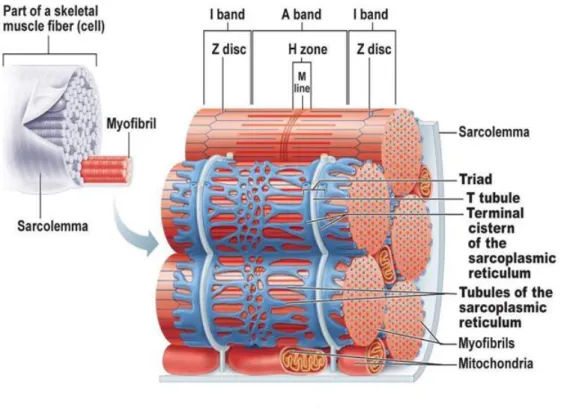
Figure 1.1. Schematic representation of a sarcomere and its structure 2
Figure 1.2. Schematic representation of the membrane structures surrounding myofibrils 3
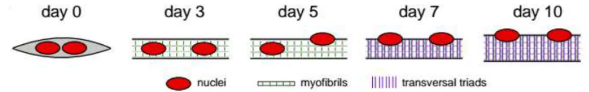
Figure 1.3. Timeline of muscle differentiation in the in vitro system used to study peripheral nuclear
positioning and transversal triad formation 4
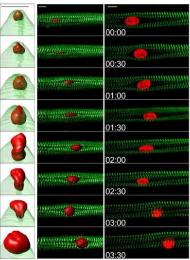
Figure 1.4. Kymograph from a time-lapse movie of a 5-day myofiber depicting peripheral movement
of a nucleus through myofibrils 6
Figure 1.5. Top: Schematic representation of the Lamin protein structure and the assembly process 9
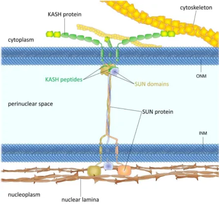
Figure 1.6. LINC complex structural organization and binding partners 11
Figure 1.7. Desmin and Plectin subcellular localization and predicted organization in a myofiber 13
Figure 1.8 Transversal cut of a deltoid muscle. Most nuclei are centrally located which is characteristic
of this disorder 14
Figure 3.1. Theoretical model of peripheral nuclear movement 25
Figure 3.2. Nuclear stiffness is involved in nuclear movement to the periphery 27
Figure 3.3. Lamin B is not involved in nuclear movement to the periphery 28
Figure 3.4. Lamin distribution during nuclear squeezing 29
Figure 3.5. Lamin A/C distribution before and after nuclear movement to the periphery 30
Figure 3.6. Lamin dynamics during nuclear movement 31
Figure 3.7. Myofibril crosslinking by Desmin drives nuclear movement to the periphery 33
Figure 3.8. Arpc5L and γ actin organize Desmin to cross-link myofibrils for nuclear movement 34
Figure 3.9. Plectin is involved in Desmin organization at the z-lines 36
Figure 3.10. Nesprin1 is involved in nuclear movement to the periphery 38
XI Indexes of Tables
Table 2.1. siRNAs used for protein knockdown 21
Table 2.2. List of antibodies used in Immunofluorescence and Western Blotting 22
XIII List of abbreviations, acronyms and symbols
BAF – Barrier to autointegration factor
CH – Calponin homology
CNM – Centronuclear myopathies
EC coupling – Excitation-contraction coupling
FRAP – Fluorescence recuperation after photobleaching
FRET – Forester resonance energy transfer
IF – Intermediate filament
KASH – Klarsicht, ANC-1 and Syne homology
LBR – Lamin B receptor
LEM – LAP2-Emerin-MAN1
LINC – Linker of nucleoskeleton and cytoskeleton
MuSK – Muscle specific receptor tyrosine kinase
Nesprins – Nuclear envelope spectrin repeat protein
NLS – Nuclear localization signal
PFA – Paraformaldehyde
PH – Pleckstrin homology
PI3P – Phosphatidylinositol 3-phosphate
PR – Proline rich
SUN – Sad1 and UNC-84
1 1. Introduction
1.1. Skeletal muscle cell
Skeletal muscle cells, also called skeletal muscle fibers, are long, cylindrical and multinucleated cells
and the main components of muscle. They usually present a diameter between 10 and 100 µm and a
length that can extend up to 30 cm. These cells possess a large quantity of myoglobin which can store
oxygen similarly to hemoglobin and glycogen granules called glycosomes that provide glucose to the
cell, both of which vital for muscle cell activity. Some structures such as myofibrils, sarcoplasmic
reticulum and T-tubules are specific to this type of cells and play an important role in muscle
contraction (Elaine N. Marieb and Katja N. Hoehn, 2015).
When observing a muscle fiber it is possible to see striations, a sequence of lighter and darker bands
called I bands and A bands respectively. The I band has a darker area called the Z disc or Z line while
the A band has a lighter area called H zone which in turn is divided by a darker M line, shown in
Figure 1.1.
Skeletal muscle fibers are composed of rodlike structures called myofibrils longitudinally distributed
throughout each fiber. The number of myofibrils per muscle fiber varies with its size, though they are
always densely packed. Desmin, an intermediate filament (IF), has a role of crosslinking myofibrils.
Myofibrils contain the contractile units of the muscle, the sarcomeres. A sarcomere is the region of the
myofibril comprised between two consecutive Z lines and it is composed of two different
myofilaments. Thick filaments containing myosin that are restricted to the A band whereas thin
filaments containing actin extend along all the I band and into a small area of the A band (Figure 1.1.)
with the latter ones being anchored at Z-lines by α-actinin. In the areas where the myofilaments
overlap each thick myosin filament is surrounded by 6 thin actin filaments and each of these is flanked
by three thick filaments (Elaine N. Marieb and Katja N. Hoehn, 2015).
The myosin molecule is constituted by two heavy polypeptide chains which compose the rodlike tail
and four light polypeptide chains that compose the two globular heads (Fig. 1.1.). The rodlike tails
form the central part of the filament while the globular heads, responsible for linking thick and thin
filaments forming cross bridges during muscle contraction, are facing outward at the end of the
filaments. Actin monomers called globular actin polymerize to form the actin filaments that, when
intertwined, compose thin actin filaments (Fig. 1.1.)(Elaine N. Marieb and Katja N. Hoehn, 2015).
Even though this interaction between thick and thin filaments is the engine for muscle contraction,
such process would be impossible without the regulatory role played by troponin and tropomyosin.
2
therefore impeding the linking with myosin; binding calcium ions after calcium release due to an
action potential which cause troponin to detach from actin allowing the formation of the cross bridges;
and binding tropomyosin aiding its binding to actin. Tropomyosin is rod shaped protein that helps
stiffen and stabilize actin but when the muscle is relaxed it binds and blocks myosin binding sites
stopping the formation of cross bridges (Elaine N. Marieb and Katja N. Hoehn, 2015).
Titin is the protein that forms the core of the thick filaments, anchoring them to the Z lines and M lines
maintaining A band organization and helping muscle resist excessive stretching and recover after
contraction (Fig. 1.1.). Thin filaments are anchored to integrin proteins in the sarcolemma (plasma
membrane) through dystrophin.
Figure 1.1. Schematic representation of a sarcomere and its structure. Adapted from (Elaine N. Marieb
and Katja N. Hoehn, 2015)
According to the sliding filament model of contraction, thin actin filaments slide past thick myosin
filaments and overlap to a greater extent during muscle contraction. When stimulated by the motor
nerve, thick filaments link to myosin binding site of thin filaments forming the cross bridges and
beginning simultaneous contraction of all sarcomeres. During this, cross bridges are formed and
destroyed several times increasing the overlap of the filaments and increasing tension bringing thin
filaments closer to center of the sarcomere. When contraction ends, cross bridges become inactive and
there is a decrease in tension making the sarcomere return to its original relaxed state (Elaine N.
3
The sarcoplasmic reticulum and T-tubules also play a pivotal role in skeletal muscle cell contraction
regulation (Fig. 1.2.). The first is very similar to smooth endoplasmic reticulum with a tubule network
adjacent to each myofibril which is responsible for the regulation of intracellular ionic calcium levels.
This network also has terminal cisternae that flank T-tubules at the A band-I band junction (Fig. 1.2.).
Calcium is released when an action potential stimulates a muscle fiber to contract (Elaine N. Marieb
and Katja N. Hoehn, 2015).
The plasma membrane of the myofiber (sarcolemma) forms invaginations at the A band-I band
junction and forms the T-tubules which increase the muscle fiber surface area and since they are
contiguous with the sarcolemma it facilitates the propagation of the impulse (Fig. 1.2.). T-tubules
stretch in between two terminal cisternae and form structures called triads, where T-tubule proteins
function as voltage sensors and regulate calcium release from the terminal cisternae of the SR to the
whole fiber (Fig. 1.2.). These structures are vital for simultaneous calcium release along the fiber
ensuring proper signal transmission from the sarcolemma to reach the myofilaments and therefore
precise contraction progression. This whole mechanism is called excitation-contraction coupling (EC
coupling) (Elaine N. Marieb and Katja N. Hoehn, 2015).
Figure 1.2. Schematic representation of the membrane structures surrounding myofibrils. Adapted
4 Skeletal muscle cell differentiation
Each muscle fiber results from the fusion of hundreds of myoblasts which need to exit cell cycle in
order to gain the ability to fuse with each other (Abmayr and Pavlath, 2012). Initially they form small
myotubes that lack the major structures characteristic of a developed myofiber, such as triads (Fig.
1.3.). Upon fusion with a myotube, the myoblast nucleus moves towards the center of the myotube,
driven by microtubules (Cadot et al., 2012). This nuclear centration movement is regulated by the
small Rho GTPase Cdc42 and Par3 and Par6 polarity proteins with dynein/dynactin motor complex
also playing an important role. It is predicted that this movement results from nuclei pulling the
microtubules anchored in other nuclei through action of dynein/dynactin (Cadot et al., 2012; Wilson
and Holzbaur, 2012).
During myotube development, nuclei spread evenly throughout the longer axis of the myotube in a
microtubule dependent movement. In this case, nuclei move considerably slower and it is possible to
observe pausing events and nuclear rotation during this movement (Cadot et al., 2012; Englander and
Rubin, 1987; Roman et al., 2016 under revision). Three different mechanisms have been proposed to
be responsible for nuclear spreading, the first one relying on kif5b/kinesin-1 interaction with
microtubule associated protein MAP7. Microtubules anchored at the nuclear envelope by their minus
ends form an antiparallel network which is maintained by kinesin-1/MAP7 complex. The force exerted
by kinesin-1 moving towards the plus end of microtubules pushes nuclei apart (Bruusgaard et al.,
2003; Metzger et al., 2012). Kinesin-1 was reported to localize to the nuclear envelope through its
binding to Nesprin2 bound KLC-2 and therefore it was hypothesized that this would be responsible for
nuclear rotation during nuclear spreading (Wilson and Holzbaur, 2012; Wilson and Holzbaur, 2015).
Another mechanism involves the motor protein dynein anchored at microtubule poles and capable of
pulling the microtubules. Dynein also presents a similar function to kinesin-1 when anchored to the
nuclear envelope, influencing nuclear rotation during nuclear movement (Folker et al., 2012).
Figure 1.3. Timeline of muscle differentiation in the in vitro system used to study peripheral nuclear
positioning and transversal triad formation. Nuclei are in red, myofibrils in white (with z-lines in
green) and transversal triads as purple lines. Day 3: myofibril formation. Day 5: initiation of peripheral
nuclear positioning. Day 7: transversal triad formation. (Adapted from Roman et al., 2016 under
5
After nuclear spreading, these centrally located nuclei begin their movement towards the periphery of
the muscle fiber (Fig. 1.3., 1.4.) (Shichiji et al., 2013; White et al., 2010; Harris et al., 1989). This
process is not exclusive to muscle development since it also happens after injury as a part of a repair
mechanism that involves nuclear movement to the center of the fiber and back to the periphery
(Pastoret and Sebille, 1995; Maxwell et al., 1984). It was recently shown by our laboratory that
nuclear movement to the periphery of skeletal muscle cells is an actin and Nesprin dependent process
(Falcone et al., 2014). N-Wasp is an actin nucleation factor pivotal for nuclear movement to the
periphery and functions downstream of amphiphysin-2, a protein involved in T-tubule and triad
formation (Falcone et al., 2014). The role actin plays in this mechanism suggests the involvement of
linker of nucleoskeleton and cytoskeleton (LINC) complex proteins, Nesprin and Sad1 and UNC-84
(SUN) proteins, in the movement to the periphery, in addition to anchoring nuclei at the periphery of
the myofiber (Lei et al., 2009; Elhanany-Tamir et al., 2012; Zhang et al., 2010). Desmin was also
reported to play a part in nuclear positioning, responsible for maintaining the distance between nuclei
(Ralston et al., 2006; Chapman et al., 2014).
The movement of the nucleus to the periphery of the cell begins with the emergence of an elongated
nuclear wrinkle through a narrow gap in between myofibrils (Fig. 1.4.). This wrinkle gradually
increases in size forming a bud, with the nucleus undergoing dramatic deformation until it is finally
expelled to the periphery of the cell (Fig. 1.4.). Before this movement takes place, there is an area near
the nucleus devoid of myofibrils. As nuclear movement to the periphery ensues, this area starts to
decrease in size as though myofibrils zip together towards the nucleus (Roman et al., 2016 under
6
Figure 1.4. Kymograph from a time-lapse movie of a 5-day myofiber depicting peripheral movement
of a nucleus (H2B-iRFP, red) through myofibrils (YFP-α-actinin, green). Left: view from the right
side, with transparent myofibrils three-dimensional rendering. Middle: view from the top, surface
three-dimensional rendering. Time, hh:mm. Scale bar, 10 μm. Right: 2D view of the central plane of a
kymograph from a time-lapse movie of a 5-day myofiber depicting peripheral movement of a nucleus
(H2B-iRFP, red) through myofibrils (YFP-α-actinin, green). Scale bar, 10 μm.(Roman et al., 2016
under revision)
After nuclear movement to the periphery, the myofiber enters the final stages of differentiation at
which time transversal triads are formed and nuclei start to cluster in the neuromuscular junction (Fig.
1.3.)(Merlie and Sanes, 1985). These neuromuscular junctions are mainly formed in the central region
of muscles where axons specifically connect due to the clustering of acetylcholine receptors in that
area. This process depends on the complex formed between muscle specific receptor tyrosine kinase
(MuSK) and LRP4 regulated by agrin; the myoclustering mechanism itself might rely on MuSK
patterns in association with Nesprins at neuromuscular junctions (Kim and Burden, 2008; Apel et al.,
7 1.2. Lamins
IFs protein family is composed by 73 members distributed through 5 distinct groups based on
assembly properties, structure and their expression pattern throughout the different tissues (Fig. 1.5)
(Eriksson et al., 2009). Type I and II IFs are keratins and they always form heteropolymers while type
III IFs, like Desmin, form homopolymers. Neurofilaments like nestin belong to type IV IFs group
while nuclear Lamins are type V IFs and type VI IFs group is composed by eye lens proteins, such as
Phakinin and filensin (Coulombe and Wong, 2004).
Lamins are the only nuclear IF proteins, responsible for providing structural stiffness and transcription
regulation at the nuclear envelope (Stuurman et al., 1998). These proteins also influence nuclear pore
positioning as well as nuclear envelope protein anchoring and positioning (Zuleger et al., 2011).
Mammals have three Lamin genes which are LMNA, LMNB1 and LMNB2. LMNA gene encodes all
type A Lamins, both major splicing variants Lamin A and Lamin C and the minor splicing variants
A10 and C2. Type B Lamins are encoded by two genes; LMNB1 which encodes one major isoform
Lamin B1 and LMNB2 which encodes the other major isoform Lamin B2 and the minor isoform
Lamin B3. B-type Lamins are ubiquitously expressed while A-type Lamins are predominantly
expressed in differentiated cells (Butin-Israeli et al., 2012).
Both A and B-type Lamins are composed of an N-terminal globular head, a central alpha helical rod
domain and C-terminal tail domains. The latter contains a nuclear localization signal (NLS) and an Ig
fold domain possibly involved in protein-protein interactions (Dhe-Paganon et al., 2002; Shumaker et
al., 2008). In vitro, the alpha helical domains of Lamin monomers interact with each other to form head-to-head dimers, which in turn form a head-to-tail polymer structure. These polymers then
assemble side by side in an anti-parallel fashion to create a 5 to 6 µm protofilament, which is the main
assembly unit of the Lamin meshwork (Fig. 1.5.) (Foeger et al., 2006; Ben-Harush et al., 2009).
Despite this, little is known about the assembly process in vivo. Given that Lamins go through
extensive posttranslational modifications and interact with the nuclear membrane, chromatin and a
vast number of proteins, there are a lot of factors that were not taken into account during the in vitro
studies and perhaps play a crucial role in Lamin assembly. In vivo, A- and B-type Lamins form
separate filament networks with different function, unlike in vitro where they can coassemble.
Regardless of their differences in mechanic and biochemical functions, these filament networks are not
independent, having several contact points and interacting with each other (Shimi et al., 2008). In
terms of mechanical properties, A-type Lamins provide nuclear stiffness enabling nuclei to resist
mechanical stress while B-type Lamins are responsible for providing elastic properties which allows
nuclei to deform to a certain extent (Broers et al., 2004; Lammerding et al., 2006). Changes in the
levels of either A- or B-type of Lamins will result in different nuclear properties. For instance, high
8
movement and cell migration while low levels of Lamin A make nuclei more susceptible to ruptures
(De Vos et al., 2011; Vargas et al., 2012). Furthermore, recent studies suggest that Lamin A levels
might be regulated by force transmission via LINC complex and affect the differentiation process
(Swift et al., 2013).
Lamins interact with a large number of proteins in the nuclear envelope and the nucleoplasm. In
addition to LINC complex proteins, Lamin B receptor (LBR) and LAP2-Emerin-MAN1 (LEM)
domain proteins are also known to interact with Lamins (Fig. 1.5.)(Schirmer et al., 2003). LBR is an
inner nuclear membrane transmembrane protein that binds heterochromatin and is able to impacts
gene silencing mechanisms. In mammals there are 5 LEM proteins in the nucleus: Emerin, LAP2
which has two main isoforms LAP2α and LAP2β, MAN1, LEM2 and LEMD1; most of them requiring
Lamin A for their proper localization (Brachner and Foisner, 2011). These proteins also possess the
ability to bind to heterochromatin, which, depending on the LEM domain protein, might happen
directly or indirectly. LAP2 is able to tether DNA directly due to a LEM like motif while MAN1 and
LEM2 are able to do it due to a winged helix motif in the C-terminal domain (Caputo et al., 2006).
Indirect binding of DNA is achieved by all LEM domain proteins through interactions between a
chromatin binding protein called barrier to autointegration factor (BAF) and the LEM domain (Cai et
al., 2001). One of the LAP2 isoforms, LAP2α, lacks the transmembrane domain and localizes to the
nucleoplasm where it interacts with both chromatin and A-type Lamins (Brachner and Foisner, 2011).
Moreover this particular LEM domain protein might affect cell cycle and chromatin organization
(Dorner et al., 2006). Even though some molecular mechanisms behind chromatin tethering to Lamins
9
Figure 1.5. Top: Schematic representation of the Lamin protein structure and the assembly process.
Adapted from Ihalainen et al., 2015. Bottom: Schematic representation of the nuclear envelope
showing Lamin meshwork and LEM domain Lamin-binding proteins. Adapted from Barton et al.,
2015.
1.3. LINC complex
The LINC complex is composed of outer nuclear membrane Klarsicht, ANC-1 and Syne homology
(KASH) domain proteins and inner nuclear membrane SUN domain proteins (Fig. 1.6.). This complex
provides a mechanical link between the nucleus and the cytoskeleton and plays a role in nuclear
movement and positioning, mechanotransduction and chromosomic movement (Burke and Roux,
2009; Starr and Fridolfsson, 2010). KASH domain proteins, also known as Nesprins (Nuclear
10
elements, to the perinuclear space where the KASH domain interacts with SUN proteins (Fig. 1.6.)
(Sosa et al., 2012). SUN proteins on the other hand are located in the inner nuclear membrane where
they anchor the LINC complex to the nuclear lamina through interaction with A type Lamins and other
proteins like Emerin, while the SUN domain interacts with Nesprin KASH domain in the perinuclear
space (Fig. 1.6.) (Sosa et al., 2012). SUN2 in particular, multimerises to forms a trimer with a triple
helical coiled coil and a globular head, essential for KASH domain to bind along a hydrophobic
groove in between SUN domains (Sosa et al., 2012; Zhou et al., 2012). The existence of numerous
interactions between KASH and SUN proteins demonstrates how the LINC complex is capable of
resisting the mechanical forces applied on the nucleus. There are several Nesprin isoforms that differ
in length and their functional domains, with specific Nesprin isoforms bind to specific cytoskeletal
elements like actin, microtubules and even IFs (Starr and Fridolfsson, 2010). The giant isoforms
Nesprin 1G and Nesprin 2G bind directly to actin filaments through their calponin homology (CH)
domains and are necessary for nuclear movement and positioning at the periphery (Starr and Han,
2002). Nesprins that interact with microtubules usually do so through kinesin or dynein motor proteins
(Starr and Fridolfsson, 2010). Nesprin1 and Nesprin2 directly bind to dynein through specific regions
of their cytoplasmic domains while Nesprin4 binds directly to kinesin light chains and plays an
important role in the early development of myofibers, more specifically in nuclear centration and
spreading (Roux et al., 2009). The only Nesprin isoform known to bind IFs is Nesprin 3α, that binds
Plectin’s actin binding domain which in turn binds IFs through the plakin domain (Wilhelmsen et al.,
2005). Nesprin3α is also known to bind Nesprin1G actin binding domain possibly exerting some sort
11
Figure 1.6. LINC complex structural organization and binding partners. KASH proteins bind to
cytoskeletal elements such as microtubules actin filaments and IFs while SUN proteins anchor the
complex at the INM and interact with the Lamin meshwork. Adapted from Chang et al., 2015.
1.4. Desmin
Mature myofibers possess an IF cytoskeleton which is mainly composed by Desmin. The IFs are
mainly localized at the Z lines or associated with the sarcolemma in structures called the costameres
(Fig. 1.7.) (Lazarides and Hubbard, 1976). After myoblast fusion and during myotube elongation,
desmin interacts with vimentin to form longitudinal strands along the myotube, which after
development give rise to transversal filaments localized to the Z lines (Barbet et al., 1991). It was
reported that desmin depletion affects myoblast fusion hindering myotube formation, however results
obtained in mouse myofibers lacking the Desmin gene suggest that Desmin is not essential for
myotube differentiation since they still develop normal myofibers (Schultheiss et al., 1991). This
suggests that there might be another IF that can compensate the lack of Desmin.
In mature skeletal muscle fibers, Desmin forms scaffolds around the myofibrils at the Z lines and
connects myofibrils to the sarcolemma at the costameres, where Desmin is linked to Plectin. γ-actin
links the costameres to the contractile units due to its capability of binding dystrophin at the level of
the sarcolemma (Rybakova et al., 2000). Desmin filaments also link several organelles like the nucleus
12
that make the crosslink between Desmin and organelles, possibly influencing organelle positioning in
the fiber as well as nuclear shape and positioning (Konieczny et al., 2008). Nevertheless, it is not
certain that nuclear movement to the periphery depends on Desmin transversal scaffold organization.
1.5. Plectin
Plectin is a cytolinker protein, responsible for the anchoring IFs to several cellular structures such as Z
lines, costameres and nuclei (Fig. 1.7.). This protein binds to several different IFs from all subgroups,
being Desmin and Lamin B interactions noteworthy for nuclear positioning in muscle fibers (Fig. 1.7.)
(Foisner et al., 1991; Reipert et al., 1999) . It was previously shown that these interactions are mainly
regulated by phosphorylation, with Plectin phosphorylation triggering its dissociation from the binding
partner. Phosphorylation of the binding partner might also promote this dissociation as is the case with
Lamin B and Plectin interaction (Herrmann and Wiche, 1987; Foisner et al., 1991). Even though its
main function is to bind IFs providing structural stability to this particular network, Plectin is also
know to bind microtubules and actin (Herrmann and Wiche, 1987; Foisner et al., 1995). This might
also help stabilize these cytoskeletal structures and perhaps influence their dynamic regulation, since
Plectin deficiency conditions microtubule dynamics and actin filament polymerization. Plectin is
widely expressed, however it presents higher levels of expression in cells under great mechanical
stress, like muscle cells (Wiche et al., 1983). The Plectin gene contains 41 exons encoding an actin
binding domain and a plakin domain in the N-terminal region, a central coiled coil domain and the
C-terminal domain. The small N-C-terminal domain is a variable region between isoforms and defines their
subcellular localization (Wiche et al., 1991). One of these isoforms, Plectin1, is localized to the
nuclear envelope/endoplasmic reticulum where it is predicted to bind Nesprin3α (Ketema et al., 2007).
A study developed in primary dermal fibroblasts from Plectin1 deficient mice showed actin
cytoskeleton abnormalities and impaired migration, which in turn suggests an involvement of this
isoform in nuclear positioning mechanisms (Abrahamsberg et al., 2005). Alongside Plectin1, Plectin1d
isoform, which localizes to the Z lines (Fig. 1.7.), might also play an important role in nuclear
13
Figure 1.7. Desmin and Plectin subcellular localization and predicted organization in a myofiber.
Adapted from Staszewska et al., 2015.
1.6. Centronuclear Myopathies
Centronuclear myopathies (CNM) are a diverse group of neuromuscular disorders that are
characterized by centrally positioned nuclei, muscle weakness and atrophy (Fig. 1.8.) (Pierson et al.,
2005; Nicot et al., 2007). There are three different genetic forms of this disorder: The X-linked form,
that occurs due to mutations in the gene encoding myotubularin, MTM1 (Laporte et al., 1996); the
autosomal-dominant which is caused by mutations in the amphiphysin-2 and dynamin-2 genes BIN1
and DNM2 respectively (Bitoun et al., 2005); and the autosomal-recessive caused by mutations in
skeletal muscle ryanodine receptor, titin and also amphiphysin-2 encoding genes (RYR1, TTN and
14
Figure 1.8 Transversal cut of a deltoid muscle. Most nuclei are centrally located which is characteristic
of this disorder.
1.6.1. MTM1 related CNM
The X-linked CNM is the most common form and presents a severe phenotype characterized by
neonatal onset, muscle weakness, atrophy and accompanying respiratory involvement with the
necessity of invasive respiratory and nasogastric tube feeds in most cases (Herman et al., 1999). This
disorder affects 2/100000 male births and is usually fatal within the first year of life, although there are
some milder cases in which the individual survives until adolescence or even adulthood.
Myotubularin family of phosphoinositide phosphatases is composed by 14 members in humans, with
several of these proteins mutated in neuromuscular diseases or associated to other conditions like
metabolic disorders and cancer, with mutations in MTM1 being the cause for X-Linked CNM (Begley
and Dixon, 2005; Lorenzo et al., 2005). To date, more than 300 mutations in MTM1 have been
reported, most of them resulting in a significant reduction of myotubularin protein (Biancalana et al.,
2003; Tsai et al., 2005).
These proteins are responsible for the dephosphorylation of phosphatidylinositol 3-phosphate (PI3P)
and phosphatidylinositol 3,5-phosphate, both of which are essential second messengers in membrane
trafficking (Backer, 2008; Blondeau et al., 2000). The influence of PI3P regulation exerted by
myotubularin is not exclusive to membrane trafficking and endocytosis; it also affects autophagy,
formation of autophagosomes and autophagosome-lysosome fusion which depend on PI3P synthesis
(Cebollero et al., 2012; Funderburk et al., 2010). Abnormalities in muscle autophagy, T-tubule,
sarcoplasmic reticulum and triad formation have been reported in several animal models for X-Linked
15
1.6.2. BIN1 related CNM
Amphiphysin 2 related CNM is a rare condition restricted to a few families that is caused by recessive
mutations. The phenotype is usually mild and it is characterized by progressive muscle weakness and
atrophy starting from a young age, although homozygous mutations have been reported to present a
lethal phenotype. Alternative splicing of amphiphysin has been associated to myotonic dystrophy,
which presents some common features with CNM, particularly centrally located nuclei and triad
defects (Fugier et al., 2011).
BIN1 gene encodes amphiphysin 2, a protein localized at the T-tubules and involved in their
formation. It has an N-terminal BAR domain involved in membrane binding and an SH3 domain
responsible for protein-protein interaction, namely actin nucleation promoting factors (Butler et al.,
1997; Lee et al., 2002; Toussaint et al., 2011). N-WASP is an actin nucleation promoting factor known
to act downstream of amphiphysin 2 regulating nuclear positioning and triad formation in skeletal
muscle fibers. These functions are usually disrupted in cases of CNM since N-WASP is probably
misslocalized due to mutations in amphiphysin (Falcone et al., 2014).
1.6.3. DNM2 related CNM
DNM2 related autosomal-dominant form of CNM presents, in most cases, a milder phenotype than the
X-linked and recessive CNM forms and it manifests during adolescence or early adulthood. Identical
to other CNM forms, this disorder presents with general muscle weakness which in this case mainly
affects proximal muscles. It may also present ptosis with ophthalmoplegia, localized muscle
hypertrophy, axonal involvement, neutropenia and cataracts which indicates an influence in other
tissues (Liewluck et al., 2010).
Dynamin protein family is composed by three members; dynamin-1 which is mostly expressed in the
brain, dynamin-2 which is ubiquitously expressed and dynamin-3 which is expressed in the brain and
testes (Praefcke and McMahon, 2004). These proteins are mainly involved in membrane fission and
endocytosis but further roles have been proposed in microtubule network, centrosome cohesion and
actin cytoskeleton assembly, all of which can explain some of the aberrations in nuclear positioning
observed in this condition . Furthermore some abnormalities in autophagy pathways were observed in
mouse models of a common human DNM2 dominant mutation, similar to the ones observed in animal
models for the X-Linked CNM form.
Dynamin-2 is a large GTPase with five major functional domains; a C-terminal proline rich (PR)
domain, a pleckstrin homology (PH) domain, a GTPase effector domain, a middle domain and an
N-terminal GTPase domain (Gu et al., 2010; Thompson et al., 2004). The most severe phenotypes
reported about this form have been related to heterozygous de novo mutations in the PH domain,
16
domain mutations, with the PR domain able to bind to SH3 domain proteins such as amphiphysin
(Bitoun et al., 2005; Bitoun et al., 2007). Some studies suggest that DNM2 mutations hinder N-WASP
localization at the triads and therefore triad biogenesis, thus affecting proper muscle contraction
(Falcone et al., 2014).
1.6.4. RYR1 related CNM
This particular form of CNM is caused by recessive mutations in the RYR1 gene and presents an
intermediate phenotype in terms of severity. The phenotype is very similar to other forms referred
above, with the particularity of having significantly less respiratory impairment (Wilmshurst et al.,
2010). Mutations in this gene are known to be involved in a number of neuromuscular disorders other
than CNM and it is not unusual to have more than one pathogenic mutation in the same allele which
reflects the complexity of RYR1 related myopathies (Klein et al., 2012).
Ryanodine receptors are a family of intracellular calcium channels divided in three main isoforms that
are tissue specific. Ryanodine receptor 1 is mainly expressed in skeletal muscle cells, ryanodine
receptor 2 is primarily expressed in the myocardium while ryanodine receptor 3 is more widely
expressed that the first two but has increased expression in the brain (Takeshima et al., 1989; Nakai et
al., 1990). These ryanodine receptors mediate calcium release from the sarcoplasmic reticulum and
endoplasmic reticulum which is of extreme importance for excitation-contraction coupling in skeletal
muscle (Inui et al., 1987).
1.6.5. TTN related CNM
This is a very rare form of CNM with only 5 cases of CNM linked to recessive mutations in the titin
encoding gene (TTN) so far. Mutations in TTN gene are also responsible for other neuromuscular
disorders and truncated variants of this protein are relatively common. The 5 individuals with this
condition presented generalized muscle weakness, respiratory impairment but with no cardiac
involvement (Ceyhan-Birsoy et al., 2013).
Titin anchors thick filaments at the Z and M lines maintaining sarcomere organization (Fürst et al.,
1988). In TTN related CNM cases, interaction between titin and M lines is usually affected, disturbing
sarcoplasmic reticulum linkage through obscurin, which in turn impacts the organization of the
sarcomere (Bagnato et al., 2003; Charton et al., 2010). C-terminal truncations are also common in this
type of pathology, culminating in a reduction of proteins like nebulin and calpain-3. The latter is vital
for the correct localization of ryanodine receptors to the triad structure thus affecting proper EC
17 1.7. Laminopathies
This group of rare genetic disorders is caused by mutations in Lamin encoding genes. Laminopathies
are usually cell type specific but can affect several tissues at the same time with some similarities in
the phenotype, for example in progeroid syndromes (Worman and Foisner, 2010). The fact that most
laminophaties are tissue specific has a few proposed explanations and yet the mechanism remains
elusive. One hypothesis states that mutant Lamins lead to changes in Lamin meshwork structure
weakening it and making nuclei more susceptible to mechanical force (Brosig et al., 2010). On the
other hand, Lamin mutation might impact overall gene expression during differentiation and it has
been shown that Lamin A deficient myoblasts have reduced expression of proteins, such as Desmin,
which are essential for muscle differentiation (Columbaro et al., 2005; Furukawa et al., 2009). These
disorders most commonly affect striated muscle usually due to mutations in LMNA gene
(Zaremba-Czogalla et al., 2012), although some Lamin B related laminopathies have been reported
(Padmakumar et al., 2005). These mutations can be missense, nonsense, splice site mutations, in-frame
and out-of-frame insertions/deletions, however nonsense and small out-of-frame insertions/deletions
are very characteristic of muscle related laminopathies. One of the most common muscle related
laminopathies is the Emery-Dreifuss muscular dystrophy, which can be caused by mutations in LMNA
gene as well as mutations in SYNE1, SYNE2, Emerin (EMD) and FHL1 (Bonne et al., 1999; Gueneau
et al., 2009). Nevertheless the same mutation in the LMNA gene might present with phenotypic
variations which, coupled with the large number of genes involved, suggests the involvement of
several major players in this type of laminopathies.
1.8. Desminopathies
Desminopathies are characterized by the existence of Desmin aggregates and deficiencies in
sarcomeric organization (Goebel, 1995). Desmin mutations most commonly occur in the alpha-helix
intermediate domain and tail domain, with 37 of the 42 reported mutations. Alpha-helix domain
mutations usually impair Desmin filament assembly, both in Desmin dimerization and dimer-dimer
interaction to form filaments (Kaminska et al., 2004). On the other hand, tail domain mutations have
no reported impact in Desmin filament assembly, affecting Desmin interactions with other
cytoskeleton components like Plectin instead (Bär et al., 2007; Dalakas et al., 2003). Even so, both
18 1.9. Plectinopathies
Plectinopathies usually display with muscular dystrophy, skin blistering and neuropathy. The most
common disorder associated to Plectin mutations is epidermolysis bulbosa simplex with muscular
dystrophy (Gache et al., 1996). Patients suffering from this condition present Desmin aggregates,
deficient myofibrils and overall cytoskeletal organization of myofibers similar to what happens in
mouse models. Even though there are several Plectin isoforms no correlation between site of mutation
and displayed phenotype has been made (Konieczny et al., 2008). Nonsense mutations and
out-of-frame insertions/deletions compose the great majority of Plectin mutations, resulting in truncated
proteins that downregulate their own mRNA through nonsense mediated mRNA decay (Baker and
Condon, 2004). In muscle cells, the phenotype of plectinopathies and desminopathies is very similar
since both Plectin and Desmin play major roles in myofiber cytoskeletal organization and might even
play a role in nuclear positioning mechanisms.
1.10. Objectives
We recently found that nuclei are moved to the periphery of myofibers by an unexpected mechanism
involving the crosslinking and contraction of myofibrils. Furthermore we demonstrated that Arp2/3 complexes containing Arpc5L together with γ-actin are involved in the crosslinking of myofibrils that act as closing zippers on both sides of the nucleus (Roman et al., 2016 under revision). Myofibrils
induce growing centripetal forces on centrally located nuclei. These centripetal forces eventually
squeeze and extrude the nuclei to the cell periphery (Roman et al., 2016 under revision). We
hypothesize that myofibril crosslinkers such as Desmin are regulated by Arp2/3 complexes containing Arpc5L with γ-actin allowing force to be exerted in the nucleus. We also predict that nuclear stiffness, which is mainly regulated by nuclear Lamins, is required for nuclear squeezing to the periphery of the
myofiber. In this work we further explore the mechanism of nuclear movement to the periphery of
myofibers, focusing on determining the role of myofibril crosslinkers, nuclear stiffness and LINC
19 2. Materials and Methods
2.1. Myoblast Isolation
All procedures using animals were approved by the Institutional ethics committee and followed the
guidelines of the National Research Council Guide for the care and use of laboratory animals. Hind
limb muscle tibialis anterior, extensor digitorum longus, gastrocnemius and quadriceps from P3-P7
newborn mice were isolated (Figure 2.1) and placed in ice cold Dulbecco’s PBS (Sigma-Aldrich® cat#
D8537-500ML). Exceeding PBS was removed using a 10 ml pipette in order to facilitate the mincing
process. Isolated muscle was minced with a dissection scissor and digested for 1h 30 minutes at 37ºC
in 5 ml Digestion mixture (0,5mg/ml collagenase (Sigma-Aldrich® cat: C0130-500MG) and 3.5mg/ml
dispase (Invitrogen® cat# 17105041) in Dulbecco’s PBS; mixture was then filtered with 0.22 mm
Minisart® high flow Syringe Filter(Sartorius cat# 16541-K)). Digestion reaction was stopped by
adding 6 ml Dissection medium (IMDM with Glutamax (Invitrogen cat# 31980022);
penicillin/streptomycin 1% (Alfagene cat# 15140-122); Fetal Bovine Serum (FBS) 10% (Eurobio cat#
CVFSVF00-01) previously heated to 37ºC and the obtained suspension was centrifuged at 600 rpm
during 5 minutes. The fat residues and cell debris present in the supernatant were aspirated using a
vacuum pump and the suspension was centrifuged again at 600 rpm during 5 minutes. The supernatant
was centrifuged at 1400 rpm during 5 minutes after which the supernatant was discarded and the pellet
containing the cells was ressuspended in 10 ml of Dissection medium. The obtained cell suspension
was filtered using a 40 µm cell strainer (Enzifarma cat# 352340) followed by the addition of 15 ml
Dissection medium to reach 25 ml total volume that was then pre-plated on 150 mm petri dishes
(SARSTEDT cat# 83.3903) for 3.5 to 4 hours. One hour before the end of pre-plating, fluorodishes
(WPI cat# FD35-100) and 35 mm dishes ((LabClinics cat# 153066) number of fluorodishes and 35
mm dishes used in each experiment depends on the number of newborn mice utilized; 1 newborn
mouse is equivalent to 2 fluorodishes or 1.5 35 mm dishes) were coated with Matrigel Reduced Factor
(Corning cat# 354230) diluted 1:100 in 500 ml of IMDM with Glutamax and left for one hour at room
temperature. Matrigel Reduced Factor must be kept on ice during this procedure since it will start to
polymerize at 10ºC. After pre-plating, the supernatant of the 100 mm petri dish was collected and
centrifuged at 1400 rpm during 5 minutes. The supernatant was then discarded and the pellet was
ressuspended in a suitable volume of Growth medium (IMDM with Glutamax; penicillin/streptomycin
1%; FBS 20%; Chicken Embryo Extract (produced in the laboratory); (The suitable volume of Growth
medium varies according to the number of mice used in each procedure; suitable volume of Growth
medium equal to number of mice used)) keeping the suspension with a higher cell concentration for
cell counting, with further addition of growth medium until a concentration of approximately 150000
20
dishes were washed with 500 ml Dulbecco’s PBS and cells were plated; 150000 in fluorodishes and
225000 in 35 mm dishes.
2.2. Myoblast Differentiation
Primary myoblasts usually take between 2 to 5 days to reach confluence. They start to fuse
spontaneously when they reach 70% to 80% confluence and at this time we switched Growth medium
with Differentiation medium (IMDM with Glutamax; 2% HyClone Donor Equine Serum (GE
Healthcare Life Sciences cat# SH3007402); penicillin/streptomycin 1%). This is done by aspirating
the Growth medium and washing once with 1 ml Differentiation medium before adding it. The day
after switching mediums we started the full differentiation procedure by putting a Matrigel cryotube
containing 1 ml to thaw at 4ºC. After thawing, Matrigel was diluted 1:1 with chilled Differentiation
medium and cells were covered with this mixture immediately after medium aspiration (150 µl and
225µl total volume for fluorodishes and 35 mm dishes respectively). The cells were placed in the
incubator at 37ºC and 5% CO2 for 30 to 40 minutes until Matrigel polymerizes. After this, 1 µl of
recombinant rat agrin (R&D Systems cat# 550-AG-100) was added per 1 ml of Differentiation
medium (previously warmed up to 37ºC) which was then added to the cells (1 ml per fluorodish and
1.5 ml per 3,5 mm dish). Every two days, half the medium was changed by discarding 500 µl and
adding 500 µl new Differentiation medium with twice the agrin in order to ensure same agrin
concentration. The first day of agrin is considered day 1 of differentiation and fibers take between 7 to
10 days to fully mature.
2.3. Transfections
In muscle cells, the transfection procedure has to be performed at the correct stage of development in
order to increase transfection efficacy and also cell survivability. When cells start to fuse and the first,
small myotubes start to appear they are ready for transfection. First of all, DNA/RNA and
Lipofectamine mixtures were prepared in cryotubes (Fisher Scientific cat# 1000-4220) by adding 1 µl
DNA/RNA or Lipofectamine to 49 µl of Opti-MEM medium (Life Technologies cat# 31985-047)
respectively, mixed carefully and were then incubated at room temperature for 5 minutes (Each 50 µl
of DNA/RNA mixture is prepared in separate cryotubes and used to transfect cells in a single
fluorodish; all steps involving pipetting Lipofectamine must be done carefully so that liposome
formation is not affected). Following the incubation step, 50 µl of Lipofectamine mixture were added
to DNA/RNA mixture and gently mixed; Lipofectamine 3000 (Life Technologies cat# L3000-008)was
used for plasmids and co-transfections while Lipofectamine RNAiMAX (Life Technologies cat #
15338-100) was used for siRNA transfection. The mixture containing DNA/RNA and the respective
21
suitable volume of Transfection Medium (IMDM with Glutamax, 1% FBS) was heated up to 37ºC,
with 400 µl being required for each cryotube. After the incubation time, 400 µl of Transfection
medium are added to each cryotube and Growth medium is switched with the Transfection mixture
obtained. Cells were kept in this mixture for 5 hours at which time it was discarded. Cells were
washed once with Differentiation medium and 1 ml of Differentiation medium was added immediately
after washing. Cells were maintained in culture until full maturation using Myoblast Differentiation
protocol as shown before. All siRNAs used are described in Table 2.1.
2.4. Plasmids
YFP-α-actinin plasmid was a gift from Pekka Lappalainen. iRFP-H2B was a gift from Mathieu
Coppey. EmGFP-Desmin was obtained through addgene (plasmid #54059). mCherry-Lamin A/C was
obtained through addgene (plasmid#55068).
Table 2.1. siRNAs used for protein knockdown.
Target Ambion
ID Sequence (sense) Sequence (anti-sense)
Scrambled genecust UUCUCCGAACGUGUCACGUtt ACGUGACACGUUCGGAGAAtt
γ-actin s61904 AGAUAAUGUUUGAAACCUUtt AAGGUUUCAAACAUUAUCUgc
β-actin s200989 UGACGUUGACAUCCGUAAAtt UUUACGGAUGUCAACGUCAca
Nesprin1 genecust CCAUCGAGUCUCACAUCAAtt UUGAUGUGAGACUCGAUGG
Arpc5 s206017 AGAUGAUGCUAUAAGUAtt UACACUUAUAGCAUCAUCUgg
Arpc5L s92445 GCGUGGAUAUCGACGAAUUtt AAUUCGUCGAUAUCCACGCgg
Lamin A/C
1 S69252 GGCUUGUGGAGAUCGAUAAtt UUAUCGAUCUCCACAAGCCgc
Lamin A/C
2 s69253 CCACCGAAGUUCACCCUAAtt UUAGGGUGAACUUCGGUGGga
Lamin B1 S69255 GACUUGGAGUUUCGUAAAAtt UUUUACGAAACUCCAAGUCct
Desmin s64942 GAGGAGAUCCGACACCUUAAt UUAGGUGUCGGAUCUCCUCct
Plectin 1 s201801 GGAGUGACCGCAAUACCAAtt UUGGUAUUGCGGUCACUCCaa
Plectin 2 S201802 CGAGUACACCUUUGAGGGAtt UCCCUCAAAGGUGUACUCGgg
Plectin 3 s71823 GGCCGUCUCUUCAAUGCUAtt UAGCAUUGAAGAGACGGCCat
2.5. Immunofluorescence
Immunofluorescence was performed in order to determine protein localization, function in nuclear
movement and to do statistical analysis of specific phenotypes. When myofibers reached the desired
stage in development they were fixed using 200 µl of 4% paraformadehyde (PFA) (Science Services
22
day at which cell were fixed depended on the goal of the experiment. For instance, for quantifications
and statistical analysis only cells fixed between day 7 and day 10 of development were used while for
protein localization and function in nuclear movement assays cells were fixed at day 4 to 5 of
development. After fixing, cells were washed two times with PBS and then permeabilized with a 0.5%
triton (Sigma-Aldrich cat# X100-100ML) solution for 5 minutes. The cells were then washed two
times with PBS before adding 200 µl of a blocking mixture composed by 50% BSA (5 g BSA
(Sigma-Aldrich cat# A7906-50G) in 10 ml MiliQ water) diluted 1:10 in Goat Serum 10% during one hour.
Next, cells were washed once with 200 µl of PBS to remove the blocking mixture and the primary
antibody solution, composed by 50% BSA and 1% Saponin (0.1 g Saponin (Sigma-Aldrich cat#
47036-50G-F) and 5 g BSA in 10 ml MiliQ water) diluted 1:10 in goat serum 10% plus primary
antibodies (diluted 1:5 to 1:200 depending on the primary antibody used), was added. Cells are then
left at 4ºC overnight. To remove the primary antibody solution the cells were washed two to three
times with PBS for 5 minutes each time. This wash was done with agitation in order to reduce
unspecific binding and antibody clustering. Subsequently, 200 µl of the secondary antibody solution
was added to the cells and incubated for one hour at room temperature, protected from light with tin
foil. This solution was composed by 50% BSA and 1% Saponin diluted 1:10 in goat serum 10% plus
secondary antibodies, DAPI and Phalloidin (with secondary antibodies diluted 1:200, DAPI diluted
1:1000 while Phalloidin was diluted 1:200 depending on the phalloidin used). Following this
incubation period, the secondary antibody solution was discarded and cells were washed three times
with 200 µl of PBS for 10 minutes each. Once again this wash was done with agitation. Finally 200 µl
of mounting medium Fluoromout G (Southern Biotech cat# 0100-01) were added to the cells and left
overnight to dry before image acquisition. The described immunofluorescence protocol is referent to
the staining of only one fluorodish. All antibodies and antibody dilutions are described in Table 2.2
and Table 2.3.
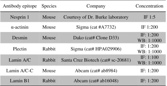
Table 2.2. List of antibodies used in Immunofluorescence and Western Blotting.
Antibody epitope Species Company Concentration
Nesprin 1 Mouse Courtesy of Dr. Burke laboratory IF 1:5
α-actinin Mouse Sigma (cat #A7732) IF 1:200
Desmin Mouse Dako (cat# Clone D33) IF: 1:200
WB: 1:1000
Plectin Rabbit Sigma (cat# HPA029906) IF: 1:200
WB: 1:1000
Lamin A/C Rabbit Santa Cruz Biotech (cat# sc-20681) IF: 1:100
WB: 1:1000
Lamin A/C-C Mouse Abcam (cat# ab8984) IF: 1:200
23
Table 2.3. List of secondary antibodies used in Immunofluorescence
Secondary antibodies Species Company Concentration
Anti-Rabbit IgG (H+L) 555 Alexa Fluor Goat Life Technologies
(cat# A21429) IF 1:200
Anti-Mouse IgG (H+L) 488 Alexa Fluor Donkey Life Technologies
(cat# A21202) IF 1:200
Phalloidin - Thermo Scientific
(cat# A12379) IF: 1:200
2.6. Microscopy and Image analysis
Live imaging was performed in a Zeiss Cell Observer fully motorized inverted microscope equipped
with a spinning disk confocal unit, a with a large cage incubator and a small stage incubator for
temperature control and CO2 supply, a 63x oil immersion objective, a Definite Focus unit and an
Evolve 512 EMCCD camera. Cells were maintained at 37ºC and 5% CO2 in Differentiation medium
supplied with agrin during image acquisition which was done at 5 minute intervals for spinning disk
confocal microscopy and at 15 minute intervals for widefield. For fluorescence recuperation after
photobleaching (FRAP) experiments, cells were photobleached for 5 to 10 minutes by scanning half a
nucleus with 100% intensity of 555 nm laser. Acquired images were analyzed using the ZEN software
(Blue edition), Fiji and Imaris 8. Confocal images of fixed cells were acquired using a Zeiss LSM 710
and a Zeiss LSM 880 confocal point scanning microscopes both with a 63X oil immersion objective.
Images were analyzed using ZEN software (Gray edition), Fiji and Icy. 3D rendering was done with
the software Imaris 8, by manually or automatically creating a Surface in the Surpass function. All
image acquisition for one specific experiment was done using the same settings for data to be
25 3. Results
The theoretical model developed in the laboratory takes into account known biophysical parameters of
myofiber components and predicts the role of myofiber contraction, myofibril cross-linking and
nuclear stiffness for nuclear movement to the periphery (Fig. 3.1.). In this project, we explore the role
of nuclear Lamins, main determinants of nuclear stiffness and plasticity; Desmin, the main myofibril
crosslinker; and Nesprins, components of the LINC complex with predicted involvement in this
movement (Falcone et al., 2014). Firstly, we tried to determine if Lamin absence and overexpression
caused a phenotype through siRNA mediated knockdown and transfection with mCherry-Lamin A/C
respectively, followed by fixing and immunofluorescence staining. Furthermore we looked into Lamin
distribution throughout the nuclear envelope with further immunofluorescences and FRAP
experiments. Desmin and Nesprin function was characterized by siRNA mediated knockdown and
overexpression coupled with live imaging and fixed cell imaging with further image analysis.
Figure 3.1. Theoretical model of peripheral nuclear movement. A) Schematic of a nucleus during
26
undeformed nucleus. ΔR = amplitude of radial deformation. Fn = force applied by myofibrils on the nucleus. L = length between the crosslinkers (blue) on each side of the nucleus. h = height of a nuclear
wrinkle formed by myofibril pressure. B) Model prediction of the stability of wrinkles relative to
global nuclear stiffness. The scaled wrinkle size h/w is plotted as a function of global E.
3.1. Lamins
3.1.1. Nuclear stiffness in nuclear movement to the periphery
The goal was to manipulate nuclear stiffness through Lamin siRNA mediated knockdown and by
mCherry-Lamin A/C overexpression to assess possible effects in nuclear movement to the periphery.
This was done by transfecting cells at day 0 of differentiation and waiting for them to reach full
maturity at day 7 to 10. The cells were then fixed with PFA and stained for Lamin A/C and Phalloidin.
We started by transfecting cells with two different LMNA siRNAs in order to test their efficacy and
assess the phenotype they originated compared to a negative control. The negative control used was a
Silencer Negative control that has no significant similarities with mouse gene sequences, furthermore
the influence in cell fusion and development is negligible. One of them (Lamin A/C 2) triggered
substantial cell death soon after transfection, which affected myoblast fusion and hindered further cell
development. Despite reducing siRNA concentration to half in subsequent experiments, cell death
remained a significant issue making it impossible to use in this experiment. On the other hand, Lamin
A/C 1 siRNA did not induce such substantial cell death letting myoblasts fuse and differentiate and in
turn allowing for proper image acquisition and analysis. One possible explanation for the difference in
results between both siRNAs might be the existence of off-target effects in the case of Lamin A/C 2
siRNA that might have led to an increase in cell mortality. For this experiment, only the Lamin A/C 1
siRNA was used. We found that Lamin A/C downregulation leads to a decrease of peripheral nuclei
(Fig. 3.2.A, B). The Lamin A/C knockdown was confirmed by immunofluorescence (Fig. 3.2.A) and
by western blotting (Fig. 3.2.C). Similarly, Lamin A/C overexpression led to a decrease of peripheral
nuclei suggesting that both the increase and decrease of nuclear stiffness impair nuclear movement to