ABSTRACT
Objective: To compare two amblyogenic risk factors screening modalities in children from an urban area.
Design: Prospective
Subjects: children from pre-schools and elementary schools from an urban area.
Materials and methods: Two independent amblyogenic risk factors screening programs, from April till September 2014. Pre-school screening was performed with photoscreening technology and the school-aged screening program in accordance with a validated protocol, regularly used in routine Infantile and Juvenile Health appointments, in Primary Care setting. Children with referral criteria in pre-school program were sent an invitation for a Paediatric Ophthalmology appointment; in the school-aged group these subjects were given a report letter for their General practitioner, recommending Ophthalmology referral.
Results: Pre-school program: four hundred and nine children were screened and 47 (11.6%) had referral criteria. From the 25 children who came to the appointment, all presented ocular movements, slit lamp and fundus exam without significant alterations, 18 cases presented refractive amblyopia risk factors, giving this screening program a positive predictive value of 72%. After treatment all children improved visual acuity. In school-age program: from the 664 screened children, 102 (15.4%) had suspected ophthalmic pathology. From the 43 subjects evaluated in the after-screening appointment, 27 had a normal ophthalmological exam. Among the 16 altered exams, 9 had amblyogenic potential or amblyopia, giving this screening program a positive predictive value of 20.9 %.
Conclusion: Photoscreening revealed to be particularly effective in assessing amblyopia risk factors in preliterate children, with a higher positive predictive value than the Primary Health Care protocol in older children.
RESUMO
Objetivo: Comparar duas modalidades de rastreio de fatores de risco ambliogénicos em crianças numa área urbana.
Desenho: Prospetivo.
Participantes: crianças das pré-escolas e escolas básicas de uma área urbana.
Materiais e métodos: Comparação de dois rastreios de fatores ambliogénicos independentes, de Abril a Setembro de 2014. O rastreio pré-escolar foi realizado com tecnologia de fotorrastreio. O rastreio escolar foi realizado de acordo com o protocolo validado usado nas consultas de rotina de Saúde Infantil e Juvenil dos Cuidados de Saúde Primários. Crianças com critérios de referenciação no rastreio pré-escolar eram convocadas a consulta de Oftalmologia Pediátrica, no rastreio escolar, era recomendada a referenciação.
Resultados: Rastreio pré-escolar: quatrocentas e nove crianças rastreadas, destes, 47 (11,6%) cumpriam critérios de referenciação. Das 25 que compareceram à consulta, todas apresentavam movimentos oculares externos, biomicroscopia e fundoscopia sem alterações significativas, 18 casos apresentavam fatores de risco ambliogénicos de causa refrativa, apresentando um valor preditivo positivo de 72%. Após tratamento todos melhoraram a acuidade visual. No rastreio escolar: das 664 crianças rastreadas, 102 (15,4%) tinham suspeita de patologia oftalmológica. Das 43 crianças avaliadas em consulta, 27 tinham um exame oftalmológico normal, 16 tinham alterações oftalmológicas, das quais 9 com potencial ambliogénico ou ambliopia instalada, conferindo a este rastreio um valor preditivo positivo de 20,9 %.
Conclusões: O fotorrastreio revelou ser superior à forma de rastreio vigente nas consultas de saúde infantil nos Cuidados de Saúde Primários Portugueses. Foi particularmente efetivo na identificação de fatores de risco ambliogénicos em crianças pré-literadas, com um valor preditivo positivo mais elevado.
Palavras Chave: ambliopia, fotorrastreio, crianças, tecnologia automática.
INTRODUCTION
Amblyopia is still the main cause of monocular vision loss in children and young adults, with an incidence of 1 – 3.5 % in developed countries.1 It is potentially reversible if timely identified and treated, with ample evidence of the direct relation between age and severity of the amblyopia, if not treated.2,3 The most common cause of amblyogenic visual disparity are refractive errors, ocular misalignment, or pathologies occluding the visual axis. Refractive causes and strabismus represent 90 % of all amblyopia cases.1 Amblyopia treatment is highly effective, with around 75 % of the children
with less than 7 years old achieving a normal visual acuity with optimal optical correction, occlusion or penalization.1 Because of this, efforts to detect amblyogenic risk factors (ARF) and its timely intervention during the critical period of visual development are widely recognized as an integrant part of primary paediatric health care.4-10 Screening methods may be divided in subjective and objective tests. The former require child’s cooperation, but are more effective in older children. Among the latter type, photoscreening proved to be efficient, especially in preverbal children.11-17 Since the appearance of the first automatic screening machines, these devices became more portable, faster and easier to use, without committing accuracy.
Moreover these devices have added other resources over subjective traditional screening tests, with less time per child and less dependency their cooperation. Photoscreening devices are autorefractors, measure pupil size and symmetry, interpupillary distance, gaze direction and also detect means opacities. Photoscreening is a validated, recognized and recommended screening method in pre-school aged children.8,9,17
The main purpose of this work is to compare two methods of screening in two age groups of children: one is the protocol currently used for every Portuguese child in the general practitioner’s and paediatrician’s office, in the regular
Preventive Medicine and Infantile and Juvenile Health visits. The other is a photoscreening protocol in a strategically targeted age group for ARF detection.
MATERIAL AND METHODS
Comparative analysis based on a prospective study of two different and independent screening programs on an urban area. The defined region has a 20.3 km of area and 16,049 of resident population, accordingly to Portugal 2011 general population census.18
Positive predictive value (PPV) of each screening program was calculated. Residual amblyopia cases, defined as an inter-ocular difference of at least two lines of visual acuity at the last visit, were identified. Follow-up time was defined as the number of months between the first and last Paediatric Ophthalmology appointment. When more than one ARF was present, it was only considered the most amblyogenic, for classification purposes. To estimate the prevalence of ARF, screening negative results were assumed as true negative cases. All subjects’ enrolment and informed consent were in accordance with the tenets of the Declaration of Helsinki.
Screening protocols
• Pre-school photoscreening protocol
All pre-schools of the defined area were contacted for scheduling the screening session, 8 out of 9 consented and were able to set dates for the sessions (the 9th could not find a suitable date). Photoscreening sessions were conducted between June to September 2014 and performed at each kindergarten. Legal representatives of the children received an informative leaflet of the study, an informed consent and a short questionnaire with sex, date of birth and if already followed on an ophthalmologist and why. The screening team
on the field was composed of two elements of a group of three Paediatric and Ophthalmology residents, trained in the handling
of the photoscreener device. The training consisted of an overview and explanation of the purpose and protocol of the study, followed by 3 hours of actual training with examples of readable and unreadable exams. The exams were performed in an available room, in a dim light environment; the equipment was set on a table; the subject was positioned facing away bright light sources and the laptop screen. The used device was PlusoptiX®S04, PlusoptiX Inc. Atlanta USA, an infrared coaxial video camera capable of simultaneous binocular measurement of refractive error, pupil size, interpupillary distance, and evaluate ocular alignment, the presence of ptosis or media opacities. It is portable, handheld, with non-contact measurements at a 1 m distance and connected to a laptop. Fixation on the camera is encouraged by light and sound stimuli. The PlusoptiX camera was used accordingly to the manufacturer’s recommendations.19 Image acquisition time averaged 5-10 seconds and at least 3 readings were obtained per subject. Two independent paediatric ophthalmologists evaluated the screening results using Arthur et al modified referral criteria,20 (Table 1.) blinded for other subject’s information. The possible results were positive, negative and unreadable. Subjects who had a positive or unreadable exam were sent a letter with an ophthalmology appointment date within 2 – 3 months after the screening date. Negative screenings were classified as normal and were not referred. The Paediatric Ophthalmology after-screening appointment was performed at (Ophtalmology Department of Hospital Pediátrico, Centro Hospitalar e Universitário de Coimbra, Coimbra, Portugal), by two paediatric ophthalmologists, blinded for the screening result and personal data. It included visual acuity (Sloan, tumbling E, Allen pictures), pupillary reflexes, ocular motility, cover tests, binocular stereopsis tests (Lang I and II and Titmus), cycloplegic refraction, dilated fundus examination and slit lamp examination. Positive screening exams that were found positive at the appointment were considered a true positive. Follow-up appointments were scheduled at the examiners discretion depending on clinical evaluation.
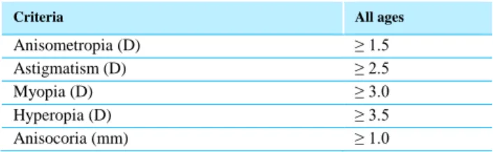
Table 1 – Referral criteria for the PlusoptiX S04 (Arthur modification 2).21
Criteria All ages
Anisometropia (D) ≥ 1.5
Astigmatism (D) ≥ 2.5
Myopia (D) ≥ 3.0
Hyperopia (D) ≥ 3.5
• School-age protocol
The population was comprised of children from every elementary school (12 schools) (6 to 10 years old) of the defined area. Screening sessions were conducted from
April to June 2014 and performed at each school. Legal representatives of the children received an informative leaflet of the study and an informed consent was obtained. A previously validated Paediatric Ophthalmologic Screening exam (by the Portuguese Paediatric Society and the Portuguese Ophthalmology Society) was applied as in the Primary Health Care setting.5,6This protocol includes: ocular external examination; cover and Hirschberg tests; red reflex; eye movements, pupillary light reflexes, visual acuity and stereopsis. (Annex I) External ocular exam evaluates cornea transparency, structural anomalies, ocular surface, iris changes and horizontal corneal diameter. Red reflex was observed with direct ophthalmoscope, at 50 cm of distance, evaluating its colour and symmetry (Bruckner test) and for leucocory presence. Hirschberg test was performed at the same distance evaluating its symmetry. Cover-uncover test for a near target, ocular movements in the nine gaze positions, convergence and the presence of nystagmus was evaluated. Monocular distance¾ visual acuity was tested with tumbling E scale adapted for 3 m distance, of the last line with at least 50 % correct, registered in decimal values. Monocular near visual acuity was tested with Rossano and Weiss scale. Pinhole was used to detect potential refractive errors. Stereopsy was evaluated with Lang 2 test, and considered normal if 200 sec of arch were registered (Annex I). Two Paediatric residents, familiar with the protocol, performed the exams at the schools. The training consisted of an overview and explanation of the purpose and protocol of the study, followed by 3 hours of training in the primary care setting, in scheduled preventive medicine Infantile and Juvenile Health visits. Children who failed the screening exam received a letter to inform their primary care physician with a report recommending referral to a Paediatric Ophthalmology appointment for further evaluation. The after-screening Paediatric Ophthalmology appointment was performed in a similar way as to the pre-schoolers’ protocol.
Statistical analysis
For statistical analysis, SPSS software version 20.0 (SPSS Inc., Chicago, IL, USA) was used. All values are presented as mean ± standard deviation. For the level of
statistical significance calculated with Chi-square test, a p-value less than 0.05 or 0.001, was considered statistically significant.
RESULTS
Pre-school screening
Four hundred and nine children, from 8 schools, were screened. The exam had a mean estimated duration of 30 seconds per child. They were 3.9 ± 1.3 years old (0.3 – 6.64); no unreadable exams were verified and 47 (11.6 %) exams met referral criteria. Since 15 of these children were already followed on an ophthalmologist, 32 were sent an invitation for the after-screening appointment. Twenty-five subjects came and 7 missed the appointment, accounting for 78.1 % of adherence. All cases had normal ocular motility, slit lamp and dilated fundus exam, except for one girl with anisometropic hyperopia who had bilateral optic disc drusen and no further alterations. Refractive ARF with spectacle correction need was identified in 18 cases (7 cases with astigmatism; 11 with anisometropia) (Table 2.). None of the cases presented strabismus or media opacities. This screening exam showed a PPV of 72 % for ARF and an estimated prevalence of ARF of 4.4 % in this population. In the 13.8 ± 6.8 months follow-up, only one child needed amblyopia treatment (penalization with atropine) and all children improved visual acuity with the optic correction and amblyopia treatment. No residual amblyopia was registered at the last visit.
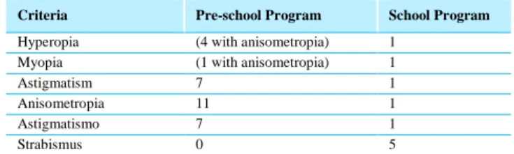
Table 2 – Amblyopia risk factors and causes in the two groups.
Criteria Pre-school Program School Program
Hyperopia (4 with anisometropia) 1
Myopia (1 with anisometropia) 1
Astigmatism 7 1
Anisometropia 11 1
Astigmatismo 7 1
Strabismus 0 5
School screening
A total of 664 children, from 12 schools were screened. With this protocol, each exam took an estimated average of 10 minutes per child. They had a mean age of 7.9 ± 1.3 years old (6-11 years). A hundred-and-two (15.4 %) had a positive test result, with ophthalmological pathology suspected, 20 of which already used optical
correction. Forty-three came to the after-screening Paediatric Ophthalmology appointment, accounting for 42.2 % of adherence; from these, 27 had a normal ophthalmological exam, and 16 had an abnormal exam. Strabismus was identified in 5 cases and ametropia with optical correction need in 12 cases: 3 myopia, 1 hyperopia, 8 astigmatism (3 of these had also strabismus) and 1 anisometropia. However risk factors with amblyogenic potential were only present in 9 of these cases (Table 2.). Including all cases with an abnormal ophthalmological exam, this screening program showed a PPV of 37.2 %, when including only cases with amblyogenic potential this rate descended to 20.9 %, with an estimated prevalence of ARF of 1.36% in this population. In the follow-up of 14.4 ± 7.0 months, only two children needed occlusive treatment: one with strabismus and the other with
anisometropia; both had residual ambliopia in the last visit, despite best treatment and compliance.
Comparison between screening programs
The proportion of screening tests with referral criteria was not significantly different between the two groups (p = 0.085, Chi-square test), achieving a similar referral rate. Photoscreening was much quicker to perform, with a very faster test per child, needing less screening sessions per school. There was also a significantly larger proportion of pre-school children with a positive screening test that came to the after screening appointment (78.1 % vs. 42.2 %, p < 0.001, Chi-square test). Moreover, while none of the affected cases in photoscreening group had residual amblyopia, two cases were seen in the school-aged group despite best treatment and compliance. (Table 3.)
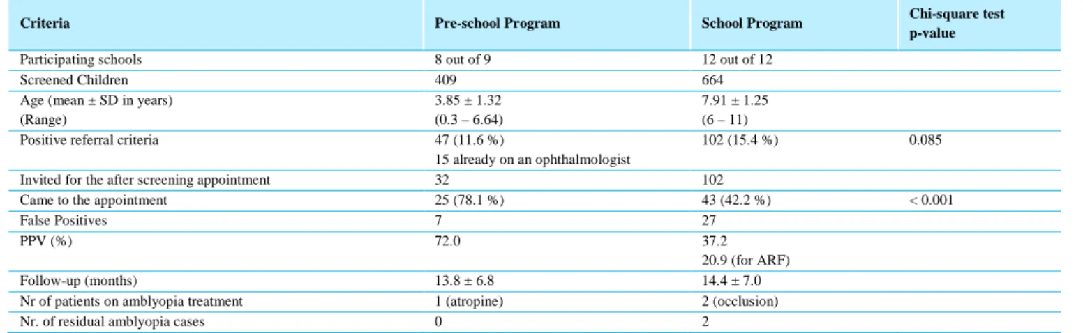
Table 3 – Comparison between the two screening programs
Criteria Pre-school Program School Program Chi-square test
p-value
Participating schools 8 out of 9 12 out of 12
Screened Children 409 664
Age (mean ± SD in years) (Range)
3.85 ± 1.32 (0.3 – 6.64)
7.91 ± 1.25 (6 – 11)
Positive referral criteria 47 (11.6 %)
15 already on an ophthalmologist
102 (15.4 %) 0.085
Invited for the after screening appointment 32 102
Came to the appointment 25 (78.1 %) 43 (42.2 %) < 0.001
False Positives 7 27
PPV (%) 72.0 37.2
20.9 (for ARF)
Follow-up (months) 13.8 ± 6.8 14.4 ± 7.0
Nr of patients on amblyopia treatment 1 (atropine) 2 (occlusion)
Nr. of residual amblyopia cases 0 2
PPV – positive predictive value; NA – non applicable; ARF – Amblyogenic risk factors
DISCUSSION
Traditional vision screening is challenging because it requires children’s cooperation, is time consuming and requires trained personnel; thus accurate and fast objective methods to improve amblyopia detection are necessary.
The sensitivity and specificity of the two protocols are different. Arthur et al modified criteria for this photoscreening protocol have shown a sensitivity of 87% and a specificity of 90% for ARF. In contrast, the school-aged group protocol presented values around 85% and 100% of sensitivity and specificity respectively, but for ophthalmological pathology in general, not only ARF.6,20,21
The screening coverage was excellent, since it was performed directly at the schools, and only one pre-school was unable to schedule the sessions. Resident children in the pre-school age who do not attend these kindergartens were not included in the screening program. The difference between these populations (kindergarten population and the same age resident population) may translate into different results, but this difference was not quantified because it was not the objective of this study.
Referral rates were similar with both methods, as in Salcido and colleagues work.14 Similarly to other studies that compared photoscreening with traditional screening techniques, our photoscreening exam took much less time than the traditional protocol.14 Consequently, it involves less expenses in human resources and time, with more
screened children per session. In what concerns to costs, it wasn’t necessary to invest on a photoscreening device, since it was lend by the Paediatric Ophthalmology Department for the study. However this type of devices cost around 5.800 to 7.500 € (manufacturer price) depending on the monitor resolution and interface options. As predicted, photoscreening also needed less cooperation by the subjects making it ideal in preverbal and preliterate children.11-17
The highest adhesion to the after-screening appointment in the photoscreening group is explained by the different referral method and by the not exclusion of children already followed by an ophthalmologist in the school-aged group. Automatic scheduling by the screening team, not being dependent on further evaluation by the primary care physician and on his referral led to the highest appointment adhesion. In some children of the school-aged program the report may not have reached its destination, his primary care physician might have disagreed with the indication and did not refer the child, or the child sought a different ophthalmologist.
The evaluation of visual acuity with pinhole was not part of the original protocol. Despite not changing the screening referral/pass indication, it was included in the school-aged group because the result could influence the priority of referral by the primary care physician.
Other works have shown similar PPV with photoscreening techniques for the same age group.14 Donahue and colleagues have also shown that PPV of this photoscreening protocol improves with age, being described as 38 % in children with around 6 months old and around 76 % in 5-year-olds. We had similar results, with a mean PPV of 78.1 % even without separating the population in age classes.22 In contrast, traditional screening methods have shown very low PPV in children of this age group, with many false positives, improving its performance with age.14 Despite this, in our school-aged population, the traditional screening method revealed to be much less efficient with a PPV for ARF of 20.9 % in comparison with the photoscreening program in kindergarteners.
In the pre-school follow-up no residual amblyopia cases were registered. Only one case was on amblyopia treatment, with a good response to atropine penalization. In the school-aged group two cases had residual amblyopia notwithstanding the correct treatment and compliance. Despite this small number of cases, this
difference might be due to the more timely intervention and the higher cerebral plasticity in the pre-schoolers and the established amblyopia in the older group. These results emphasize the importance of an early diagnosis and intervention with optical correction and amblyopia treatment when needed, to full visual potential recovery.
The school program intended to simulate the regular screening on the Primary Care setting as in scheduled Preventive Medicine and Infantile and Juvenile Health visits. Therefore the school screening was performed by trained Paediatric residents and not by ophthalmologists. Nonetheless some kind of bias might be introduced by the choice of the screening team, since in primary care setting a General Practitioner or a Paediatrician performs this exam. The different experience component was reduced with the training period before the study. Moreover, since the team on the field screened more children per session than if in the regular Primary Care visits, in a very systemized protocol, this bias was further minimised and might even be overtaken. Other difference was the setting, as the screening sessions were not on regular scheduled visits at the Health Centre, but directly at the school in a shorter period of time and with a higher coverage. Other relevant aspect is that not all Portuguese Primary Care offices are equipped with the required material for this exam (visual acuity charts, Lang I or II plates), further improving our results in comparison with our national clinical practice reality.
Although a direct comparison between the two programs is not possible, since we are comparing different age groups with different methods, indirect comparisons allowed taking some conclusions and reflections. Other limitation of our work is the population size; despite being a population study of the defined area, it represents a convenience sample of a wider urban area.
This is the first study of the Portuguese reality comparing the screening method routinely offered to every child in the Primary Care and an organized photoscreening program in a strategically targeted age group. This is a population study, which enrolment was made at schools, and not in an Ophthalmology appointment, avoiding sample biases.
In conclusion, refractive causes were the most prevalent ambliogenic risk factor in our population. Photoscreening is more time efficient and has a significantly higher PPV than traditional screening methods for ARF, particularly in preverbal and preliterate
children in our urban population. A high sensitivity is desirable to detect as many affected children as possible, but a high specificity is also important to lower false-positives and over-referrals.
This screening method is promising respecting all principles of screening by the World Health Organization: adequate sensitivity and specificity, low cost, ease of administration, safe, imposes minimal discomfort, and is acceptable to both patients and practitioners.23 But photoscreeners detect ARF rather than amblyopia, and the
natural history of many of these amblyogenic factors and its role on emmetropization process remains a mystery. Nonetheless, children with ARF if not identified and adequately treated in the first years of life would probably progress to irreversible vision loss. If these results can be replicated in similar age groups, support for traditional vision screening must undergo intense scrutiny, and attention should be turned toward creating an adjusted photoscreening programme feasible for widespread implementation.
Annex 1
Registo do Rastreio Oftalmológico Infantil (>6 anos)
NOME: _________________________________________________________________ TM: _______________________________ DATA: ______ - ______ - ______________ IDADE: _______ ANOS / MESES MÉDICO: _______________________________________________________________
Exame Ocular Externo
Posição viciosa da cabeça S / N Coloboma da íris S / N
Ptose S / N Coloboma da pálpebra S / N
Nistagmo S / N Blefarite S / N
Aspecto transparente do segment anterior S / N Conjuntivite S / N
Córnea ≤ 12 mm S / N Outras S / N
Refelexo vermelho pupilar S Vermelho S Róseo S Branco Simétrico S / N
Reflexo na córnea (Hirschberg) Centrado S / N
Movimentos oculares conjugados (partindo do olhar primário)
OD OE
Para cima S / N Para cima S / N
Para baixo S / N Para baixo S / N
Para dentro S / N Para dentro S / N
Para for a S / N Para for a S / N
Para cima e para dentro S / N Para cima e para dentro S / N
Para cima e para fora S / N Para cima e para fora S / N
Para baixo e para dentro S / N Para baixo e para dentro S / N
Para baixo e para fora S / N Para baixo e para fora S / N
Reflexo pupilar fotomotor
OD OE Direto S / N Direto S / N Consensual S / N Consensual S / N Convergência S / N Teste de cover OD OE Movimento S / N Movimento S / N
Teste de cover / uncover OD OE
Movimento S / N Movimento S / N
Visão estereoscópica Lang S Bom S Mau S Duvidoso
Visão estereoscópica Fly Teste Mosca S / N Animais (segundos de arco)
_____ - _____ - _____
Círculos (segundos de arco) _____ - _____ - _____ Acuidade Visual
de Perto Teste
OD OE
Valor decimal _____ - _____ Valor decimal _____ - _____
Acuidade Visual
de Longe Teste
OD OE
Valor decimal _____ - _____ Valor decimal _____ - _____
REFERENCES
1. Gunton KB. Advances in amblyopia: what have we learned from PEDIG trials? Pediatrics 2013;131:540-7. 2. Donahue SP. Relationship between anisometropia,
patient age, and the development of amblyopia. Am J Ophthalmol 2006;142:132-40.
3. Holmes JM, Lazar EL, Melia BM, et al. Effect of age on response to amblyopia treatment in children. Arch Ophthalmol 2011;129:1451-7.
4. Saúde DGd. Circular Normativa: Programa Nacional para a Saúde da Visão. In: Saúde Md, ed.2005.
5. Pinto F, Guerra I, Maia I, Rodrigues S. Rastreio Oftalmológico Infantil nos Cuidados Primários. Acta Pediátrica Portuguesa 2007;38:99-102.
6. Pinto F, Rodrigues S, Pessoa B, Coelho P. Estudo piloto para validação de um Protocolo de Rastreio Oftalmológico Infantil em Cuidados de Saúde Primários. Acta Pediátrica Portuguesa 2007;38:93-8. 7. 2010 GdtdCpoPNdS-. Bases de reflexão para um
programa nacional de saúde da visão. Sociedade Portuguesa de Oftalmologia; 2003.
8. Force UPST. Vision screening for children 1 to 5 years of age: US Preventive Services Task Force Recommendation statement. Pediatrics 2011;127:340-6. 9. Miller JM, Lessin HR. Instrument-based pediatric
vision screening policy statement. Pediatrics 2012;130:983-6.
10. Araújo FMF. Despacho n.o 5868-B/2016. In: Saúde GdSrdEAed, ed.: Diário da República; 2016:13942(2) -(3).
11. Donahue SP, Baker JD, Scott WE, et al. Lions Clubs International Foundation Core Four Photoscreening: results from 17 programs and 400,000 preschool children. J AAPOS 2006;10:44-8.
12. Leman R, Clausen MM, Bates J, Stark L, Arnold KK, Arnold RW. A comparison of patched HOTV visual acuity and photoscreening. J Sch Nurs 2006;22:237-43. 13. Paff T, Oudesluys-Murphy AM, Wolterbeek R, et al.
Screening for refractive errors in children: the plusoptiX S08 and the Retinomax K-plus2 performed by a lay screener compared to cycloplegic retinoscopy. J AAPOS 2010;14:478-83.
14. Salcido AA, Bradley J, Donahue SP. Predictive value of photoscreening and traditional screening of preschool children. J AAPOS 2005;9:114-20.
15. Silbert DI, Matta NS, Andersen K. Plusoptix photoscreening may replace cycloplegic examination in select pediatric ophthalmology patients. J AAPOS 2013;17:163-5.
16. Vieira B, Ribeiro I, Menezes C, et al. Fotorrefracção no rastreio refractivo visual da criança: substituto ou complemento? Oftalmologia 2014;38:13-8.
17. Ruao M, Almeida I, Leitao R, et al. Photoscreening for amblyogenic risk factors in 1-year-olds: results from a single center in Portugal over a 9-year period. J AAPOS 2016.
18. INE INdE. População residente por Local de residência, Sexo e Grupo etário (Decenal). 2011.
19. GmbH P. PlusoptiX S04 Hand-Held Binocular Auto Refractometer - INSTRUCTION MANUAL. Version 4.6.15 ed: Plusoptix GmbH; 2005:1 - 38.
20. Arthur BW, Riyaz R, Rodriguez S, Wong J. Field testing of the plusoptiX S04 photoscreener. J AAPOS 2009;13:51-7.
21. Nathan NR, Donahue SP. Modification of Plusoptix referral criteria to enhance sensitivity and specificity during pediatric vision screening. J AAPOS 2011;15:551-5.
22. Donahue SP, Johnson TM. Age-based refinement of referral criteria for photoscreening. Ophthalmology 2001;108:2309-14; discussion 14-5.
23. Wilson JMG, Jungner G, Organization WH. Principles and practice of screening for disease. . Geneve: World Health Organization; 1968.
CONTACT
Cátia AzenhaCentro de Responsabilidade Integrado de Oftalmologia Centro Hospitalar Universitário de Coimbra
Faculdade de Medicina da Universidade de Coimbra E-mail: c.azenha@hotmail.com
The authors have no financial or proprietary interest in any material or method presented herein.