Joana Teixeira da Costa Lucas
Novel Strategies to Combat Gram-Negative
Bacterial Pathogens Using Bacteriophage
Proteins
Joana Teixeira da Costa Lucas
Novel Strategies to Combat Gram-Negative
Bacterial Pathogens Using Bacteriophage
Proteins
Dissertação de Mestrado
Mestrado em Bioengenharia
Trabalho efetuado sob a orientação de
Doutor Sílvio B Santos
DECLARAÇÃO Nome: Joana Teixeira da Costa Lucas
Endereço eletrónico: [email protected] Telefone: 912405399/227834320 Cartão do Cidadão: 14221126
Título da dissertação: Novel Strategies to Combat Gram-Negative Bacterial Pathogens Using Bacteriophage Proteins / Novas Estratégias para Combater Bactérias Patogénicas Gram-Negativos Usando Proteínas Bacteriofágicas
Orientadores:
Doutor Sílvio Roberto Branco dos Santos
Professora Doutora Joana Cecília Valente Rodrigues Azeredo Ano de conclusão: 2015
Mestrado em Bioengenharia
DE ACORDO COM A LEGISLAÇÃO EM VIGOR, NÃO É PERMITIDA A REPRODUÇÃO DE QUALQUER PARTE DESTA TESE/TRABALHO.
Universidade do Minho, _____/_____/_________ Assinatura:
A
CKNOWLEDGMENTS
“Agradeço aos meus orientadores, Dr. Sílvio Santos e Prof.ª Dra. Joana Azeredo, por me terem proporcionado a realização deste projecto. Um especial agradecimento ao Dr. Sílvio Santos por todo o incentivo, esclarecimentos e sugestões prestados durante a realização desta tese, e também, pelo à vontade e constante animação em laboratório, principalmente na presença do rádio.”
“Aos restantes colegas do DEB, um muito obrigada por todos os momentos e camaradagem, em particular ao Carlos e ao Macedo, que tornaram os dias no laboratório mais divertidos, ao Zé por todas as explicações, histórias e conversas e à Cláudia, pela companhia no ginásio.”
“Aos meus amigos, particularmente à Sílvia, à Inês e ao Manel por, quando em momentos de desespero, sempre me terem ignorado, levando-me a perceber que não havia motivo para tal.”
“Aos meus pais e à minha irmã, devo-lhes a minha eterna gratidão, por sempre me terem apoiado e incentivado e nunca terem desistido de mim. Um obrigada a todos os familiares por toda a preocupação e interesse demonstrados.”
“Obrigada Hugo, por me conheceres como mais ninguém conhece e por todos os momentos fantásticos que só tu me sabes proporcionar.
A
BSTRACT
The infections caused by Gram-negative pathogens are a concern to global health, resulting in high rates of morbidity, mortality and high costs in health care. Once formed, biofilms cause reduction of host defenses and increase resistance to antimicrobial products 10 to 1000 times more than the cells in their planktonic state.
According to recent studies, in 2050, bacterial infections, associated with the growing resistance of pathogens to antimicrobial agents, will cause more than 300 million premature deaths and a loss of € 90 trillion to the global economy. At this scenario is extremely important the research and the development of new methodologies to eliminating bacteria and prevent or eradicate biofilm formation. The use of phage proteins presents several advantages as a potential alternative to antibiotics, since they are natural products with a low environmental impact and able to reduce aggregation and bacterial growth.
The aim of this project was to select and study the use of phage proteins to treat three Gram-negative bacteria: Pseudomonas, Salmonella and Escherichia. From the 18 proteins studied, 10 were expressed and these 4 had positive results. The holin from phage Lambda and the spanin Rz from phage T1 eliminated, with efficiency, planktonic cells of ATCC 10145 and protein T1_146 showed to be effective when applied to PAO1 planktonic cells. Biofilms of BL21 were effectively removed by Alginate Lyase. The results showed the studied phage protein present high potential in the control of Gram-negative bacteria turning them an important alternative to antibiotics in the treatment of infection caused by these bacteria.
R
ESUMO
As infeções causadas por bactérias patogénicas Gram-negativas são uma preocupação para a saúde global, resultando em taxas elevadas de morbidade, mortalidade e em altos custos em cuidados de saúde. Os biofilmes bacterianos provocam a redução das defesas do hospedeiro e o aumento da resistência a produtos antimicrobianos entre 10 a 1000 vezes mais que nas células no seu estado plantónico.
Segundo estudos recentes, em 2050, as infeções bacterianas, associadas à crescente resistência dos patogénicos aos agentes antimicrobianos, provocarão mais de 300 milhões de mortes prematuras e uma perda de €90 triliões para a economia global. Tendo em conta este cenário é da máxima importância a aposta e desenvolvimento de novas metodologias que permitam o controlo de bactérias e a formação de biofilmes. A utilização de proteínas fágicas apresenta várias vantagens, uma vez que são produtos naturais, com baixo impacto ambiental, capazes de reduzir a agregação e o crescimento bacteriano com baixa probabilidade de as bactérias desenvolverem resistência.
O objectivo deste projecto foi a seleção e estudo de proteínas (bacterio)fágicas para o controlo de três bactérias Gram-negativas com relevância clínica: Pseudomonas, Salmonella e Escherichia. De entre as 18 proteínas estudadas, 10 foram expressas e dessas, 4 apresentaram resultados positivos. A holina do fago Lambda e a proteína Rz do fago T1 eliminaram, com eficiência, as bactérias plantónicas de P. aeruginosa ATCC 10145 e a proteína T1_146 mostrou ser eficaz quando aplicada a células plantónicas de P. aeruginosa PAO1. Os biofilmes de E. coli BL21 foram os mais eficazmente removidos pela Alginato Liase.
Os resultados obtidos mostraram que as proteínas de origem fágica aqui estudadas apresentam um grande potencial no controlo de bactérias Gram-negativas constituindo uma alternativa aos antibióticos no tratamento de infecções provocadas por estas bactérias.
I
NDEX
Acknowledgments ... v
Abstract... vii
Resumo... ix
List of Figures ... xiii
List of Tables ... xv
List of Abbreviations ... xvii
1. State of the art ... 1
1.1 Gram-Negative Bacterial Pathogens ... 3
1.1.1 Planktonic Cells and Associated Problems ... 4
1.1.1.1. Identification of Gram-negative Bacteria with Clinical Relevance ... 5
1.1.2 Bacterial Biofilms ... 8
1.1.2.1. Biofilm formation ... 9
1.1.2.2. Identification of Gram-negative Biofilm with clinical relevance ... 10
1.2 Clinical treatment of bacterial infection ... 11
1.3 Bacteriophages ... 15
1.3.1 Bacteriophage Therapy ... 17
1.3.2 Bacteriophage proteins in the treatment of Gram-negative Bacterial Pathogens ... 20
2. Materials and Methods ... 23
2.1 Cloning and Expression of Bacteriophage Proteins ... 25
2.1.1 Bacteria, Plasmid, Phages and Growth Conditions ... 25
2.1.2 Phage DNA Extraction and Gene Amplification ... 25
2.1.3 Gene Insertion in the pET-28a Cloning Vector ... 30
2.1.4 Gene cloning in E. coli ... 32
2.1.5 Protein expression ... 33
2.1.6 Protein purification ... 34
2.2 Evaluation of proteins activity against planktonic and sessile cells ... 35
2.2.1 Test on Planktonic Cells ... 35
3. Results and Discussion ... 39
3.1 Bioinformatics Analysis ... 41
3.1.1 Prediction of transmembrane helices and signal peptides ... 41
3.1.2 Prediction of isoelectric point, molecular weight and proteins domains ... 44
3.1.3 Codon usage ... 46
3.2 Laboratory Tests ... 47
3.2.1 Colony PCR ... 47
3.2.2 Protein expression and Purification ... 48
3.2.3 Protein activity tests ... 50
4. Conclusion and Futures Perspectives ... 57
Bibliography ... 61
Annexes ... 79
Annex I – Expression vector pET-28a ... 81
Annex II – Proteins’ Amino Acid sequences ... 82
Annex III – Genes’ DNA Sequences ... 83
Annex IV – Competent culture CaCl2 method ... 85
Annex V – Cells Transformation ... 86
Annex VI – Preparation of SDS-PAGE ... 87
L
IST OF
F
IGURES
Figure 1- Components of the GN cell, detailing the structures of the envelope. ... 4
Figure 2- Stages of biofilm development33. ... 9
Figure 3- Drug efflux, the great responsible for antibiotic resistance in GN organisms84 ... 12
Figure 4- Adsorption and penetration of the phage to the bacterial cell114 ... 16
Figure 5-Lytic and lysogenic cycles. The replication of lytic bacteriophages follows the lytic cycle, while lysogenic bacteriophages may followed by lytic or lysogenic cycles122 ... 17
Figure 6- A 96 well plate scheme: wells on blue represent distilled water; grey wells schematize biofilms: line C - P. aeruginosa ATCC 10145, line D - P. aeruginosa PAO1, line E - S. enterica S1400 and line F - E. coli BL21. On columns are represented the proteins tested on biofilms: column 4 - dispersin B, column 5 - Dispersin B_146, column 6 - gp49, column 7 - Alginate Lyase and column 8 - the control. ... 37
Figure 7- Composion of the 1% agarose gels resulted from Colony PCR from each gene. Column 1 – 1 Kb ladder; column 2 – holin T1; column 3 – holin T1_146; column 4 – holin T4; column 5 – holin T4_146; column 6 – holin T7; column 7 – holin T7_146; column 8 – holin Lambda; column 9 – holin Lamda_146; column 10 – Rz T1; column 11 – Rz T1_146; column 12 - RZ1 Lambda; column 13 – RZ1 Lambda_146; column 14 – dispersin B; column 15 – dispersin B_146; column 16 – gp146; column 17 – gp49. ... 47
Figure 8- SDS-PAGE of the expressed proteins. M - protein marker, 1- dispersin B (40 kDa); 2- dispersin B_146 (67 kDa); 3- holin T1_146 (36 kDa); 4- holin Lambda_146 (39 kDa); 5- gp146 (28 kDa); 6- holin T4 (26 kDa); and 7- gp49 (78 kDa). ... 50
Figure 9- CFU/ml, on a logarithmic scale, from the proteins tests on planktonic cells. ... 51
Figure 10- CFU/ml, on a logarithmic scale, of the sessile cells released due to the proteins activity. The ‘#’ represents an amount of CFU higher than 200. ... 53
Figure 11- CFU/ml, on a logarithmic scale, of the cells in biofilm, after the proteins activity. The ‘#’ represents an amount of CFU higher than 200. ... 54
Figure 12- Restriction map of cloning vector used, obtained from http://biochem.web.utah.edu/hill/links/pET28.pdf. ... 81
Figure 13- Multiple Cloning site of expression vector pET-28a, obtained from http://biochem.web.utah.edu/hill/links/pET28.pdf. ... 81
Figure 14- Identifying transmembrane domains and the signal peptides of dispersin B, using the Phobius database. ... 88 Figure 15 - Identifying transmembrane domains and the signal peptides of gp49, using the Phobius database. ... 89 Figure 16- Identifying transmembrane domains and the signal peptides of gp146, using the Phobius database. ... 89 Figure 17 - Identifying transmembrane domains and the signal peptides of holin Lambda, using the Phobius database. ... 90 Figure 18 - Identifying transmembrane domains and the signal peptides of holin T1, using the Phobius database. ... 90 Figure 19 - Identifying transmembrane domains and the signal peptides of holin T4, using the Phobius database. ... 91 Figure 20 - Identifying transmembrane domains and the signal peptides of holin T7, using the Phobius database. ... 91 Figure 21 - Identifying transmembrane domains and the signal peptides of RzT1, using the Phobius database. ... 92 Figure 22 - Identifying transmembrane domains and the signal peptides of R1 Lambda, using the Phobius database. ... 92
L
IST OF
T
ABLES
Table 1- Organisms and the clinical features associated1 ... 7
Table 2- Humans infections involving GN biofilms77 ... 11
Table 3- Examples of new strategies to inhibit or disrupt biofilms at different stages of their development33 ... 14
Table 4- Some of the problems with early therapeutic phage research and the ways they have been addressed in more recent studies122. ... 19
Table 5- List of bacteriophage proteins to express... 26
Table 6- Primers sequences of the proteins and its characteristics ... 27
Table 7- Parameters used to the PCR ... 30
Table 8- Quantities of components used to make a digestion ... 31
Table 9- Quantities of each element added to the plasmid ... 31
Table 10- Quantities used to insert the genes into the plasmid vector ... 31
Table 11- Parameters used to the Colony PCR ... 32
Table 12- Different conditions tested to optimize the protein expression ... 33
Table 13- Scheme of the proteins tested on planktonic cells. ... 36
Table 14- Proteins tested on biofilms ... 37
Table 15- Predicted TM topology and SP. CYT and green represent the residues present on cytoplasm; TMH (dark grey) illustrates the amino acids located within the membrane; NCYT, at blue, show the residues situated at periplasm or extracellular; and the NCYT, at red SP, represent the signal peptides. The AA No is the amino acid boundary between the different locations of each segment. ... 42
Table 16- Predicted domains of the proteins. MW represents the molecular weight, in dalton; pI is the isoelectric point; DOM indicates the designation of the different domains and AA No is the number of amino acids associated to those domains ... 45
Table 17- Rare codons on E. coli strains present on each protein... 47
Table 18- CFU/ml of planktonic cells, after proteins action. ... 51
Table 19- CFU/ml of the sessile cells released from the biofilm. The UC (uncountable) represent an amount of CFU higher than 200. ... 53
Table 20- CFU/ml of the cells composing the biofilm. The UC represent an amount of CFU higher than 200. ... 54
Table 21- Amino Acid sequence of each protein ... 82 Table 22- DNA sequence of each gene ... 83 Table 23 – Components of SDS-PAGE ... 87
L
IST OF
A
BBREVIATIONS
AHLs – acyl homoserine lactones AIs – autoinducers
AIPs – autoinducing peptides APS – ammonium persulfate BSA – bovine serum albumin CFU – colony-forming unit DNA – deoxyribonucleic acid
EDTA – ethylenediamine tetraacetic acid EO – essential oils
EPS – extracellular polymeric substances GI – gene identification Gi – gastrointestinal GN – Gram-negative GP – Gram-positive IM – inner membrane Kan – kanamycin LB – lysogeny broth LPS – lipopolysaccharide MCS – multiple cloning site MW – molecular weight O.D. – optical density OM – outer membrane ORF – open reading frame PBS – phosphate-buffered saline PCR – polymerase chain reaction PES – polyethersulfone
PG – peptidoglycan
PGHs – peptidoglycan hydrolases pH – potential hydrogen
pI – Isoelectric point QS – quorum-sensing
SDS – sodium dodecyl sulfate
SDS-PAGE – sodium dodecyl sulfate polyacrylamide gel electrophoresis SP – signal peptide TAE – Tris-acetate-EDTA TEMED – tetramethylethylenediamine TM – transmembrane TMH – transmembrane helices UC – uncountable
1.1
Gram-Negative Bacterial Pathogens
Bacteria are prokaryotes (unicellular organisms with no nuclear membrane, mitochondria, Golgi bodies, or endoplasmic reticulum) that reproduce by asexual division1.
Bacteria, based on the Gram stain technique, are differentiated in two major categories: Gram-positive (GP), which remain stained as purple by crystal violet on washing and the Gram-negative (GN), which lose the crystal violet-iodine complex during decolorization with the alcohol rinse, but retain the counterstain safranin, appearing reddish or pink1–4.
Gram-negative bacilli (GNB), that can be commensal organisms among normal intestinal flora, are responsible for numerous infections in community and nosocomial settings5–7.
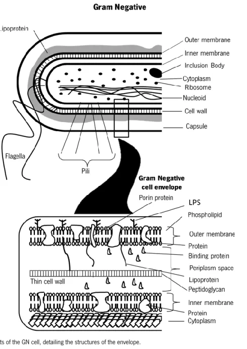
Structurally, the envelope of the GN organisms is free from teichoic or lipoteichoic acids and contains an inner or cytoplasmic membrane, the periplasm space and the outer membrane. The periplasm space contains peptidoglycan (PG) and comprises a variety of hydrolytic enzymes, which are important for the breakdown of large macromolecules during metabolism (phosphatases, lipases, nucleases, and carbohydrate-degrading enzymes) and may represent lytic virulence factors (collagenases, hyaluronidases, proteases and beta-lactamase). The outer membrane allows to distinguish the GN from Gram-positive bacteria and from spirochetes and is the major permeability barrier in GN bacteria. The outer surface of this membrane is composed predominantly by lipopolysaccharides (LPS), or endotoxins, and the inner leaflet of the membrane and the entire inner membrane are composed of phospholipids. Both bilayers can contain a range of different types of membrane proteins. In addition to conferring a GN staining characteristic, these structures are important virulence factors and partially determine antibiotic susceptibility1,8,9. The mentioned structures are presented on Figure 1.
Figure 1- Components of the GN cell, detailing the structures of the envelope.
1.1.1 Planktonic Cells and Associated Problems
The human body provides the temperature, moisture, and resources necessary for bacterial growth. Moreover, many of the traits used by bacteria to enable them to invade the environment, to remain adhered or colonize, to gain access to resources (degradative enzymes), to escape clearance by host immune and nonimmune protective responses and the byproducts of bacterial growth are virulence factors, which enhance the ability of bacteria to cause disease10.
The numerous microbes that colonize the human body, generally the gastrointestinal (Gi) tract, mouth, skin, and upper respiratory tract, compose the normal flora. Various microorganisms belonging to the
normal flora of hosts aid in important functions, such as: digestion of food, production of vitamins, protection from colonization with pathogenic microbes and activation of host innate and immune responses. An alteration of the normal flora, due to antibiotic treatment, diet, stress, and changes in the host response to the flora, can lead to inappropriate immune responses, causing inflammatory diseases7.
Damages caused by bacteria are commonly due to the directly tissue damage, or to the release and dissemination of toxins through the blood. Those damages associated with the consequences of the innate and immune responses to the infection may result in a disease. Not all bacteria or bacterial infections cause disease; however, some always do it1,11,12.
The importance of the affected organ and the extent of the damage caused by the bacterial infection gives an estimative of the seriousness of the disease. The gravity of the disease is also influenced by host factors, namely, congenital defects, immunodeficiency states and other disease-related conditions that increase the susceptibility to infection1,13.
The number of bacteria, the capacity to spread, the potential to cause tissue damage and disease, and the host response determines the residence time of a bacterium in the organism1,11.
1.1.1.1. Identification of Gram-negative Bacteria with Clinical Relevance
The GN bacteria are responsible for, among others, several respiratory tract infections, sexually transmitted diseases and Gi diseases. These microorganisms are the major cause of nosocomial (healthcare-associated) infections. The interactions of the host’s susceptibility to infection, the organism’s virulence potential, and the opportunity for interaction between host and organism mediate the development of an infection1,11,12,14. Some of the most important GN pathogens include:
- Pseudomonas, belonging to the Pseudomonadaceae family, an aquatic bacterium specie. As opportunistic pathogen, this organism can cause devastating chronic infections in compromised hosts and bacteraemia in immunocompromised hosts, representing an important source of nosocomial infections, wherein, the P. aeruginosa is the most important specie among more than 20013,15–17. It is the
most common pathogen isolated from patients who have been hospitalized longer than 1 week18–20.
- The family Neisseriaceae englobes three genera of medically important bacteria: Neisseria, Eikenella, and Kingella. Among the ten species of Neisseria found in humans, two are strictly human pathogens: N. gonorrhoeae, responsible for gonorrhea and N. meningitides that can colonize the nasopharynx of healthy people without producing disease or can cause community acquired meningitis, overwhelming
and rapidly fatal sepsis, or bronchopneumonia21–23. Eikenella corrodens and Kingella kingae are
opportunistic pathogens that colonize the human oropharynx24.
- The largest and most heterogeneous collection of medically important GN rods is the Enterobacteriaceae family. One third of all bacteremias, more than 70% of urinary tract infections (UTIs), and many intestinal infections are due to Citrobacter freundii and C. koseri, Enterobacter aerogenes and E. cloacae, Escherichia coli, Klebsiella pneumonia and K. oxytoca, Morganella morganii, Proteus mirabilis, Salmonella enterica, Serratia marcescens, Shigella sonnei, and S. flexneri, Yersinia pestis, Y. enterocolitica and Y. pseudotuberculosis. Those bacteria could be associated with human disease, with members of the normal commensal flora that can cause opportunistic infections or with commensal organisms that become pathogenic when they acquire virulence genes present on plasmids, bacteriophages, or pathogenicity islands24.
- Vibrio and Aeromonas, belonging, respectively, to the families Vibrionaceae and Aeromonadaceae, are the second major group of GN rods. These organisms are found in water and are able to cause Gi diseases25. Within the genus Vibrio three species are particularly important human pathogens: V.
cholerae, V. parahaemolyticus, and V. vulnificus. The genus Aeromonas englobes 30 species, many of which associated with human disease1,26.
- Campylobacter from the Campylobacteraceae family and Helicobacter belonging to the Helicobacteraceae family include the most important human pathogens27,28.
- Haemophilus is a genus from the Pasteurellaceae family, responsible for a broad spectrum of diseases, with emphasis to the specie H. influenzae which commonly colonizes the human upper respiratory tract, although introduction of the H. influenzae type b vaccine has dramatically reduced the incidence of the disease particularly in the pediatric population29.
- Legionella pneumophila, from Legionellaceae family, is an organism responsible for multiple epidemics and sporadic infections of pneumonia30.
Table 1- Organisms and the clinical features associated1
Organism Clinical Features
Pseudomonas aeruginosa
Pulmonary; primary skin and soft-tissue infection: burn wounds, folliculitis, osteochondritis; urinary tract infections; ear or eye infections; bacteremia; endocarditis
Neisseria gonorrhoeae Gonorrhea, septic arthritis; pelvic inflammatory disease; perihepatitis; septicemia
Neisseria meningitides Meningitis, septicemia (meningococcemia); pneumonia; arthritis; urethritis
Citrobacter Meningitis; brain abscesses; hospital acquired infections Enterobacter Hospital acquired infections
Morganella Hospital acquired infections Escherichia coli
Watery diarrhea; diarrhea with mucus; hemorrhagic colitis; vomiting; hemolytic uremic Syndrome; Cystitis; pyelonephritis; acute meningitis
Klebsiella pneumoniae Pneumonia; urinary tract infections Proteus mirabilis Urinary tract infections, wound infections Salmonella enterica Diarrhea; enteric fever (serovar Typhi)
Serratia, Enterobacter Pneumonia; urinary tract infections; wound infections
Shigella Bacillary dysentery
Yersinia
Bubonic and pulmonary plague; gastroenteritis (acute watery diarrhea or chronic diarrhea); transfusion related sepsis; mesenteric lymph nodes and mimic acute appendicitis
Vibrio cholera Severe watery diarrhea; septicemia Vibrio parahaemolyticus Water diarrhea; wound infection Vibrio vulnificus Wound infections; primary septicemia Aeromonas Wound infections; gastroenteritis Campylobacter jejuni,
C. coli, C. upsaliensis Gastroenteritis
Campylobacter fetus Septicemia; meningitis; gastroenteritis; spontaneous abortion Helicobacter pylori Gastritis, peptic, and duodenal ulcers; gastric adenocarcinoma
Haemophilus influenza Meningitis, septicemia, cellulitis, epidlottis; otitis media, sinusitis, bronchitis, pneumonia
Legionella pneumophila Legionnaires’ disease (pneumonia); Pontiac fever (flulike illness)
1.1.2 Bacterial Biofilms
Biofilms are complex sessile bacterial communities embedded in a matrix of extracellular polymeric substances (EPS), such as proteins, nucleic acids and polysaccharides31,32.
The microbial population comprising a biofilm can be made up of a single or multiple bacterial species19,33,34.The structures formed by biofilms are not static and cells may detach, leading to dispersion
of the biofilm and formation of new ones35.
Once developed, biofilms are harder to be removed completely36,37. Sessile bacteria are less susceptible
to host defenses and more resistant to antimicrobial products (10-1000 times) than planktonic forms. This fact can be explained by the differences between planktonic and sessile cells physiology, gene expression and morphology and the different conditions (gaseous, nutrient stratifications) that the cells are exposed38–42.
Diseases related with biofilms are typically persistent infections characterized by slow development, an ability to resist host immune defenses and a transient response to antimicrobial therapy43.
Biofilm formation by human bacterial pathogens on implanted medical devices causes major morbidity and mortality among patients, and leads to billions of dollars in healthcare cost44. It’s estimated that
1.1.2.1. Biofilm formation
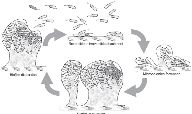
The process of biofilm formation occurs in response to environmental changes, and involves multiple regulatory networks, which translate signals to concerted gene expression changes thereby mediating the spatial and temporal reorganization of the bacterial cell47. Biofilm development is a dynamic and
multicellular process with several stages (Figure 2)48,49.
Figure 2- Stages of biofilm development33.
Biofilm formation starts with the attachment of bacteria to a surface, a random process mediated by Brownian motion and gravitational forces, and influenced by surrounding hydrodynamic forces 50,51. The
structure of biofilms can be influenced by various bacterial activities such as cell growth or death, nutrient acquisition, waste product accumulation, motility mechanisms and exopolysaccharide synthesis52,53.
Once microorganisms are attached to a surface, they are submitted to a series of changes to adapt at life on a surface, such as the expression of large quantities of exopolysaccharides that may protect the biofilm and lead to biocide resistance and the development of complex architectural features54–58.
Bacteria with flagella have a competitive advantage to overcome hydrodynamic and repulsive forces. After intercepting the surface, additional extracellular adhesive appendages and secreted adhesion mediate the adherence. Initial attachment is dynamic and reversible, during which bacteria can detach
and rejoin the planktonic population if perturbed by hydrodynamic forces, repulsive forces, or in response to nutrient availability59–62.
Surface contact leads to gene expression changes and up-regulating factors favoring sessility, such as those implicated in the formation of the extracellular matrix51,63–66. Within the mature biofilm there is a
bustling community that actively exchanges and shares products that play a pivotal role in maintaining biofilm architecture and providing a favorable living environment for the resident bacteria. Despite this, in mature biofilms, dispersal becomes an option. Besides passive dispersal, brought about by shear stresses, bacteria have evolved ways to perceive environmental changes and evaluate whether it is still beneficial to reside within the biofilm or whether it is time to resume a planktonic lifestyle. Biofilm dispersal can be the result of several cues, such as alterations in nutrient availability, oxygen fluctuations and increase of toxic products, or other stress-inducing conditions. Once dispersed, bacteria can reinitiate the process of biofilm formation, on encountering a suitable environment67,68.
Different classes of autoinducers (AI), signaling molecules, are responsible for mediation of the quorum-sensing (QS), a form of cell-to-cell interaction in bacteria, in response to the increase in cell density. GP bacteria employ autoinducing peptides while GN biofilms are mediated by acyl homoserine lactones (AHL)69,70. QS is an important event related to bacterial biofilm growth and differentiation71–73.
In conclusion, biofilm formation is mediated by a combination of adhesion mechanisms, bacterial motility and QS phenomenon that protect the cells and increase its resistance to the antibiotics74.
1.1.2.2. Identification of Gram-negative Biofilm with clinical relevance
Both GN and GP bacteria can form biofilms on indwelling medical devices such as catheters, mechanical heart valves and prosthetic joints75. E. coli, K. pneumoniae, P. mirabilis, and P. aeruginosa
are responsible for the majority of the clinical cases involving biofilms from GN bacteria75,76.
Table 2- Humans infections involving GN biofilms77
Infection or disease Common biofilm bacterial species
Periodontitis Porphyromonas gingivalis
Otitis media Nontypable strains of Haemophilus influenza Biliary tract infection E. coli
Osteomyelitis P. aeruginosa, Acinetobacter spp. and Enterobacter spp. Bacterial prostatitis E. coli, Pseudomonas spp. and Klebsiella oxitoca Cystic fibrosis pneumonia P. aeruginosa and Burkholderia cepacia
Meloidosis Pseudomonas pseudomallei
Nos ocom ial i nfe ct ion
s Intensive care unit pneumonia P. aeruginosa, Acinetobacter spp., and Stenotrophomonas maltophilia
Contact lens P. aeruginosa
Urinary catheter cystisis E. coli, P. mirabilis, P. aeruginosa and Klebsiella spp. Peritoneal dialysis peritonitis P. aeruginosa, E. coli, and Klebsiella spp.
Endotracheal tubes P. aeruginosa, K. pneumonia, P. mirabilis and E. coli Biliary stent blockage E. coli and Klebsiella spp.
It is estimated that 60% of nosocomial infections are derived from biofilm-related infections, many of which are caused by coagulase-negative staphylococci78–81. However, not only GP or GN bacteria form
biofilms on indwelling medical devices, also yeasts cause this aggregate76.
1.2
Clinical treatment of bacterial infection
Antibiotics are one of the most important forms of therapy for bacterial infections, caused by planktonic and sessile cells.
However, the efficiency of antibiotics is compromised by a growing number of antibiotic-resistant pathogens and by the failure on the discovery of new antibiotics to keep pace with the emergence of the pathogenic resistance, becoming one of the most serious and grievous challenge of the 21st century82.
In the United States, according to the Centers for Disease Control and Prevention at least 23.000 people die and more than 2 million are sickened annually as a result of an infection with an antibiotic-resistant organism. According to a recent report from the United Kingdom, the antibiotic-resistance crisis, in 2050, will be responsible for a loss of up to €90 trillion to the global economy83.
Multidrug-resistant GN organisms have emerged as a major threat to hospitalized patients and have been associated with mortality rates ranging from 30 to 70%14.
Resistance to antibiotics in GN bacteria is due to various molecular and biochemical mechanisms, with special attention to the chromosomally encoded drug efflux mechanisms that are ubiquitous in these bacteria and play an important role in both intrinsic and acquired multidrug resistance of clinical relevance. The drugs can be extruded out of the cell by efflux transporters, which exist as either single-component pumps or multisingle-component pumps (typically contain a pump, an OM channel protein (OMP), and an accessory membrane fusion protein (MFP)). The drug efflux, schematized on Figure 3, also interplays with other resistance mechanisms, such as the membrane permeability barrier, enzymatic inactivation or modification of drugs, and/or antibiotic target changes, increasing the levels of resistance84.
Figure 3- Drug efflux, the great responsible for antibiotic resistance in GN organisms84
β-lactam and aminoglycosides are two of the most widely available class of clinically important antibiotics for the treatment of various bacterial infections, whose effectiveness is now compromised85.
Antipathogenic drugs are an alternative to the antibacterial drugs (i.e., most traditional antibiotics) which target key regulatory bacterial systems that govern the expression of virulence factors, leading to the organism inability to establish successful infection86.
Furanones, antipathogenics compounds similar to the natural AIs, inhibit the QS and presented positive results when tested to control P. aeruginosa infections in animal models. However, this form of therapy
is too reactive, and therefore presumably too toxic for the treatment of bacterial infections in humans. QS antagonist has a narrow spectrum, and, therefore, specific antagonists have to be developed for each organism targeted, allowing the attenuation of a single pathogenic organism living in a mixed population of normal bacterial flora, leaving the rest of the bacterial population unaffected. Although, some AHL signal molecules function as virulence factors, affecting the muscle tissue and tracheal gland cells86.
The application of the drugs mentioned above, due to associated problems and disadvantages, is inappropriate and leads to the necessity to discovery and practice new forms of treatment to bacterial infections.
Dietary phytochemicals, such as essential oil (EO), phenolics, glucosinolates and their hydrolysis products, could represent a natural antimicrobial strategy with significant impact not only against planktonic bacteria but also on bacterial biofilm formation and development87. Some therapeutic
hypotheses for sessile and planktonic bacteria are described below. Planktonic
Borris and Sakanaka showed that catechins, a simple phenol present in the oolong green teas, inhibited, in vitro, the growth of V. cholerae and Shigella spp, and inactivated specific bacterial enzymes (toxin and glucosyltransferases) from the firsts88,89.
Tannins, polymeric phenolic substances, present antimicrobial activity, responsible for the inactivation of microbial adhesions, enzymes and cell envelope transport proteins from several bacteria, including E. coli90.
Copper, zinc, magnesium and especially silver and gold nanoparticles display antibacterial activity and are used for various healthcare. Rai and Chopra proved that Ag+ (ionic silver) was active against E. coli,
S. aureus, Klebsiella sp. and Pseudomonas sp.91,92.
Thitiporn Anunthawan proposed that cationic peptides KT2 and RT2 bind to negatively-charged LPS to enable self-promoted uptake and, subsequently interact with cytoplasmic membrane phospholipids through their hydrophobic domains enabling translocation across the bacterial membrane, entry to the cells within minutes and liaison to DNA and other cytoplasmic membrane. These peptides, due to their antimicrobial and anti-biofilm activities may be an alternative to (or in conjunction with) conventional antibiotics to treat acute infections caused by planktonic bacteria and chronic biofilm-related infections93.
Biofilms
Prevention of the biofilms’ development is the first step for their control or eradication and the removal of the biofilm from contaminated device is an effective strategy for treating these infections74,94. However,
prevention can not always be applied95.
Coating the surface of biomaterial with bactericidal/bacteriostatic substances is one of the approaches to make it resistant to biofilms’ formation96.
The emergent resistance of biofilms to a variety of antimicrobial agents, including antibiotics, antiseptics and industrial biocides leads to the treatment failure without removing the bacterial infection38,40.
Another parameter that can be used to remove biofilms is mechanical forces. This strategy is implemented with chemical agents, since they tend to leave the biofilm intact when no mechanical treatment is applied37.
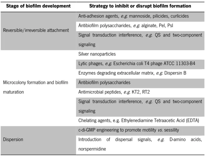
Different strategies to inhibit or disrupt biofilms at different stages of their development are summarized on Table 3.
Table 3- Examples of new strategies to inhibit or disrupt biofilms at different stages of their development33
Stage of biofilm development Strategy to inhibit or disrupt biofilm formation
Reversible/irreversible attachment
Anti-adhesion agents, e.g. mannoside, pilicides, curlicides Antibiofilm polysaccharides, e.g. alginate, Pel, Psl
Signal transduction interference, e.g. QS and two-component signaling
Microcolony formation and biofilm maturation
Silver nanoparticles
Lytic phages, e.g. Escherichia coli T4 phage ATCC 11303-B4 Enzymes degrading extracellular matrix, e.g. Dispersin B Antibiofilm polysaccharides
Antimicrobial peptides, e.g. KT2, RT2
Signal transduction interference, e.g. QS and two-component signaling
Chelating agents, e.g. Ethylenediamine Tetraacetic Acid (EDTA) Dispersion
c-di-GMP engineering to promote motility vs. sessility
Introduction of dispersal signals, e.g. D-amino acids, norspermidine
Terpenoids are derived from terpenes (based on an isoprene structure) and due to their composition have a lot of biological functions and are applied as pharmaceuticals, fragrances and colorants. In 2005, Ren demonstrated that ursolic acid, a triterpenoid, inhibited biofilms of E. coli and P. aeruginosa and Vibrio harveyi97,98.
Methyl eugenol, an EO with an aromatic ring, inhibit motility, QS, EPS production and biofilm formation by P. aeruginosa99.
Borges demonstrated that gallic and ferulic acids (two phenolic compounds) have potential to inhibit bacterial motility, adhesion and to prevent and control biofilms of P. aeruginosa100.
Davies proved that an unsaturated fatty acid, cis-2-decenoic acid, produced by P. aeruginosa in biofilm cultures, is responsible for inducing a dispersion response in biofilms formed by a range of GN bacteria, including P. aeruginosa101.
Kolodkin-Gal concluded that the D-Amino acids, produced by many bacteria in stationary phase, prevent biofilm formation by P. aeruginosa102.
The potential clinical value of antimicrobial agents that control and prevent P. aeruginosa infections by interruption of QS and inhibition of the transcription of biofilm-controlling genes or genes involved in cell attachment might also prove to be a successful strategy in inhibiting biofilm infections by interfering with various stages of biofilm maturation95.
Various reports have suggested that macrolides act through effects on the immune system, modifying the inflammatory response to infection (as immunomodulatory), or through a direct effect in decreasing the virulence of Pseudomonas40,95,103–111.
Despite the large numbers of approaches, neither can lead to completely inhibition or eradication of bacterial infection associated with planktonic or sessile cells, but only to the attenuation of their formation or effects. However, some of those therapies may become important in the control and decrease of these infections, being necessary additional research work and financial support.
The best results on the combat of problems related with bacteria are obtained when more than one strategy is implemented.
1.3
Bacteriophages
Bacteriophages (phages) are viruses that infect Bacteria and Archaea, also known as viruses of prokaryotes.
Approximately 96% of the 5500 reported phages have tailed morphology, which consist on a protein head, with a linear double-stranded DNA genome, enclosed by capsid, and a tail that promotes the phage attachment to the host cell and enable DNA injection112.
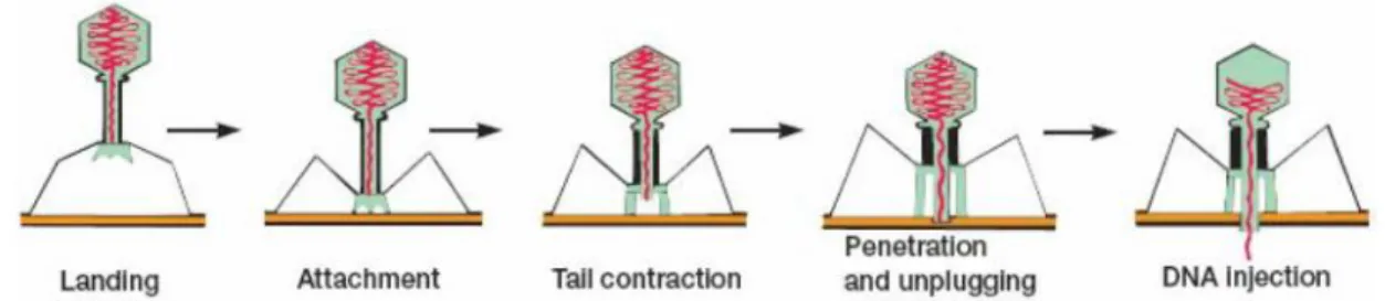
In these phages, the first step in the infection process is the adsorption of the phage to the bacterial cell, mediated by the tail fibers or by some analogous structure that specifically recognize receptors on the bacterial cell such as proteins on the outer surface of the bacterium, LPS, pili and lipoprotein, in a reversible process113. Afterwards, one or more of the components of the base plate mediates the
irreversible binding of the phage to the bacterium. This results in the contraction of the sheath and the hollow tail fiber is pushed through the bacterial envelope. Some phages have enzymes that digest various components of the bacterial envelope enabling the nucleic acid to pass into the bacterial cell. The rest of the phage remains on the outside of the bacterium. This process of infection can be made artificially even in a non-susceptible bacterium by injecting phage DNA through transfection114.
The firsts steps on infection process are presented on Figure 4.
Figure 4- Adsorption and penetration of the phage to the bacterial cell114
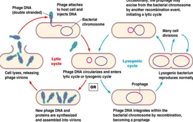
The replication of the phages may occur in two different life cycles, lytic and lysogenic. Depending on the followed pathway, phages can be distinguished on strictly lytic and temperate.
Temperate phages may reproduce through the lytic or the lysogenic cycle. During the lysogenic pathway they are able to establish a persistent infection on the cell without killing it, since the phage DNA becomes integrated with the bacterial genome, originating the prophages which replicate synchronously along with the host chromosome for many generations, causing no harm to the host cell, as opposed to the lytic cycle114–120.
The strictly lytic phages always follow the lytic pathway, infecting and inducing the bacterial cell lysis and resulting in the release of the progeny virions114,116,117. The liberated progeny phages are then ready to
start another cycle by infecting new neighboring bacterial cells. The whole cycle can be completed in 20 to 40 minutes, depending on a variety of factors such as temperature, nutrients, light and other environmental forces, and during that time 50 to 200 viruses are released115,121. Once the phage
reproduction is much faster than typical bacterial reproduction, so entire colonies can be destroyed very quickly116.
The life cycles are represented on Figure 5.
Figure 5-Lytic and lysogenic cycles. The replication of lytic bacteriophages follows the lytic cycle, while lysogenic bacteriophages may followed by lytic or lysogenic cycles122
1.3.1 Bacteriophage Therapy
The emergence of multiple drug-resistant bacteria has prompted interest in alternatives to conventional antimicrobials. Bacteriophage therapy, which consists on the use of bacteriophages as antimicrobial agents for the treatment of bacterial infections, has high potential as an alternative to antibiotics123 due
to abundance of phages in nature, their easy isolation, effectiveness to kill bacteria (even antibiotic-resistant bacteria at least in controlled laboratory experiments113.
In opposite to antibiotics which usually target both pathogens and patients normal flora and travel throughout the body decreasing its concentration through time, bacteriophages are very specific to their hosts, minimizing the chance of secondary infections and tend to only minimally disrupt normal flora, replicating at the site of infection where they are mostly needed to lyse the pathogens.
It was demonstrated that the consumption of large amounts of phages has not led to any immunological complications, and topical application has not shown any adverse effects124–127. Contrarily,
the antibiotics may cause allergies, sometimes even fatal anaphylactic reaction, and secondary infections128.
Bacteriophages seem to be capable of disrupting bacterial biofilms, are environmentally friendly and are selected and isolated in a very rapid process128,129, whereas the development of new antibiotic may take
several years, may cost millions of dollars for clinical trials, and may also not be very cost effective 130.
Multiple experiments that focused on the therapeutic use of phages demonstrated an effective elimination of pathogenic bacteria from Gi diseases131. However, although the dynamics may differ, the
evolution of bacterial resistance to a particular phage, just as to an antibiotic, is inevitable 132.
The predominance of lytic among temperate phages is an advantage to phage therapy, once that the lytic phages infect, lyse, and kill bacteria, until the infection is cleared or reduced to a level where the host immune system can effectively remove the remaining infection133–135.
The temperate phages are unsuitable for phage therapy, as they do not lyse their host bacteria, and due to their ability to integrate into the genome of bacteria, may transduce the resistance genes from one bacterium to another, and thus paradoxically contribute to the spread of resistance between bacteria113.
Bacteriophage therapy requires specific knowledge of phage biology. Some of the main problems arising from the use of phage therapy have recently been studied and are reported in Table 4113,122,135.
Table 4- Some of the problems with early therapeutic phage research and the ways they have been addressed in more recent studies122.
Problem Comments
Narrow host range of phages
Because of the high specificity of phages, many negative results may have been obtained because of the failure to
select phages lytic for the targeted bacterial species
Insufficient purity of phage preparations
Early therapeutic phages were crude lysates of host bacteria, and they contained numerous contaminants (including endotoxins) that may have counteracted the
effect of phages
Poor stability and/or viability of phage preparations
Some commercial phage preparations were supplemented with mercurial or oxidizing agents or were heat treated to
ensure bacterial sterility, many of these treatments also may have inactivated the phages, resulting in some
ineffective phage preparations Lack of understanding of the
heterogeneity and mode of action of phages (i.e., lytic or lysogenic phages)
Failure to differentiate between lytic and lysogenic phages may have resulted in the use of lysogenic phages, which
are much less effective than lytic phages Bacterial resistance to phages
This will unquestionably develop, although according to some authors the rate of developing resistance to phages
is approximately 10-fold lower than that to antibiotics. Additionally, there are some obstacles to phage therapy on biofilm infection, such as, difficulties of phage to penetrate the biofilm matrix and the presence of proteolytic enzymes and endoglucanases that can lead to inactivation of bacteriophages, the reduction of metabolic activity of biofilm cells and in the case of bacterial aggregate be formed by several species, these may bind and occlude phage receptors136.
Although the use of phages to the treatment of bacterial infections present advantages relatively to antibiotics, the problems associated to their viral nature lead to the necessity of using another type of procedure, particularly proteins from phage origin.
1.3.2 Bacteriophage proteins in the treatment of Gram-negative Bacterial Pathogens
An interesting and promising phage-based therapeutic advance is centered in the use of phage encoded enzymes, produced actively during the lytic cycle, using the machinery of the host cell, which lysis the bacterial cell wall, from inside, allowing the release of phages137–141.
Endolysins, also termed phage lysins or murein hydrolases, are phage-encoded peptidoglycan hydrolases (PGHs). These enzymes are employed by the majority of bacteriophages, at the end of their replication cycle, to enzymatically degrade the PG of the bacterial cell wall from within (by hydrolyzing the four major bonds), resulting in cell lysis and release of progeny virions. Lysis can be accomplished in two different ways: inhibition of PG synthesis by a single protein or enzymatic cleavage of PG by lysins or holin–lysin systems142.
Historically, application of endolysins as antimicrobials has been limited to GP pathogens, due to the absence of an OM in cell wall, allowing the access of endolysins to the peptidoglycan from the outside (or from without). However, recent developments involving peptides with OM–disrupting properties fused to phage lysins have raised hopes of also tackling GN organisms with PGH enzymes143–145. In fact,
Briers et al. recently reported the development of Artilysins®, a protein-engineered endolysin that passes through the OM and subsequently kill the cells through PG degradation and cell lysis146. Several
published studies prove the influence and effectiveness of endolysins to lyse the cells, acting independently from others enzymes.
- The recombinant expression of endolysin Lys1521, from phage IAM 1521 of the Bacillus amyloliquefaciens, in E. coli, resulted in cell lysis, indicating that this enzyme is able to pass the cytoplasmic membrane independently. External application of Lys1521 on E. coli W3110 and P. aeruginosa reduced the number of cells with 98.75% and 99.78% in 10 min, respectively147
- LysAB2, an endolysin produced by Acinetobacter baumannii phage ϕAB2, when used in a high final concentration, show antibacterial activity against A. baumannii, E. coli, Citrobacter freundii and S. enterica, with a reduction between 67.5 and 99.9%, after 1 h of incubation148.
- It was demonstrated that the P7 protein, from the Pseudoalteromonas PM2 phage, has bacteriolytic activity and is involved in the PG penetration process, causing a limited depolarization of the cytoplasmic membrane149.
- A similar phenomenon was also observed with phages Φ13 (from P. syringae), and ΦKMV and ΦKZ (infect P. aeruginosa strains) which contain a thermosensitive lytic enzyme involved in PG penetration150– 152.
- A study developed by Hanlon et al. found that a P. aeruginosa bacteriophage was able to penetrate the inner layers of the biofilm due to the reduction of the viscosity of the alginate matrix by enzymatic degradation106.
Beside those enzymes, some phages encode a second PGH, the virion-associated peptidoglycan hydrolase (VAPGH). In contrast to endolysins, VAPGH degrade localized peptidoglycan during infection, being able to generate small holes through which the phage tail tube crosses the cell envelope to eject the phage genetic material at the beginning to the infection cycle153,154.
- It was shown that the P5 protein from phage ф6 is active against P. phaseolicola, P. aeruginosa, P. fluorescens and P. putida, after destabilization of the outer membrane by incubation of the cells in chloroform-saturated buffer at room temperature. The VAPGH from phage ΦKZ, Gp181, also has a wide lytic spectrum including P. aeruginosa, P. fluorescens and P. putida155.
During phage development in the infected bacterium, lysin accumulates in the cytoplasm in anticipation of phage maturation. Meanwhile, phage encoded small hydrophobic membrane proteins termed holins, which promote the membrane disruption, allowing that lysins access the PG, causing cell lysis and the release of progeny phage142,156.
Holins control the activity of bacteriophage-encoded endolysins and the timing of lysis during bacteriophage infection by regulating the access of these enzymes to their substrate. This regulation can be achieved by one of two proposed mechanisms: through control of murein hydrolase transport across the cytoplasmic membrane or by mediating the release and activation of membrane-associated murein hydrolases157–161.
The GN specific phages developed other protein responsible for crossing the outer membrane, whose encoding gene is typically located near the endolysin and holin genes, creating a three-component lysis cassette, the spanin162. Without spanin function, lysis is blocked and progeny virions are trapped in dead
spherical cells, suggesting that the outer membrane has considerable tensile strength163.
Biofilm polysaccharide protects the bacterial cells against the majority of bacteriophage. However, if a phage possesses a specific polysaccharide hydrolase, the depolymerase, it may be able to degrade macromolecule carbohydrates within extracellular polysaccharides and LPS surrounding bacterial cells and gain access to the bacterial surfaces. Consequently, it could cause biofilm disruption through cell infection and lysis, as well as EPS degradation108,111,164.
Different polysaccharide depolymerases are known, such as, endorhamnosidases that hydrolyze outer membrane LPS of GN species, endolsialidases that degrade capsular polysaccharides of E. coli,
alginate lysases that degrade the capsules of P. aeruginosa and hyaluronidases that degrade capsules of streptococcal species153.
In fact, bacteriophage-encoded proteins with antimicrobial activity and combinations between different enzymes and between enzymes and other biocide agents represent a promising alternative to antibiotics, as potential new antimicrobials against infectious diseases.
2.1
Cloning and Expression of Bacteriophage Proteins
The aim of this work was to express bacteriophage proteins alone and fused with Lysin gp146 and tested them on GN bacteria, on sessile and planktonic forms.
2.1.1 Bacteria, Plasmid, Phages and Growth Conditions
The bacteria strains used to test the proteins were Escherichia coli strains BL21 Gold (DE3) purchased from Epicurian Coli line of Stratagene; E. coli OverExpress C43(DE3) from Lucingen, ArcticExpress (DE3) from Agilent Technologies, Salmonella Enteritidis strains 1400, isolated by the group in the scope of the European project PhageVet-P165; and P. aeruginosa strains ATCC 10145 and PAO1166.
All bacteria were grown at 37 ºC on Lysogeny Broth (LB Broth- nzytech), at 120 rpm.
The plasmid pET-28a acquired from Novagen was used to accomplish the desired constructions. The restriction map and Multiple Cloning Site (MCS) are presented in Annex I, Figure 12 and Figure 13, respectively.
The DNA (deoxyribonucleic acid) from phages lambda and T7 were purchased from FRILABO. The DNA from phages T1 and T4, acquired from DSMZ, were extracted from phage lysate using the kit NucleoSpin Tissue (250), from Macherey Nagel Bioanalysis- Fisher Scientific, according to the manufacturer’s instructions. The phage PVP-SE1, isolated by the group in the scope of the European project PhageVet-P165.
2.1.2 Phage DNA Extraction and Gene Amplification
The bacteriophage proteins tested in the treatment of biofilms and planktonic bacteria above mentioned are listed in Table 5. The enzyme Alginate Lyase, from Sigma-Aldrich® (Product Number: A1603) was also tested against biofilms.
Table 5- List of bacteriophage proteins to express
Host Proteins type Phage Name Gene Identification (GI) on NCBI E. Coli Holins T1 holin T1 45686347 T4 holin T4 9632830 T7 holin T7 9627478
lambda holin Lambda 160380505
Spanin T1 RzT1 45686349 lambda Rz1Lambda 160338810 S. enteritidis Lysin PVP-SE1 gp146 363539667 Colonic Acid degrading enzyme gp49 363539570
A glycosyl hydrolase, dispersin B (GI: 30420959), was acquired from a previously plasmid construction and cloned into pET-28a, expressed and tested. The referred proteins (Table 5), excepting the gp49, were expressed and tested in fusion with gp146, originating: holin T1_146, holin T4_146, holin T7_146, holin Lambda_146, holin Lambda_L30_146, RzT1_146, Rz1Lambda_146 and dispersin B_146.
The amino acidic and nucleotide sequences from each protein were obtained and can be found in Annex II and Annex III (Table 21 and Table 22).
The sequences encoding each protein were amplified using genomic DNA as template and specific primers, whose quality and specificity was analyzed by the online computer tools OligoAnalyzer 3.1 (https://eu.idtdna.com/calc/analyzer) and BLAST (http://blast.ncbi.nlm.nih.gov/Blast.cgi?PROGRAM= blastn&PAGE_TYPE=BlastSearch&LINK_LOC=blasthome), respectively (Table 6).
2. M ate ria ls a nd M ethod s 27
Table 6- Primers sequences of the proteins and its characteristics
Protein Primer Sequence GC Content (%) Tm (°C) PCR (bp) Specificity (%)
holin T1/
holin T1_146
Forward (NcoI) 5’GGCATCCATGGCAATGAAAGAGTTTTTAA
CGGCTGCTAC3’ 40 55.8 246
1036
100 Reverse (XhoI/NdeI) 5’CGCCGCTCGAGCATATGTCTCCCCCTGAT
CTTAAGCG3’ 55 56.1 100
holin T4/
holin T4_146
Forward (NcoI) 5’GGCATCCATGGCAATGGCAGCACCTAGAA
TATCATTTTC3’ 38.5 55.4 687
1477
100 Reverse (XhoI/NdeI) 5’CGCCGCTCGAGCATATGTTTAGCCCTTCCT
AATATTCTGGC3’ 41.7 54.6 100
holin T7/
holin T7_146
Forward (NcoI) 5’GGCATCCATGGCAGTGCTATCATTAGACT
TTAACAACGAATT3’ 31 54.2 234
1024
100 Reverse (XhoI/NdeI) 5’CGCCGCTCGAGCATATGCTCCTTATTGGCT
TTCTTCCAGTC3’ 45.8 55.5 100
holin Lambda/
holin Lambda_146
Forward (NcoI) 5’GGCATCCATGGCAATGCCAGAAAAACATG
ACCTGTTG3’ 41.7 56.4 348
1138
100 Reverse (XhoI/NdeI) 5’CGCCGCTCGAGCATATGTTGATTTCTACCA
2. M ate ria ls a nd M ethod s 28 holin Lambda_L30_146
Forward (NcoI) 5’GGCATCCATGGCAATGCCAGAAAAACATGA
CCTGTTG3’ 41.7 56.4 1176 100 Reverse (SacI) 5’CGCGGGAGCTCGCCGCCCGCGGAGCCGG ACGCCGCGCCCGCGGAGCCGCCCGCGCCG GAGGACGCGGAGCCCGCGCCCGCCGCGCC GGACGCGGAGCCCGGTTGATTTCTACCATCT TCTACTCCG3’ 40 54.1 100 Rz T1/ Rz T1_146
Forward (NcoI) 5’GGCATCCATGGCAATGAAACTTAAGAAAA
CGTGCATTGCAATT3’ 30 57 432
1222
100 Reverse (XhoI/NdeI) 5’CGCCGCTCGAGCATATGCGCCTCCTTTTTT
TCGTGCTTAC3’ 47.8 57 100
Rz1 Lambda/
Rz1 Lambda_146
Forward (NcoI) 5’GGCATCCATGGCAATGCTAAAGCTGAAAA
TGATGCTCTG3’ 38.5 56.1 213
1003
100 Reverse (XhoI/NdeI) 5’CGCCGCTCGAGCATATGGCCTCTCTCTGA
2. M ate ria ls a nd M ethod s 29 gp146
Forward (NcoI /SacI) 5’GGCATCCATGGCACGGGAGCTCATGAATG
CTGCAATTGCGGAGATT3’ 41.7 58
744
100 Reverse (XhoI) 5’CGCCGCTCGAGCGAGGTTAGAACAGATTT
TGCCTTTT3’ 38.5 56 100
gp49
Forward (NdeI) 5’GGGCCCATATGATGGCAGATCTATTACCT
ACCGT3’ 43.5 55.1
2161
100 Reverse (BamHI) 5’GGGCCGGATCCTTAAGTCCTTTCGCTGTA
TACTACG3’ 40 54.2 100
dispersin B/
dispersin B_146
Forward (NcoI) 5’GGCATCCATGGCAATGAACTGCTGCGTGA
AGGG3’ 55 58.1 1060
1850
100 Reverse (XhoI/NdeI) 5’CGCCGCTCGAGCATATGGATGGTCTCGTC
CTTCAGGG3’ 60 57.1 100
- Associated with fused Protein - Associated with singled Protein
- Associated with both kind of Proteins
Holin Lambda_146 and holin Lambda_L30_146 differs on the linker between the holin and gp146, wherein the second one has the linker identified by White et al.167.
The PCR amplification of each CDS was made through the use of the proof reading polymerase Phusion Green High-Fidelity DNA Polymerase from Thermo Scientific (#F-534L), to a final volume of 100 µl, following the manufacturer instructions using the parameters presented on Table 7.
Table 7- Parameters used to the PCR
Step Temperature (°C) Time (sec)
1st 98 30
2nd 98 10
3th 55 30
4th 72 60
5th Back to the 2nd step, 34 times
6th 72 600
7th 4 ∞
Once finished, 5 µl of each amplified sample and 5 µl of 1kb DNA Ladder acquired from New England BioLabs®Inc. were loaded on 1% agarose gel electrophoresis to confirm gene amplification. Gels were
visualized in a ChemiDoc™ XRS+ System with Image Lab™ Software (Version 5.1 Bio-Rad Laboratories, Inc.)168.
The commercial kit DNA, RNA and protein purification: PCR Clean up, from Nucleospin® was used to clean up the PCR amplified genes. The concentration as well the 260/280 and 260/230 ratios of the purified genes were obtained by loading 1.5 µl of the cleaned up sample in nanoDrop 1000
Spectrophotometer (Thermo SCIENTIFIC).
2.1.3 Gene Insertion in the pET-28a Cloning Vector
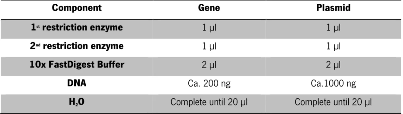
Taking into account the concentration, the genes and the plasmid were digested for 3 h, at 37 °C, following the amounts presented in Table 8, with the FastDigest enzymes (NcoI, XhoI, NdeI, BamHI and SacI) specified on Table 6 and purchased to Thermo SCIENTIFIC.
Table 8- Quantities of components used to make a digestion
Component Gene Plasmid
1st restriction enzyme 1 µl 1 µl
2nd restriction enzyme 1 µl 1 µl
10x FastDigest Buffer 2 µl 2 µl
DNA Ca. 200 ng Ca.1000 ng
H2O Complete until 20 µl Complete until 20 µl The OPTIZYME™ Alkaline Phosphatase, from Fisher BioReagents®, (Table 9) was added to the plasmid,
to remove the phosphate groups from the cutting ends of the plasmid to avoid self-ligation. Incubation was carried for 1 h at 37 °C.
Table 9- Quantities of each element added to the plasmid
Component Quantities (µl)
Plasmid Digestion (Table 8- Quantities of components used to
make a digestionTable 8) 20
10x OPTIZYME AP Buffer 3
OPTIZYME Alkaline Phosphatase 1
H2O 6
Final volume 30
Inactivation of the restriction enzymes and the alkaline phosphatase was carried at 80 °C for 15 min. The T4 DNA ligase, from Thermo SCIENTIFIC, was used as specified on Table 10 and incubated 1 h, at 22 °C to make the ligation between genes and plasmid.
Table 10- Quantities used to insert the genes into the plasmid vector
Component Quantities
pET-28a DNA (linear vector) 20-100 ng
Protein DNA (insert) 1:1 to 5:1 molar ratio over vector
10X T4 DNA Ligase Buffer 2 µl
T4 DNA Ligase 0.5 µl
H2O Complete until 20 µl
To the fused proteins, the procedure for cloning into plasmid was the same, with the difference that the plasmid used was a construction of pET-28a with gp146.
2.1.4 Gene cloning in E. coli
The constructions obtained were used to transform chemically competent cells (previously prepared as specified in Annex IV) of E. coli CTOP10 through heat shock (Annex V).
The transformants were selected in a LB with kanamycin (kan) Petri dish, and 10 colonies were randomly chosen and incubated with 50 µl of LB and kan, for 1 h 30 min, at 37 °C.
A Colony PCR, using DreamTaq Green DNA Polymerase (Thermo SCIENTIFIC – #EP0713) and T7 primers was done to confirm transformation, following the conditions presented on Table 11.
Table 11- Parameters used to the Colony PCR
Step Temperature (°C) Time (sec)
1st 95 300
2nd 95 45
3th 45 30
4th 72 60
5th Back to the 2nd step, 34 times
6th 72 600
7th 4 ∞
Colonies presenting a band with the correct size were recovered in solid LB with kan incubated overnight, at 37 °C.
The plasmids from each positively and grown clone were extracted by DNA, RNA and protein purification: Plasmid kit, from Nucleospin® and sent to sequencing to confirm the correct insertion of the genes.
Plasmids with a positive result were transferred to the expression strains, E. coli C43, E. coli BL21 and E. coli Artic Express as described in Annex V.
Three colonies of each transformation were submitted to colony PCR to confirm the correct transformation of the cells and a positive clone was selected for further preservation.
2.1.5 Protein expression
For a given protein, a pre-inoculum with the corresponding clone (bacteria with the correct plasmid) in 1 ml of LB with kan was incubated overnight, at 37 °C and 120 rpm.
At this phase, multiples approaches were tested to optimize the conditions of protein expression. To each essay, detailed on Table 12, inoculum of 100 ml of LB with kan was transferred to sterilized 250 ml Erlenmeyer flasks and 1 ml of the pre-inoculum was added, followed by the incubation, until the defined O.D.600 (Optical density). To induce the protein expression, 100 µl of 1M IPTG (1 µM final) were
added to the inoculum and incubated overnight, at the conditions listed below. Table 12- Different conditions tested to optimize the protein expression
Essay Host O.D. 600
Before Induction
Induction
with IPTG Additional approaches
°C min °C rpm
1 BL21 gold (DE3) 0.450 - - 37 120 -
2 BL21 gold (DE3) 0.450 - - 21 150 -
3 BL21 gold (DE3) 0.450 - - 16 150 -
4 BL21 gold (DE3) 0.600 4 10 16 150 NaCl 2 M and Ethanol
5 BL21 gold (DE3) 0.900 4 10 16 150 1% glucose
6 BL21 gold (DE3) 0.600 4 10 16 150 1% glucose
7 C43 (DE3) 0.600 4 10 16 150 1% glucose
8 Artic express (DE3) 0.600 4 10 16 150 1% glucose
9 BL21 gold (DE3) 0.400 4 10 21 200 1% glucose
10 C43 (DE3) 0.400 4 10 21 200 1% glucose
11 Artic express (DE3) 0.400 4 10 21 200 1% glucose
12 C43 (DE3) 0.600 4 10 16 250 1% glucose and azide
13 BL21 gold (DE3) 0.600 4 10 16 250 1% glucose and azide
14 BL21 gold (DE3) 0.600 4 10 16 250 1% glucose
The NaCl 2 M and the ethanol were added to the culture, at the essay 4, after the O.D.600 reached
0.600. At the essays 5 to 14, 1% glucose was mixed with the inoculums in the Erlenmeyer flasks. The compound with azide, used on essays 12 and 13, was added to the Erlenmeyer before the induction with IPTG.