U
m
inho | 20
1
6
Vânia da Silva Gaio
Study of t
he suscep
tibility to antibio
tics of cells released from St
aph
y
lococcus epidermidis biofilm
s
October 2016
Universidade do Minho
Escola de Engenharia
Vânia da Silva Gaio
Study of the susceptibility to antibiotics of
cells released from Staphylococcus
Master Thesis
Integrated Master in Biomedical Engineering
This work was realized under supervision of:
Doctor Nuno Miguel Dias Cerca
Vânia da Silva Gaio
Study of the susceptibility to antibiotics of
cells released from Staphylococcus
epidermidis biofilms
October 2016
Universidade do Minho
Escola de Engenharia
III
DECLARAÇÃO
Nome: Vânia da Silva Gaio
Endereço eletrónico: [email protected]
Título da dissertação:
Study of the susceptibility to antibiotics of cells released from Staphylococcus epidermidis biofilms
Orientador:
Doutor Nuno Miguel Dias Cerca
Ano de conclusão: 2016
Mestrado Integrado em Engenharia Biomédica
É AUTORIZADA A REPRODUÇÃO INTEGRAL DESTA DISSERTAÇÃO APENAS PARA EFEITOS DE INVESTIGAÇÃO, MEDIANTE DECLARAÇÃO ESCRITA DO INTERESSADO, QUE A TAL SE COMPROMETE.
Universidade do Minho, _____/_____/_________
V
A
CKNOWLEDGMENTS
Chega, assim, ao fim a última e mais desafiante etapa desta longa caminhada conducente ao grau de Mestre em Engenharia Biomédica. Contudo, a realização desta dissertação de Mestrado apenas foi possível graças ao apoio e contributo, direto e indireto, de várias pessoas a quem gostaria de dirigir algumas sinceras palavras de apreço e gratidão.
Em primeiro lugar quero agradecer ao meu orientador, Doutor Nuno Cerca, pela oportunidade que me deu para realizar um trabalho tão interessante e por me ter orientando ao longo do desenvolvimento do mesmo. Pela sua disponibilidade, transmissão de conhecimentos e, sobretudo, pela confiança que depositou em mim para que a concretização de todas as tarefas fosse possível, o meu muito obrigada. Agradeço-lhe ainda pelas oportunidades que me proporcionou, pelo seu constante interesse e por todas as sugestões que me deu, desafiando-me sempre a fazer mais e melhor, pois só assim foi possível evoluir e aprender tanto ao longo deste ano.
Agradeço a todos os elementos do Grupo NC pela forma como me receberam e integraram, pela partilha de conhecimentos, pela paciência para ouvir e esclarecer as minhas dúvidas e pela gentileza para me ensinar, ajudar e apoiar em tudo o que precisei ao longo desta etapa. Obrigada também por todos os momentos de convívio e descontração, que em muito contribuíram para que este percurso ficasse marcado por boas memórias.
Expresso também o meu agradecimento ao Departamento de Engenharia Biológica da Universidade do Minho pela disponibilização das instalações e equipamentos que foram imprescindíveis para a realização do trabalho apresentado ao longo desta dissertação.
Aos colegas do LIBRO e da “Biblioteca”, obrigada pelo bom ambiente de trabalho, pela camaradagem e disponibilidade que sempre demonstraram para ajudar em tudo o que fosse preciso, pelos ensinamentos que me transmitiram e também pelos animados momentos de convívio.
Às “Danielas” da minha vida, obrigada por terem estado ao meu lado durante todo este percurso! Foram cinco anos a aprender convosco, a partilhar conhecimentos, aventuras e muitos bons momentos. Agradeço-vos, sinceramente, por todo o vosso apoio e amizade e por terem estado sempre presentes, mesmo quando o meu mau feitio superou a minha boa disposição. Daniela A., a tua força de vontade para superar obstáculos foi, sem dúvida, uma inspiração para mim. Daniela S., obrigada especialmente pela força e bondade para me apoiar ao longo dos momentos mais difíceis.
VI
Mariana, obrigada também por todo o teu apoio e amizade ao longo dos quatro anos que tivemos o prazer de partilhar. Tenho pena que tenhamos seguido caminhos diferentes, mas sei que ambas nos sentimos concretizadas naquilo que fazemos, e isso é o mais importante.
Aos amigos de longa data (Mónica, Henrique, Hugo, Joana, Bryan, Bruna, Abel e Carlos), obrigada por terem feito parte do meu percurso académico e por fazerem parte da minha vida. Obrigada por todas as alegrias e tristezas partilhadas, é bom ter-vos por perto.
Tiago, a ti devo-te um agradecimento muito especial. Ficarei sempre profundamente grata por todos os anos de conquistas e por todos os obstáculos que me ajudaste a superar, por todo o teu apoio e paciência comigo, pelo esforço para me compreenderes e por acreditares sempre em mim.
Por último, o agradecimento mais importante é direcionado à minha família, especialmente aos meus Pais e Irmãs. Mãe e Pai, nunca conseguirei transmitir por palavras o quão importantes são para mim e o quanto vos agradeço por todo o esforço que fizeram para que eu pudesse alcançar este feito. Obrigada por acreditarem em mim e por sempre me incentivarem a seguir os meus sonhos.
Sónia e Daniela, mais do que “amiguinhas do coração”, foram e sempre serão um exemplo para mim. Admiro a vossa generosidade, a vossa garra e coragem, e a vossa disponibilidade para ajudarem sempre o próximo. Ainda que tenhamos personalidades completamente distintas, a vocês devo muito daquilo que sou hoje. Obrigada por tudo!
Dedico esta tese à minha família, e a quem dela infelizmente já partiu, por sempre me fazerem acreditar que:
“Pessoas com nome de pássaro devem saber voar por bons céus.”
Albertina Fernandes.
Este estudo foi suportado pela Fundação para a Ciência e a Tecnologia (FCT) Portuguesa no âmbito do fundo estratégico da unidade UID/BIO/04469/2013 e COMPETE 2020 (POCI-01-0145-FEDER-006684).
A Vânia da Silva Gaio usufruiu de uma bolsa ANICT para o desenvolvimento da Dissertação de Mestrado com o título “Study of the susceptibility to antibiotics of cells released from Staphylococcus epidermidis biofilms”.
VII
A
BSTRACT
Worldwide, Staphylococcus epidermidis has been recognized as a leading cause of several clinically relevant infections, primarily associated with its notable ability to colonize surfaces and form biofilms, especially in the surface of medical indwelling devices. The formation of bacterial biofilms, which is a major concern in health care systems due to their high tolerance to antibiotics, may be divided in three mains stages: 1) adhesion, 2) maturation and 3) biofilm disassembly. During the last stage, cells are released from the biofilm to the surrounding environment by both active and passive mechanisms, often being associated with the development of serious complications as bacteremia and embolic events of endocarditis. Despite the clinical relevance of biofilm-released cells (Brc), disassembly remains the least studied stage of the biofilm lifecycle and little is known concerning the phenotypic changes that these cells undergo after being released from the biofilm. Thus, this study aimed to provide a better characterization of S. epidermidis Brc phenotype, in particular its susceptibility to different classes of antibiotics (cell wall, nucleic acids and protein synthesis inhibitors). By directly quantifying the susceptibility of Brc and comparing to that of biofilm and stationary planktonic cells, this study allowed to demonstrate that Brc exhibit a distinct antibiotic tolerance profile. Moreover, it was found that Brc seem to have a transient phenotype, strengthening the vision of a biofilm lifecycle with individual cell physiology changing overtime. Overall, this study provided some clinically relevant outcomes in the pathogenesis of biofilm-related infections, demonstrating that the metabolic state of S. epidermidis cells has an important impact on antimicrobial susceptibility, and this is not only related to the distinct features of intact biofilms and planktonic cells. A better characterization of the Brc phenotype may help in the development of more efficient therapeutic measures against S. epidermidis biofilm-related infections.
KEYWORDS: STAPHYLOCOCCUS EPIDERMIDIS, BIOFILM DISASSEMBLY, BIOFILM-RELEASED CELLS, ANTIBIOTIC TOLERANCE
IX
S
UMÁRIO
A espécie Staphylococcus epidermidis tem sido reconhecida, a nível mundial, como uma das principais causas de infeções clinicamente relevantes, principalmente devido à sua capacidade eminente para colonizar superfícies e formar biofilmes, especialmente em dispositivos médicos invasivos. A formação de biofilmes bacterianos, que está associada a um aumento da tolerância a antibióticos, pode ser dividida em três etapas: 1) adesão, 2) maturação e 3) dispersão do biofilme. Durante a última etapa, as células são libertadas do biofilme para o ambiente envolvente por mecanismos ativos e passivos, sendo frequentemente associadas ao desenvolvimento de complicações sérias como bacteriemia e eventos embólicos relacionados com endocardite. Apesar da relevância clínica da dispersão das células libertadas do biofilme (Brc), esta etapa continua a ser a menos estudada do ciclo de vida do biofilme e pouco é sabido acerca das alterações fenotípicas das Brc. Assim, este estudo teve como objetivo proporcionar uma melhor compreensão acerca do fenótipo das Brc de S. epidermidis, em particular a sua suscetibilidade a diferentes classes de antibióticos (inibidores da síntese da parede celular, de ácidos nucleicos e de proteínas). Ao quantificar diretamente a suscetibilidade das Brc em comparação à das células do biofilme e planctónicas estacionárias, este estudo permitiu demonstrar que as Brc exibem um perfil distinto de tolerância aos antibióticos. Adicionalmente, foi verificado que as Brc parecem apresentar um fenótipo transiente, reforçando a ideia de um ciclo de vida do biofilme com uma particular fisiologia das células que é alterada ao longo do tempo. De uma forma geral, este estudo forneceu conclusões clinicamente relevantes acerca da patogénese de infeções associadas aos biofilmes, demonstrando que o estado metabólico das células de S. epidermidis tem um impacto importante na suscetibilidade a antimicrobianos, facto que não está apenas relacionado com as caraterísticas distintas dos biofilmes intactos e das células planctónicas. Uma melhor caracterização do fenótipo das Brc pode auxiliar no desenvolvimento de medidas terapêuticas mais eficientes contra infeções relacionadas com biofilmes de S. epidermidis.
PALAVRAS-CHAVE:STAPHYLOCOCCUS EPIDERMIDIS, DISPERSÃO DO BIOFILME, CÉLULAS LIBERTADAS DO BIOFILME, TOLERÂNCIA A ANTIBIÓTICOS
XI
T
ABLE OF
C
ONTENTS
Acknowledgments ... v
Abstract ... vii
Sumário ... ix
Index of Figures ... xiii
Index of Tables ... xv
List of Abbreviations ... xvii
List of Publications ... xix
1. Introduction ... 1
1.1 Staphyloccus genus ... 3
1.1.1 Staphylococcus epidermidis ... 4
1.2 Biofilms ... 6
1.2.1 Staphylococcus epidermidis biofilms ... 7
1.2.2 Quorum-sensing ... 11
1.2.3 Biofilm tolerance to antibiotics ... 12
1.2.4 Biofilm-released cells (Brc) ... 16
1.3 Aims and objectives ... 17
2. Materials and Methods ... 19
2.1 Isolates and growth conditions ... 21
2.1.1 Biofilm formation and biofilm-released cells collection ... 21
2.1.2 Planktonic growth ... 22
2.1.3 Cell homogenization ... 22
2.2 Characterization of the antimicrobial profile of planktonic S. epidermidis ... 23
2.3 Comparison of the antimicrobial susceptibility of the distinct S. epidermidis populations ... 24
3. Results and Discussion ... 25
3.1 Study of the antibiotic susceptibility of cells released from Staphylococcus epidermidis 9142 biofilms with 48 hours of maturation (Brc48H) ... 27
3.1.1 Preliminary MIC assay ... 27
3.1.2 Susceptibility assays ... 29 3.2 Study of the antibiotic susceptibility of cells released from biofilms with different stages of maturation . 35
XII 3.3 Study of the antibiotic susceptibility of cells released from different Staphylococcus epidermidis isolates
with 28 hours of maturation (BRC28H) ... 37
3.3.1 Preliminary MIC assay ... 37
3.3.2 Study of the biofilm formation ability of the six different S. epidermidis isolates selected ... 39
3.3.3 Vancomycin susceptibility of different S. epidermidis isolates and populations ... 42
4. Conclusions and Future work ... 45
4.1 Main conclusions ... 47
4.2 Suggestions for future work ... 49
XIII
I
NDEX OF
F
IGURES
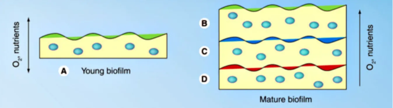
Figure 1.1 - Scanning electron microscopy (SEM) image of a grape-like cluster of S. epidermidis. Adapted from [5]. ... 3 Figure 1.2 - Scanning electron microscopy (SEM) of a Staphylococcus epidermidis biofilm. Adapted from [38]. ... 7 Figure 1.3 - Representation of S. epidermidis biofilm cycle and some of the molecules involved in the different phases of biofilm formation and disassembly. The process begins with the initial attachment to the surface, followed by the adhesion of cells to each other, forming clusters. Maturation of the biofilm is achieved by the growth of the bacteria clusters and production of the polymeric matrix by those aggregates, which will accumulate and surround bacteria. Lastly, a mature biofilm is obtained and bacteria can detach and disperse from this biofilm and colonize other surfaces. [33,44] Adapted from [12]. ... 8 Figure 1.4 - Low vacuum secondary electron image of a S. epidermidis biofilm, with evidence to the polymeric matrix surrounding bacteria. Adapted from [55]. ... 9 Figure 1.5 - Some hypothesis that attempt to explain the decreased susceptibility of biofilm cells to antibiotics. Adapted from [84]. ... 13 Figure 1.6 - Schematization of the heterogeneity of Staphylococcus epidermidis biofilms over the depth. Young biofilms (A) provide a high availability of nutrients and oxygen (O2) to all the bacteria,
while mature biofilms are characterized by deeper layers (D) with a small amount of nutrients and O2, and upper layers (B) with a great accessibility of nutrients and oxygen. Adapted from [40]. 14
Figure 1.7 - Schematization of the resistance mechanism due to resistant/persister cells. Although antimicrobial therapies can eradicate part of the biofilm cells, some resistant variants are not affected by the antimicrobial drugs and are able to persist and maintain the biofilm survival. After antimicrobial therapy discontinuation, the resistant fraction is able to develop a new biofilm that will grow and reach maturation. Adapted from [95]. ... 15
XIV
Figure 3.1 - Base 10 logarithmic CFU/mL reduction of S. epidermidis 9142 populations upon 2 hours of incubation with peak serum concentrations of distinct antibiotics. The columns represent the mean plus or minus standard error deviation, of at least three independent experiments. Statistical differences between groups were analysed with one-way ANOVA multiple comparisons, with * representing statistically significant differences (p <0.05) between biofilm cells and Brc and ◻ between Brc and their planktonic counterparts. ... 30 Figure 3.2 - Base 10 logarithmic CFU/mL reduction of S. epidermidis 9142 populations upon 6 hours of incubation with peak serum concentrations of distinct antibiotics. The columns represent the mean plus or minus standard error deviation, of at least three independent experiments. Statistical differences between groups were analysed with one-way ANOVA multiple comparisons, with * representing statistically significant differences (p <0.05) between biofilm cells and Brc and ◻ between Brc and their planktonic counterparts. ... 31 Figure 3.3 - Base 10 logarithmic CFU/mL reduction of S. epidermidis 9142 different Brc populations upon 2 hours of incubation with peak serum concentrations of five distinct antibiotics. The columns represent the mean plus or minus standard error deviation, of at least three independent experiments. Statistical differences between groups were analysed with one-way ANOVA multiple comparisons and no significant differences (p <0.05) were found among the different populations. ... 35 Figure 3.4 - Optical density of 24, 28, 48 and 72 hour-old mature biofilms (BIO) and 28 and 48-hours bulk fluid containing Brc (BRC) of seven S. epidermidis isolates, measured at 640 nm. (A) OD of high biofilm producing isolates; (B) OD of medium biofilm producing isolates; and (C) OD of low biofilm producing isolates. The columns represent the mean plus or minus standard error deviation, of at least three independent experiments ... 40 Figure 3.5 - Normalized base 10 logarithmic CFU/mL reduction of three populations of several S. epidermidis isolates upon 2 hours of incubation with peak serum concentrations of vancomycin. The results were normalized according to the results obtained for the biofilm population for each isolate, where the biofilm cultivability decrease was considered equal to 1 log10 CFU/mL. The
second and third columns represent the mean plus or minus standard error deviation, of at least three independent experiments. Statistical differences between groups were analysed with one-way ANOVA multiple comparisons, with * representing statistically significant differences (p <0.05) between biofilm cells and Brc and ◻ between Brc and their planktonic counterparts. ... 43
XV
I
NDEX OF
T
ABLES
Table 2.1 - Origin of the Staphylococcus epidermidis isolates used in this study ... 21 Table 2.2 - Mechanism of action and peak serum concentration (PSC) in mg/L of the ten antibiotics used in this study ... 23 Table 3.1 - Determination of the MIC ranges in mg/L of ten antibiotics against S. epidermidis 9142 and evaluation, by EUCAST, CLSI and BSAC standards, of the susceptibility to the antibiotics tested ... 28 Table 3.2 - Determination of the MIC ranges in mg/L of five antibiotics against six different S. epidermidis isolates ... 38
XVII
L
IST OF
A
BBREVIATIONS
Agr. Accessory gene regulator ANOVA. Analysis of variance Brc. Biofilm-released cells
BSAC. British Society for Antimicrobial Chemotherapy CFU. Colony forming units
CLSI. Clinical and Laboratory Standards Institute CoNS. Coagulase-negative staphylococci DNA. Deoxyribonucleic acid
EPS. Extracellular polymeric substances
EUCAST. European Committee on Antimicrobial Susceptibility Testing I. Clinically intermediate
MIC. Minimum inhibitory concentration
NCCLS. National Committee for Clinical Laboratory Standards OD. Optical density
PIA. Polysaccharide intercellular adhesin PNAG. Poly-N-acetyl-glucosamine PSC. Peak serum concentration PSMs. Phenol-soluble modulins QS. Quorum-sensing
R. Clinically resistant RNA. Ribonucleic acid S. Clinically susceptible
SEM. Scanning electron microscopy TSA. Tryptic soy agar
TSB. Tryptic soy broth
XIX
L
IST OF
P
UBLICATIONS
Abstracts and Posters
Gaio V, Acúrcio V, França A, Cerca N. (2016). Preliminary studies on the susceptibility of Staphylococcus epidermidis biofilm-released cells to antibiotics and ability to survive in the presence of human blood. In Biofilms 7. No. P3: 83, Porto, Portugal, June 26-28, 2016
França A, Gaio V, Carvalhais V, Perez-Cabezas B, Correia A, Pier GB, Vilanova M, Cerca, N. (2016). S. epidermidis biofilm-released cells: the final frontier? In 3rd International Conference Pathophysiology of Staphylococci, Tubingen, Germany, September 15-17, 2016
1
3
1.1
Staphyloccus
genus
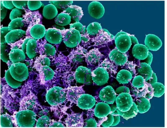
Staphylococcus genus belongs to the Staphylococcaceae family and contains around 50 species and more than 20 subspecies, many of which can be found in humans and other mammals [1,2]. Staphylococci are gram-positive bacteria, characterised by their spherical shape, with a diameter generally ranging from 0.5 to 1.5 μm [3]. Their tendency to be arranged in clusters that reminds clusters of grapes is a distinguish feature of these bacteria (Figure 1.1), owing the name from the Greek staphylé that means “bunch of grapes” [3,4].
Figure 1.1 - Scanning electron microscopy (SEM) image of a grape-like cluster of S. epidermidis. Adapted from [5].
Gram-positive cocci are known for being very heterogeneous and, concerning catalase activity, Staphylococcus spp. are classified as catalase-positive, i.e., they produce catalase, an enzyme responsible for the catabolization of peroxide hydrogen into water and oxygen gas [3,6].
Along with other bacterial species, staphylococci are important pathogens of several mammals, including humans, and are responsible for a wide spectrum of infections, commonly termed “Staph infections”, including a variety of life-threatening systemic diseases [3,7]. Skin and urinary tract infections, as well as infections of the soft tissues and bones, are common examples of injuries caused by several staphylococci, including by some opportunistic Staphylococcus species [3,7]. Opportunistic staphylococci owe the designation to their interactions with the host tissues, since these commensal
4
bacteria usually interact with the host in a probiotic way and, despite taking benefits from the host, they are not considered harmful to the same. Usually, these microorganisms only cause disease under specific circumstances, taking advantage of opportunities that are not generally available, as compromised physical barriers and compromised immune systems, generally in patients with predisposing factors [8,9].
1.1.1 Staphylococcus epidermidis
Staphylococcus epidermidis is part of the wide range of bacteria from the Staphylococcus genus and can be found on the skin and mucous membranes of humans [3,6]. These bacteria are able to grow and possibly cause disease in a great variety of conditions, as they have a remarkable ability to propagate in mediums with high levels of salts, besides being facultative anaerobic and being able to grow in a wide range of temperatures, from 18 to 40 °C [3,6].
Colonization by S. epidermidis is considered frequent and can be harmful to humans, however, this species is known to perform an important role in the maintenance of a healthy skin flora by competing with similar microorganisms which can be considerably more harmful, for instance S. aureus [10,11]. Being a common inhabitant of the skin, S. epidermidis can easily invade this physical barrier through wounds and follicles. This happens mainly when the skin barrier is compromised, for example due to medical practices as the insertion and removal of catheters and other medical devices, or upon fissures on the skin resulting from surgical procedures [7].
It has been argued that S. epidermidis is an accidental pathogen, based on diverse characteristics of the non-infectious lifestyle of this bacterium, for instance, this microorganism presents a benign relationship with the host and acts on a probiotic way to prevent the colonization by more harmful bacteria [12]. Hereupon, the occurrence of some chronic infections and diseases can be justified by the facility of this staphylococcal species to overcome some physiological barriers, as the skin, and to evade antimicrobial therapies and form biofilms that can lead to severe and recurrent infections [12–14].
S. epidermidis is a pathogen with some virulence factors in which interest has been increasing since these bacteria are pointed as one of the leading causes of nosocomial infections [11,15]. This species can be distinguished from S. aureus, one of the most pathogenic and well-known staphylococcal bacteria, due to its inability to produce coagulase, an enzyme that coagulates fibrin in blood, since S. aureus is coagulase-positive and S. epidermidis has a lack in the production of this
5
enzyme, being part of the coagulase-negative staphylococci (CoNS), which are usually less virulent and pathogenic than coagulase-positive species [6, 7].
Prosthesis, medical implants, catheters and shunts are some examples of indwelling medical devices that are becoming more and more common on medical practices, due to their great ability to improve the quality of life of many persons. These medical devices are becoming increasingly sophisticated, however, that does not prevent them from being colonized by several microorganisms. As a result, the surface of these biomedical devices often serves as a microbial reservoir and may lead to several infections, contributing to the increased number of biofilm-related infections [17,18]. Once an indwelling medical device is introduced into the human body, a variety of molecules will quickly coat the biomedical device, forming a film on its surface [17]. Fibronectin, vitronectin, albumin and immunoglobulins are some of the proteins and glycoproteins produced by the human body that, in the presence of a medical device, will allow the attachment of cells, potentially facilitating the formation of biofilms in the surface of the indwelling devices [19].
Although the majority of staphylococcal infections are local, they can evolve to systemic diseases, especially due to the release of bacteria from the infection sites and their consequent entrance in the bloodstream, being able to damage a diversity of organs [4,7]. A great deal of bloodstream infections related to the insertion of catheters, vascular grafts and other indwelling medical devices, since these surgical procedures enhance the exposure of patients to a large amount of bacteria [20]. Staphylococcal bacteremia is one of the most common systemic staphylococcal infections, being one of the major causes of mortality in hospitalized patients with chronic diseases, representing an increased concern due to the lack of effective ways of treatment [21,22]. Moreover, several antimicrobial therapies target S. epidermidis bacterium, since this is one of the most frequent microorganisms causing primary bacteremia and infections on indwelling medical devices, especially in ill patients and neonates [20,23].
S. epidermidis has some virulence factors that allow these bacteria to infect the human tissues and promote the occurrence of infections and diseases, being a major threat to immunocompromised patients [11,24]. Among these virulence factors, the capacity to form biofilms is highlighted, since bacteria within biofilms present some interesting particularities, including higher tolerance to several antimicrobial therapies [11,25]. Furthermore, a few studies have also shown that S. epidermidis biofilms present a higher tolerance to mechanisms of host defense, contributing to the evasion of the immune system and persistence of infections [26,27].
6
1.2 Biofilms
Biofilms are recognized as ubiquitous in nature, being the most common form of organization of several microorganisms, overcoming the number of microorganisms living in a planktonic form [28].
There are a few definitions of the term biofilm, yet, one of the most popular was given by Costerton et al., defining biofilm as an aggregation of microorganisms and their extracellular products, forming a well structured population, generally attached to a surface [29]. An organic film, alternative designation for biofilms, can also be briefly described as an agglomeration of adhered microorganisms surrounded by a macromolecular matrix [19].
Human health can be deeply affected by the development of biofilms, not only because of the high tolerance towards antimicrobial therapies, but also because biofilms can serve as a continuous reservoir of several opportunistic bacteria that are able to colonize different surfaces [19, 31].
Although prevention is the main strategy referring to biofilm infection control, it is not always possible to avoid contamination of medical devices inserted in the human body, despite all the aseptic care in surgical interventions [6]. As a result, it is considerably frequent that contaminations by S. epidermidis occur after a surgical procedure [31].
Formation of bacterial biofilms is accepted as a survival strategy of bacteria and occurs in a spontaneous way, being accounted as responsible for several chronic and acute infections, from which can be pointed out bacterial wound infections, endocarditis and respiratory tract infections [18,32]. The existence of a polymeric matrix surrounding bacteria has some benefits in protecting bacteria towards environmental changes, as pH or temperature, and also protecting them from being removed from the surface, by washing or scraping [28,33]. Besides the contribution to the survival of the biofilm under assorted environmental adverse conditions, as the lack of nutrients, the biofilm matrix is also fundamental for the maintenance of the tridimensional structure of the biofilm [14,34]. Other benefits of residing within a polymeric matrix are the highest protection against exposure to antimicrobial therapies, compared to bacteria in the planktonic state, and the improved assess to nutrients [35,36].
Biofilms are very common in nature and present a crucial role in what refers to the occurrence and persistence of infectious diseases, since these biofilms can prosper on medical implants (Figure 1.2), as well as in the tissue of a large number of mammals [15,37].
7
Figure 1.2 - Scanning electron microscopy (SEM) of a Staphylococcus epidermidis biofilm. Adapted from [38].
1.2.1 Staphylococcus epidermidis biofilms
The process of biofilm formation is the result of a controlled process that comprises multiple steps, being commonly divided in three main phases: attachment, maturation, and disassembly [33,39]. It is important to take into consideration that some authors divide the biofilm formation process in more than three phases, once they subdivide attachment and maturation into multiple stages, however these stages are interconnected and can overlap, being irrelevant to clinically distinguish these multiple stages [40].
Structural and metabolic heterogeneity is common among biofilms, with S. epidermidis biofilms being formed by very heterogeneous populations of cells, in which are involved live, dead, dormant, and persistent bacteria [27,41,42].
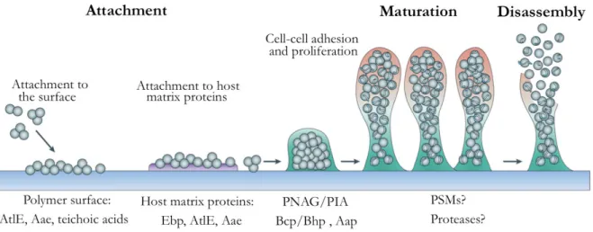
S. epidermidis biofilms are formed according to the general process of biofilm formation, presenting some particular molecules involved in the different stages of biofilm formation [12], as demonstrated in Figure 1.3. The biofilm formation process of this species is regulated by a system of cell-to-cell communication known as staphylococcal accessory gene regulator (agr) [43], as will be further described.
The first phase of biofilm development is generally termed initial adhesion or attachment and comprises bacterial adhesion to surfaces as a result of the contact of bacteria with those surfaces [28]. Non-specific interactions, as hydrophobic and electrostatic interactions, generally command this primary attachment to inert surfaces, in which bacteria adhere straightly to the surface of medical
8
devices in the body [28]. However, bacteria can also adhere to films of host-derived matrix molecules coating a surface, as the surface of biomedical devices, and, in this case, the surface proteins will mediate the adhesion of bacteria to the coated surface of the medical devices [15,28].
The AtlE autolysin is part of the specific proteins that mediate primary attachment, facilitating the adhesion of bacteria to surfaces or to previously attached host matrix proteins [15,45]. Moreover, Bap/Bhp protein is also involved in the first stage of biofilm formation, by increasing the hydrophobicity of the cell surface that facilitate the initial adhesion process [47].
Figure 1.3 - Representation of S. epidermidis biofilm cycle and some of the molecules involved in the different phases of biofilm formation and disassembly. The process begins with the initial attachment to the surface, followed by the adhesion of cells to each other, forming clusters. Maturation of the biofilm is achieved by the growth of the bacteria clusters and production of the polymeric matrix by those aggregates, which will accumulate and surround bacteria. Lastly, a mature biofilm is obtained and bacteria can detach and disperse from this biofilm and colonize other surfaces. [33,44] Adapted from [12].
Succeeding stages of biofilm formation require specific interactions and molecules to allow the growth of bacteria into clusters [44,45]. For this reason, not all of the bacteria that initially adhere to a surface will be able to develop a biofilm. Some of the bacteria will detach from the surface, while only a part of those will enter the next phase and be able to form the biofilm, by influence of specific molecules, as intercellular adhesins and autolysins [39,48].
The second phase, called maturation, refers to the accumulation of several bacteria and formation of the hydrated polymeric matrix that surrounds cells in biofilms [12, 45]. During this phase, multicellular structures, namely clusters, are formed due to the aggregation of cells [12]. Therefore, some molecules, for instance adhesive and exopolysaccharide macromolecules, are secreted to enhance cell-to-cell communication and aggregation [12,44].
9
The icaADBC operon is often present in S. epidermidis bacteria and accomplish an important function in the aggregation of bacteria into clusters [50,51]. These proteins produce a polymer of N-acetyl glucosamine (PNAG) [50]. PNAG is commonly defined as a polysaccharide intercellular adhesin (PIA), which is pointed as the major responsible for the biofilm development of this species since it mediates the intercellular adhesion [50,52].
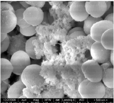
The development of the biofilm continues with the maturation of these agglomerates that grow and produce the extracellular polymeric substances (EPS) which will lodge between cells [33,53]. The major components of these polymeric substances are polysaccharides, proteins and nucleic acids that result from cellular metabolism and/or cell death process, however the composition of the matrix varies among different biofilms [39,54]. This complex extracellular matrix surrounds the bacteria attached to the surface and to each other [53], as shown in Figure 1.4.
Figure 1.4 - Low vacuum secondary electron image of a S. epidermidis biofilm, with evidence to the polymeric matrix surrounding bacteria. Adapted from [55].
During maturation of the biofilm, the increasing number of bacteria and the production of the polymeric matrix lead to the expansion of the biofilm thickness [18,56]. However, the thickness of the biofilm does not increase infinitely and a disassembly process may occur in order to regulate the cell density of the biofilm [57,58]. The availability of nutrients and oxygen [59,60] and environmental parameters, as the pH, temperature and nature of the surfaces to which bacteria are attached [56, 59], cause an active release of biofilm cells, known as dispersion, that contributes to the regulation of the biofilm cell density. Furthermore, a passive release process may also contribute to the regulation of the extent of the biofilm, since shear forces are able to induce the detachment of biofilm cells [61,62].
10
The active and passive events of release may occur throughout the entire biofilm cycle, nevertheless, the remaining cells of the biofilm undergo further stages of maturation. It is important to have in consideration that biofilm infections are clinically relevant not only when they reach a mature state, but they can also be threatening in previous phases of the biofilm cycle. This may happen, for instance, because some of the clusters that are formed during maturation process may detach from the surface and enter blood circulation, introducing a potential danger of causing thromboembolisms that can, ultimately, culminate in patient death [63].
Later in this phase, a biofilm structure containing channels is formed. This event is dependent on adhesive and disruptive forces and allow the communication of cells with the exterior, enabling the circulation of nutrients and oxygen into the deeper layers of the biofilm [33,64].
Finally, the mature biofilm, characterized by a thicker film of bacteria and a more protuberant matrix, reaches a state that no longer allow the growth and division of cells due to nutritional and physicochemical limitations and, thereby, biofilm cells undergo a final disassembly process in a greater extent, by active or passive processes, as previously explained [44,57]. The previously formed channel-containing structure facilitates the evolution of the biofilm to achieve the disassembly phase [39,64]. The cells disassembled from the biofilm may be designated as biofilm-released cells (Brc) [58] and have the ability to colonize other sites, contributing to the spreading of infections among the host and to the occurrence of inflammation processes [65,66].
Disassembly remains the least understood phase of biofilm lifecycle and, therefore, some of its molecular mechanisms are not completely established [66]. Although the promoters of the disassembly are not entirely known, it is currently accepted that shear forces, associated with detachment, and/or specific gene expression, related to disassembly, can be the cause of the release of these new colonizers, leading to the propagation of the infection and to an increasing number of biofilms [57]. Furthermore, proteases and PSMs (phenol-soluble modulins) are thought to participate in the degradation of S. epidermidis biofilm matrix, contributing to the disassembly process, being modulated by a quorum-sensing mechanism that will be succeeding described [57,67].
It is known that disassembly involves some alterations in the biofilm, as the degradation of the extracellular polymeric matrix, as well as some physiological changes that allow the preparation of Brc to the environmental conditions outside the biofilm [57,68]. Therefore, Brc are believed to present distinct phenotypic features from both biofilm and planktonic cells [65], as will be further addressed.
11 1.2.2 Quorum-sensing
Despite a certain lack of knowledge about the mechanisms of maturation and detachment of the biofilm, it is known that there are several mechanisms of intercellular signalling among bacteria that result from the ability of microorganisms to produce molecules that can be recognized by specific receptors [44,69]. Quorum-sensing (QS) is an example of those mechanisms, though to be responsible for the transition of planktonic to biofilm lifestyle in bacteria and can be defined as a regulatory mechanism that exists in microorganisms to control gene expression, being dependent on cellular density [56]. This system allows cell-to-cell communication and mediates the secretion of molecules that act as signals to control the synchronization of gene expression and functional coordination among populations of microorganisms, as biofilms [57, 64].
The initiation of biofilm formation is triggered when, by quorum-sensing signalling, bacteria sense unfavourable or stress conditions, as the lack of nutrients and alterations in environmental parameters [56,71]. Due to different signalling by QS, biofilm formation and development differs according to distinct environmental conditions, as different temperatures, pH, and nutritional availability, among others [56,69]. Moreover, quorum-sensing mechanisms are involved in the monitoring and regulation of biofilm density, acting as a control to promote either the maturation of the biofilm, to increase its extent and thickness, or the inhibition of biofilm formation and stimulation of the dispersion phase, leading to a decrease in the amount of bacteria residing within the biofilm structure [56,70].
Similarly to what happens with other species, the formation and regulation of staphylococcal biofilms is a complex process, influenced by the environmental conditions and by the genotype of the microorganisms [72]. In S. epidermidis, biofilm formation is controlled by a system named agr (accessory gene regulator) [43], wherein the expression of targets regulated by agr is dependent on the density of cells, as it is characteristic of QS mechanisms [73]. agr was once viewed as a regulator of virulence factors, however, findings on the existence of this gene in non-pathogenic species lead to the assumption that this system is a quorum-sensing regulator, which includes the control of some virulence factors in pathogenic species, playing an important role in the species pathogenesis [43], but, as well, the control and regulation of other non-virulent mechanisms [74,75]. Consequently, agr is involved in the invasiveness of bacteria, by upregulating the expression of virulence factors and downregulating the production of surface proteins, contributing to the invasiveness of bacteria on the hosts [43].
12
When there is few agr activity, as a result of a low density of cells, the quorum-sensing mechanisms emit signals to increase the expression of surface proteins that allow bacterial colonization, so that they can divide and increase cellular density [76,77]. As a result of augmented cellular density, agr activity raises and the secretion of surface proteins decreases, reestablishing the balance by a mechanism of negative regulation [76,77]. However, the aging of the biofilm leads to the loss of viability of bacteria and to a reduction in the expression of agr, affecting the chronicity of the infections since the decrease in the production of signaling molecules will compromise the regulation mechanisms and have a negative effect on the balance of the number of bacteria [72,78].
It is believed that, besides the previously described functions, QS also performs a considerably important role in the release of cells from the biofilms and may influence the resistance to some antimicrobial drugs [43,71]. Some studies have already reported that agr expression is involved in the dispersion of staphylococci biofilms [58,78]. Furthermore, agr expression has been associated with decreased antibiotic susceptibility for staphylococcal biofilms [71], which may, as well, influence the tolerance of biofilm cells and Brc to antibiotics.
Quorum-sensing mechanisms, namely agr in what refers to staphylococci, have been pointed as potential targets for prophylaxis and therapy. An approach that have been suggested is the inhibition of genes directly involved in QS, since this would reduce the pathogenicity of several bacteria as a result of the attenuation of the expression of virulence factors commanded by QS [43,70]. However, the upregulation of adhesion mechanisms caused by agr inhibition may enhance cell adhesion and lead to a higher persistence and chronicity of biofilm infections, increasing biofilm formation [43,71,72]. For that reason, it is still unknown whether the advantages of the inhibition of QS would overlap the disadvantages, so that further studies need to be accomplished in this matter.
1.2.3 Biofilm tolerance to antibiotics
Biofilm infections are very threatening and the decrease in the susceptibility to antibiotics is a very concerning issue [15,31]. This increased tolerance often leads to situations where it is unsuitable to treat infections with common antibiotic therapies, since the concentration of antibiotic needed to kill bacteria within biofilms is higher than the peak serum concentration (PSC), which is the maximum concentration of antibiotic that the human body can endure after administration [79–81]. Different mechanisms that attempt to explain this feature will be presented ahead.
Biofilms are thought to admit a higher tolerance to antibiotics by a diversity of factors, from which can be highlighted the diffusional barrier to antibiotics, the existence of a more resistant
13
phenotype and a slow growth-rate of cells within the biofilm [25,36], as represented in Figure 1.5. Moreover, the existence of persister cells, with an increased tolerance to antibiotics, can also partially explain the inefficacy of antibiotic treatments in biofilms [82].
Figure 1.5 - Some hypothesis that attempt to explain the decreased susceptibility of biofilm cells to antibiotics. Adapted from [83].
The structure of the extracellular polymeric matrix acts as a physical diffusional barrier reducing and/or delaying the penetration of antibiotics into the biofilm, whereby antibiotics can no longer reach a great amount of bacterial cells [14,81]. For the same reason, the increased number of bacteria in biofilms, which result from cell division, contribute to the expansion of the thickness of the biofilm and, consequently, hinders the penetration of antimicrobial substances into the deeper layers of the biofilm [38,81].
Furthermore, the negatively charged polymeric matrix may also behave as a chemical barrier to the positively charged antimicrobial agents, since these agents tend to bind to the matrix and, thus, the amount of antimicrobial drugs that successfully reach biofilm cells is limited [18,84]. Moreover, some of the polysaccharides and proteins that constitute the matrix perform an important role in the protection of bacterial biofilm cells against antimicrobial therapies by acting as a protective barrier and/or inactivating some antibiotics [14,82,85].
Among the modifications that bacteria experience upon adaptation to biofilm mode, phenotypic changes are one of the most important, considering they may influence the susceptibility to antibiotics within the biofilm environment [14,81]. It is now accepted that bacteria residing within the biofilm are phenotypically different from free-floating bacteria, whereby some bacteria may experience a
14
differentiation process which leads to a resistant phenotype, contributing to the higher tolerance of biofilms against antimicrobials [65,81].
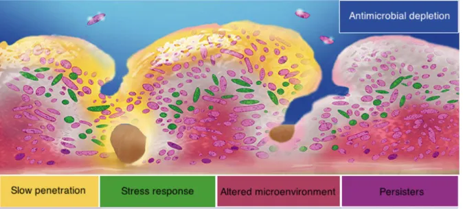
A slower growth-rate of bacteria is found in altered environment zones, since in the deeper layers of the biofilm the concentrations of oxygen and nutrients are reduced, leading to distinct growth conditions [76, 79], as represented in Figure 1.6.
The reduced bacterial growth-rate, as well as the resulting alteration of metabolic processes and reduced metabolic activity, present a limitation to the action of some antibiotic classes in biofilm cells, since it increases their tolerance to these chemical agents [25,82,86].
Figure 1.6 - Schematization of the heterogeneity of Staphylococcus epidermidis biofilms over the depth. Young
biofilms (A) provide a high availability of nutrients and oxygen (O2) to all the bacteria, while mature biofilms are
characterized by deeper layers (D) with a small amount of nutrients and O2, and upper layers (B) with a great
accessibility of nutrients and oxygen. Adapted from [40].
The heterogeneity of cells within biofilms, from which can be highlighted the wide range of metabolic activities between cells [87], also contributes to the increased tolerance to antimicrobials [81,82]. Dormant and persister cells are characterized for becoming metabolically less active than other cells, mainly upon facing stressful conditions, and for presenting an increased tolerance to antibiotics, contributing to recalcitrant infections [88–90].
Dormant cells exist in a non-replicative state that is reversible, i.e., these cells are in a temporary dormancy state where they slowdown metabolic processes and are not able to replicate [89,90]. On the other hand, persistence refers to a state in which some bacteria survive antimicrobial treatments [89,91]. Thus, persisters are often defined as a sub-population of cells that entered a spontaneous dormant state in which they do not proliferate, presenting a substantial tolerance to antibiotics, being, however, able to restore their function when inoculated into fresh medium without antimicrobial substances [81,92]. Therefore, 9ipersistence may not be directly associated with dormancy, which means that not all dormant cells are persisters, especially taking into account that
15
persistence is mainly associated with antibiotic stress and dormancy often occurs in response to unfavorable environmental conditions rather than to antimicrobial therapies [91,93].
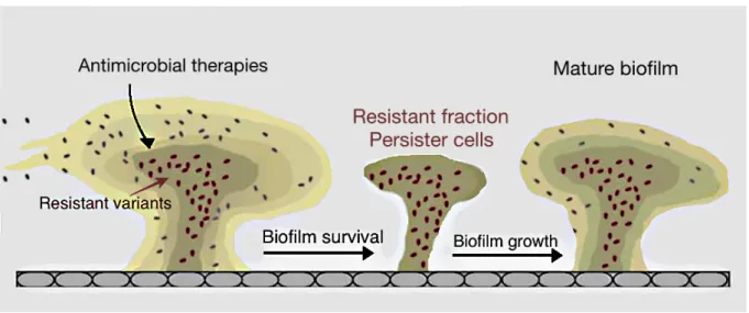
Although persister cells can exist in planktonic state, their frequency is higher in slow-growing biofilms, partially explaining the higher tolerance of biofilms against antibiotics, compared to planktonic cells [82]. These cells can be pointed as a cause of relapsing biofilms after antimicrobial treatments, since in the persistent state these cells survive antimicrobial drugs (resistant variants) and, afterwards, are able to proliferate and lead to the growth of the biofilm, culminating in a mature biofilm [81,82,94], as represented in Figure 1.7.
Figure 1.7 - Schematization of the resistance mechanism due to resistant/persister cells. Although antimicrobial
therapies can eradicate part of the biofilm cells, some resistant variants are not affected by the antimicrobial drugs and are able to persist and maintain the biofilm survival. After antimicrobial therapy discontinuation, the resistant fraction is able to develop a new biofilm that will grow and reach maturation. Adapted from [94].
As a consequence of the reduced susceptibility of biofilms to common antibiotics, it is often necessary to use a combination of different antibiotics and substances capable of degrading the matrix that envelops bacteria, in order to expose cells to the antibiotics [80, 78]. However, due to the inefficacy of several therapies, the treatment of medical devices-related infections may result in failure and, in those cases, the removal of the infected medical devices is required, resulting in high health costs and great inconvenience to patients [96,97].
16 1.2.4 Biofilm-released cells (Brc)
Brc are cells that suffered disassembly from the biofilm, by either dispersion (active process) or detachment (passive process) during its lifecycle, being capable to trigger inflammatory events [65,66]. These cells may also act as new colonizers and are able to form biofilms in different loci after being released from the biofilm [65,66].
Surprisingly, little is known about the phenotypic alterations that these cells undergo, as well as about the impact of these alterations in the clinical field [67]. It was primarily thought that, soon after being released from the biofilm, Brc would revert the phenotypic alterations and become similar to planktonic cells again [65,81]. However, some studies have reported that cells released from the biofilms were different from both biofilm and planktonic bacteria, denying the previous assumption of immediate phenotype reversion [57, 58, 65, 99].
Recently, studies published by França et al. confirmed suspicions about phenotypic differences of Brc comparing to the biofilm and planktonic counterparts, regarding the inflammatory response and the reaction to antimicrobial therapies, that help explain the relapsing nature of infections of S. epidermidis biofilm-related infections [58,98]. These researchers have shown that S. epidermidis Brc may be more effective in the activation of the inflammatory response, since Brc induced a particular gene expression on mouse splenocytes, with an increased expression of several genes related to cell death, and induced a higher stimulation of pro-inflammatory cytokines [98]. They also showed that Brc present a higher tolerance than their planktonic counterparts against some antibiotics, retaining their tolerance when growing in the presence of the originating biofilm [58]. However, their transient phenotype was reverted when these bacteria proliferated planktonically in the absence of the originating biofilm [58].
This specific bacterial population merits special attention, as the disassembly of cells from the biofilm may provide a pathway to the occurrence of diverse injurious events and to the spreading of biofilm infection, particularly since these cells present a different behaviour against antimicrobials [58]. The determination of the antibiotic profile of Brc would provide significant insights to the pathophysiology of biofilm infections and facilitate the development of effective strategies to the control of infections related to biofilm disassembly [67]. Undoubtedly, a depth investigation on the properties of Brc should be performed in order to proficiently target, prevent and treat Staphylococcus epidermidis biofilm-related infections.
17
1.3 Aims and objectives
The aim of the present work was to determine the antibiotic tolerance profile of clinical strains of S. epidermidis Brc. To accomplish this goal, the work was divided into three main tasks.
The first task consisted in the study of the antibiotic susceptibility of cells released from 48-hour mature biofilms (Brc48H) of S. epidermidis 9142. The main objective of this task was the comparison of
the antibiotic effects in Brc48H with the effects in 48 hour-biofilm cells and in stationary planktonic cells.
The aim of the second phase was to determine if cells released from biofilms with different stages of maturation presented distinct susceptibilities to antibiotics.
Finally, the purpose of the last phase of this work was to determine if the results of antibiotic susceptibility in the three different bacterial populations of different S. epidermidis isolates were consistent with the results obtained in the previous phases, for the control strain 9142. It was assumed that the results of this study would help to understand if the phenomenon of antibiotic tolerance of Brc is common to distinct isolates of this species.
19
21
2.1 Isolates and growth conditions
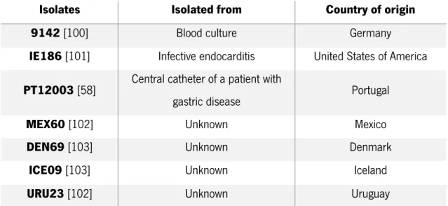
Staphylococcus epidermidis 9142, a blood clinical isolate known by its strong ability to form biofilms and generally used as a biofilm positive control [99,100], was the isolate selected for the majority of the experiments of this thesis. Furthermore, other clinical isolates (see Table 2.1) were used in order to compare the antibiotic susceptibility among different S. epidermidis and assess if the pattern behaviour remains constant in all the isolates tested.
Table 2.1 - Origin of the Staphylococcus epidermidis isolates used in this study
Isolates Isolated from Country of origin
9142 [100] Blood culture Germany
IE186 [101] Infective endocarditis United States of America
PT12003 [58] Central catheter of a patient with
gastric disease Portugal
MEX60 [102] Unknown Mexico
DEN69 [103] Unknown Denmark
ICE09 [103] Unknown Iceland
URU23 [102] Unknown Uruguay
2.1.1 Biofilm formation and biofilm-released cells collection
An inoculum was done by adding one S. epidermidis colony into 2 mL of Tryptic Soy Broth (TSB) (Liofilchem, Teramo, Italy) and incubated in an orbital shaker overnight at 37 °C and with agitation at 120 rpm. Later, the overnight cells were diluted in TSB medium until an optical density at 640 nm (OD640) of 0,250 ± 0,05 was reached, corresponding to an approximate concentration of 2 ×
108 CFU (colony forming units ) / mL [104]. Biofilms were formed through the inoculation of 15 µL of
the adjusted suspension into a 24-well microtiter plate (Orange Scientific, Braine-l’Alleud, Belgium), with 1 mL of TSB medium supplemented with 0.4 % (v/v) glucose (TSBG) to induce biofilm formation, being incubated at 37 °C with shaking at 120 rpm for as long as 72 (±1) hours in an orbital shaker. After each 24 (± 1) hours of incubation, spent medium was carefully removed and the biofilms were washed twice with a saline solution (0.9 % (m/v) NaCl in distilled water) in order to remove unattached cells, followed by the careful addition of 1 mL of fresh TSBG and subsequent incubation in the same
22
conditions. Finally, biofilms were washed twice with the saline solution, suspended in 1 mL of the same by scraping the cells from the plastic surface, and bacteria from either 24, 28, 48 or 72-hour biofilms were collected into a flask, pooling together at least 4 different biofilms to decrease the variability inherent to biofilm formation [105].
Biofilm-released cells (Brc) were collected, from at least 4 different wells, by careful aspiration, at different timepoints, from the biofilm bulk fluid of 28 or 48-hour biofilms, depending on the study concerned, and stored into a flask, as described previously [58].
2.1.2 Planktonic growth
From an overnight inoculum grown in the same temperature and agitation conditions previously mentioned (section 2.1.1), a dilution with TSB medium was performed in order to adjust the optical density to a cellular concentration of 2 × 10⁸ CFU/mL. Following, 150 µL of this suspension were
inoculated into a 25 mL Erlenmeyer containing 10 mL of TSBG and incubated at 37 °C with agitation at 120 rpm during 24 (± 1) hours. Stationary planktonic cells were, then, collected into a flask.
2.1.3 Cell homogenization
The three suspensions (disrupted biofilm cells, Brc and stationary planktonic cells) were submitted to a pulse of 5 seconds of sonication with 40 % amplitude (Ultrasonic Processor Model CP-750, Cole-Parmer, Illinois, U.S.A.) in order to homogenize the suspensions and disassociate possible existing clusters. As previously demonstrated [106], this sonication cycle did not have a significant effect on cell viability.
23
2.2 Characterization
of the antimicrobial profile of planktonic
S.
epidermidis
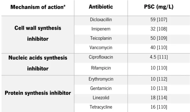
A total of 10 antibiotics (see Table 2.2) with different mechanisms of action were used to assess the susceptibility of the three cell populations under study. A preliminary study was performed to characterize the antimicrobial profile of the S. epidermidis isolates under study, through the determination of the minimum inhibitory concentration (MIC) for each antibiotic.
Inocula from all the populations were diluted into TSB to obtain a concentration of about 2 × 10⁸ CFU/mL, by measuring the OD
640, after calibrating for CFU/mL [104]. Following, 2 µL of each
suspension were added to different wells containing 200 µL of TSB medium with antibiotics, whereas different gradients of concentrations were used according to each antibiotic. Simultaneously, a positive control was performed by inoculating the same quantity of suspension into 200 µL of TSB without antibiotics. The MIC was determined as the lowest concentration of antibiotic that inhibited a visual growth of bacteria and the determination was based on at least two consistent replicates.
Table 2.2 - Mechanism of action and peak serum concentration (PSC) in mg/L of the ten antibiotics used in this study
Mechanism of action
aAntibiotic
PSC (mg/L)
Cell wall synthesis
inhibitor
Dicloxacillin 59 [107]
Imipenem 32 [108]
Teicoplanin 50 [109]
Vancomycin 40 [110]
Nucleic acids synthesis
inhibitor
Ciprofloxacin 4.5 [111]
Rifampicin 10 [110]
Protein synthesis inhibitor
Erythromycin 10 [112]
Gentamicin 10 [113]
Linezolid 18 [114]
Tetracycline 16 [110]
aThe mechanism of action of the antibiotics was determined by the information sheet provided by the antibiotics manufacturer.
24
2.3 Comparison of the antimicrobial susceptibility of the distinct
S.
epidermidis
populations
Following the treatment and homogenization of the different cell populations, according to the previously described process (section 2.1), the suspensions were diluted in TSB medium in order to reach a concentration of about 2 × 10⁸ CFU/mL. Next, 200 µL of the adjusted suspensions were
inoculated into TSB medium, in a total of 2 ml, achieving a concentration of approximately 2 × 10⁷
CFU/mL. Then, each antibiotic was added to the previous suspensions, at the peak concentration, and the tubes were incubated at 37 °C and 120 rpm agitation for a period up to 6 hours. Simultaneously, controls were performed by the inoculation of the same suspensions in TSB medium, without the addition of any antibiotic, and further incubation under the same conditions. All the tubes were prepared in duplicate, for all the conditions tested.
After 2 and 6 hours of incubation, one mL of each tube was collected and centrifuged at 4 °C and 16,000 g for 10 minutes. Next, the supernatant was carefully discarded and the pellet was resuspended in 1 mL of 0.9 % NaCl solution, with the aid of a pulse of 5 seconds of sonication at 40 % amplitude.
Finally, 10-fold serial dilutions were performed, vortexing each sample before each dilution, and plated onto Trypticase Soy Agar (TSA), which was prepared by the addition of 30 g/L of TSB (Liofilchem) and 15 g/L Agar (Liofilchem). The plates for CFU counting were incubated at 37 °C until the colonies were grown enough to allow the counting.
25
27
3.1 Study of the antibiotic susceptibility of cells released from
Staphylococcus epidermidis
9142 biofilms with 48 hours of maturation
(Brc
48H)
Biofilms, communities of bacteria embedded in a polymeric matrix, follow a lifecycle with three main stages: attachment, maturation and disassembly [12,29]. Over the disassembly stage, the biofilm release cells to the surrounding environment, namely biofilm-released cells (Brc), which are thought to be responsible for serious complications as, for instance, bacteremia [20]. Although several studies have been performed to compare the antibiotic susceptibility of bacteria in biofilms with their planktonic counterparts, little is know regarding the tolerance of Brc to antibiotics.
To overcome the lack of knowledge on the efficiency of antibiotics against Brc, the first studies of this thesis consisted in the determination of the susceptibility to antibiotics of biofilm-released cells from 48-hours mature biofilms (Brc48H) and the comparison with both 48-hours mature biofilm cells and
planktonic cells in the stationary phase, grown for 24 hours.
3.1.1 Preliminary MIC assay
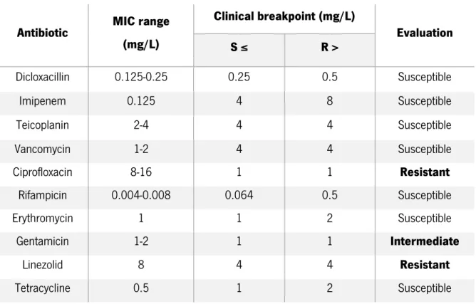
First, a preliminary assay was performed by determining the minimum inhibitory concentrations (MIC) of all the antibiotics against S. epidermidis 9142, in order to verify if this control strain would be susceptible to the antibiotics under study. A standard MIC assay was conducted as previously described (section 2.2) and the results are presented in Table 3.1.
According to EUCAST, bacteria may be considered as clinically susceptible (S), clinically resistant (R) or clinically intermediate (I) to an antibiotic. When the MIC value is equal to or below the lower breakpoint value, bacteria are considered susceptible, meaning that the level of antimicrobial activity is associated with a significant chance of therapeutic success, while when the MIC value is higher than the upper breakpoint value, bacteria as defined as resistant, what is an evidence that there is a high probability of therapeutic failure with the antibiotic concerned. However, in some cases, the MIC range is in the middle of the breakpoint values, which may include the breakpoint limits, and bacteria are considered intermediate, meaning that the therapeutic effect is uncertain [115].
The results obtained in the MIC assays were compared with the clinical breakpoints for S. epidermidis described in the literature by the European Committee on Antimicrobial Susceptibility Testing (EUCAST)[116] for the majority of the antibiotics, and by the Clinical and Laboratory Standards
28
Institute (CLSI)[117] and British Society for Antimicrobial Chemotherapy (BSAC)[118] for dicloxacillin and imipenem, respectively, since EUCAST did not provide the MIC breakpoints for these antibiotics.
Table 3.1 - Determination of the MIC ranges in mg/L of ten antibiotics against S. epidermidis 9142 and evaluation, by EUCAST, CLSI and BSAC standards, of the susceptibility to the antibiotics tested
Antibiotic MIC range (mg/L) Clinical breakpoint (mg/L) Evaluation S ≤ R > Dicloxacillin 0.125-0.25 0.25 0.5 Susceptible Imipenem 0.125 4 8 Susceptible Teicoplanin 2-4 4 4 Susceptible Vancomycin 1-2 4 4 Susceptible Ciprofloxacin 8-16 1 1 Resistant Rifampicin 0.004-0.008 0.064 0.5 Susceptible Erythromycin 1 1 2 Susceptible Gentamicin 1-2 1 1 Intermediate Linezolid 8 4 4 Resistant Tetracycline 0.5 1 2 Susceptible
From the analysis of Table 3.1 and according to the CLSI breakpoints, 9142 was classified as susceptible to dicloxacillin. Similar, comparing the results to the BSAC breakpoints, this strain was found to be susceptible to imipenem. Through the comparison with the EUCAST clinical breakpoints, S. epidermidis 9142 was classified as susceptible to teicoplanin, vancomycin, rifampicin, erythromycin and tetracycline. On the other hand, this strain is thought to be resistant to ciprofloxacin and linezolid. Moreover, the MIC range obtained with gentamicin comprised both the susceptible and resistant limits of the clinical breakpoints, thus, this strain was classified as clinically intermediate to this antibiotic.
29 3.1.2 Susceptibility assays
To assess the effect of antibiotics among the three different populations of cells, the suspensions were simultaneously incubated under the same conditions with and without antibiotics (control), in order to evaluate the changes on the cultivability of the suspensions after having contacted with the antibiotics. The concentration chosen to accomplish these comparisons was the PSC for each antibiotic, which is thought to be the concentration that presents the highest relevance from the clinical point of view, since it is an estimation of the maximum concentration of antibiotic reached in the human bloodstream [79,110]. Furthermore, all the suspensions were adjusted to the same concentration prior to the incubation with the antibiotics, allowing to accomplish a more accurate comparison between the susceptibility of the distinct populations of cells, as previously described [79]. Since the antibiotics are frequently dependent on the cellular density of the population, the initial adjustment of the optical density is advantageous in what refers to a suitable comparison between populations, yet if the number of cells is too high or too low in comparison with the ideal range of action, the antibiotic may not be able to act as expected and present a lower efficacy of killing [119]. It is, however, important to take into consideration that the measurement of the OD only provides an estimation of the number of cells, since the extracellular products may also affect the OD value, meaning that the number of cells of the adjusted suspensions may continue to present some variability.
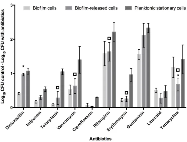
The evaluation of the different susceptibilities of the three populations over time was performed with two different times of incubation with PSC of antibiotics, namely 2 and 6 hours, and the results are represented in Figure 3.1 and Figure 3.2, respectively. Based on the MIC results, it is predicted that S. epidermidis 9142 will experience a significant reduction on the cultivability upon exposure to seven out of ten of the antibiotics, to which showed to be susceptible, and a smaller or negligible cultivability decrease with the three antibiotics to which was considered intermediate or resistant. However, it is important to recall that the MIC assay was performed with planktonic cells, whereby the conclusions may not be applied to biofilm cells and Brc, meaning that these populations may present a different reaction upon contacting with the antibiotics, as shown before with a limited number of antibiotics [58].
To confirm those earlier findings, this study was conducted with ten antibiotics with different mechanisms of action, namely cell wall synthesis inhibitors (dicloxacillin, imipenem, teicoplanin and vancomycin), nucleic acids synthesis inhibitors (ciprofloxacin and rifampicin) and antibiotics that act as inhibitors of protein synthesis (erythromycin, gentamicin, linezolid and tetracycline), being expected that different classes of antibiotics could generate different responses in the populations of bacteria tested [120]. Moreover, antibiotics with the same mechanism of action may also produce different effects on