Pearls
The Skin Microbiome: A Focus on Pathogens and Their
Association with Skin Disease
Keisha Findley1, Elizabeth A. Grice2*
1Social and Behavioral Research Branch, National Human Genome Research Institute, Bethesda, Maryland, United States of America,2Department of Dermatology, University of Pennsylvania, Perelman School of Medicine, Philadelphia, Pennsylvania, United States of America
What Is the Human Microbiome and Why Is It Important?
Microbes are present in every environmental niche and live in close association with humans and each other. Members of these communities include bacteria, archaea, viruses, fungi, and protists. In the environment, microbes perform various roles and are essential in agriculture and food production. In humans and animals, microbes provide protection against foreign invaders, educate and stimulate the immune response, produce antimicro-bials, aid in digestion, and produce vitamins, among a host of other functions.
In the last decade, researchers have actively investigated the impact of microbes and their gene products in human health and disease. This field of research is defined as the human microbiome, which is the totality of the microbes and their genes in and on the human body. To better understand the impact of microbes on health and disease, multiple body sites have been investigated, including skin, lungs, the nasal passage, the oral cavity, and the gastrointestinal and urogenital tracts. The results of these studies have revealed that each body site is home to a unique microbial community. Shifts in microbial communities can result from multiple factors, including environment, genetic variation, lifestyle and hygienic factors, and the immune system, and have been associated with many diseases.
What Is the Difference between Culture-Dependent and –Independent Methods of Microbial Community Characterization?
Up until the 1980s, microbiologists routinely relied on culture-dependent methods for microbial isolation, identification, and characterization. Colony morphology, stains (i.e., Gram stain), biochemical characteristics (i.e., coagulase test), motility tests, antibiotic resistance profiles, and other characteristics guided bacterial and/or fungal identification and taxonomy. However, this approach has several limitations, including an inability to mimic in vivo conditions and selection against slow-growing and/ or fastidious organisms. With recent advances in sequencing technologies and development of bioinformatics tools and reference databases, researchers are now better equipped to capture microbial diversity without the biases of culture-based approaches.
Culture-independent methods of microbial identification rely on a targeted amplicon strategy, which employs highly conserved microbe-specific molecular markers and does not rely on growing isolates in pure culture. The 16S ribosomal RNA (rRNA) gene is used for bacterial identification, while fungi and other micro-eukaryotes are identified using either the 18S rRNA gene or the Internal Transcribed Spacer (ITS) region. A complementary approach to amplicon-based surveys is whole genome shotgun metagenomics. With this approach, one can identify the
micro-biota present and gain insight into the functional potential of the microbiota in an untargeted manner.
What Is the Diversity of the Human Skin
Microbiota, as Revealed by Culture-Independent Methods?
The skin is our first line of defense against foreign invaders and is also home to a diverse population of microbes. The majority of these microbes are commensals (nonpathogenic permanent residents) or transients (temporary residents) of the human microbiota. In pathogenic interactions, only the microbe benefits, while the host is eventually harmed. Many skin pathogens can be typically found living on the skin as commensals, but microbial dysbiosis (or microbial imbalance), host genetic variation, and immune status may drive the transition from commensal to pathogen.
Analysis of bacterial diversity on human skin employing 16S rRNA sequencing revealed that multiple skin sites exhibited greater bacterial diversity than in the gut and oral cavity; interpersonal variation varied significantly within the population studied, and the temporal stability of the analyzed skin microbial communities remained relatively stable [1]. Physiological charac-teristics of various skin sites are associated with different levels of bacterial diversity [2]. Spatially, the skin microbiota may extend to subepidermal compartments [3]. Findley et al. characterized fungi on healthy human skin using sequencing of the ITS region and showed the greatest diversity of fungi was found on the feet, with intermediate levels of diversity on the hand and forearm [4]. The core body sites were less diverse but more stable over time and were primarily colonized by the most common skin commensal,
Malassezia. Additionally, using shotgun metagenomic sequencing techniques, several viruses have been reported in association with healthy skin and skin disorders, including human papillomavirus, human polyomaviruses, circoviruses, and bacteriophages [5,6].
Citation:Findley K, Grice EA (2014) The Skin Microbiome: A Focus on Pathogens and Their Association with Skin Disease. PLoS Pathog 10(11): e1004436. doi:10. 1371/journal.ppat.1004436
Editor:Virginia Miller, University of North Carolina at Chapel Hill School of Medicine, United States of America
PublishedNovember 13, 2014
This is an open-access article, free of all copyright, and may be freely reproduced, distributed, transmitted, modified, built upon, or otherwise used by anyone for any lawful purpose. The work is made available under the Creative Commons CC0 public domain dedication.
Funding:The authors received no funding specific to the preparation of this review.
Competing Interests:The authors have declared that no competing interests exist.
* Email: [email protected]
What Are Some Key Pathogenic Skin Microbes, the Diseases They Cause, and Their Relationship to the Skin Microbiome?
In the following sections we highlight key findings that implicate specific microbes in skin disease, but whose pathogenesis may be complicated by microbial community interactions and/or host-microbe interactions. The specific host-microbes discussed include
Staphylococcus aureus, Propionibacterium acnes, and Malassezia
spp., all of which are known skin commensals but also exhibit pathogenic potential under certain conditions. There are other well-characterized skin pathogens that have been definitively linked to dermatological disorders, but will not be examined in depth here. These include, but are not limited to, the dermato-phyte Trichophyton (causing onychomycosis and tinea pedis),
Corynebacterium minutissimum (causing erythrasma), papilloma-viruses (causing warts),Candidafungal species (causing cutaneous candidiasis and diaper rash), and Pseudomonas aeruginosa
(implicated in green nail syndrome, gram-negative folliculitis, and toe web infections).
Staphylococcus aureus
Staphylococcus aureusis a gram-positive bacteria and facultative anaerobe. In some cases, it may be a skin commensal and colonizes the nares in approximately 20% of the population [7]. However, S. aureus is a major cause of skin and soft tissue infections (SSTI) (Table 1). The sequenced genome ofS. aureus
has revealed multiple virulence factors encoded by phages, plasmids, and pathogenicity islands [8]. In order to evade detection by the host’s immune system, S. aureus produces a variety of enzymes and toxins to successfully establish infection [8].
S. aureuscolonization and infection are associated with atopic dermatitis (AD), a chronic and highly inflammatory skin disease in children and adults (Table 1). Kong et al. recently showed, using 16S rRNA sequencing, that in a pediatric population of AD patients, temporal shifts in the skin microbiota occur over three disease stages, baseline, flare (active disease), and post flare (after treatment has been administered), as compared to healthy age-matched controls. In particular, lesional skin bacterial diversity decreased during the flare stage, parallel with increased relative abundance ofS. aureus, but increased during the post flare status, indicative of a link between disease severity and microbial diversity [9]. An explanation for the observed increase inS. aureusin lesional skin could in part be due to a reduction in the host’s ability to produce antimicrobial peptides, which normally prevent invasion byS. aureus[10].
Propionibacterium acnes
Propionibacterium acnes is a gram-positive, oxygen tolerant anaerobe, and one of the most common skin commensal bacteria.
P. acnesresides in hair follicles and sebaceous glands where it metabolizes sebum triglycerides to release free fatty acids. P. acneson skin may inhibit invasion by pathogenic microbes likeS. aureus and Streptococcus pyogenes through the production of these short-chain fatty acids [11]. P. acnes also produces propionic acid and secretes bacteriocins such as thiopeptide, which suppress the growth of S. aureus and other pathogenic microbes [12,13].
P. acnes is associated with the common adolescent skin condition acne vulgaris (Table 1) [14], though the disease is likely multifactorial with contributions from the immune system, genetics, and the environment. The colonization of skin withP. acnes is age-dependent [15], increasing in parallel with maturation of sebaceous glands during puberty. Sequencing of the bacterial rRNA gene indicates that certain strains of P. acnesmay be associated with acne and contain unique genetic elements that contribute to their virulence [11,16]. Probiotic applications may offer some benefit for treatment, as P. acnes
growth is inhibited by succinic acid, a fatty acid fermentation product of the commensalStaphylococcus epidermidis[17]. Acne may also be an ideal candidate for bacteriophage therapy, asP. acnes bacteriophage are limited in genetic diversity, have a broad host range, and are unable to form stable lysogens within their hosts [18].
Malassezia
Malassezia is a genus of dimorphic and primarily lipophilic fungi formerly known as Pitysporum. It is the most abundant fungal skin commensal, representing 50%–80% of total skin fungi, most common in oily areas such as the face, scalp, and back.
Malassezia species live in the infundibulum of the sebaceous glands where they feed on lipids found in human sebum [19].
Malasseziahas been hypothesized as the infectious agent in several skin disorders, namely dandruff, atopic dermatitis, tinea versicolor, and to a lesser extent psoriasis (Table 1) [20].Malasseziaproduces lipases, phospholipases, and allergens (Mala genes), which can damage the integrity of the skin by inducing inflammation and triggering an immune response [21].
Dandruff is a common skin disorder marked by abnormal flaking and itching of the scalp. Antifungal agents are quite effective in treatment, thus suggesting a fungal component to the disease. Free fatty acids such as oleic acid produced by
Malassezia lipases may be responsible for altering skin barrier permeability, leading to irritation and inflammation of the scalp in predisposed individuals [22]. A recent culture-independent study on French subjects with and without dandruff suggested that disequilibrium between bacteria and fungi, including
Malassezia spp., on the scalp is associated with this condition [23].
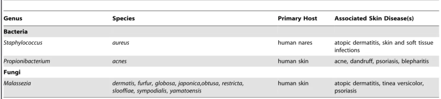
Table 1.Skin microbes discussed and their associated skin diseases.
Genus Species Primary Host Associated Skin Disease(s)
Bacteria
Staphylococcus aureus human nares atopic dermatitis, skin and soft tissue infections
Propionibacterium acnes human skin acne, dandruff, psoriasis, blepharitis
Fungi
Malassezia dermatis,furfur,globosa,japonica,obtusa,restricta, slooffiae,sympodialis,yamatoensis
human skin atopic dermatitis, tinea versicolor, psoriasis
doi:10.1371/journal.ppat.1004436.t001
Exciting New Directions in Skin Microbiome Research
The skin microbiome in healthy adults has been defined using culture-independent methods, and changes in microbial popula-tions associated with disease have been identified. Future efforts will need to focus on proving causation and functional relevance of shifts in microbial populations that are associated with certain conditions, such as those described here. It is clear that many factors other than the microbiome underlie transitions between health and disease states, including the immune system, the environment, and genetic variation. Recent findings suggest a
complex interplay between our cutaneous immune system and the microbiome. For example, skin commensals were shown to control the cutaneous inflammatory milieu and tune resident T cell function [24]. The skin commensalS. epidermidishas also been shown to inhibit inflammation via pattern recognition receptor– mediated cross talk [25]. On the other hand, cutaneous immunity has been shown to impact microbial communities [26]. Additional exciting new directions may focus on the identification of novel treatments and diagnostic and prognostic tools that leverage our knowledge of skin microbial communities.
References
1. Costello EK, Lauber CL, Hamady M, Fierer N, Gordon JI, et al. (2009) Bacterial community variation in human body habitats across space and time. Science 326: 1694–1697.
2. Grice EA, Kong HH, Conlan S, Deming CB, Davis J, et al. (2009) Topographical and temporal diversity of the human skin microbiome. Science 324: 1190–1192.
3. Nakatsuji T, Chiang HI, Jiang SB, Nagarajan H, Zengler K, et al. (2013) The microbiome extends to subepidermal compartments of normal skin. Nat Commun 4: 1431.
4. Findley K, Oh J, Yang J, Conlan S, Deming C, et al. (2013) Topographic diversity of fungal and bacterial communities in human skin. Nature 498: 367– 370.
5. Foulongne V, Sauvage V, Hebert C, Dereure O, Cheval J, et al. (2012) Human skin microbiota: high diversity of DNA viruses identified on the human skin by high throughput sequencing. PLoS ONE 7: e38499.
6. Antonsson A, Erfurt C, Hazard K, Holmgren V, Simon M, et al. (2003) Prevalence and type spectrum of human papillomaviruses in healthy skin samples collected in three continents. J Gen Virol 84: 1881–1886.
7. Peacock SJ, de Silva I, Lowy FD (2001) What determines nasal carriage of Staphylococcus aureus? Trends Microbiol 9: 605–610.
8. Baba T, Bae T, Schneewind O, Takeuchi F, Hiramatsu K (2008) Genome sequence of Staphylococcus aureus strain Newman and comparative analysis of staphylococcal genomes: polymorphism and evolution of two major pathoge-nicity islands. J Bacteriol 190: 300–310.
9. Kong HH, Oh J, Deming C, Conlan S, Grice EA, et al. (2012) Temporal shifts in the skin microbiome associated with disease flares and treatment in children with atopic dermatitis. Genome Res 22: 850–859.
10. Ong PY, Ohtake T, Brandt C, Strickland I, Boguniewicz M, et al. (2002) Endogenous antimicrobial peptides and skin infections in atopic dermatitis. N Engl J Med 347: 1151–1160.
11. Tomida S, Nguyen L, Chiu BH, Liu J, Sodergren E, et al. (2013) Pan-genome and comparative genome analyses of propionibacterium acnes reveal its genomic diversity in the healthy and diseased human skin microbiome. MBio 4: e00003– 00013.
12. Shu M, Wang Y, Yu J, Kuo S, Coda A, et al. (2013) Fermentation of Propionibacterium acnes, a commensal bacterium in the human skin microbiome, as skin probiotics against methicillin-resistant Staphylococcus aureus. PLoS ONE 8: e55380.
13. Wieland Brown LC, Acker MG, Clardy J, Walsh CT, Fischbach MA (2009) Thirteen posttranslational modifications convert a 14-residue peptide into the antibiotic thiocillin. Proc Natl Acad Sci U S A 106: 2549–2553.
14. Leyden JJ, McGinley KJ, Vowels B (1998) Propionibacterium acnes colonization in acne and nonacne. Dermatology 196: 55–58.
15. Oh J, Conlan S, Polley EC, Segre JA, Kong HH (2012) Shifts in human skin and nares microbiota of healthy children and adults. Genome Med 4: 77. 16. Fitz-Gibbon S, Tomida S, Chiu BH, Nguyen L, Du C, et al. (2013)
Propionibacterium acnes strain populations in the human skin microbiome associated with acne. J Invest Dermatol 133: 2152–2160.
17. Wang Y, Kuo S, Shu M, Yu J, Huang S, et al. (2013) Staphylococcus epidermidis in the human skin microbiome mediates fermentation to inhibit the growth of Propionibacterium acnes: implications of probiotics in acne vulgaris. Appl Microbiol Biotechnol 98: 411–24.
18. Marinelli LJ, Fitz-Gibbon S, Hayes C, Bowman C, Inkeles M, et al. (2012) Propionibacterium acnes bacteriophages display limited genetic diversity and broad killing activity against bacterial skin isolates. MBio 3: e00279–e00312. 19. Gaitanis G, Velegraki A, Mayser P, Bassukas ID (2013) Skin diseases associated
with Malassezia yeasts: facts and controversies. Clin Dermatol 31: 455–463. 20. Nakabayashi A, Sei Y, Guillot J (2000) Identification of Malassezia species
isolated from patients with seborrhoeic dermatitis, atopic dermatitis, pityriasis versicolor and normal subjects. Med Mycol 38: 337–341.
21. Saunders CW, Scheynius A, Heitman J (2012) Malassezia fungi are specialized to live on skin and associated with dandruff, eczema, and other skin diseases. PLoS Pathog 8: e1002701.
22. Dawson TL, Jr. (2007) Malassezia globosa and restricta: breakthrough understanding of the etiology and treatment of dandruff and seborrheic dermatitis through whole-genome analysis. J Investig Dermatol Symp Proc 12: 15–19.
23. Clavaud C, Jourdain R, Bar-Hen A, Tichit M, Bouchier C, et al. (2013) Dandruff is associated with disequilibrium in the proportion of the major bacterial and fungal populations colonizing the scalp. PLoS ONE 8: e58203. 24. Naik S, Bouladoux N, Wilhelm C, Molloy MJ, Salcedo R, et al. (2012)
Compartmentalized control of skin immunity by resident commensals. Science 337: 1115–1119.
25. Lai Y, Di Nardo A, Nakatsuji T, Leichtle A, Yang Y, et al. (2009) Commensal bacteria regulate Toll-like receptor 3-dependent inflammation after skin injury. Nat Med 15: 1377–1382.
26. Chehoud C, Rafail S, Tyldsley AS, Seykora JT, Lambris JD (2013) Complement modulates the cutaneous microbiome and inflammatory milieu. Proc Natl Acad Sci U S A 110: 15061–15066.