Biomarker Discovery in Human
Prostate Cancer: an Update in
Metabolomics Studies
Ana Rita Lima*, Maria de Lourdes Bastos*, Márcia Carvalho*,†and Paula Guedes de Pinho* *UCIBIO-REQUIMTE, Laboratory of Toxicology, Department of Biological Sciences, Faculty of Pharmacy, University of Porto, Rua Jorge Viterbo Ferreira, 228, 4050-313, Porto, Portugal;†FP-ENAS, CEBIMED, Fundação Ensino e Cultura Fernando Pessoa, Universidade Fernando Pessoa, Porto, Portugal
Abstract
Prostate cancer (PCa) is the most frequently diagnosed cancer and the second leading cause of cancer death among men in Western countries. Current screening techniques are based on the measurement of serum prostate specific antigen (PSA) levels and digital rectal examination. A decisive diagnosis of PCa is based on prostate biopsies; however, this approach can lead to false-positive and false-negative results. Therefore, it is important to discover new biomarkers for the diagnosis of PCa, preferably noninvasive ones. Metabolomics is an approach that allows the analysis of the entire metabolic profile of a biological system. As neoplastic cells have a unique metabolic phenotype related to cancer development and progression, the identification of dysfunctional metabolic pathways using metabolomics can be used to discover cancer biomarkers and therapeutic targets. In this study, we review several metabolomics studies performed in prostatic fluid, blood plasma/serum, urine, tissues and immortalized cultured cell lines with the objective of discovering alterations in the metabolic phenotype of PCa and thus discovering new biomarkers for the diagnosis of PCa. Encouraging results using metabolomics have been reported for PCa, with sarcosine being one of the most promising biomarkers identified to date. However, the use of sarcosine as a PCa biomarker in the clinic remains a controversial issue within the scientific community. Beyond sarcosine, other metabolites are considered to be biomarkers for PCa, but they still need clinical validation. Despite the lack of metabolomics biomarkers reaching clinical practice, metabolomics proved to be a powerful tool in the discovery of new biomarkers for PCa detection.
Translational Oncology (2016) 9, 357–370
Introduction
Systems biology applied to cancer research encompasses the“omics” tools, including genomics, transcriptomics, proteomics, and metabo-lomics, which complement each other and are capable of measuring changes in several entities (genes, transcripts, proteins, or metabolites, respectively) simultaneously, providing an overview of various physiological or pathological conditions[1–3].
Metabolomics can provide an idea of the physiological status of a biological system, and therefore, alterations in the “normal” metabolome may be indicative of disease. These alterations in the “normal” metabolome have the potential to deliver new diagnostic markers for the detection and prognosis of diseases and to monitor the response to therapeutic interventions[4]. Metabolomics also has the potential to give new understanding of the phenotypic changes
resultant from genetic alterations, environmental influence, and toxicological influence[5].
The term metabolomics includes the assessment of all the endogenous metabolites produced by the organism including small molecule intermediates and end products of biochemical reactions in www.transonc.com
Address all correspondence to: Ana Rita Lima or Paula Guedes de Pinho, UCIBIO-REQUIMTE, Laboratory of Toxicology, Department of Biological Sciences, Faculty of Pharmacy, University of Porto, Rua Jorge Viterbo Ferreira, 228, 4050-313 Porto, Portugal. E-mails:[email protected]@ff.up.pt
Received 5 April 2016; Revised 21 May 2016; Accepted 31 May 2016
© 2016 The Authors. Published by Elsevier Inc. on behalf of Neoplasia Press, Inc. This is an open access article under the CC BY-NC-ND license (http://creativecommons. org/ licenses/by-nc-nd/4.0/).
1936-5233/16
a cell (approximately≤1500 Da), as well as exogenous metabolites, such as drugs, products from the body flora, and food. Instead of genes and proteins that participate in these biological processes, the metabolites produced are indicators of what is happening in the metabolism of a cell in physiological or pathophysiological conditions. Thus, metabolites can be altered in such diseases as cancer[1,6–9].
As neoplastic cells have a unique metabolic phenotype related to cancer development and progression, the identification of dysfunc-tional metabolic pathways through metabolomics can be used to identify cancer biomarkers and discover therapeutic targets[5,6,10]. Prostate Cancer
Prostate cancer (PCa) is the second most diagnosed cancer in men [11], principally affecting men over 50 years old[12], and is the fifth leading cause of cancer-related deaths in men worldwide [11]. Statistically, in 25% of men worldwide with PCa that develop metastatic disease [13], the bones are the principal targets of PCa metastasis [14]. Given that PCa has a long latency period and is potentially curable, it is essential to develop efficient and precocious screening methods for its early detection and characterization[12].
The quantification of prostate serum antigen (PSA) and the digital rectal examination are the most common screening techniques used for PCa diagnosis. However, performing a prostate biopsy is mandatory for a final diagnosis[12]. Serum PSA levels higher than 4.0 ng/ml are a sign of PCa [14], although PSA is not able to differentiate patients with aggressive PCa from those with indolent disease[15]. The value of PSA screening is also controversial because of its limited sensitivity and specificity [1,16]. Recent studies suggested that certain PCa patients may present with PSA levels below 4.0 ng/ml[14]. This fact leads to false negatives, as no reliable cutoff values exist to demonstrate the unequivocal presence of PCa [16,17]. Furthermore, PSA levels may be affected by several other factors, such as age, prostatitis, urinary tract infection, and benign prostate hyperplasia, leading inevitably to false-positive results [14,16,18,19].
The biopsy analysis can also provide false-negative results when the tumor is small; when the cancer cells are distributed heterogeneously; and in early PCa stage when, histologically, the tumor appears benign [20–22]. Thus, samples obtained during the biopsy for histopatho-logic analysis may not be representative of the cancer[23].
The lack of a consistent biomarker for PCa diagnosis and monitoring highlights the need for novel, specific, sensitive, and cost-effective biomarkers to implement the best treatment approach in a precocious state of the disease[14].
Altered Metabolism of PCa Cells
In 1920, Otto Warburg discovered that cancer cells, unlike nonmalignant cells, preferentially produce ATP through the glycolytic pathway (anaerobic pathway) instead of the Krebs cycle, even in the presence of oxygen. This capability of cancer cells to sustain high rates of glycolysis for ATP generation is known as the Warburg effect[24–26].
Despite the relevance of the Warburg effect in cancer cells, the Krebs cycle and oxidative phosphorylation also play an important role in many types of cancer, including PCa. Recent evidence suggests that increased citrate oxidation is an important metabolic characteristic in PCa that supports the high cellular energy demand[27]. One of the major functions of prostate cells is the production of citrate, PSA, and polyamines, such as spermine, which are the major components of
prostatic fluid. Therefore, prostate cells have a distinct metabolic profile as they produce specific compounds[1,16]. The production of citrate by prostate cells is very high in comparison with other organs[28]. Unlike other cells in the organism, prostate cell metabolism significantly favors citrate synthesis over citrate utilization, which makes the prostate peripheral zone epithelium unique among human cells[29]. Usually, cells degrade citrate in aerobic ATP production, with citrate being oxidized during the Krebs cycle as part of the intermediary metabolism of glucose. However, nonmalignant prostate cells accumulate and secrete citrate. The oxidation of citrate is catalyzed by mitochondrial aconitase (m-aconitase). In normal prostate cells, m-aconitase is inhibited by the high intracellular concentrations of zinc, leading to citrate accumulation (Figure 1)[28,29]. Extensive metabolic alterations occur when prostate cells experience neoplastic transformation. One of the most relevant alterations is citrate oxidation, because cancer cells are unable to accumulate zinc, and without elevated levels of zinc, m-aconitase is no longer inhibited and can catalyze citrate oxidation[2,4,27,28,30]. This transformation of citrate accumu-lation in healthy prostate cells to oxidized citrate in malignant prostate cells results in more efficient energy production. This is probably an early event in the progression to malignancy and precedes the histopathological identification of malignant cells[27,31,32].
For citrate synthesis, oxaloacetate and acetyl-coenzyme A (acetyl-CoA) are essential, but whereas oxaloacetate is regenerated in the Krebs cycle, acetyl-CoA is consumed. To ensure that cancer cells have the needed energy for rapid proliferation, it is necessary to maintain elevated rates of citrate oxidation, and thus, the availability of acetyl-CoA is required. Some studies suggested that to maintain this accelerated citrate oxidation, alterations in fatty acid metabolism are needed to provide both ATP and acetyl-CoA[27,33,34](Figure 1). Beyond the Krebs cycle and glycolysis, glucose also can be degraded by the pentose phosphate pathway. This metabolic pathway provides NADPH and ribose-5-phosphate (important for the synthesis of nucleic acids and nucleotides), thus promoting anabolic reactions and redox homeostasis. In a recent study, Tsouko et al. (2014) demonstrated that androgen receptor (AR) signaling augmented the levels of glucose-6-phosphate dehydrogenase (G6PD) (key enzyme for pentose phosphate pathway), NADPH, and ribose synthesis in hormone-sensitive PCa cells and castrate-resistant PCa (CRPC) cells. After inhibition of mammalian target of rapamycin with rapamycin, the upregulation of G6PD is abolished. Hence, these studies revealed a relationship between the upregulation of G6PD via AR and mammalian target of rapamycin. These results suggested the importance of pentose phosphate pathway for PCa growth[35].
Cell proliferation and intercellular signaling are dependent on increased lipid biosynthesis. Acetyl-CoA also plays an important role in this metabolic alteration because it is a precursor for lipogenesis and cholesterogenesis and can be produced by transformation of citrate in the cytosol [1]. Sterol regulatory element-binding protein–1, an essential transcription factor for lipogenesis, is also implicated in AR transcriptional regulation. Beyond increased lipogenesis, sterol regula-tory element-binding protein–1 also increased reactive oxygen species production and the expression of NADPH oxidase, which leads to proliferation, migration, and invasion of PCa cells [36–38]. In PCa cells, the levels of choline and creatine are increased because there is an augmentation of membrane synthesis for cell proliferation[16].
Glutamine also has an important role in the maintenance of lipogenesis, as well as to provide intermediates for the Krebs cycle through glutaminolysis (where glutamine is transformed into glutamate by glutaminase and then glutamate is transformed into
α-ketoglutarate). The observation that the glutamine transporter and glutaminase are both overexpressed in PCa cells was proof of the importance of this mechanism in PCa[38–40]. Theα-ketoglutarate derivate from glutamine can contribute to the formation of citrate when incorporated into the Krebs cycle (oxidation pathway); however, α-ketoglutarate can also be transformed into citrate by the reversal of the tricarboxylic acid cycle through reductive carboxylation. This alteration of oxidation to reductive carboxylation is promoted by hypoxia and leads to lipid synthesis and tumor growth [41]. The tumor-stromal interactions also have an important role in PCa development. The myofibroblastic microenvironment, formed from the interaction of cancer cells with “cancer-associated fibroblasts”, is important for the reverse Warburg effect. Cancer-associated fibroblasts in the myofibro-blastic microenvironment undergo the Warburg effect, induced by epithelial cancer cells, and secrete lactate and pyruvate. The lactate and pyruvate are taken up by the PCa cells and used for the Krebs cycle, anabolic metabolism, and cell proliferation[38,42,43].
Metabolomics Studies in PCa Model Systems and Biological Fluids
The most common models and biological fluids used to perform metabolomics studies are tissue and cultured cell lines and human urine, serum/plasma, prostatic fluid/seminal fluid, respectively.
The use of urine as a sample for metabolomics studies has many advantages compared with serum: urine is easier to obtain and handle, needs less sample preparation, and has higher amounts of metabolites and a lower protein content [12,44,45]. Blood plasma/serum has some advantages compared with urine, as the diurnal variation and the intra- and intervariability are lower. However, serum and plasma are more complex matrices than urine, having a higher concentration of proteins, and sample collection is more invasive[5].
Seminal fluids, obtained by ejaculation, come from the seminal vesicles, prostate, and epididymis. Prostatic fluid is collected after prostate massage, and the composition of this biofluid is simpler than seminal fluid[46,47]. The use of seminal/prostatic fluids has some advantages compared with the use of other biofluids, as these samples are richer in prostatic metabolites because the metabolites do not need to cross blood-tissue barriers once they are naturally secreted into the seminal/prostatic fluid. Thus, seminal/prostatic fluids are less affected by confounding factors. However, these biofluids may be difficult to collect in men with erectile dysfunction, and a portion of men may have personal or ethical problems with giving these types of samples[48].
The collection of tissue samples is more invasive than the collection of other matrices; however, the use of matched malignant and normal Figure 1. Schematic illustration of the most significantly altered metabolic pathways in PCa cells. Dashed lines = downregulated pathway; continuous line = upregulated pathway. Metabolites overexpressed in PCa cells are shown in bold. TCA, tricarboxylic acid (cycle); AAs, amino acids; DNA, deoxyribonucleic acid; GNMT, glycine N-methyltransferase; SARDH, sarcosine dehydrogenase; G6P, glucose-6-phosphate; 6P, 6-phosphate; 3PG, 3-phosphoglycerate; CoA, coenzyme A.
adjacent prostate tissue is a good strategy to reduce intraindividual variability in metabolomics studies[49].
In vitro models are increasingly used because of interindividual variability, and difficulties enrolling patients in these models are nonexistent. In addition, cell lines have a perfectly defined cell state which allows the analysis of a targeted metabolic status [2,50,51].
However, they do not efficiently simulate the complex cell-cell and cell-matrix interactions occurring within an organism[2,52].
There are two different metabolomics approaches to discover biomarkers for cancer: the top-down approach and the bottom-up approach. Both approaches have advantages and disadvantages. The top-down approach has the advantage of starting the metabolome
Table 1. Metabolomic Studies Performed in Urines from PCa Patients PCa Subject
Group
Control Group Analytical Platform Statistical Methods Total Metabolites Found/Discrimina-t i v e M e Found/Discrimina-t a b o l i Found/Discrimina-t e s Found
Discriminatory Metabolites/Biomarkers Metabolic Pathways Dysregulated Ref.
n= 13 n= 24 GS-MS Binary strings, Similarity coefficients
91/21 Butyrolactone, methyl vinyl ketone, methylamine, N-ethylformamide, acetonitrile dimethylamino, pyridine, N-methyl-formamide, acetaldehyde, acetamide, 1-methyl-piperidine, 1-piperidineacetonitrile, dimethylamine, pyrrole, methacrolein, N-N-dimethylamine, 3-methyl-pyridine, 2-methyl-1H-pyrrole, 2-octanone, 1-ethyl-1H-pyrrole,
2-n-butylacrolein and methyl propyl disulfide
NS [65] n= 59 n= 51 LC-MS GS-MS Wilcoxon P test, hierarchical clustering, nonparametric tests
583/34 Sarcosine (+) Alterations in glycine synthesis and degradation [53] n= 106 n= 57 (33 patients with no evidence of malignancy plus 24 HC) GC-MS Nonparametric statistical tests and ROC
NS/0 No relevant differences in sarcosine levels between patients with and without PCa
[61] n= 3 n= 5 LC-MS NS NS/5 1.Sarcosine 2.Proline 3.Kynurenine (+) 4.Uracil (+) 5.Glycerol 3-phosphate (+)
1. Alterations in glycine synthesis and degradation
Sarcosine is an intermediate
compound in the metabolism of choline. 2. Alteration in amino acids metabolism 3. Alteration in kynurenine pathway 4. Alteration in pyrimidine metabolism 5. Alteration in energetic metabolism
[55] n= 25 PCa patients developing biochemical recurrence n= 29 PCa patients who remained recurrence-free GC-MS ROC 8/2 Sarcosine (+) Cysteine (+) (in the group developing biochemical recurrence)
Alterations in glycine synthesis and degradation [56] n= 33 n= 23 (13 HC plus 10 patients with BPH) GC-MS Nonparametric statistical tests and ROC
NS/1 Sarcosine (+) Alterations in glycine synthesis and degradation
[57]
n= 86 n= 45 LC-MS ROC NS/1 Diagnostic value of sarcosine was modest; relationship with clinicopathologic parameters was not found
Alterations in glycine synthesis and degradation [58] n= 20 n= 28 (8 patients with BPH plus 20 HC) GC-MS PCA ROC
81/5 Dihydroxybutanoic acid (+), xylonic acid (+), pyrimidine (−), ribofuranoside(−), and xylopyranose(−)
Alterations in carbohydrate and energy metabolism
[60]
n= 211 n= 134 GC-MS ROC NS/1 Sarcosine (+) Alterations in glycine synthesis and degradation
Sarcosine is an intermediate
compound in the metabolism of choline.
[59]
n= 32 n= 32 LC-MS
GC-MS PCA PLS-DA
1132/15 1. Glycine (−), serine(−), threonine (−), alanine (−)
2. Glutamine (−), citrate (−), aconitate (−), succinate (−)
3. Sucrose (−), sorbose (−), arabinose (−), arabitol (−), inositol (−), galactaric acid (−) 4.Carnitines (−)
1. Alteration in amino acids metabolism 2. Disturbance in energy metabolism 3. Dysregulation in carbohydrates degradation
4. Alteration in long-chain fatty acids metabolism [63] n= 59 n= 43 GC-MS RF LDA 196/4 1. 2,6-dimethyl-7-octen-2-ol (−), 3-octanone (−), 2-octanone (−) 2. Pentanal (+)
1. Increase of utilization of these metabolites for increased energy consumption 2. Inflammatory conditions via the excessive production of reactive oxygen species, known to induce lipid peroxidation
[64]
BPH, benign prostatic hypertrophy; GS-MS, Gas chromatography–mass spectrometry; HC, healthy controls; LDA, linear discriminant analysis; LC-MS, Liquid chromatography–mass spectrometry; NS, not specified; PCA, Principal component analysis; PLS-DA,Partial least squares discriminant analysis RF, random forest; ROC, receiver-operator characteristic.
Table 2. Metabolic Studies in Serum/Plasma from PCa patients PCa Subject
Group
Control Group Analytical Platform Statistical Methods Total Metabolites Found/ Discriminative Metabolites Found
Discriminatory Metabolites/Biomarkers Metabolic Pathways Dysregulated
Ref.
n= 962 n= 1061 HC GC-MS Conditional logistic regression
NS/5 Palmitic acid (+), stearic acid (−), myristic acid (+), linolenic acid (+), eicosapentaenoic
acids (+)
Alteration in lipid metabolism [70]
n= 85 HG plus n= 120 LG n= 114 HC F I A - M S / M S LC-MS/MS ROC and logistic regression model 112/5 1. Lysophosphatidyl-choline (C16:0 and C18:0) (−) 2. Serotonin (−) 3. Aspartic acid (+) 4. Ornithine (−) 1. Alteration in lipid metabolism 2. Alteration in growth inhibition and induction of apoptosis 3. Alteration in protein biosynthesis 4. Ornithine decarboxylase overexpression [75] n= 561 n= 1034 HC GC-MS LC-MS Wilcoxon signed rank tests and χ2tests 7/3 Choline (+), vitamin B2 (+), methylmalonic acid (−) Alteration in membrane phospholipidic metabolism [71]
n= 134 n= 666 HC LC-MS ROC 19/7 Glutamine (−), alanine (+), valine (−), isoleucine (+), tryptophan (−), ornithine (+), lysine (+)
Alteration in free amino acid metabolism [73] n= 28 Serum from patients developing biochemical recurrence n= 30 Serum from patients with recurrence free 5 years after prostatectomy LC-MS GC-MS ROC 9/3 Cystathionine (+), homocysteine (+), cysteine (+) Alteration in methionine metabolism [56] n= 36 Fasting plasma from PCa patients 3 months after the therapy initiation
n= 36 Fasting plasma from PCa before starting androgen deprivation therapy
LC-MS GC-MS
t tests 504/56 1. DHEAS (−), epiandrosterone sulfate (−), androsterone sulfate (−), cortisol (−), 4-androsten-3β (−), 17β-diol disulfates 1 & 2 (−), 5α-androstan-3β (−) 17β-diol disulfate (−), pregnen-diol disulfate (−), pregn steroid monosulfate (−) and andro steroid monosulfates 1 & 2 (−). 2. Cholate (+), glycocholate (+), taurocholate (+), chenodeoxycholate (+), taurochenodeoxycholate (+), ursodeoxycholate (+), hyodeoxycholate (+), deoxycholate (+), taurodeoxycholate glycodeoxycholate (+), glycochenodeoxycholate (+), 7-ketodeoxycholate (+) glycochenodeoxycholate (+), glycolithocholate sulfate (+), taurolithocholate 3-sulfate (+), glycocholenate sulfate (−), and taurocholenate sulfate (−) 3. Carnitines (−), ketone bodies (−), dicarboxylic acids
4. 2-hydroxybutyrate (−) and branched-chain keto-acid
dehydrogenase complex products (−)
1. Steroids metabolism 2. Bile acids and intermediates of bile acid metabolism 3. Lipid oxidation
4. Markers of insulin resistance
[78]
n= 290 n= 312 Fluorometric assay ROC 1/1 Sarcosine (+) Alterations in glycine synthesis and degradation
[66]
n= 105 n= 36 ESI-MS/MS PCA and
HCA
390/35 Phosphatidylethano-lamine (+), ether-linked phosphatidylethanola mine (+), ether-linked
phosphatidylcholine (+)
Alteration in lipid metabolism [72]
n= 1122 (813 serum from patients with nonaggressive PCa plus 309 serum from patients with aggressive PCa (Gleason score 8))
n= 1112 LC-MS ROC NS/1 Sarcosine (+) Alterations in glycine synthesis and degradation
[67]
n= 25 n= 100 HC Immunoassay NS 4/1 Insulin (+) Alteration in energetic
metabolism
[76]
evaluation using a real sample (urine or plasma) that could be used in a clinical practice. However, urine and plasma are complex biological matrices and have metabolites from different origins, and the metabolites may be more diluted. The advantage of the bottom-up approach (starts with the metabolic analyses of cell lines) is that cultured cell lines are a simpler biological system with less interference factors. In fact, studies with immortalized cultured cells are important to eliminate confounding factors, such as the age of patients, smoking habits, diet, and other diseases that influence the intervariability in plasma and urine. Nevertheless, the findings in cultured cell lines may not be directly extrapolated to the real disease as it is practically impossible to simulate complex cell–cell and cell–matrix interactions in cell cultures of PCa. Both approaches used together can be useful tools to obtain metabolic information that can discriminate the metabolic pathways in PCa cells.
Studies with Human Fluids and Model Systems
Urine Studies. Table 1 summarizes the major metabolites and metabolic pathways that have been found to be dysregulated in metabolomics studies performed on urine samples from PCa patients. One of the most relevant metabolomics studies was performed by Seekumar et al. (2009), where sarcosine (N-methylglycine) was discovered
as possible biomarker in urine for PCa. Sarcosine, an intermediate product in the synthesis and degradation of glycine, was found to be highly elevated during PCa progression to metastasis and was not detected or was presented at very low concentrations in the urine of healthy individuals [53]. Carcinogenesis alters the biosynthesis of sarcosine, although the importance of sarcosine in carcinogenesis remains unknown. It is known that glycine N-methyltransferase (GNMT) has a significant role in the metabolism of PCa tissues. This enzyme catalyzes the conversion of glycine to sarcosine and also participates both in the metabolism of methionine and in gluconeogenesis[54]. Similar results were achieved in other studies performed with different analytical platforms using urine as the matrix (Table 1)[55–59]. However, other studies concluded that urinary sarcosine levels were not significantly different between PCa patients and healthy controls (Table 1)[60,61]. According to Issaq and Veenstra (2011), several causes for such divergences can be associated to different study design and methods, which can have diverse specificities and sensitivities, and also to interindividual differences[62].
Others common metabolic alterations detected in urine from PCa patients are alterations in amino acids, organic acids, sphingolipids, fatty acids, and carbohydrates. Dysregulation of carbohydrate degradation occurs because carbohydrates can be used by cancer
TABLE 2 (continued) PCa Subject
Group
Control Group Analytical Platform Statistical Methods Total Metabolites Found/ Discriminative Metabolites Found
Discriminatory Metabolites/Biomarkers Metabolic Pathways Dysregulated Ref. PLS (18:0/0:0), 3-oxo-9,11-tridecadienoic acid, 3-hydroxy-tetradecanedioic acid, 6-hydroxy-pentadecanedioic acid, 5-(2-methylpropyl)-2-oxooxolane-3-carboxylic acid, 5-butyl-2-oxooxolane-3-carboxylic acid, lysoPE (0:0/18:2), LysoPE (18:2/0:0), lysoPC (18:2/0:0), cortolone-3-glucuronide, pregnanetriol glucuronide, androstenedione, decanoic acid, menthol glucuronide, citronellol glucuronide,
l-α-amino-1H-pyrrole-1-hexanoic acid, lysoPC (0:0/18:2), phenylalanyl phenylalanine 3β,16α-dihydroxyandrostenone sulfate, 2-tert-butyl-1,4-benzenediol sulfate, indoxyl sulfuric acid,10-dihydroxy-12Z,15Z-octadecadienoic acid,12,13-dihydroxy-9Z, 15Z-octadecadienoic acid, 5,16-dihydroxy-9Z,12Z-octadecadienoic acid,
27-nor-5β-cholestane-3α,7α,12α,24,25-pentol glucuronide, hexadecanedioic acid phenylacetylglutamine, heptadecanoic acid, n-[(3α,5β,7β)-7-hydroxy-24-oxo-3-(sulfooxy)cholan-24-yl]-glycine, n-[(3 α,5β,7α)-3-hydroxy-24-oxo-7-(sulfooxy)cholan-24-yl]-glycine, glycochenodeoxycholate-3-sulfate, 5-isopropyl-2-methylphenol, sulfate, 5-carboxy-α-chromanol glucuronide, indole-3-carboxaldehyde, androsterone sulfate,
5α-dihydrotestosterone sulfate and etiocholanolone sulfate
metabolism, amino acids metabolism, lysophospholipids metabolism, and bile acids metabolism and alteration in steroid hormone biosynthesis pathway n= 70 40 LG PCa plus 30 HG PCa n= 32 HC 1H-NMR PCA, OPLS-DA and ROC NS/4 1.Alanine (+), pyruvate(+) 2.Sarcosine (+) glycine (−) 1. Alteration in energetic metabolism and lipogenesis 2. Alterations in glycine synthesis and degradation
[68] n= 29 n= 21 BPH NMR LC-MS GC-MS PCA and ROC 348/53 1. Acylcarnitines 2. Choline (glycerol-phospholipids) 3. Arginine
1. Alteration in fatty acids metabolism
2. Alteration in membrane phospholipidic metabolism 3. Alteration in amino acids metabolism
[74]
BPH, Benign prostatic hypertrophy; DHEAS, dehydroepiandrosterone sulfate; HC, Healthy Controls; HCA, hierarchical clustering analysis; GS-MS, Gas chromatography–mass spectrometry; HG, high grade; LC-MS, Liquid chromatography–mass spectrometry; LG, low grade; NS, Not specified; OPLS-DA, orthogonal partial least squares discriminant analysis; PCA, principal component analysis; PLS, Partial least squares; ROC, Receiver-Operator Characteristic.
cells for energy production. Alterations in carnitine profiles were also detected; carnitines and their derivatives are important for conserva-tion of regular mitochondrial funcconserva-tion as well as the transport of activated long-chain fatty acids from the cytoplasm to the mitochondrial compartment. The results also suggested disturbances in energy metabolism, including the Krebs cycle, which were expected given the Warburg effect and the alteration of the activity of m-aconitase, as previously explained (Table 1)[60,63].
Potentiality of urinary volatile organic compounds to discriminate between PCa samples and controls was also evaluated in several different studies. In these studies, volatile organic compounds were able to differentiate urine from PCa patients and from control individuals (Table 1)[64,65].
Plasma and Serum Studies. A summary of metabolomics studies performed on serum and plasma samples can be found inTable 2.
Studies evaluating sarcosine as a biomarker for PCa were performed in serum samples using different analytical platforms [fluorometric assay, liquid chromatography–mass spectrometry (LC-MS), and 1H-nuclear magnetic resonance (NMR)]. The results showed that PCa samples had elevated levels of sarcosine and could distinguish low-grade from high-grade PCa, suggesting plasmatic level of sarcosine as a good biomarker for PCa (Table 2)[66–68].
Beyond the alteration in sarcosine levels, metabolomics studies of serum/plasma from PCa patients also revealed alterations in fatty acids, amino acids, lysophospholipids, bile acids, and metabolites related to the steroid hormone biosynthesis pathway. The alteration in fatty acids is related to changes in lipidβ-oxidation necessary to provide energy for abnormal cell proliferation (Table 2) [69,68,70–75]. Alterations in energetic metabolism are also common[68,76](Table 2), and increased levels of glucose in serum samples at the time of PCa diagnosis were associated with an increased risk of recurrences after therapy with radical prostatectomy or radiation therapy[77].
Serum and plasma metabolomics studies can also be used to assess alterations in the metabolic profile caused by medical treatment. The metabolic profile of fasting plasma from PCa patients before starting androgen deprivation therapy and 3 months after the therapy initiation was analyzed [78]. As expected, steroid levels decreased during androgen deprivation therapy, whereas the levels of most bile acids and their metabolites increased with therapy. Bile acids have an important role in the control of serum lipids, glycemic regulation, and energy homeostasis. Lower levels of metabolites related to lipid
metabolism after 3 months of treatment were also observed. Carnitines, ketones, dicarboxylic acids, and the levels of 2-hydroxybutyrate and branched-chain keto-acid dehydrogenase complex products (bio-markers of insulin resistance) were also present in lower levels after the therapy, indicating a reduction in the catabolic state[78].
Prostatic Fluid and Seminal Fluid Studies. Table 3 presents a summary of metabolomics studies performed in prostatic fluid and seminal fluid from PCa patients.
Prostatic and seminal fluids are other biofluids that may be used to perform PCa metabolomics studies to discover alterations in cancer cell metabolism and noninvasive biomarkers for PCa detection.
As explained previously, normal prostate cells have the ability to accumulate zinc and consequently accumulate citrate. However, PCa cells lose this ability. Several metabolomics studies support this theory. The analysis of prostatic and seminal fluid using different analytical platforms (fluorescence technique and NMR) revealed reduced levels of zinc and citrate in PCa groups when compared with the controls (Table 3)[46,79–82]. Kline et al. (2006) also concluded that citrate level tests outperform the PSA test in PCa detection. Additionally, the analysis of citrate in semen has the same efficacy as the analysis of citrate in prostatic secretion for detecting PCa[81].
Another important function of normal prostate cells is the synthesis of polyamines, such as spermine and myo-inositol. The analysis of prostatic and seminal fluid from PCa patients showed significantly decreased levels of spermine and myo-inositol (Table 3)[80,82]. Serkova et al. (2008) also demonstrated that the reduction in citrate, spermine, and myo-inositol levels is independent of the patient’s age[82].
Ex Vivo Tissue Studies. Table 4 presents a summary of metabolomics studies performed in PCa tissues.
The value of sarcosine as a PCa biomarker was also evaluated in prostate tissue samples using different analytical platforms. The levels of sarcosine were increased in PCa samples (Table 4)[15,53,59,83]. Results also revealed that sarcosine levels were significantly elevated in metastatic PCa and clinically localized PCa tissue samples, whereas in benign samples, sarcosine was not detected. These results indicate that sarcosine may be a good biomarker for monitoring disease progression and aggressiveness[53].
Other metabolites, namely citrate, lactate, and alanine, were frequently altered in PCa tissue samples. These results suggest alterations in citrate synthesis (Krebs cycle) and in energetic metabolism. As previously explained, the PCa cells switch from
Table 3. Metabolomic Studies in Prostatic and Seminal Fluid from PCa Patients PCa Subject Group Control Group Analytical
Platform
Statistical Methods Total Metabolites Found/Discriminative Metabolites Found
Discriminatory Metabolites/Biomarkers
Metabolic Pathways Dysregulated Ref.
n= 13 (prostatic fluid) n= 28 chronic prostatitis, n= 28 adenoma n= 22 HC
Fluorescence Student’s t test and Kolmogorov-Smirnov test
1/1 Zinc (−) Lose capability to accumulated zinc [79]
n= 4 (prostatic fluid) n= 10 BPH n= 12
NMR Multiple regression NS/3 1. Citrate (−) 2. Spermine (−) and myo-inositol (−)
1. Alteration in energetic metabolism 2. Alteration in polyamines synthesis
[80]
n= 3 (seminal fluid) n= 3 n= 1 BPH
NMR NS NS/1 Citrate (−) Alteration in energetic metabolism [46]
n= 21 (seminal fluid) n= 7
(prostatic fluid)
n= 16 (seminal fluid) n= 17 (prostatic fluid)
NMR ROC NS/1 Citrate (−) Alteration in energetic metabolism [81]
n= 52
(prostatic fluid)
n= 26 NMR LR and ROC 9/3 1. Citrate(−)
2. Myo-inositol (−) and spermine (−)
1. Alteration in energetic metabolism 2. Alteration in polyamines synthesis
[82]
BPH, Benign prostatic hypertrophy; LR, logistic regression; NMR, Nuclear magnetic resonance; NS, Not specified; ROC, Receiver-operator characteristic.
Table 4. Metabolomic Studies in Prostate Cancer Tissue PCa Subject Group Control Group Analytical
Platform
Statistical Methods Total Metabolites Found/Discrimi-native Metabolites Found
Discriminatory Metabolites/Biomarkers Metabolic Pathways Dysregulated Ref.
n= 21 n= 66
Benign prostate tissue
MRS LDA NS/6 1. Citrate (−)
2. Taurine (+), glutamate (+)
1. Reduced citrate synthesis (Krebs cycle)
2. Alteration in energy metabolism
[85] n= 10 Malignant human prostate tissue n= 10 BPH 1H-NMR Nonparametric test of Kruskal-Wallis NS/3 1. Citrate (−) 2. Choline (+) 3. Myo-inositol (+)
1. Reduced citrate synthesis (Krebs cycle)
2. Increased membrane turnover 3. Altered membrane metabolism
[86]
n= 15
Adenocarcinoma n= 1 HC
1H-NMR Linear regression analysis NS/2 1. Citrate (−)
2. Spermine (−)
1. Reduced citrate synthesis 2. Reduced spermine synthesis (amino acid synthesis)
[87] n= 27 Adenocarcinoma n= 44 BPH MRS NS 22/3 1. Citrate (−) 2. Choline (+) 3. Lipid/lysine ratio (+)
1. Reduced citrate synthesis 2. Increased membrane turnover 3. Increased cellular proliferation
[88] n= 20 n= 33 Benign tissue 1H-NMR Nonparametric Spearman correlation coefficients NS/8 1. Choline (+), phosphocholine (+), glycerophospho-choline (+) 2. Taurine (+) 3. Myo-inositol (+), scyllo-inositol (+) 4. Citrate (−), polyamines (−) 1. Alteration in phospholipid membrane synthesis and hydrolysis 2. Alteration in energy metabolism 3. Altered membrane metabolism 4. Reduced citrate and polyamines synthesis
[89]
n= 15 n= 32
Benign prostatic tissue
1
H-NMR Z statistics NS/5 Phosphocholine (+), glycerol-phosphocholine (+), phosphor-ethanolamine (+), glycerophospho-ethanolamine (+), ethanolamine (−)
Alterations on phospholipid membrane assembly and catabolism
[92] n= 16 n= 82 Benign prostate biopsies 1H-NMR NS NS/2 1. Lactate (+) 2. Alanine (+) 1.“Warburg effect” 2. Intensification in glycolytic flux and increased protein synthesis in cancer cells
[90]
n= 18 n= 30 1H-NMR LR NS/7 tCho/Cit (+), Cho/Cr (+),
GPC + PC)/Cr (+), Lac/Al (+) Cit/Cr (−)
Alterations in citrate synthesis (Krebs cycle), in membrane turnover, and in energetic metabolism [84] n= 12 Localized PCa n= 14 metastatic PCa n= 16 Benign tissue adjacent to PCa LC-MS GC-MS Wilcoxon P test, hierarchical clustering, nonparametric tests 626/60 1. Sarcosine (+) 2. Uracil (+) 3.Kynurenine (+) 4. Glycerol-3-phosphate (+), 5. Leucine (+), proline (+) 1. Alterations in glycine synthesis and degradation Sarcosine is an intermediate compound in the metabolism of choline.
2. Alterartion in pyrimidine metabolism
3.Alteration in Kynurenine pathway 4. Alteration in energetic metabolism 5. Alteration in amino acids metabolism
[53]
n= 27 n= 54 NMR NS NS/NS Omega-6 PUFA (+) Alteration in lipid metabolism [95]
n= 16 patients with chemical failure n= 32 Patients without recurrence after prostatectomy 1H-NMR PCA NS/6 1. Spermine
2. Myo-inositol, phosphoryl, scyllo-inositol 3. Choline
4. Glutamate, glutamine
1. Reduced spermine synthesis (amino acid synthesis) 2. Altered membrane metabolism 3. Alteration in phospholipid membrane synthesis and hydrolysis 4.Alteration in energy metabolism
[93]
n= 41 n= 108 Benign adjacent prostate tissue
1H-NMR Binary logistic regression
and multivariate linear regression
13/6 1. Choline compounds (+) 2. Myo-inositol (−), scyllo-inosito (+)
1. Alteration in phospholipid membrane synthesis and hydrolysis 2. Altered membrane metabolism
[94]
n= 92 n= 92 GC-MS Nonparametric statistical tests and ROC
NS/1 Sarcosine (+)
(sarcosine levels were not related with tumor stage, grade or
biochemical recurrence)
Alterations in glycine synthesis and degradation. Sarcosine is an intermediate compound in the metabolism of choline. [83] n= 331 n= 178 GC-MS LC-MS ROC 469/200 1. Sarcosine (+) 2. Kynurenine (+) 3. Proline (+) 4. Uracil (+) 5.Glycerol-3-phosphate (+), threonine (+) citrate (−) ADP-ribose (−) 15-hydroxyeicosatetraenoic acid(−) 6.Polyanines (−) 1. Alterations in glycine synthesis and degradation Sarcosine is an intermediate
compound in the metabolism of choline. 2. Alteration in kynurenine pathway 3.Alteration in amino acids metabolism 4.Alterartion in pyrimidine metabolism 5.Alteration in energetic metabolism 6.Reduced polyamines synthesis
[15] n= 11 Localized PCa plus n= 10 metastatic PCa n= 11 Benign adjacent prostate samples
GC-MS ROC NS/1 Sarcosine (+) (progressive elevation from benign tissue to localized tumors and metastatic disease)
Alterations in glycine synthesis and degradation
Sarcosine is an intermediate
compound in the metabolism of choline.
[59]
citrate accumulation to citrate oxidation when becoming malignant. PCa cells also undergo the Warburg effect, all of which explain these alterations (Table 4)[15,84–91].
It is also well established that cancer cells have elevated proliferation rates, and this is reflected in alterations in membrane metabolism. Several metabolomics studies performed in PCa tissue revealed an increase of choline levels in PCa samples, which indicates alterations in phospholipid membrane synthesis and hydrolysis (Table 4) [84,86,88,89,92–94]. Because this elevated proliferation rate also increases cell energy requirements, PCa samples also have alterations in lipid metabolism (as lipids may be used by the cells to produce energy) (Table 4)[49,95].
Beyond the study of the metabolic pathways involved in cancer development, it is also important to assess which metabolic pathways are involved in the growth of bone metastases. The results obtained from a study[78]revealed a significant increase in cholesterol levels in bone metastases tissues from PCa patients. The metabolic profile from PCa bone metastases indicates high energy metabolism, which may be related to highly proliferating cells. This conclusion was based on the elevated levels of certain metabolites, such as threonine, glutamate, phenylalanine, citrate, fumarate, glycerol-3-phosphate, and fatty acids. Another relevant metabolic alteration in PCa bone metastases tissue was the elevated levels of myo-inositol-1-phosphate, which may indicate active cell signaling involving inositol-based compounds as second messengers. Inositol-based molecules are related to the activation of protein kinase C, and the activation of this molecule is important for cell proliferation, apoptosis, differen-tiation, invasion, and angiogenesis. The concentration of sarcosine was increased in bone metastases from PCa and from other cancers, which reveals that sarcosine may not be specific to PCa. Metabolites such as threonine, asparagine, fumarate, and linoleic acid are present in high levels in samples from bone metastases. The levels of these metabolites were also increased in samples of primary prostate tissues from patients with confirmed bone metastases. Linoleic acid, an essential fatty acid, was associated with PCa progression. Further-more, linoleic acid may also be associated with the inflammatory response because linoleic acid is transformed into arachidonic acid. Arachidonic acid is a precursor for prostaglandins, which have an important role in inflammation[96].
In Vitro Studies. Table 5summarizes general information on in vitro metabolomics studies in PCa-derived cell lines.
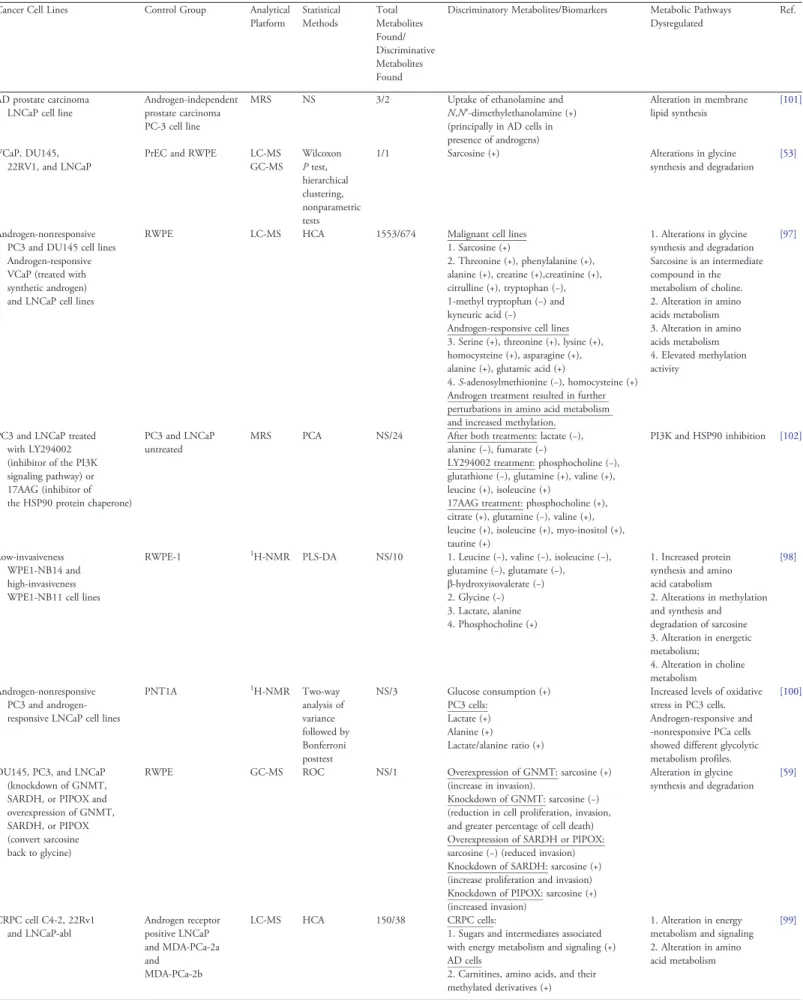
The biological relevance of sarcosine was evaluated in four immortalized PCa cell lines, in primary benign prostate epithelial cells, and in an immortalized benign prostate epithelial cell line. Sarcosine levels were increased in malignant cell lines when compared with benign cell lines [53,97]. Furthermore, alterations in the expression of the enzymes involved in sarcosine metabolism influence cell proliferation, invasion, and cell death, which suggest the importance of sarcosine in PCa metabolism (Table 5)[53,59]. In an effort to understand the role of sarcosine in PCa progression, Sreekumar et al. (2009) evaluated the role of androgen signaling and the genes ERG and ETV1 (important mediators of PCa progression). The results showed that after treatment with androgens, cell lines that were ERG positive and ETV1 positive had increased GNMT expression and decreased sarcosine dehydrogenase (SARDH) expression[53].
In agreement with the results from metabolomics studies performed in other matrices (urine, plasma/serum, and tissues), presented here previously, alterations in amino acid metabolism were also observed in the studies performed in PCa cell lines. PCa cells lines revealed alterations in the levels of certain amino acids, such as leucine, valine, or isoleucine (Table 5)[97–99].
The increase of lactate and alanine levels in PCa cell lines is frequently observed in metabolomics studies. These alterations suggest changes in cellular energy metabolism (Table 5)[97–100].
As reported for other matrices, metabolomics studies in PCa also revealed that PCa cells experience alterations in membrane metabolism with changes, for example, in choline metabolite levels (Table 5)[98,101]. Androgen signaling has an important role in the development and progression of PCa. In fact, one current therapy for metastatic PCa is the use of antiandrogen agents; however, with the progression of the disease, patients normally develop resistance to this therapy, and it is currently impossible to predict if the cancer will progress into a castration-resistant state. The androgen-responsive cell lines can be characterized by increased levels of spermine, N-acetyl-spermine, serine, threonine, lysine, homocysteine, asparagine, alanine, and glutamic acid, as well as decreased levels of S-adenosylmethionine, with a simultaneous increase in levels of its breakdown product, homocysteine. These findings indicate that androgen-responsive cell lines have an elevated methylation activity. Androgen treatment resulted in further perturbations in amino acid metabolism and in a shift toward increased methylation.
TABLE 4 (continued) PCa Subject Group Control Group Analytical
Platform
Statistical Methods Total Metabolites Found/Discrimi-native Metabolites Found
Discriminatory Metabolites/Biomarkers Metabolic Pathways Dysregulated Ref.
n= 95 n= 95 Normal adjacent prostate tissue GC-MS LC-MS ROC,
Univariate Cox regression, Kaplan-Meier analyses, and multivariate Cox regression analyses
820/9 1. Gluconic acid (−), maltotriose (−) 2. Aminoadipic acid (+) 3. Cerebronic acid (+), glycerophosphoethanol-amine (+),
2-hydroxybehenic acid (+), tricosanoic acid (+) 4. Isopentenyl pyrophosphate (+)
5. 7-methylguanine (+)
1. Alteration in carbohydrate metabolism 2. Increased fatty acid synthesis 3. Alteration in lipid metabolism 4. Intermediate in the steroid synthesis pathway, indicates an increase of cholesterol levels 5. DNA damage [49] n= 30 LG PCa plus n= 81 HG PCa n= 47 Normal adjacent tissue
MRS PCA, PLS and PLS-DA 23/2 1. Citrate (−) 2. Spermine (−)
1. Reduced citrate synthesis 2. Reduced spermine synthesis
[91]
BPH, Benign prostatic hypertrophy; Cho/Cr, choline/creatinine; Cit/Cr, citrate/creatinine; GS-MS, Gas chromatography–mass spectrometry; (GPC + PC)/Cr, (glycerol-phosphocholine + phosphoryl-choline)/creatinine; HC, Healthy Controls; HG, High grade; Lac/Al, lactate/alanine; LC-MS, Liquid chromatography–mass spectrometry; LDA, Linear discriminant analysis; LG, Low grade; LR, Linear regression MRS, magnetic resonance spectroscopy; NMR, Nuclear magnetic resonance; NS, Not specified; PCA, Principal component analysis; PLS, partial least squares; PLS-DA, partial least squares discriminant analysis; tCho/Cit, total choline/citrate.
Table 5. Metabolomic Studies Performed in Human PCa-Derived Cell Lines Cancer Cell Lines Control Group Analytical
Platform Statistical Methods Total Metabolites Found/ Discriminative Metabolites Found
Discriminatory Metabolites/Biomarkers Metabolic Pathways Dysregulated
Ref.
AD prostate carcinoma LNCaP cell line
Androgen-independent prostate carcinoma PC-3 cell line
MRS NS 3/2 Uptake of ethanolamine and N,N′-dimethylethanolamine (+) (principally in AD cells in presence of androgens) Alteration in membrane lipid synthesis [101] VCaP, DU145, 22RV1, and LNCaP
PrEC and RWPE LC-MS GC-MS Wilcoxon P test, hierarchical clustering, nonparametric tests
1/1 Sarcosine (+) Alterations in glycine synthesis and degradation
[53]
Androgen-nonresponsive PC3 and DU145 cell lines Androgen-responsive VCaP (treated with synthetic androgen) and LNCaP cell lines
RWPE LC-MS HCA 1553/674 Malignant cell lines 1. Sarcosine (+)
2. Threonine (+), phenylalanine (+), alanine (+), creatine (+),creatinine (+), citrulline (+), tryptophan (−), 1-methyl tryptophan (−) and kyneuric acid (−)
Androgen-responsive cell lines 3. Serine (+), threonine (+), lysine (+), homocysteine (+), asparagine (+), alanine (+), glutamic acid (+)
4. S-adenosylmethionine (−), homocysteine (+) Androgen treatment resulted in further perturbations in amino acid metabolism and increased methylation.
1. Alterations in glycine synthesis and degradation Sarcosine is an intermediate compound in the metabolism of choline. 2. Alteration in amino acids metabolism 3. Alteration in amino acids metabolism 4. Elevated methylation activity [97]
PC3 and LNCaP treated with LY294002 (inhibitor of the PI3K signaling pathway) or 17AAG (inhibitor of the HSP90 protein chaperone)
PC3 and LNCaP untreated
MRS PCA NS/24 After both treatments: lactate (−), alanine (−), fumarate (−)
LY294002 treatment: phosphocholine (−), glutathione (−), glutamine (+), valine (+), leucine (+), isoleucine (+)
17AAG treatment: phosphocholine (+), citrate (+), glutamine (−), valine (+), leucine (+), isoleucine (+), myo-inositol (+), taurine (+)
PI3K and HSP90 inhibition [102]
Low-invasiveness WPE1-NB14 and high-invasiveness WPE1-NB11 cell lines
RWPE-1 1H-NMR PLS-DA NS/10 1. Leucine (−), valine (−), isoleucine (−),
glutamine (−), glutamate (−), β-hydroxyisovalerate (−) 2. Glycine (−) 3. Lactate, alanine 4. Phosphocholine (+) 1. Increased protein synthesis and amino acid catabolism
2. Alterations in methylation and synthesis and degradation of sarcosine 3. Alteration in energetic metabolism; 4. Alteration in choline metabolism [98] Androgen-nonresponsive PC3 and androgen-responsive LNCaP cell lines
PNT1A 1H-NMR Two-way analysis of variance followed by Bonferroni posttest NS/3 Glucose consumption (+) PC3 cells: Lactate (+) Alanine (+) Lactate/alanine ratio (+)
Increased levels of oxidative stress in PC3 cells. Androgen-responsive and -nonresponsive PCa cells showed different glycolytic metabolism profiles.
[100]
DU145, PC3, and LNCaP (knockdown of GNMT, SARDH, or PIPOX and overexpression of GNMT, SARDH, or PIPOX (convert sarcosine back to glycine)
RWPE GC-MS ROC NS/1 Overexpression of GNMT: sarcosine (+) (increase in invasion).
Knockdown of GNMT: sarcosine (−) (reduction in cell proliferation, invasion, and greater percentage of cell death) Overexpression of SARDH or PIPOX: sarcosine (−) (reduced invasion) Knockdown of SARDH: sarcosine (+) (increase proliferation and invasion) Knockdown of PIPOX: sarcosine (+) (increased invasion)
Alteration in glycine synthesis and degradation
[59] CRPC cell C4-2, 22Rv1 and LNCaP-abl Androgen receptor positive LNCaP and MDA-PCa-2a and MDA-PCa-2b LC-MS HCA 150/38 CRPC cells:
1. Sugars and intermediates associated with energy metabolism and signaling (+) AD cells
2. Carnitines, amino acids, and their methylated derivatives (+)
1. Alteration in energy metabolism and signaling 2. Alteration in amino acid metabolism
[99]
Androgen-nonresponsive cell lines and androgen-responsive cell lines also have differences in their glycolytic metabolism profiles. Androgen-dependent PCa cells and androgen-independent PCa cells also show differences in membrane lipid synthesis (Table 5)[97,100,101]. Metabolomics studies in cell lines may also be used to evaluate the alterations that occur after a pharmacological therapy. The treatment of PCa cells appears to lead to changes in energetic metabolism and choline metabolism (Table 5)[102]. Dichloroacetate (DCA) is an inhibitor of pyruvate dehydrogenase kinase, and inhibition of this enzyme has the potential to reverse the Warburg effect due to the increased pyruvate uptake into mitochondria. After treatment with this drug, the highly metastatic cells showed significantly lower levels of lactate/metabolite ratios [Lac/Cr, Lac/Cho, Lac/Al, and Lac/(Cho + Cr + Al)], whereas in poorly metastatic cells, no changes in lactate/metabolite ratios were found after the treatment. These findings suggest that highly metastatic cells are more dependent on lactate production (Table 5)[103].
Conclusions and Future Directions
The introduction of PSA testing has radically altered how PCa is diagnosed and managed. However, this test may lead to a false-positive or false-negative diagnosis. This drawback has given rise to serious efforts toward the discovery of new biomarkers, preferentially noninvasive, which have better specificity and sensitivity.
Because metabolic alterations are the last step in the cellular response to diseases, metabolomics can be successfully used to discover new biomarkers for cancer. In this regard, several studies have been conducted to characterize the metabolic profile of PCa.
One of the major obstacles in data interpretation is that the metabolic profile is influenced by various factors, such as age, diet, drugs, and chronobiological variations, among others. Additional problems include sample preparation, the analytical procedures, and the statistical platforms used. Major differences in these conditions can compromise the comparison of results among different studies and consequently compromise the discovery of new biomarkers. Another intrinsic difficulty with metabolomics studies is the massive amount of data produced that is difficult statistically to analyze. Despite these difficulties, metabolomics is a powerful tool for the discovery of new biomarkers for PCa detection, biomarkers indicative of cancer prognosis, disease progression, and therapeutic response, as well as identifying new therapeutic targets.
The metabolomics studies described in this review revealed different results, but almost all studies in urine, plasma/serum,
prostatic fluids, tissues, and cell lines associated PCa with decreased levels of citrate and polyamines and increased levels of choline, lactate, and amino acids.
Further studies are still needed to confirm the results of these studies and identify an inexpensive, noninvasive, sensible, and specific biomarker for PCa.
Acknowledgements
This work received financial support from the European Union (FEDER funds POCI/01/0145/FEDER/007728) and National Funds (FCT/MEC, Fundação para a Ciência e Tecnologia and Ministério da Educação e Ciência) under the Partnership Agreement PT2020 UID/MULTI/04378/2013.
References
[1] Trock BJ (2011). Application of metabolomics to prostate cancer. Urol Oncol 29, 572–581.
[2] Halama A (2014). Metabolomics in cell culture—a strategy to study crucial metabolic pathways in cancer development and the response to treatment. Arch Biochem Biophys564, 100–109.
[3] He JC, Chuang PY, Ma'ayan A, and Iyengar R (2012). Systems biology of kidney diseases. Kidney Int81, 22–39.
[4] Ramautar R, Berger R, van der Greef J, and Hankemeier T (2013). Human metabolomics: strategies to understand biology. Curr Opin Chem Biol17, 841–846. [5] Monteiro M, Carvalho M, de Lourdes Bastos M, and de Pinho P (2014). Biomarkers in renal cell carcinoma: a metabolomics approach. Metabolomics10, 1210–1222. [6] Aboud OA and Weiss RH (2013). New opportunities from the cancer
metabolome. Clin Chem59, 138–146.
[7] Monteiro MS, Carvalho M, Bastos ML, and de Pinho PG (2013). Metabolomics analysis for biomarker discovery: advances and challenges. Curr Med Chem20, 257–271.
[8] Weiss RH and Kim K (2012). Metabolomics in the study of kidney diseases. Nat Rev Nephrol8, 22–33.
[9] Monteiro M, Carvalho M, Henrique R, Jeronimo C, Moreira N, de Lourdes Bastos M, and de Pinho PG (2014). Analysis of volatile human urinary metabolome by solid-phase microextraction in combination with gas chromatography–mass spectrometry for biomarker discovery: application in a pilot study to discriminate patients with renal cell carcinoma. Eur J Cancer50, 1993–2002.
[10] Monteiro MS, Carvalho M, Bastos MdL, and Pinho PG (2015). Potentiality of volatile organic compounds to discriminate patients with cancer by using chemometric tools. Identification and data processing methods in metabolo-mics: Future Science Ltd; 2015. p. 166–184.
[11] Ferlay J, Soerjomataram I, Dikshit R, Eser S, Mathers C, Rebelo M, Parkin DM, Forman D, and Bray F (2015). Cancer incidence and mortality worldwide: sources, methods and major patterns in GLOBOCAN 2012. Int J Cancer136, E359-386.
TABLE 5 (continued) Cancer Cell Lines Control Group Analytical
Platform Statistical Methods Total Metabolites Found/ Discriminative Metabolites Found
Discriminatory Metabolites/Biomarkers Metabolic Pathways Dysregulated
Ref.
Highly metastatic LNCaP-LN3 treated with DCA and poorly metastatic LNCaP treated with DCA
LNCaP-LN3 and LNCaP untreated
NMR Nonparametric test
NS/3 LNCaP-LN3 treated with DCA: Lac/Cr (−), Lac/Cho (−), Lac/Al (−), and
Lac/(Cho + Cr + Al) (−)
LNCaP (poorly metastatic) treated with DCA: no change in lactate/metabolite ratios
Reversion of Warburg effect (LNCaP-LN3 cells respond better to DCA)
[103]
AD, Androgen dependent; CRPC, castrate-resistant prostate cancer; DCA, Dichloroacetate; GC-MS, Gas chromatography–mass spectrometry; GNMT, Glycine-N-methyl transferase; HCA, Hierarchical clustering analysis; Lac/Al, lactate/ alanine; Lac/Cr, lactate/creatine; Lac/Cho, lactate/choline; Lac/(Cho + Cr + Ala), lactate/(choline + creatine + alanine); LC-MS, Liquid chromatography–mass spectrometry; NMR, Nuclear magnetic resonance; MRS, Magnetic resonance spectroscopy; NS, Not specified; PCA, Principal component analysis; PIPOX, Pipecolic acid oxidase; PLS-DA, Partial least squares discriminant analysis; ROC, Receiver-operating characteristics analysis; SARD, Sarcosine dehydrogenase.
[12] Rigau M, Olivan M, Garcia M, Sequeiros T, Montes M, Colas E, Llaurado M, Planas J, Torres Id, Morote J, et al (2013). The present and future of prostate cancer urine biomarkers. Int J Mol Sci14, 12620–12649.
[13] Aoun Fouad, Peltier Alexandre, and van Velthoven Roland (2014). A Comprehensive Review of Contemporary Role of Local Treatment of the Primary Tumor and/or the Metastases in Metastatic Prostate Cancer. BioMed Research International, vol. 2014, Article ID 501213, 12 pages, 20142014, 501213.http://dx.doi.org/10.1155/2014/501213.
[14] Dimakakos Andreas, Armakolas Athanasios, and Koutsilieris Michael (2014). Novel Tools for Prostate Cancer Prognosis, Diagnosis, and Follow-Up. BioMed Research International, vol. 2014, Article ID 890697, 9 pages, 2014 2014, 890697.http://dx.doi.org/10.1155/2014/890697.
[15] McDunn JE, Li Z, Adam KP, Neri BP, Wolfert RL, Milburn MV, Lotan Y, and Wheeler TM, et al (2013). Metabolomic signatures of aggressive prostate cancer. Prostate73, 1547–1560.
[16] Roberts MJ, Schirra HJ, Lavin MF, and Gardiner RA (2011). Metabolomics: a novel approach to early and noninvasive prostate cancer detection. Korean J Urol52, 79–89. [17] Barry MJ (2009). Screening for prostate cancer—the controversy that refuses to
die. N Engl J Med360, 1351–1354.
[18] Abrate A, Lughezzani G, Gadda GM, Lista G, Kinzikeeva E, Fossati N, Larcher A, Dell'Oglio P, Mistretta F, and Buffi N, et al (2014). Clinical use of [−2]proPSA (p2PSA) and its derivatives (%p2PSA and Prostate Health Index) for the detection of prostate cancer: a review of the literature. Korean J Urol55, 436–445.
[19] Link RE, Shariat SF, Nguyen CV, Farr A, Weinberg AD, Morton RA, Richardson B, Bernard D, and Slawin KM (2004). Variation in prostate specific antigen results from 2 different assay platforms: clinical impact on 2304 patients undergoing prostate cancer screening. J Urol171, 2234–2238.
[20] Wu CL, Jordan KW, Ratai EM, Sheng J, Adkins CB, Defeo EM, Jenkins BG, Ying L, McDougal WS, and Cheng LL (2010). Metabolomic imaging for human prostate cancer detection. Sci Transl Med2, 16ra18.
[21] Andriole GL, Crawford ED, Grubb III RL, Buys SS, Chia D, Church TR, Fouad MN, Isaacs C, Kvale PA, Reding DJ, et al (2012). Prostate cancer screening in the randomized Prostate, Lung, Colorectal, and Ovarian Cancer Screening Trial: mortality results after 13 years of follow-up. J Natl Cancer Inst 104, 125–132.
[22] Spur EM, Decelle EA, and Cheng LL (2013). Metabolomic imaging of prostate cancer with magnetic resonance spectroscopy and mass spectrometry. Eur J Nucl Med Mol Imaging40(Suppl. 1), S60–S71.
[23] Ukimura O, Coleman JA, de la Taille A, Emberton M, Epstein JI, Freedland SJ, Giannarini G, Kibel AS, Montironi R, Ploussard G, et al (2013). Contemporary role of systematic prostate biopsies: indications, techniques, and implications for patient care. Eur Urol63, 214–230.
[24] Warburg O (1956). On the origin of cancer cells. Science123, 309–314. [25] Warburg OPK and Negelein F (1924). Über den Stoffwechsel der
Carcinom-Zelle. Biochem Z152, 319–344.
[26] Soga T (2013). Cancer metabolism: key players in metabolic reprogramming. Cancer Sci104, 275–281.
[27] Pertega-Gomes N and Baltazar F (2014). Lactate transporters in the context of prostate cancer metabolism: what do we know? Int J Mol Sci15, 18333–18348. [28] Costello LC and Franklin RB (1991). Concepts of citrate production and
secretion by prostate. 1. Metabolic relationships. Prostate18, 25–46. [29] Costello LC and Franklin RB (2009). Prostatic fluid electrolyte composition for
the screening of prostate cancer: a potential solution to a major problem. Prostate Cancer Prostatic Dis12, 17–24.
[30] Dakubo GD, Parr RL, Costello LC, Franklin RB, and Thayer RE (2006). Altered metabolism and mitochondrial genome in prostate cancer. J Clin Pathol59, 10–16. [31] Costello LC, Liu Y, Franklin RB, and Kennedy MC (1997). Zinc inhibition of mitochondrial aconitase and its importance in citrate metabolism of prostate epithelial cells. J Biol Chem272, 28875–28881.
[32] Costello LC and Franklin RB (2000). The intermediary metabolism of the prostate: a key to understanding the pathogenesis and progression of prostate malignancy. Oncology (Williston Park)59, 269–282.
[33] Zha S, Ferdinandusse S, Hicks JL, Denis S, Dunn TA, Wanders RJ, Luo J, De Marzo AM, and Isaacs WB (2005). Peroxisomal branched chain fatty acid beta-oxidation pathway is upregulated in prostate cancer. Prostate 63, 316–323.
[34] Liu Y, Zuckier LS, and Ghesani NV (2010). Dominant uptake of fatty acid over glucose by prostate cells: a potential new diagnostic and therapeutic approach. Anticancer Res30, 369–374.
[35] Tsouko E, Khan AS, White MA, Han JJ, Shi Y, Merchant FA, Sharpe MA, Xin L, and Frigo DE (2014). Regulation of the pentose phosphate pathway by an androgen receptor-mTOR–mediated mechanism and its role in prostate cancer cell growth. Oncogenesis3, e103.
[36] Huang WC, Zhau HE, and Chung LW (2010). Androgen receptor survival signaling is blocked by anti-beta2-microglobulin monoclonal antibody via a MAPK/lipogenic pathway in human prostate cancer cells. J Biol Chem285, 7947–7956.
[37] Huang WC, Li X, Liu J, Lin J, and Chung LW (2012). Activation of androgen receptor, lipogenesis, and oxidative stress converged by SREBP-1 is responsible for regulating growth and progression of prostate cancer cells. Mol Cancer Res10, 133–142. [38] Lucarelli G, Rutigliano M, Galleggiante V, Giglio A, Palazzo S, Ferro M, Simone C, Bettocchi C, Battaglia M, and Ditonno P (2015). Metabolomic profiling for the identification of novel diagnostic markers in prostate cancer. Expert Rev Mol Diagn15, 1211–1224.
[39] Wang Q, Hardie RA, Hoy AJ, van Geldermalsen M, Gao D, Fazli L, Sadowski MC, Balaban S, Schreuder M, Nagarajah R, et al (2015). Targeting ASCT2-mediated glutamine uptake blocks prostate cancer growth and tumour development. J Pathol236, 278–289.
[40] Pan T, Gao L, Wu G, Shen G, Xie S, Wen H, Yang J, Zhou Y, Tu Z, and Qian W (2015). Elevated expression of glutaminase confers glucose utilization via glutaminolysis in prostate cancer. Biochem Biophys Res Commun456, 452–458. [41] Li Z and Zhang H (2015). Reprogramming of glucose, fatty acid and amino
acid metabolism for cancer progression. Cell Mol Life Sci73(2), 377–392. [42] Pavlides S, Whitaker-Menezes D, Castello-Cros R, Flomenberg N, Witkiewicz
AK, Frank PG, Casimiro MC, Wang C, Fortina P, Addya S, et al (2009). The reverse Warburg effect: aerobic glycolysis in cancer associated fibroblasts and the tumor stroma. Cell Cycle8, 3984–4001.
[43] Fiaschi T, Marini A, Giannoni E, Taddei ML, Gandellini P, De Donatis A, Lanciotti M, Serni S, Cirri P, and Chiarugi P (2012). Reciprocal metabolic reprogramming through lactate shuttle coordinately influences tumor-stroma interplay. Cancer Res72, 5130–5140.
[44] Wilkosz J, Brys M, and Rozanski W (2011). Urine markers and prostate cancer. Cent Eur J Urol64, 9–14.
[45] Zhang T, Watson DG, Wang L, Abbas M, Murdoch L, Bashford L, Ahmad I, Lam NY, Ng AC, and Leung HY (2013). Application of holistic liquid chromatography–high resolution mass spectrometry based urinary metabolomics for prostate cancer detection and biomarker discovery. PLoS One8, e65880. [46] Averna TA, Kline EE, Smith AY, and Sillerud LO (2005). A decrease in 1H nuclear
magnetic resonance spectroscopically determined citrate in human seminal fluid accompanies the development of prostate adenocarcinoma. J Urol173, 433–438. [47] Kumar V, Dwivedi DK, and Jagannathan NR (2014). High-resolution NMR
spectroscopy of human body fluids and tissues in relation to prostate cancer. NMR Biomed27, 80–89.
[48] Roberts MJ, Richards RS, Gardiner RA, and Selth LA (2015). Seminal fluid: a useful source of prostate cancer biomarkers? Biomark Med9, 77–80. [49] Jung K, Reszka R, Kamlage B, Bethan B, Stephan C, Lein M, and Kristiansen G
(2013). Tissue metabolite profiling identifies differentiating and prognostic biomarkers for prostate carcinoma. Int J Cancer133, 2914–2924.
[50] Zhang A, Sun H, Xu H, Qiu S, and Wang X (2013). Cell metabolomics. OMICS17, 495–501.
[51] Cuperlovic-Culf M, Barnett DA, Culf AS, and Chute I (2010). Cell culture metabolomics: applications and future directions. Drug Discov Today15, 610–621. [52] Keshari KR, Sriram R, Van Criekinge M, Wilson DM, Wang ZJ, Vigneron DB, Peehl DM, and Kurhanewicz J (2013). Metabolic reprogramming and validation of hyperpolarized 13C lactate as a prostate cancer biomarker using a human prostate tissue slice culture bioreactor. Prostate73, 1171–1181. [53] Sreekumar A, Poisson LM, Rajendiran TM, Khan AP, Cao Q, Yu J, Laxman B,
Mehra R, Lonigro RJ, Li Y, et al (2009). Metabolomic profiles delineate potential role for sarcosine in prostate cancer progression. Nature457, 910–914. [54] Cernei N, Heger Z, Gumulec J, Zitka O, Masarik M, Babula P, Eckschlager T,
Stiborova M, Kizek R, and Adam V (2013). Sarcosine as a potential prostate cancer biomarker—a review. Int J Mol Sci 14, 13893–13908.
[55] Jiang Y, Cheng X, Wang C, and Ma Y (2010). Quantitative determination of sarcosine and related compounds in urinary samples by liquid chromatography with tandem mass spectrometry. Anal Chem82, 9022–9027.
[56] Stabler S, Koyama T, Zhao Z, Martinez-Ferrer M, Allen RH, Luka Z, Loukachevitch LV, Clark PE, Wagner C, and Bhowmick NA (2011). Serum methionine metabolites are risk factors for metastatic prostate cancer progression. PLoS One6, e22486.