Mediator of Alcohol-Induced Brain Inflammation
Salil R. Rajayer1, Asha Jacob1,2, Weng-Lang Yang1,2, Mian Zhou1,2, Wayne Chaung1,2, Ping Wang1,2*
1Department of Surgery, Hofstra North Shore-LIJ School of Medicine, Manhasset, New York, United States of America,2Center for Translational Research, The Feinstein Institute for Medical Research, Manhasset, New York, United States of America
Abstract
Binge drinking has been associated with cerebral dysfunction. Ethanol induced microglial activation initiates an inflammatory process that causes upregulation of proinflammatory cytokines which in turn creates neuronal inflammation and damage. However, the molecular mechanism is not fully understood. We postulate that cold-inducible RNA-binding protein (CIRP), a novel proinflammatory molecule, can contribute to alcohol-induced neuroinflammation. To test this theory male wild-type (WT) mice were exposed to alcohol at concentrations consistent to binge drinking and blood and brain tissues were collected. At 5 h after alcohol, a significant increase of 53% in the brain of CIRP mRNA was observed and its expression remained elevated at 10 h and 15 h. Brain CIRP protein levels were increased by 184% at 10 h and remained high at 15 h. We then exposed male WT and CIRP knockout (CIRP2/2) mice to alcohol, and blood and brain tissues were collected at 15 h post-alcohol infusion. Serum levels of tissue injury markers (AST, ALT and LDH) were significantly elevated in alcohol-exposed WT mice while they were less increased in the CIRP2/2mice. Brain TNF-amRNA and protein expressions along with IL-1bprotein levels were significantly increased in WT mice, which was not seen in the CIRP2/2mice. In cultured BV2 cells (mouse microglia), ethanol at 100 mM showed an increase of CIRP mRNA by 274% and 408% at 24 h and 48 h respectively. Corresponding increases in TNF-aand IL-1bwere also observed. CIRP protein levels were markedly increased in the medium, suggesting that CIRP was secreted by the BV2 cells. From this we conclude that alcohol exposure activates microglia to produce and secrete CIRP and possibly induce pro-inflammatory response and thereby causing neuroinflammation. CIRP could be a novel mediator of alcohol-induced brain inflammation.
Citation:Rajayer SR, Jacob A, Yang W-L, Zhou M, Chaung W, et al. (2013) Cold-Inducible RNA-Binding Protein Is an Important Mediator of Alcohol-Induced Brain Inflammation. PLoS ONE 8(11): e79430. doi:10.1371/journal.pone.0079430
Editor:Charles C. Caldwell, University of Cincinnati, United States of America
ReceivedSeptember 13, 2013;AcceptedSeptember 30, 2013;PublishedNovember 1, 2013
Copyright:ß2013 Rajayer et al. This is an open-access article distributed under the terms of the Creative Commons Attribution License, which permits unrestricted use, distribution, and reproduction in any medium, provided the original author and source are credited.
Funding:This study was supported in part by National Institutes of Health (NIH) grant, R21 AA021496 to P.W. The funders had no role in study design, data collection and analysis, decision to publish, or preparation of the manuscript. No additional external funding was received for this study.
Competing Interests:The authors have declared that no competing interests exist. * E-mail: [email protected]
Introduction
In the United States, over fifty percent of the adult population consumes alcohol on a regular basis [1]. In 2006, the CDC had estimated the economic cost of excessive drinking as about$220 billion [2]. Binge drinking was responsible for .70% of these costs. Binge drinking is defined by the National Institute on Alcohol Abuse and Alcoholism (NIAAA) as $4 drinks for a woman and$5 drinks for a man on a single occasion. Typically, these results of alcohol concentrations are in the range of the legal intoxication limit (i.e., a blood alcohol level of 80 mg/dL). Thus, the cost incurred while treating any of the acute conditions related with intoxication can be fully attributed to binge drinking [2]. A number of studies have shown that binge drinking leads to impairment of cognitive function and several mechanisms have been proposed to account for this brain dysfunction [3–7]. Among these, central nervous system (CNS) inflammation is one of the proposed explanations for alcohol induced brain dysfunction.
The primary effectors of neuroinflammation are microglia cells, which are the resident macrophages of the brain. They form a significant portion of the CNS cell population, constituting approximately 20% of the total glial cell population and are almost as numerous as neurons [8]. At rest, microglia seem to perform various homeostatic functions, involving themselves in
microarrays. They found a two fold increase in mRNA of a gene termed AA818118 which is similar to CIRP [31]. Our recent study showed that healthy animals injected with recombinant murine CIRP (rmCIRP) were found to have significantly elevated serum levels of liver enzymes (AST and ALT) and cytokine TNF-a indicating CIRP as a potent proinflammatory agent [32]. Based on these findings, we postulated that CIRP may play a role in the alcohol induced proinflammatory cascade in the brain.
Materials and Methods
Mouse model of acute binge alcohol
Male C57BL/6 mice (20–25 g, Taconic, Albany, NY) were used as Wild-type (WT) mice in all experiments. CIRP2/2mice on a background of C57BL/6 were a gift from Dr Jun Fujita (Kyoto University, Japan) and are bred in our animal facility. Upon acquisition, WT mice were allowed to acclimate to the environment in our facility for 5–7 days before the experiment. All mice were housed in cages of five members each and subjected to 12 h light/dark cycles. Each type of mouse (WT or CIRP2/2) was randomly divided into 2 groups: Saline (Sham) or Alcohol. Anesthesia was induced with 2.5% inhalational isoflurane. The right internal jugular vein was exposed with a 0.5-cm neck incision and a PE-10 catheter was inserted via a venotomy. The catheter was sutured in place and connected via a harness (SAI Infusion Technologies, Libertyville, IL) to an infusion pump (KD Scientific, Holliston, MA). The harness allowed for free movement of the mice in the cage while receiving a continuous infusion. Alcohol group animals received an initial bolus of 43.7 mg/25 g ethanol followed by 7.5 mg/25 g/h for 15 h bringing the total amount to 156 mg/25 g of ethanol. At the end of 15 h, animals were anesthetized and blood and brain tissue samples were harvested and stored at280uC. For determining time-course changes, only WT animals were used. They were anesthetized and cannulated identical to the procedure as described above. Alcohol group animals received the same initial bolus of ethanol followed by continuous infusion for 5, 10 or 15 h. At the end of each corresponding time period, animals were anesthetized and blood and brain tissue samples were harvested. Serum alcohol levels were measured by a commercially available alcohol kit (Pointe Scientific, MI).
All experiments were performed in strict accordance with the guidelines for the use of experimental animals by the National Institute of Health and were approved by the Institutional Animal Care and Use Committee of The Feinstein Institute of Medical Research. All surgery was performed under 2.5% isoflurane anesthesia and all efforts were made to minimize suffering.
Measurement of serum levels of injury markers
Blood samples were centrifuged at 2,000 g for 15 min to collect serum. The serum levels of alcohol and activity of organ injury markers aspartate aminotransferase (AST), alanine aminotransfer-ase (ALT) and lactate dehydrogenaminotransfer-ase (LDH) were measured by using assay kits from Pointe Scientific (Canton, MI).
Measurement of tissue cytokine levels
Tissue lysate protein concentrations were determined using Bio-Rad DC protein assay kit (BIO-RAD, Hercules, CA). TNF-aand IL-1blevels were determined in the samples by ELISA kits from BD Biosciences (San Jose, CA) as per the manufacturer’s protocols and represented as pg/mg protein.
Culture of microglial cells
The immortalized murine BV2 cell line (BV2 cells), which exhibits both the phenotypic and functional properties of reactive microglia cells [33,34] were obtained as a kind gift from Dr Philippe Marambaud (Feinstein Institute for Medical Research, NY) and cultured in Dulbecco’s Modified Eagle’s Medium (DMEM, Invitrogen) supplemented with 10% fetal bovine serum (FBS), 1% penicillin-streptomycin, and 1% glutamine, as previ-ously described [35,36].
Alcohol stimulation of BV2 cells
BV2 cells in their third passage were plated in triplicates at 26106per well (six well plates) in complete DMEM. Plated cells were then incubated at 37uC, 5% CO2 overnight to enable
attachment. The next day the culture medium was changed to OPTI-MEM (reduced serum medium, Life technologies, Grand Island, NY). One hour later, these plates were exposed to either sterile phosphate buffered saline (PBS) or endotoxin free 99.9% Ethanol (Sigma-Aldrich, St. Louis, MO) at concentrations of 50 mM and 100 mM. These concentrations were chosen because they were in the range of blood alcohol levels found among alcoholics [37]. The plates were incubated for 24 h or 48 h at 37uC, 5% CO2. Cell lysates and supernatants were collected at the
end of the corresponding time-period and stored in280uC before being subjected to analysis. Three separate BV2 microglia-alcohol stimulation experiments were performed.
Reverse transcriptase-polymerase chain reaction (RT-PCR) analysis
Total RNA was extracted from brain tissues and cell lysates using Trizol (Invitrogen, Carlsbad, CA). cDNA was reverse-transcribed from 2mg of total RNA using Oligo (dT)12–18primer
(Life Technologies, Grand Island, NY) and murine leukemia virus reverse transcriptase (Applied Biosystems, Foster City, CA). A PCR reaction was done in 25ml of final volume containing 0.08mmol of each forward and reverse primer, cDNA, and 12.5ml SYBR Green PCR Master Mix (Applied Biosystems, Foster City, CA). Amplification was conducted in an A lied Biosystems 7300 real-time PCR machine under the temperature of 50uC for 2 min, 95uC for 10 min and 45 cycles of 95uC for 15 seconds and 60uC for 1 min. Relative expression of each mRNA was calculated using DD
Ct threshold model. Mouseb-actin was used for normalization. Relative expression of mRNA was represented as fold change in comparison to sham/control levels. The primers used are listed in Table 1.
Western blotting for CIRP protein
system 2800 (LI-COR, Lincoln, NE) and the band intensity measured using the NIH Image J densitometric software.
Supernatant
Cell supernatant proteins were precipitated using the DOC-TCA method. The precipitate was then subjected to Western blotting for CIRP as described above. The band densities were equalized using Ponceau S Solution (0.1% Ponceau S (w/v) in 5% acetic acid (v/v), Sigma-Aldrich, St. Louis, MO).
Statistical analysis
All data are expressed as mean6SE and compared by one-way analysis of variance (ANOVA) and the Student-Newman-Keuls test. Differences in values were considered significant if p,0.05.
Results
Serum alcohol levels after the continuous infusion The blood alcohol level was measured as 66 mg/dL at 5 h after the initiation of the continuous alcohol infusion, and significantly increased to 132 mg/dL at 10 h and finally at the end of the full infusion was about 156 mg/dL (Table 2). The level of blood alcohol at 15 h is consistent with reported levels of binge drinking habits [38] and it was about twice the legal intoxication limit of 80 mg/dL.
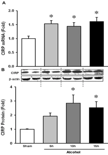
Acute binge alcohol increased brain CIRP level in a time dependent manner
Prior studies have shown that CIRP is highly expressed in stress conditions such as hypothermia, hypoxia and UV irradiation [27,28]. Our recent study showed that CIRP is induced and released into the circulation in hemorrhagic shock and sepsis [32]. Based on these findings, we examined whether CIRP is increased in the brain in WT mice exposed to alcohol. Towards that end, we evaluated the mRNA and protein levels of WT mice exposed to alcohol for 5, 10 and 15 h. We found that there was a significant 53% induction of CIRP mRNA level at 5 h and it remained
elevated until 15 h (Fig. 1A). A corresponding significant increase by 184% in protein level was observed at 10 h and remained high at 15 h as compared to unexposed mice (Fig. 1B). This suggests that acute binge alcohol induces CIRP expression in the brain. Table 1.Real-Time PCR primers used in this study.
Gene GenBank# Forward (59-39) Reverse (59-39)
CIRP NM_031168 CCAGAGGAGACTTCACAG CAGAATTGCCATTGCACAAC
TNF-a NM_007705 AGGACTCAGCTTCGACACCA CGTCCACAGACTTCCCATTC
IL-1b NM_008361 CAGGATGAGGACATGAGCACC CTCTGCAGACTCAAACTCCAC
b-Actin NM_007393 CGTGAAAAGATGACCCAGATCA TGGTACGACCAGAGGCATACAG
doi:10.1371/journal.pone.0079430.t001
Table 2.Serum alcohol levels after the continuous infusion.
Serum Alcohol Level (mg/dL)
Sham Not Detectable
5 h 6664.6
10 h 13262.7*
15 h 156623.0*
Data presented as means6SE (n = 3–6/group) and compared by one-way ANOVA and SNK method.
*p,0.05 vs. 5 h group.
doi:10.1371/journal.pone.0079430.t002
Figure 1. Alcohol increased brain CIRP level in a time dependent manner. WT mice were infused intravenously with alcohol as described in Materials and Methods. Whole brain tissue from WT mice was collected at 5, 10 and 15 h after the initiation of continuous infusion of alcohol. (A) CIRP mRNA expression was determined by real time RT-PCR analysis and expression levels were normalized tob-actin. (B) Tissue lysates were collected and analyzed for CIRP by Western blotting. Blots were scanned and quantified by densitometry. Band intensity of CIRP was normalized to the corre-sponding band intensity ofb-actin. The ratio of the control group is designated as 1 for comparison. Data presented as means6SE (n = 3– 4/group) and compared by one-way ANOVA and SNK method; *p,0.05 vs. Sham.
Clinical markers of organ injury were attenuated in CIRP2/2mice following alcohol exposure
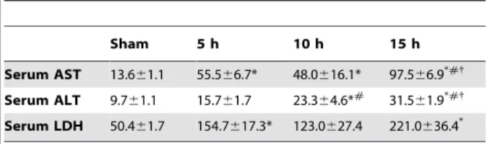
To delineate the importance of the increased CIRP expression in the brain, we evaluated the effects of alcohol on mice in the absence of the CIRP protein. WT and CIRP2/2 mice were infused intravenously with alcohol for 15 h as previously described. As systemic markers of organ injury, serum levels of enzymes AST, ALT and LDH were measured. Alcohol infusion in the WT animals caused significant increases of 467%, 157% and 321% in serum AST, ALT and LDH respectively (Figs. 2A–C). In the CIRP2/2mice, these values were decreased by 66%, 42% and 47% respectively, when compared to the alcohol-exposed WT mice, indicating a significantly decreased degree of tissue injury (Figs. 2A–C). We also measured AST, ALT and LDH at 5 h and 10 h after alcohol infusion in the WT mice. All three parameters increased with time after the start of the alcohol infusion (Table 3).
Brain pro-inflammatory cytokines TNF-aand IL-1bwere diminished in CIRP2/2 following alcohol exposure
Next, it was explored if the increase in CIRP was associated with an increase in the cytokine levels in the brain following alcohol infusion. We found that in the WT animals, the 15 h alcohol infusion significantly increased the brain TNF-a mRNA expression and protein levels by 127% and 34%, respectively. In the CIRP2/2 mice however, TNF-a mRNA and protein levels were attenuated by 78% and 55% respectively, as compared to alcohol exposed WT mice (Figs. 3A and 3B). Likewise, IL-1b protein levels were increased by 58% in alcohol exposed WT mice and these levels were decreased by 50% in the CIRP2/2 mice (Fig. 3C). This suggests that in the absence of CIRP, the microglial activation and by corollary, the proinflammatory effects created by alcohol exposure are significantly attenuated in mice. Serum levels of the cytokines TNF-a and IL-1bwere also measured but were undetectable.
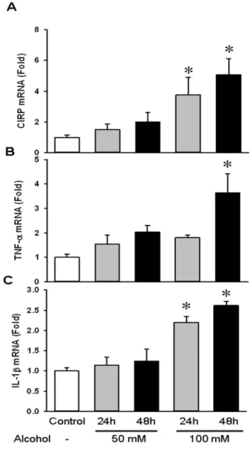
Alcohol upregulated gene expression of CIRP and cytokines TNF-aand IL-1bin BV2 cells
Alcohol exposure has been shown to recruit microglia and cause their activation. As mentioned earlier, microglial activation consists of a characteristic pattern of cellular responses including morphological and functional changes. These involve cell prolif-eration, expression of immunomolecules, recruitment to the injured region and the release of cytotoxic and/or inflammatory mediators [39]. In our study, BV2 cells were exposed to ethanol at a concentration of 50 mM and 100 mM over 24 h and 48 h. We found that gene expression of CIRP in cells treated with 100 mM ethanol was increased by 274% and 400% at 24 h and 48 h respectively (Fig. 4A). We also examined the mRNA levels of TNF-a and IL-1b. While TNF-a levels were only significantly elevated by 262% at 48 h at 100 mM ethanol dose, IL-1blevels were significantly upregulated by 120% and 161% at 24 h and 48 h respectively, at the same dose (Figs. 4B–C). Thus, acute alcohol caused microglial activation leading to increased gene expression of CIRP and increases in TNF-aand IL-1b.
CIRP protein was secreted into the cell culture medium following alcohol exposure
To determine whether CIRP could be secreted into the cell medium, we collected the supernatants and cell lysates separately from BV2 cells treated with 50 and 100 mM alcohol for 48 h and analyzed them by Western blotting. We found that while CIRP protein was decreased in the total cell lysate at 48 h, it was markedly increased in the cell culture medium at both concentrations tested (Fig. 5). Thus, alcohol causes microglial Figure 2. Clinical markers of organ injury were improved in
CIRP2/2following alcohol exposure.WT and CIRP2/2 mice were
infused intravenously with alcohol for 15 h. Serum was collected and analyzed for AST (A), ALT (B) and LDH (C) using standardized assays. Data are presented as means6SE (n = 5/group) and compared by one-way ANOVA and SNK method; *p,0.05 vs. respective Sham and
#p,0.05 vs. WT alcohol group. doi:10.1371/journal.pone.0079430.g002
Table 3.Time-course changes in tissue injury markers and TNF-aafter acute alcohol.
Sham 5 h 10 h 15 h
Serum AST 13.661.1 55.566.7* 48.0616.1* 97.566.9*#{ Serum ALT 9.761.1 15.761.7 23.364.6*# 31.5
61.9*#{ Serum LDH 50.461.7 154.7617.3* 123.0627.4 221.0636.4*
Data presented as means6SE (n = 3–5/group) and compared by one-way ANOVA and SNK method.
*p,0.05 vs. Sham,
#p
,0.05 vs. 5 h,
{
p,0.05 vs.10 h groups.
activation, leading to the secretion of CIRP protein into the medium.
Discussion
Binge drinking has become the most popular form of alcohol abuse, especially among the youth [40]. The damaging effects of alcohol abuse on the developing brain in young people, i.e. retardation of intellectual function, rational decision making and emotional maturation have been well documented [41]. The earlier paradigm that moderate alcohol consumption is neuropro-tective has now been reversed [42]. Alcohol induced immune activation in the brain has been widely considered responsible for its deleterious effects [43].
In our current study, we first showed that binge alcohol intoxication increased brain levels of CIRP, both at the transcriptional and translational level. This increase was correlated with increases in the serum organ injury markers and proin-flammatory cytokine levels in the brain. We then showed that in the absence of CIRP, i.e. in CIRP2/2mice, the serum organ injury markers and brain cytokine levels were markedly dimin-ished suggesting a critical role of CIRP in alcohol-induced neuroinflammation. Finally, similar to what was observed from the whole brain tissue, we demonstrated that alcohol exposure in BV2 cells, a mouse brain microglia cell line, increased both CIRP mRNA and protein levels indicating that the cell type responsible for CIRP upregulation could be brain microglial cells. We further showedin vitrothat alcohol exposure caused CIRP to be secreted Figure 3. Brain proinflammatory cytokines TNF-aand IL-1b
were attenuated in CIRP2/2following alcohol exposure.Whole brain samples from WT and CIRP2/2mice were collected at 15 h after
alcohol and analyzed. TNF-amRNA expression (A) was determined by real time RT-PCR analysis and expression levels were normalized tob -actin. Brain protein cytokine levels of TNF-a(B) and IL- 1b(C) were determined by subjecting cell lysates to standardized mouse ELISA (BD Biosciences). Data are presented as means6SE (n = 3–5/group) and compared by one-way ANOVA and SNK method;*p,0.05 vs. Sham,
#p,0.05 vs. WT alcohol groups. doi:10.1371/journal.pone.0079430.g003
into the medium. Thus, we implemented bothin vivoand in vitro approaches and identified CIRP as a novel mediator of alcohol-induced neuroinflammation in mice. To our knowledge, this is the first report that CIRP as a contributor to alcohol-induced neuroinflammation.
Central nervous system inflammation is one of the proposed explanations for alcohol induced brain dysfunction. The primary effectors of neuroinflammation are microglia. The activation of microglia in response to stimuli can be triggered either by families of pattern recognition receptors such as the Toll-Like receptors or due to the cessation of neuroprotective signals from receptor interactions [11]. Activation of microglia might initially be protective for neurons. When they are exposed to invading pathogens, neuronal debris, or proinflammatory cytokines and chemokines, microglia rapidly change to an activated state [12]. They play a part in regulating the regeneration of neurons and remodeling of the brain by producing a variety of cytotoxic as well as neuroprotective molecules [13]. In lesions where there is breakdown of the blood-brain barrier such as cerebral ischemia, brain abscesses and traumatic injuries causing, microglial activa-tion along with further macrophage recruitment and debris clearance takes place [14]. However, overactivation of these cells in various conditions leads to inflammatory products that eventually may cause neuronal destruction as observed in various CNS pathologies. Microglial cells play an active part in degenerative CNS diseases such as Alzheimer’s [20] and
Parkinson’s diseases [16]. A role for neuroinflammation via microglial activation is also seen in Amyotrophic Lateral Sclerosis[15]. In fact, neuroinflammation has even been seen with Down’s Syndrome [21], which may predispose these patients to undergo neurodegeneration. In most neurological autoimmune diseases such as multiple sclerosis, microglia induced phagocytosis is the pathological hallmark [17]. In the human acquired immunodeficiency syndrome (AIDS) virus infected macrophages probably introduce the virus to the CNS and in concert with microglia are involved in the pathophysiology of the AIDS dementia complex [18]. Recently, light has been shed on the role of microglial activation and neuroinflammation in neurodevelop-mental disorders such as autism [19]. It is clear that neuroin-flammation via microglial activation plays a major role in the pathology of CNS disease and dysfunction.
Excessive alcohol consumption also promote inflammation in the CNS [44]. Ethanol increases expression of brain pro-inflammatory genes through activation of the transcription factor, NF-kB [22] and transgenic mice lacking TLR4 are protected against ethanol-induced upregulation of pro-inflammatory genes, glial activation, and neurotoxicity [24]. Ethanol induces microglia activation by stimulating TLR4 response and that the inflamma-tory response induced by ethanol is completely abrogated in microglia of TLR42/2mice [45].
In the present study, we show that alcohol increased CIRP mRNA and protein expressions in the mouse brain. Without CIRP, alcohol-induced serum levels of organ injury markers, and brain TNF-a and IL-1b levels were markedly diminished. Furthermore, we demonstrated that alcohol increased CIRP mRNA and protein expressions in mouse microglial cells and caused its release into the medium. Our recent study showed that CIRP interacts with TLR4 and facilitates inflammatory responses in macrophages [32]. We therefore suggest that alcohol stimulates the production and secretion of CIRP from microglial cells in the brain, CIRP then interacts with TLR-4 on the microglial cells, activating them to release proinflammatory cytokines. Thus, CIRP plays a role in alcohol-induced neuroinflammation. In summary, as a marker for alcohol-induced neuroinflammation, or as a target for therapy in alcohol use disorders, CIRP may play an important role in the future.
Acknowledgments
The authors thank Hao-Ting Yen for his technical assistance. We also thank Andrew Godwin, MD for critical reading of the manuscript.
Author Contributions
Conceived and designed the experiments: PW AJ. Performed the experiments: SR WLY MZ WC. Analyzed the data: SR AJ. Contributed reagents/materials/analysis tools: PW. Wrote the paper: SR AJ WLY MZ PW.
References
1. Schiller JS, Lucas JW, Ward BW, Peregoy JA (2012) Summary health statistics for U.S. adults: National Health Interview Survey, 2010. Vital Health Stat 10: 1–207.
2. Bouchery EE, Harwood HJ, Sacks JJ, Simon CJ, Brewer RD (2011) Economic costs of excessive alcohol consumption in the U.S., 2006. Am J Prev Med 41: 516–524.
3. Bleich S, Bandelow B, Javaheripour K, Muller A, Degner D, et al. (2003) Hyperhomocysteinemia as a new risk factor for brain shrinkage in patients with alcoholism. Neurosci Lett 335: 179–182.
4. Climent E, Pascual M, Renau-Piqueras J, Guerri C (2002) Ethanol exposure enhances cell death in the developing cerebral cortex: role of brain-derived neurotrophic factor and its signaling pathways. J Neurosci Res 68: 213–225.
5. Dodd PR, Beckmann AM, Davidson MS, Wilce PA (2000) Glutamate-mediated transmission, alcohol, and alcoholism. Neurochem Int 37: 509–533. 6. Harper C, Matsumoto I (2005) Ethanol and brain damage. Curr Opin
Pharmacol 5: 73–78.
7. Niemela O (2001) Distribution of ethanol-induced protein adducts in vivo: relationship to tissue injury. Free Radic Biol Med 31: 1533–1538.
8. Yang I, Han SJ, Kaur G, Crane C, Parsa AT (2010) The role of microglia in central nervous system immunity and glioma immunology. J Clin Neurosci 17: 6–10.
9. Tremblay ME, Majewska AK (2011) A role for microglia in synaptic plasticity? Commun Integr Biol 4: 220–222.
Figure 5. CIRP protein was secreted into the cell culture medium following alcohol exposure.BV2 Cells were exposed to ethanol at a concentration of 50 mM and 100 mM over 48 h. The supernatant and cell lysates were collected separately and analyzed for CIRP by Western blotting.
10. Nimmerjahn A, Kirchhoff F, Helmchen F (2005) Resting microglial cells are highly dynamic surveillants of brain parenchyma in vivo. Science 308: 1314– 1318.
11. Hanisch UK, Kettenmann H (2007) Microglia: active sensor and versatile effector cells in the normal and pathologic brain. Nat Neurosci 10: 1387–1394. 12. Kreutzberg GW (1996) Microglia: a sensor for pathological events in the CNS.
Trends Neurosci 19: 312–318.
13. Nakajima K, Kohsaka S (2001) Microglia: activation and their significance in the central nervous system. J Biochem 130: 169–175.
14. Streit WJ, Mrak RE, Griffin WS (2004) Microglia and neuroinflammation: a pathological perspective. J Neuroinflammation 1: 14.
15. Appel SH, Zhao W, Beers DR, Henkel JS (2011) The microglial-motoneuron dialogue in ALS. Acta Myol 30: 4–8.
16. Chung YC, Kim YS, Bok E, Yune TY, Maeng S, et al. (2013) MMP-3 Contributes to Nigrostriatal Dopaminergic Neuronal Loss, BBB Damage, and Neuroinflammation in an MPTP Mouse Model of Parkinson’s Disease. Mediators Inflamm 2013: 370526.
17. Jack C, Ruffini F, Bar-Or A, Antel JP (2005) Microglia and multiple sclerosis. J Neurosci Res 81: 363–373.
18. Stoll G, Jander S (1999) The role of microglia and macrophages in the pathophysiology of the CNS. Prog Neurobiol 58: 233–247.
19. Theoharides TC, Asadi S, Patel AB (2013) Focal brain inflammation and autism. J Neuroinflammation 10: 46.
20. Weitz TM, Town T (2012) Microglia in Alzheimer’s Disease: It’s All About Context. Int J Alzheimers Dis 2012: 314185.
21. Wilcock DM, Griffin WS (2013) Down’s syndrome, neuroinflammation, and Alzheimer neuropathogenesis. J Neuroinflammation 10: 84.
22. Crews F, Nixon K, Kim D, Joseph J, Shukitt-Hale B, et al. (2006) BHT blocks NF-kappaB activation and ethanol-induced brain damage. Alcohol Clin Exp Res 30: 1938–1949.
23. Zou J, Crews F (2010) Induction of innate immune gene expression cascades in brain slice cultures by ethanol: key role of NF-kappaB and proinflammatory cytokines. Alcohol Clin Exp Res 34: 777–789.
24. Alfonso-Loeches S, Pascual-Lucas M, Blanco AM, Sanchez-Vera I, Guerri C (2010) Pivotal role of TLR4 receptors in alcohol-induced neuroinflammation and brain damage. J Neurosci 30: 8285–8295.
25. Nishiyama H, Itoh K, Kaneko Y, Kishishita M, Yoshida O, et al. (1997) A glycine-rich RNA-binding protein mediating cold-inducible suppression of mammalian cell growth. J Cell Biol 137: 899–908.
26. Gualerzi CO, Giuliodori AM, Pon CL (2003) Transcriptional and post-transcriptional control of cold-shock genes. J Mol Biol 331: 527–539. 27. Sheikh MS, Carrier F, Papathanasiou MA, Hollander MC, Zhan Q, et al. (1997)
Identification of several human homologs of hamster DNA damage-inducible transcripts. Cloning and characterization of a novel UV-inducible cDNA that codes for a putative RNA-binding protein. J Biol Chem 272: 26720–26726. 28. Wellmann S, Buhrer C, Moderegger E, Zelmer A, Kirschner R, et al. (2004)
Oxygen-regulated expression of the RNA-binding proteins RBM3 and CIRP by a HIF-1-independent mechanism. J Cell Sci 117: 1785–1794.
29. Xue JH, Nonoguchi K, Fukumoto M, Sato T, Nishiyama H, et al. (1999) Effects of ischemia and H2O2 on the cold stress protein CIRP expression in rat neuronal cells. Free Radic Biol Med 27: 1238–1244.
30. Morf J, Rey G, Schneider K, Stratmann M, Fujita J, et al. (2012) Cold-inducible RNA-binding protein modulates circadian gene expression posttranscriptionally. Science 338: 379–383.
31. Saito M, Smiley J, Toth R, Vadasz C (2002) Microarray analysis of gene expression in rat hippocampus after chronic ethanol treatment. Neurochem Res 27: 1221–1229.
Qiang X, Yang, W-L., Wu, R., Zhou, M., Jacob, A., et al. (in press) Release of Cold-Inducible RNA-Binding Protein triggers inflammation and organ injury. Nat Med.doi: 10.1038/nm.3368
33. Blasi E, Barluzzi R, Bocchini V, Mazzolla R, Bistoni F (1990) Immortalization of murine microglial cells by a v-raf/v-myc carrying retrovirus. J Neuroimmunol 27: 229–237.
34. Bocchini V, Mazzolla R, Barluzzi R, Blasi E, Sick P, et al. (1992) An immortalized cell line expresses properties of activated microglial cells. J Neurosci Res 31: 616–621.
35. Boscia F, Gala R, Pannaccione A, Secondo A, Scorziello A, et al. (2009) NCX1 expression and functional activity increase in microglia invading the infarct core. Stroke 40: 3608–3617.
36. Cheyuo C, Jacob A, Wu R, Zhou M, Qi L, et al. (2011) Recombinant human MFG-E8 attenuates cerebral ischemic injury: Its role in anti-inflammation and anti-apoptosis. Neuropharmacology 62: 890–900.
37. Adachi J, Mizoi Y, Fukunaga T, Ogawa Y, Ueno Y, et al. (1991) Degrees of alcohol intoxication in 117 hospitalized cases. J Stud Alcohol 52: 448–453. 38. Carson EJ, Pruett SB (1996) Development and characterization of a binge
drinking model in mice for evaluation of the immunological effects of ethanol. Alcohol Clin Exp Res 20: 132–138.
39. Gehrmann J, Matsumoto Y, Kreutzberg GW (1995) Microglia: intrinsic immuneffector cell of the brain. Brain Res Brain Res Rev 20: 269–287. 40. Archie S, Zangeneh Kazemi A, Akhtar-Danesh N (2012) Concurrent binge
drinking and depression among Canadian youth: prevalence, patterns, and suicidality. Alcohol 46: 165–172.
41. Spear L (2011) Alcohol and the developing brain. Chichester, UK: Blackwell Publishing Ltd.
42. Anderson ML, Nokia MS, Govindaraju KP, Shors TJ (2012) Moderate drinking? Alcohol consumption significantly decreases neurogenesis in the adult hippocampus. Neuroscience 224: 202–209.
43. Mayfield J, Ferguson L, Harris RA (2013) Neuroimmune signaling: a key component of alcohol abuse. Curr Opin Neurobiol 23: 513–520.
44. Zou J, Crews F (2010) Induction of innate immune gene expression cascades in brain slice cultures by ethanol: key role of NF-kB and proinflammatory cytokines. Alcohol Clin Exp Res 34: 777–789.
45. Fernandez-Lizarbe S, Pascual M, Guerri C (2009) Critical role of TLR4 response in the activation of microglia induced by ethanol. J Immunol 183: 4733–4744.