INTRODUCTION
Studies on the prevalence of dentin hyper-sensitivity show that this condition is not an un-common problem among the adult population. According to Azevedo4 (1994), six in ten patients have been reported to suffer from cervical dentin
The influence of vinegars on exposure of dentinal tubules: a
SEM evaluation
Influência de vinagres na exposição dos túbulos dentinários:
avaliação em MEV
Daniela Leal Zandim*
Fernanda Oliveira Bello Corrêa* José Eduardo Cezar Sampaio** Carlos Rossa Júnior***
hypersensitivity; although Sobral et al.14 (1995) reported approximately one in six patients.
All tooth types may be affected, but canines and premolars in both jaws are the most frequently involved teeth. Nevertheless, dentin
* Graduate Students; **Adjunct Professor; ***PhD, Assistant Professor, Department of Diagnosis and Surgery, Division of Peri-odontics – School of Dentistry of Araraquara, São Paulo State University.
ABSTRACT: Dentin hypersensitivity is a common painful condition observed in clinics. Dietary habits have been much associated with its development and persistence during and following periodontal treatment. The aim of this in vitro study was to evaluate the influence of vinegars on the removal of smear layer and exposure of dentinal tubules. Extracted human teeth were submitted to manual scaling with Gracey curettes in order to remove the cementum as well as to form a smear layer. Dentin samples with 3 mm2 were obtained and distributed into six
experimental groups: one control and five types of vinegars (alcohol, apple, rice, white wine and balsamic). Each group included two methods of vinegar application: topical and friction. After routine preparation for SEM analysis, photomicrographs were assessed by a calibrated and blind examiner using an appropriate index system. Kruskal-Wallis test indicated a significant influence of vinegars on smear layer removal. There was a statistically significant difference between groups treated with apple, white and rice vinegars and the control group (p < 0.05). Neverthe-less, Mann-Whitney test indicated that removal of smear layer did not vary with the method of application (topical versus friction) for any of the tested substances. We can conclude that the contact of vinegar may remove smear layer and expose dentinal tubules, regardless of the type of application. However, balsamic vinegar was associated with less removal of smear layer after both methods of application.
DESCRIPTORS: Dentin sensitivity; Diet; Smear layer.
RESUMO: A hipersensibilidade dentinária cervical é uma condição dolorosa muito comum nos consultórios. A dieta tem sido bastante associada ao seu aparecimento, assim como a sua persistência após o tratamento periodontal. O objetivo deste trabalho foi avaliar in vitro a influência dos vinagres na remoção de “smear layer” e exposição dos túbulos dentinários. Dentes de humanos foram instrumentados com curetas Gracey para a remoção do cemento e formação de “smear layer”. Foram obtidas amostras de dentina com 3 mm2, divididas entre o grupo controle (água
destilada) e cinco grupos de vinagre: branco, maçã, arroz, vinho branco e balsâmico. Cada grupo incluiu duas formas de aplicação da substância, tópica ou por fricção. Após o preparo para observação em MEV (microscopia eletrônica de varredura), as fotomicrografias foram avaliadas por um examinador previamente calibrado utilizan-do um índice apropriautilizan-do. O teste de Kruskal-Wallis indicou influência significativa utilizan-dos vinagres na remoção de “smear layer”. Foi constatada uma diferença estatística significante entre os grupos maçã, branco e arroz e o grupo controle (p < 0,05). O teste de Mann-Whitney, porém, indicou que a remoção de “smear layer” não variou para ne-nhuma das substâncias segundo a forma de aplicação. Conclui-se que os vinagres podem remover “smear layer” da superfície radicular e expor túbulos dentinários, não sendo influenciados pelo tipo de aplicação. Dentre os tipos de vinagres testados, o balsâmico esteve associado a menor remoção de “smear layer” após ambas as formas de aplicação.
ity is most commonly present at buccal cervical areas and does not have higher incidence in one gender2,14.
Dentin hypersensitivity has been described as an exaggerated sensitivity of exposed dentin in response to chemical, tactile or thermal stimuli16, where there is not any other form of dental defect or pathology15.
The hydrodynamic theory of Brännström is the currently accepted theory for transmission of stimuli in dentine. Fast fluid flow across the dentinal tubules activates the pain fibers at the pulpal wall and causes pain. The presence of open dentinal tubules seems to be a prerequisite for the occurrence of dentin sensitivity5.
According to Dowell, Addy9 (1983), exposure of cervical dentine can occur through the loss of covering enamel and/or gingival recession with loss of cementum.
Therefore, it is clear that there are a number of etiological factors that could potentially cause den-tin hypersensitivity12. The influence of acid in the diet as a factor in dentin sensitivity was demon-strated in some studies. Evidence in vitro indicates that weak and strong acids, which are the content of food and beverage acids, can remove smear layer and expose dentinal tubules3,8. However, authors observed a negative association between the fre-quency of ingestion of specific acidic foods and bev-erages and the persistence of dentin sensitivity6. Findings suggest that dietary counseling should be part of the treatment offered to patients with these problems, particularly regarding the control of excessive consumption of acid1,2,6,8.
The aim of this in vitro study was to evaluate, by using the scanning electron microscope (SEM), the influence of vinegars on the removal of smear layer and exposure of dentinal tubules.
MATERIAL AND METHODS
The study protocol was approved by the Re-search Ethics Committee of the School of Den-tistry of Araraquara, São Paulo State University (UNESP).
Human teeth, recently-extracted for peri-odontal reasons, were used in the present study. High-speed diamond burs were used to remove the cementum from the cervical portion of the roots. Subsequently, teeth were instrumented with forty shaving strokes on each surface using Gracey cu-rettes 5-6 by the same examiner to form the smear layer. These roots were then reduced with a dia-mond disk to obtain dentin samples of 3 x 3 mm.
Five experimental groups were formed based on the type of tested vinegar (alcohol, apple, rice, white wine, balsamic). Distilled water was also included as a negative control and the pH of the vinegars was determined. Each group was then subdivided into two other groups based on the method of ap-plication, performed as follows8:
• Topical: samples were immersed in the vinegar for five minutes and washed with a stream of tap water for fifteen seconds.
• Friction: samples were immersed in the vin-egar for five minutes, brushed with a soft tooth brush for thirty seconds and washed with tap water for fifteen seconds.
Samples were submitted to routine processing for SEM analysis to obtain two photomicrographs of the centre of the sample, with magnifications of 750 and 1,500 X. These photomicrographs were subsequently assessed by an examiner previously calibrated and blind to the experimental groups, using the following index of smear layer removal:
• Grade 1 - Complete removal of smear layer and dentinal tubules open.
• Grade 2 - Partial removal of smear layer and dentinal tubules partially open.
• Grade 3 - Smear layer present on the root sur-face and only indication of opening of dentinal tubules.
• Grade 4 - Smear layer present on the root surface and total obliteration of dentinal tu-bules.
Considering that the data were obtained using an index representing a scoring system, non-para-metric methods of analysis were applied. For the purpose of comparison among groups (different tested vinegars), these were considered to be in-dependent with respect to the method of applica-tion of the substances (topical or fricapplica-tion). Thus, a non-parametric analysis of variance was used to compare the performance among groups according to each method of application. On the other hand, the Mann-Whitney test was applied to compare results between the two methods of application of each vinegar that was tested. A 5% confidence level was used and the calculations were performed with the software Statistica version 5.1 (Statsoft Inc., Tulsa, OK, USA).
RESULTS
substances (H = 13.197, p = 0.021), and post hoc
paired comparisons demonstrated that alcohol, apple and rice vinegars were significantly different from the negative control group (p < 0.05). These three vinegar groups presented an average rank lower than that of the negative control group, indi-cating greater removal of smear layer and opening of dentinal tubules. The balsamic and white wine groups were similar to the control group. The bal-samic vinegar had a higher frequency of score 4 (Graph 1). In addition, from a comparison among vinegars, significant difference between balsamic and apple groups with topical application was veri-fied.
Results for the active application of tested vin-egar by friction were similar to those for topical application. Again, Kruskal-Wallis test indicated a significant difference among groups (H = 12.015, p = 0.034), and post hoc paired comparisons showed that only the balsamic vinegar was not sig-nificantly different from the negative control group (p > 0.05). This group had a higher frequency of scores 3 and 4, as shown in Graph 2.
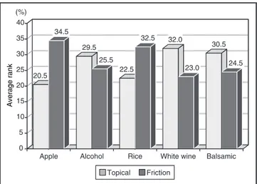
On the other hand, a comparison between the methods of application (topical versus friction) us-ing the Mann-Whitney test did not show any sig-nificant differences (Graph 3).
Index scores - alcohol 40%
20% 20% 20%
0 1 2 3 4 5 6
1 2 3 4
Index scores - apple 60% 40% 0% 0% 0 1 2 3 4 5 6
1 2 3 4
Index scores - white wine 20% 40% 20% 20% 0 1 2 3 4 5 6
1 2 3 4
Index scores - balsamic 20% 20% 0% 80% 0 1 2 3 4 5 6
1 2 3 4
Index scores - control 0% 0% 0%
100% 0 1 2 3 4 5 6
1 2 3 4
Index scores - rice 0% 100% 0% 0% 0 1 2 3 4 5 6
1 2 3 4
N umb er of o bse rva tio ns
Histogram of frequencies according to conditioning substance Application method: topic
Histogram of frequencies according to conditioning substance Application method: friction
Index scores - rice 20% 20% 60% 0% 0 1 2 3 4 5 6
1 2 3 4
Index scores - alcohol 40% 40% 20% 0% 0 1 2 3 4 5 6
1 2 3 4
Index scores - apple 20% 40% 20% 20% 0 1 2 3 4 5 6
1 2 3 4
Index scores - white wine 60% 20% 0% 20% 0 1 2 3 4 5 6
1 2 3 4
Index scores - balsamic 20% 20% 40% 20% 0 1 2 3 4 5 6
1 2 3 4
Index scores - control 0% 0% 0%
100% 0 1 2 3 4 5 6
1 2 3 4
N umb er of o bse rva tio ns
GRAPH 1 - Histogram
showing the frequency distribution of scores attributed to samples treated topically with each of the substances tested.
GRAPH 2 - Histogram
showing the frequency distribution of scores attributed to samples treated actively (friction) with each of the
DISCUSSION
It is known that cervical dentin hypersensitivi-ty has a multifactorial etiology, but the factors were not very clear. Prior studies demonstrated that one of the most important factors was the patient’s diet. Thus, frequent ingestion of acidic foods and drinks can cause the loss of dental structure or remove smear layer, followed by sensitivity.
According to Pashley11 (1992), during root planing, although cementum and some dentin are removed, the dentinal tubules remain occluded by smear layer created during manipulation of the root surface. The smear layer would also restrict stimulus transmission across dentine, so teeth do not usually present hypersensitivity immediately or shortly after root scaling. With time, however, the loss of smear layer may be caused by tooth-brushing, effects of dietary acids or both, thus allowing fluid movement in the dentinal tubules in response to stimuli.
It is evident in some studies that substances with lower pH can remove the smear layer and open dentinal tubules1,3,6,8, and it is important to check the pH to provide diet recommendations for patients who have dentin hypersensitivity. Corrêa
et al.7 (2002) determined the pH of some fruits, beverages, seasonings and the values found in the vinegars were similar to the types used in our study (Table 1).
Linkosalo, Markkanen10 (1985) researched the frequency and severity of erosions associated with diet of lactovegetarians. Erosive defects were found in 76.9% lactovegetarians, with different grades. In control groups, however, no erosion defects were
observed. Some differences were found between the dietary habits of lactovegetarians and those of the controls. The frequency of ingesting acid berries, vinegars or acid drinks and pickles was higher in lactovegetarians.
Since vinegar has low pH (Table 1) and was associated with dental erosion, we tested in vitro
some types of vinegars to obtain the one that has the most reduced effect on dentin surface. The studies of Corrêa et al.8 (2002) and Prati et al.13 (2003) compared the effect of vinegar (acetic acid) and other acid substances on smear layer removal. Vinegar was able to remove smear layer, open den-tinal tubules and increase dentin permeability in both studies. The same was observed in our study (Graphs 1 and 2).
Two methods of application were used in our study. Topical application to simulate the con-tact of cervical dentin exposure with acid foods and drinks, and the active application (friction) to check if toothbrush tends to exaggerate the erosive action of dietary acids (Graphs 1 and 2).
According to Absi et al.1 (1992), brushing in the presence of dietary acids enhanced smear layer removal and opening of tubules, for this reason toothbrushing should not take place at the same time of acidic food intake. However, Prati et al.13 (2003) observed in vitro that toothbrushing imme-diately after the exposure of dentin to acidic drinks reduced dentin permeability, creating a new fine and thin smear layer and the combination of tooth-brushing and toothpaste created a new artificial smear layer (probably dentin debris and collagen mixed with toothpaste components) that is able to close tubules. These effects were not observed in our study. The Mann-Whitney test did not show any significant difference between the methods of application (topical versus friction) for any of the tested substances (Graph 3).
Based on the index system used in the cur-rent study, representing the degree of opening of
GRAPH 3 - Average rank of vinegars. Comparison
between the methods of application (topical versus
friction).
TABLE 1 - The pH of vinegars used in the study.
Vinegars pH
Rice 2.49
White wine 2.45
Apple 2.71
Alcohol 2.79
Balsamic 3.36
Distilled water (control) 5.90
Topical Friction 0
5 10 15 20 25 30 35 40
Apple Alcohol Rice White wine 20.5
34.5
29.5 25.5
22.5
32.5 32.0
23.0 30.5
24.5
Balsamic
Ave
ra
ge
ra
nk
served in the study of Corrêa et al.8 (2002). After topical application, the rice vinegar was associ-ated predominantly with dentinal tubules partially opened while apple (Figure 2A) and alcohol vinegar (Figure 2B) presented a much higher proportion dentinal tubules, it was observed that after
topi-cal application and friction, the balsamic vinegar (Figure 1A) resulted predominantly in complete obliteration of dentinal tubules, similar to the con-trol group (Figure 1B). This condition was also
ob-FIGURE 1 - Scanning electron micrographs showing
the root surface covered with smear layer (1,500 X). A: balsamic vinegar (topical). B: negative control group (topical).
FIGURE 2 - Scanning electron micrographs showing
opened dentinal tubules (1,500 X). A: apple vinegar (topical). B: alcohol vinegar (topical).
FIGURE 3 - Scanning electron micrograph of white wine
vinegar (friction), showing opened dentinal tubules (1,500 X).
1A
1B
2A
2B
of samples with complete opening of dentinal tu-bules. The same occurred with the white wine vin-egar when the substances were applied by friction (Figure 3).
The findings of this study cannot necessarily be extrapolated to the clinical situation, but sug-gest that certain dietary factors could play a role in the etiology of dentin hypersensitivity. The oral environment is complex and a number of variables could modify the effects of acidic foods on dentin surface but, in view of these in vitro results, the
2002;11:46-9.
8. Corrêa FOB, Rossa Júnior C, Sampaio JEC. Remoção da smear layer radicular através de bebidas da dieta. Estudo in vitro. JBE 2002;3:15-20.
9. Dowell PC, Addy M. Dentine hypersensitivity: a review. Etiology, symptoms and theories of pain production. J Clin Periodontol 1983;10:341-50.
10. Linkosalo E, Markkanen H. Dental erosions in relation to lactovegetarian diet. Scand J Dent Res 1985;93:436-41. 11. Pashley DH. Dentin permeability and dentin sensitivity.
Proc Finn Dent Soc 1992;88:31-7.
12. Pereira JC. Hiperestesia dentinária: aspectos clínicos e formas de tratamento. Maxiodonto 1995;1:1-24. 13. Prati C, Montebugnoli L, Suppa P, Valdrè G, Mongiorgi R.
Permeability and morphology of dentin after erosion in-duced by acidic drinks. J Periodontol 2003;74:428-36. 14. Sobral MAP, Carvalho RCR, Garone Netto N. Prevalência
de hipersensibilidade dentinária cervical. Rev Odontol Univ São Paulo 1995;9:177-81.
15. Terezan MLF, Otero A. Hipersensibilidade dentinária: perspectivas atuais de tratamento. Rev Bras Odontol 2001;58:82-6.
16. Vale IS, Bramante AS. Hipersensibilidade dentinária: diagnóstico e tratamento. Rev Odontol Univ São Paulo 1997;11:207-13.
Received for publication on Aug 07, 2003 Sent for alterations on Nov 25, 2003 Accepted for publication on Jan 26, 2004
REFERENCES
1. Absi EG, Addy M, Adams D. Dentine hypersensitivity – the effect of toothbrushing and dietary compounds on dentine in vitro: a SEM study. J Oral Rehabil 1992;19:101-10. 2. Addy M. Clinical aspects of dentine hypersensitivity. Proc
Finn Dent Soc 1992;88:22-30.
3. Addy M, Absi EG, Adams D. Dentine hypersensitiv-ity. The effects in vitro of acids and dietary substances on root-planed and burred dentine. J Clin Periodontol 1987;14:274-9.
4. Azevedo VMNN. Avaliação clínica de pacientes portadores de lesões dentárias cervicais não cariosas relacionadas com alguns aspectos físicos, químicos e mecânicos da cavidade bucal [Tese de Doutorado]. Bauru: Faculdade de Odontologia da USP; 1994.
5. Brännström M. A hydrodinamic mechanism in the trans-mission of pain producing stimuli through the dentine. In: Anderson DJ, editor. Sensory mechanisms in dentine. London: Pergamon Press; 1962. p. 73-80.
6. Clark C, Woo G, Silver JG, Sweet D, Grisdale JC. The influ-ence of frequent ingestion of acids in the diet on treatment for dentin sensitivity. J Periodontol Res 1990;56:1101-3.
7. Corrêa AM, Zukeran DYU, Corrêa FOB, Sampaio JEC. A influência do pH de frutas, bebidas e condimentos na hipersensibilidade dentinária cervical. Rev Robrac
balsamic vinegar was the best to be consumed by patients with cervical dentin hypersensitivity.