Blood Cells Attachment after Root Conditioning and
PRP Application: An in vitro Study
Andrea Abi Rached Dantas, Lucas Amaral Fontanari, Eduardo de Paula Ishi, Fábio Renato Manzolli Leite Daniela Leal Zandim, Ricardo Samih Georges Abi Rached, José Eduardo Cezar Sampaio
ABSTRACT
Aim: Root conditioning is aimed at smear layer removal and at dental matrix collagen exposure, which may promote periodontal regeneration. This in vitro study assessed smear layer removal, collagen fiber exposure and the influence of PRP (platelet-rich plasma) application on adhesion of blood cells to the root surface using scanning electron microscopy (SEM).
Materials and methods: Scaled root samples (n = 160) were set in five groups and conditioned with: group I – control group (saline solution); group II (EDTA 24%); group III (citric acid 25%); group IV (tetracycline hydrochloride 50 mg/ml); group V (sodium citrate 30%). Eighty samples were assessed using the root surface modification index (RSMI). The other eighty samples were set in two groups. The first group (n = 40) received PRP gel application with a soft brush and the second group (n = 40) received PRP application and then a blood drop. The fibrin clot formation was assessed in the first group and the blood cells adhesion was assessed in the second group using the BEAI (blood elements adhesion index). A previously trained, calibrated, and blind examiner evaluated photomicrographs. Statistical analysis was performed using the Kruskal-Wallis’s and Dunn’s tests.
Results: Group III attained the best results for RSMI and BEAI. Moreover, it was the only group showing fibrin clot formation.
Conclusion: Citric acid was the most efficient conditioner for smear layer removal, collagen fiber exposure and blood cell adhesion. Moreover, it was the only group showing fibrin clot formation after PRP application.
Clinical significance: This study demonstrated that root conditioning followed by PRP application may favor blood cell adhesion on root surface which may optimize periodontal healing.
Keywords: Root conditioning, Blood, Platelet-rich plasma, Smear layer, Edetic acid, Citric acid, Tetracycline, Laboratory research.
How to cite this article: Dantas AAR, Fontanari LA, Ishi EP, Leite FRM, Zandim DL, Abi Rached RSG, Sampaio JEC. Blood Cells Attachment after Root Conditioning and PRP Application: An in vitro Study. J Contemp Dent Pract 2012;13(3):332-338.
Source of support: The study was financially supported by CAPES—Coordination for the Improvement of Higher Education Personnel (Brasilia, DF, Brazil).
Conflict of interest: None declared
INTRODUCTION
Microorganisms associated toxins adsorbed to dental surface may promote periodontal breakdown trough inflammatory response. Periodontal therapy involves scaling and root planning which is aimed at removing bacterial deposits and byproducts from dental surface. Nevertheless, this procedure produces a smear layer formed by residues of calculus, biofilm, contaminated cement and dentin and bacterial byproducts.1
Blood Cells Attachment after Root Conditioning and PRP Application: An in vitro Study
application on root surface on the adhesion of blood elements to it. So the objectives of this study were to evaluate using SEM:
1. Root surface modification produced by EDTA 24%, citric acid 25%, tetracycline hydrochloride 50 mg/ml and sodium citrate 30%.
2. The influence of root modification on stabilization of PRP on the root surface.
3. The influence of root modification and PRP application on the stabilization of blood elements on the root surface.
MATERIALS AND METHODS
This study was approved by the Research Ethics Committee of the School of Dentistry at Araraquara, São Paulo State University – UNESP (protocol # 10/04). A total of 80 teeth were obtained from the undergraduate oral surgery clinic after patients conveyed written consent to use their extracted teeth. Inclusion criteria for teeth were advanced periodontal breakdown (attachment loss in more than 50% of root length), caries-free cementoenamel junction (CEJ) and donator must be never smoker due to higher degree of mineralization of their teeth.16,17
Extrated teeth were stored in tubes (Eppendorf, New England, USA) containing filtered water at room temperature. Water has been renewed once a week to avoid medium acidification, microorganism growth and sample dehydration.
Sample Preparation
Samples were obtained from the cervical third of the roots by making two parallel grooves of 0.5 mm depth with a cylindrical bur (KG Sorensen, Barueri, SP, Brazil) under copious irrigation. The first groove was positioned horizontally at the CEJ and the second groove was made parallel and 4 mm apical in relation to the first. The area between the two grooves was then scaled with 50 apico-cervical strokes using a sharp #5-6 Gracey curette (Hu-Friedy, Chicago, IL, USA).
Two samples were produced from each tooth, thus, a total of 160 samples were prepared and stored in the same tubes containing filtered water.
Conditioning Procedure
The 160 samples were conditioned with different substances. Eighty samples were used to assess root surface modification and the other 80 samples were fixed into containers using utility wax to keep them stable during PRP (n = 8) or PRP+blood (n = 8) application.
Samples were divided in five groups containing 32 samples each:
• Group I (control): Irrigation with 10 ml of saline solution
• Group II: EDTA 24% gel (Farmácia Santa Paula, São Paulo, Brazil) application with a soft brush
• Group III: Citric acid 25% solution (Farmácia Santa Paula, São Paulo, Brazil) application using a cotton pellet embebed in the solution
• Group IV: Tetracycline hydrochloride 50 mg/ml solution (Farmácia Santa Paula, São Paulo, Brazil) application using vigorous burnishing of a cotton pellet embebed in the solution
• Group V: Sodium citrate 30% solution (Farmácia Santa Paula, São Paulo, Brazil) application using a soft brush. Samples in the groups II, III, IV and V were conditioned during 3 minutes and solutions were renewed every 30 seconds.18-20 After the conditioning procedure samples were washed with 10 ml of saline solution.
Preparation for SEM
Sixteen samples of each group were prepared for SEM analysis. Samples were dehydrated by the critical point method which consisted of immersion in ethanol concentrations of 30, 50, 70, 80, 95 and 100% for 1 hour each. After the final immersion in 100% ethanol concentration, samples were immersed in a 50% (v/v) solution of 100% ethanol and hexamethyldisilazane (HMDS) (Sigma, Sigma-Aldrich Inc, St Louis, MO, USA) for 30 minutes and a final immersion in HMDS only for 10 minutes. HMDS allows visualization of the collagen matrix in SEM photomicrographs. Finally, samples were dried for 48 hours in a dehydration jar (Corning, Sao Paulo, Brazil) and mounted on metallic holders (Senai, Sao Paulo, Brazil) for sputter coating with 99.99% pure gold.
Blood Elements Adhesion Evaluation
Sixteen remainig samples in each group received PRP (n = 8) or PRP+blood (n = 8) application.
PRP Application
Imeadiately after PRP application, eigth samples received a drop of fresh blood on it10 which was left to rest for 20 minutes. After this period, samples were washed three times for 5 minutes in PBS (Merck, Darmstadt, Germany), under soft agitation (60 rpm) (Mini-Rocker Shaker, Boeco, Hamburg, Germany).
Preparation for SEM
Samples were fixed in formaldehyde 1% (Merck, Darmstadt, Germany) solution in PBS for 15 minutes and washed three times for 5 minutes in PBS. After this, samples were incubated for 10 minutes in 0.02 M glycine (Merck, Darmstadt, Germany) solution in PBS and washed as described earlier. Subsequentely, samples were fixed in glutaraldehyde 2.5% solution in PBS for 30 minutes and washed as described earlier. Then they were dehydrated by immersion in ethanol concentrations of 25, 50, 75 and 95% for 10 minutes each and washed three times for 10 minutes each in 100% ethanol. The dehydration process was finished in a critical point device (Baltec CPD 030, Flórida, USA). Finally, samples were mounted on metallic holders for sputter coating with 99.99% pure gold. Table 1 illustrates sample distribution on the experimental groups.
SEM Examination
Photomicrographs were made (JEOL JSM-T330A adjusted to 20kV) of the center area of each sample. Samples that received blood application were evaluated under 500x and 1000x magnifications and all other samples were evaluated
a trained and calibrated examiner who was unaware of the experimental groups.
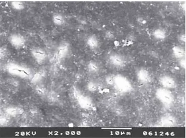
The root surface modification index (RSMI)6 was used to evaluate photomicrographs of samples that received root conditioning procedures only (Table 2). Photomicrographs of samples that received PRP application were evaluated considering presence (Fig. 1) or absence (Fig. 2) of fibrin network. Finally, photomicrographs of samples that received blood application were evaluated by the blood elements adhesion index (BEAI)21 presented on Table 3.
Each photomicrograph was evaluated three times in intervals of at least 7 days. The score attributed to each sample was the most prevalent score of the three evaluations. Good reproducibility was achieved in the use of the indexes. Kappa, k = 0.810 (BEAI) and k = 0.949 (RSMI).
Table 1: Distribution of samples in the treatment groups.
Group treatment Control (I) EDTA (II) Citric acid (III) TTCHCl (IV) Sodium citrate (V) Total
HMDS 16 16 16 15 16 79
PRP 8 8 8 8 8 40
PRP + blood 8 8 8 8 8 40
Total 32 32 32 31 32 159
Score 1: Observation of cementum/dentin and exposure of collagen of dental matrix
Score 2: Observation of cementum/dentin with no exposure of collagen of dental matrix
Score 3: Presence of smear layer
Table 2: Root suface modification index (RSMI)
Blood Cells Attachment after Root Conditioning and PRP Application: An in vitro Study
Table 3: Blood elements adhesion index (BEAI)
Score 0: Absence of fibrin network and blood cells Score 1: Scarcely distributed fibrin network and/or blood cells
Score 2: Moderate number of blood cells and thin fibrin network with poor interlacing
Score 3: Dense fibrin network with rich interlacing and presence of blood cells
Fig. 2: Absence of fibrin network
STATISTICAL ANALYSIS
Groups were compared on ordinal (RSMI and BEAI) and dichotomous variables by nonparametric analysis of
variance (Kruskal-Wallis and Dunn tests) using a significance of p ≤ 0.05. These analyses were performed using the Bioestat 3.0 statistical software package (CNPq and Sociedade Civil Mamirauá, Belém PA, Brazil).
RESULTS
Among five groups compared using the RSMI, difference was detected (p = 0.0001) between control group and all other groups. Nevertheless, group II presented score 1 frequency of 25% and score 2 of 75%; the group III presented score 1 frequency of 56.25% and score 2 of 43.75%, the best results for RSMI. Less favorable results were observed in the control group with 6.25% of score 2 and 93.75% of score 3 (Table 4). Groups IV and V presented samples with 100% of score 2 (Fig. 3).
Table 5: Frequency distribution of samples according to the presence of fibrin network
Groups P A
I-Control n(%) 0 (0) 8 (100)
II-EDTA 24% n(%) 0 (0) 8 (100)
III-Citric acid 25% n(%) 7 (87.5) 1 (12.5)
IV-Tetracycline hydrochloride
50 mg/ml n (%) 0 (0) 8 (100)
V-Sodium citrate 30% n(%) 0 (0) 8 (100)
Presence (P); Absence (A)
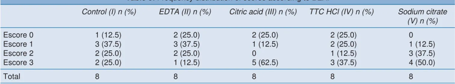
Table 6: Frequency distribution of scores according to BEAI
Control (I) n (%) EDTA (II) n (%) Citric acid (III) n (%) TTC HCl (IV) n (%) Sodium citrate (V) n (%)
Escore 0 1 (12.5) 2 (25.0) 2 (25.0) 2 (25.0) 0
Escore 1 3 (37.5) 3 (37.5) 1 (12.5) 2 (25.0) 1 (12.5)
Escore 2 2 (25.0) 2 (25.0) 0 1 (12.5) 3 (37.5)
Escore 3 2 (25.0) 1 (12.5) 5 (62.5) 3 (37.5) 4 (50.0)
Total 8 8 8 8 8
Control (I) n(%) EDTA (II) n(%) Citric acid (III) n(%) TTC HCl (IV) n(%) Sodium citrate (V) n(%)
score 1 0 4 (25.0) 9 (56.2) 0 0
score 2 1 (6.3) 12 (75.0) 7 (43.4) 15 (100.0) 16 (100.0)
score 3 15 (93.7) 0 0 0 0
Total 16 16 16 15 16
There was no statistical difference among sample who received blood application (p = 0.3380). Nevertheless, groups III and V presented higher frequency of score 3 (62.5 and 50%, respectively) (Fig. 4). Group IV presented 37.5% of score 3, followed by groups I and II (25 and 12.5% respectively) (Table 6).
DISCUSSION
To achieve periodontal regeneration, it is necessary to remove plaque, calculus and bacterial byproducts from root surface.7 Scaling and root planning are considered the best method to do it.7,8 Nevertheless, this procedure leads to smear layer formation22 which can impair periodontal healing and regeneration. Thus, attention has been given to root surface modification through demineralizing agents, which promotes additional root decontamination and dental matrix collagen exposure aiming new periodontal attachment formation.10,23
In the present study, all demineralizing agents were able to remove smear layer, but only some of them exposed the collagen from the dental matrix (Fig. 3). In accordance with Fig. 3: Frequency of scores 1, 2 and 3 in different groups of
demineralizing agents, according to the root surface modification index (RSMI)
Blood Cells Attachment after Root Conditioning and PRP Application: An in vitro Study
previous studies,24 citric acid 25% and EDTA 24% presented high capacity of root surface modification. Tetracycline hidrochloride 50 mg/ml presented high smear layer removal capacity, which is in accordance with previous studies22 and showed no capacity of dental matrix collagen exposure, what does no corroborate previous data.9,25 This unexpected observation may be attributed to the fast deterioration of this chemical after its preparation, once concentration, period of application and mode of application were standardized. Sodium citrate 30% was not able to expose collagen fibers from dental matrix, so new in vitro studies are necessary to establish the parameters for its use as dental root modification agent.
It was observed fibrin network and blood cells attached to the dental surface in all groups that received blood application. Nevertheless, there was variability in this phenomenon among groups (Fig. 4). Adhesion of blood elements to dental surfaces in the control group is in discordance with previous studies6,10 and may be explained by the sample roughness which created a mechanical retention of blood clot.
Although EDTA 24% has a high capacity of smear layer removal and dental matrix collagen exposure working in neutral pH (more biocompatible), it does not favor blood elements adhesion to the root surface,6 which is in accordance with the results of the present study. This may be explained due to its strong chelation character, which may inhibit formation and adhesion of blood clot to the dental surface and may promote early platelet fragmentation.26
Absence of blood elements on the samples conditioned with tetracycline hydrochloride 50 mg/ml may be explained by the weak capacity this chemical showed to expose dental matrix collagen (Table 6).
In vivo studies have shown the ability of citric acid to provide blood clot stabilization on dental surface27 (corroborating data of the present study) and to provide linkage between collagen from dental matrix and from periodontal connective tissue.28,29
The goods results observed in the present study on the adhesion of blood elements over sodium citrate conditioned samples determine the need of additional studies to verify this phenomenon.
Regarding samples that received PRP application, only those previously conditioned with citric acid 25% presented fibrin network (Table 5). The authors attribute this finding to increased dental matrix collagen exposure attained with citric acid 25% conditioning which may favor platelets adhesion to dental surface. The fact that platelets have affinity for collagen and that citric acid is easily washed off from dental surface after conditioning procedure are factors
that may be contributed to the citric acid conditioning performance. Chelant agents (EDTA and sodium citrate) and tetracycline hydrochloride (which presents high substantivity)30 are more difficult to be removed from the dental surface and remaints of these agents may interfere on PRP adhesion.
The encouraging results observed in the present study on the adhesion of blood elements over sodium citrate conditioned samples determine the need of additional studies to verify this phenomenon.
CONCLUSION
According to the results and considering the limitations of the present methodology, the authors conclude that:
Citric acid 25% was the most efficient conditioning agent to remove smear layer, to promote dental collagen exposure and to promote fibrin clot and blood elements adhesion to the dental surface.
CLINICAL SIGNIFICANCE
This study demonstrated that root conditioning followed by PRP application may favor blood cell adhesion on root surface which may optimize periodontal healing.
REFERENCES
1. Polson AM, Frederick GT, Ladenheim S, Hanes PJ. The production of a root surface smear layer by instrumentation and its removal by citric acid. J Periodontol 1984 Aug;55(8):443-46. 2. Babay N. The effect of EDTA on the attachment and growth of cultured human gingival fibroblasts in periodontitis-affected root surface. J Contemp Dent Pract 2001 Feb 15;2(1):13-23. 3. Bouchard P, Nilveus R, Etienne D. Clinical evaluation of
tetracycline HCl conditioning in the treatment of gingival recessions. A comparative study. J Periodontol 1997 Mar;68(3):262-69.
4. Wikesjo UM, Claffey N, Christersson LA, Franzetti LC, Genco RJ, Terranova VP, et al. Repair of periodontal furcation defects in beagle dogs following reconstructive surgery including root surface demineralization with tetracycline hydrochloride and topical fibronectin application. J Clin Periodontol 1988 Jan;15(1):73-80.
5. Bergenholtz A, Babay N. Scanning electron microscopy of the root surface texture of extracted periodontally diseased teeth following various etching and chelating regimens. Int J Periodontics Restorative Dent 1998 Apr;18(2):171-79. 6. Leite FR, Moreira CS, Theodoro LH, Sampaio JE. Blood cell
attachment to root surfaces treated with EDTA gel. Braz Oral Res 2005 Apr-Jun;19(2):88-92.
7. Lafferty TA, Gher ME, Gray JL. Comparative SEM study on the effect of acid etching with tetracycline HCl or citric acid on instrumented periodontally-involved human root surfaces. J Periodontol 1993 Aug;64(8):689-93.
hydrochloride conditioning. J Contemp Dent Pract 2008;9(5): 25-33.
10. Baker PJ, Rotch HA, Trombelli L, Wikesjo UM. An in vitro screening model to evaluate root conditioning protocols for periodontal regenerative procedures. J Periodontol 2000 Jul;71(7):1139-43.
11. Caton JG, Greenstein G. Factors related to periodontal regeneration. Periodontol 2000 1993 Feb;1(1):9-15.
12. Wikesjo UM, Nilveus R. Periodontal repair in dogs. Healing patterns in large circumferential periodontal defects. J Clin Periodontol 1991 Jan;18(1):49-59.
13. Ruggeri ZM. Mechanisms initiating platelet thrombus formation. Thromb Haemost 1997 Jul;78(1):611-16.
14. Whitman DH, Berry RL, Green DM. Platelet gel: An autologous alternative to fibrin glue with applications in oral and maxillofacial surgery. J Oral Maxillofac Surg 1997 Nov;55(11):1294-99.
15. Garg AK. The future role of growth factors in bone grafting. Dent Implantol Update 1999 Jan;10(1):5-7.
16. Bergstrom J. Tobacco smoking and subgingival dental calculus. J Clin Periodontol 2005 Jan;32(1):81-88.
17. van Winkelhoff AJ, Bosch-Tijhof CJ, Winkel EG, van der Reijden WA. Smoking affects the subgingival microflora in periodontitis. J Periodontol 2001 May;72(5):666-71.
18. Blomlof J, Blomlof L, Lindskog S. Effect of different concentrations of EDTA on smear removal and collagen exposure in periodontitis-affected root surfaces. J Clin Periodontol 1997 Aug;24(8):534-37.
19. Sterrett JD, Dhillon M, Murphy HJ. Citric acid demineralization of cementum and dentin: The effect of application pressure. J Clin Periodontol 1995 Jun;22(6):434-41.
20. Isik AG, Tarim B, Hafez AA, Yalcin FS, Onan U, Cox CF. A comparative scanning electron microscopic study on the characteristics of demineralized dentin root surface using different tetracycline HCl concentrations and application times. J Periodontol 2000 Feb;71(2):219-25.
21. Theodoro LH, Sampaio JE, Haypek P, Bachmann L, Zezell DM, Garcia VG. Effect of Er:YAG and Diode lasers on the adhesion of blood components and on the morphology of irradiated root surfaces. J Periodontal Res 2006 Oct;41(5):381-90.
22. Madison JG, 3rd, Hokett SD. The effects of different tetracyclines on the dentin root surface of instrumented, periodontally involved human teeth: A comparative scanning electron microscope study. J Periodontol 1997 Aug;68(8):739-45. 23. Chandra RV, Jagetia GC, Bhat KM. The attachment of V79
and human periodontal ligament fibroblasts on periodontally involved root surfaces following treatment with EDTA, citric acid, or tetracycline HCL: An SEM in vitro study. J Contemp Dent Pract 2006 Feb 15;7(1):44-59.
24. Blomlof L, Bergman E, Forsgardh A, Foss L, Larsson A, Sjoberg B, et al. A clinical study of root surface conditioning with an EDTA gel. I. Nonsurgical periodontal treatment. Int J Periodontics Restorative Dent 2000 Dec;20(6):560-65. 25. Babay N. SEM study on the effect of two different
demineralization methods with saturated tetracycline hydrochloride on diseased root surfaces. J Contemp Dent Pract. 2001 May 15;2(2):25-35.
conditions on platelet impedance volume. Klin Wochenschr. 1989 Oct 2;67(19):980-84.
27. Bal B, Eren K, Balos K. Effects of various root surface treatments on initial clot formation: A scanning electron microscope study. J Nihon Univ Sch Dent 1990 Dec;32(4):281-93.
28. Selvig KA, Bogle G, Claffey NM. Collagen linkage in periodontal connective tissue reattachment. An ultrastructural study in beagle dogs. J Periodontol 1988 Nov;59(11):758-68. 29. Ririe CM, Crigger M, Selvig KA. Healing of periodontal
connective tissues following surgical wounding and application of citric acid in dogs. J Periodontal Res 1980 May;15(3):314-27. 30. Baker PJ, Evans RT, Coburn RA, Genco RJ. Tetracycline and its derivatives strongly bind to and are released from the tooth surface in active form. J Periodontol 1983 Oct;54(10):580-85.
ABOUT THE AUTHORS
Andrea Abi Rached Dantas (Corresponding Author) Assistant Professor, Department of Operative Dentistry, School of Dentistry at Araraquara, State University of São Paulo, UNESP, 1680 Humaita Street, Araraquara, 14801-903, São Paulo, Brazil, Phone: 55 (16) 3301-6393, e-mail: [email protected]
Lucas Amaral Fontanari
PhD Student, Department of Diagnostic and Surgery, School of Dentistry at Araraquara, State University of São Paulo, UNESP, São Paulo, Brazil
Eduardo de Paula Ishi
PhD, Clinical Practice, São Paulo, Brazil
Fábio Renato Manzolli Leite
Adjunct Professor, Department of Semiology and Clinics, School of Dentistry of Pelotas, Federal University of Pelotas, UFPel, Rio Grande do Sul, Brazil
Daniela Leal Zandim
Assistant Professor, Department of Diagnostic and Surgery, School of Dentistry at Araraquara, State University of São Paulo, UNESP São Paulo, Brazil
Ricardo Samih Georges Abi Rached
Titular Professor, Department of Diagnostic and Surgery, School of Dentistry at Araraquara, State University of São Paulo, UNESP, São Paulo, Brazil
José Eduardo Cezar Sampaio