Update / Atualização
Perspectives of stem cell therapy in type 2 diabetes mellitus
Angela M. O. Leal 1
Júlio César Voltarelli2
Type 2 diabetes mellitus (DM2) is associated with insulin resistance and secretory dysfunction of the -cells. There is now experimental, epidemiological and clinical evidence suggesting that the immune system and inflammatory mediators are involved in the pathogenesis of DM2. The interest in regenerative therapeutics and cellular therapy for DM2 is motivated by the importance of preserving -cell mass and function. Cellular therapy with stem cells for glycemic control has been tested in experimental models for some years however, there are only a few published studies using this approach in humans. Although there are many obstacles to overcome before regenerative therapy becomes a real option to treat DM2, it may be an important strategy to attain metabolic control and prevent chronic complications in the future. Rev. Bras. Hematol. Hemoter. 2010;32 (4):329-334.
Keywords: Diabetes mellitus; cell therapy; stem cell.
Physiopathological aspects of Type-2 diabetes mellitus
Type-2 diabetes mellitus (DM2), the most common form of diabetes (approximately 90% of cases), is caused basically by two pathogenic mechanisms - resistance to insulin and secretory dysfunction of pancreatic β-cells.
The etiopathogenesis and pathophysiology of DM2 are complex and involve genetic and environmental components that are interrelated in ways that are still unknown. Genetically, DM2 has monogenic and polygenic forms. The former are rare, suffer minimal environmental influence and the genes involved in insulin resistance and insulin secretion defects have been characterized. However, more commonly DM2 is polygenic and although potential loci of susceptibility for DM2 have been identified in different populations, numerous methodological difficulties must be overcome until their participation in the genesis of the disease is properly understood.(1,2)
Obesity, particularly with visceral fat accumulation, whose metabolic behavior differs from subcutaneous fat, is
1Endocrinologist, Medicine Department of Universidade Federal de São Carlos – São Carlos (SP), Brazil
2Clinical Immunology, Clinical Medicine Department, Faculdade de Medicina de Ribeirão Preto-USP, Ribeirão Preto (SP), Brazil
Universidade Federal de São Carlos (UFScar) and Universidade de São Paulo (USP)
Correspondence: Angela Leal Rua Garibaldi, 1108/182
14010-170 – Ribeirão Preto (SP), Brazil E-mail: [email protected]
Doi: 10.1590/S1516-84842010005000088
one of the environmental determinants of DM2. Adipose tissue modulates the metabolism by releasing free fatty acids, glycerol, pro-inflammatory cytokines, chemokines and hormones, including leptin and adiponectin. The increase in most of these factors affects the action of insulin in target organs and causes insulin resistance, mainly by acting on its signaling cascade.(3)
However, most obese individuals who have insulin resistance do not evolve with hyperglycemia as, normally, the pancreatic β-cell has great plasticity and adapts to a reduction in insulin sensitivity by increasing both insulin secretion and β-cell mass. The adaptive mechanisms of
β-cells to insulin resistance appear to involve an increase in glucose metabolism, signaling by nonesterified fatty acids, increased signaling by insulin/IGF-1 (insulin-like growth factor) and the secretagogue and mitogen actions of incretin GLP- 1 (glucagon-like peptide-1).( 4,5)
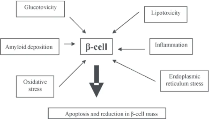
Figure 1 – Mechanisms of β-cell failure in DM2
DM2 are both functional and quantitative; the β-cell mass is reduced to approximately 50% of normal by increased apoptosis and β-cells lose 75% of their functional capacity.(6,7)
The factors involved in β-cell dysfunction are not well established, but they fail to respond to secretagogue stimuli. Many signaling routes can affect the growth and survival of
β-cells. Some of the many potential mechanisms involved in this impairment are oxidative stress, mitochondrial dysfunction, endoplasmic reticulum stress, inflammation and amyloid deposition of material, associated with genetic predisposition.(8-11) Hyperglycemia, resulting from this process and the increased concentration of free fatty acids that cause glucolipotoxicity are aggravating factors that accelerate the decline in β-cells in DM2.(12-14)
endothelial cells and monocytes, increases the recruitment of macrophages that feed the process.(23-26) Although by different mechanisms, most inflammatory factors act negatively in the process of insulin signaling by modifying its intracellular substrates, in particular phosphorylation of the protein family of insulin receptor substrates, with the help of different transcription factors, including those of the family of peroxisome proliferator-activated receptors. Thus, the metabolic inflammatory and innate immunity pathways seem to interact, leading to insulin resistance in a complex yet still unclear manner despite of the many advances in this area over the last decade. Endoplasmic reticulum stress and mitochondrial dysfunction, caused, in part, by increased metabolic demand, are considered important links between the inflammatory and metabolic pathways involved in insulin resistance associated with obesity and DM2.(16,27,28)
Although recognition that the immune system is involved in β-cell dysfunction and death in DM2 is much more recent, in vivo and in vitro clinical and experimental data indicate that inflammation of islets of Langerhans (insulitis) is a predominant feature in DM2, as well as in type 1 diabetes mellitus (DM1).(11,29) An increased number of macrophages were observed in islets of mice submitted to a high fat diet well before the onset of hyperglycemia. In addition, the production of inflammatory factors, including IL-6, IL-8, chemokine KC, G-CSF (granulocyte colony-stimulating factor) and MIP-1α (macrophage inflammatory protein 1α) has been identified in islets exposed to metabolic stress (hyperglycemia and hyperlipemia) and in isolated islets of mice submitted to a high fat diet. Some evidence suggests that the local immune system may be involved in the mechanism of apoptosis of β-cells that accompanies metabolic disorders culminating in DM2. Among the cytokines involved in this process, IL-1α is noteworthy as its local production by β-cells under glucose stimulation has been demonstrated as has its blockage in 70 patients with DM2 by the injection of a recombinant IL-1 receptor antagonist with a resulting increase in insulin secretion.(30-32) The hypothesis put forward by the group of researchers who produced most of these results is that metabolic stress (hyperglycemia, dyslipidemia and adipokines) leads to the production of IL-1α by islets, which controls their own production and the production of other inflammatory mediators and attracts macrophages, thereby perpetuating the inflammatory process. This process culminates in secretory failure and β-cell death. (33) Additionally, more than a decade ago it was shown that IL-1α can induce the expression of Fas, a cell surface receptor involved in β-cell apoptosis.(34) However, the action of IL-1αon islets is dose dependent as small concentrations may play a protective role in β-cells.(35)
Thus, it is clear that, although a few years ago, the pathophysiological differences between DM1 and DM2
Inflammation and type-2 diabetes mellitus
Currently, there is much experimental, clinical and epidemiological evidence of the involvement of immune and inflammatory mediators in the basic mechanisms of DM2, both insulin resistance and pancreatic β-cell failure.(15-20)
The increased release of TNF-α (tumor necrosis factor), IL-6 (interleukin), MCP-1 (monocyte chemoattractant protein-1) and soluble factors secreted by macrophages and other cells residing in adipose tissue play a key role in the development of insulin resistance.(21) TNF-α and IL-6 stimulate the c-Jun aminoterminal kinase (JNK) and IêB kinase
seemed so different, now they are considered fairly comparable, implying the possibility of using similar therapeutic approaches (immunological and reparative) for both diseases.(12,15)
Stem cell therapy for type-2 diabetes mellitus
Clinical trials using cell therapy for the treatment of DM1 are already at an advanced stage and the results, so far, seem promising.(36,37)
The interest in regenerative therapy and the use of cell therapy, more specifically, with pluripotent or multipotent stem cells (SC) in the treatment of DM2, derives from the aforementioned clinical evidence and the importance of preserving the functional and quantitative integrity of pancreatic β-cells whose destruction is involved in the pathogenesis of DM2. Additionally, the difficulties encountered with the use of other methods of β-cell replacement, such as islet, pancreas and insulin-producing cell line transplants(38,39) makes the possibility of using SC very attractive.
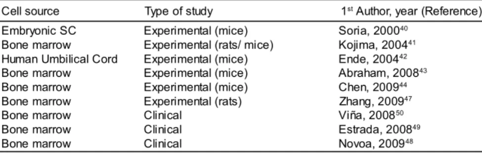
The use of SC from different sources for glucose control in experimental models of DM2 was described several years ago (Table 1). Soria et al.(40) showed that insulin-secreting cells derived from undifferentiated embryonic SC implanted into the spleens of mice with diabetes induced by streptozotocin were able to maintain normoglycemia. Kojima et al.(41) found insulin-producing cells in extrapancreatic tissues of hyperglycemic rats and mice treated with adult bone marrow SC. Additionally, Ende et al.(42) showed a reduction in glucose levels, improved survival and attenuation of renal injury in obese mice with spontaneous development of DM2 treated with human umbilical cord blood mononuclear cells (MNC). Recently, Abraham et al. reported increased glucose tolerance and plasma adiponectin concentrations in obese mice after the intraosseous transplantation of bone marrow SC, associated with the induction of heme oxygenase (43) and Chen et al. reported improved insulin sensitivity, and vascular and renal function after the infusion of bone marrow cells of non-diabetic mice in diabetic db/db mice.(44)
Different production strategies and sources of SC to form insulin-producing cells are being intensively investigated and could become an option in regenerative therapy for DM2
in the future. Among them are differentiation from embryonic SC,(45) pluripotent SC reprogrammed using transcription factors to a differentiated state known as iPS (induced pluripotent SC) (46) and bone marrow mesenchymal SC.(47) The authors of the last study reported in vitro differentiation of bone marrow mesenchymal cells into insulin-producing cells, which were transplanted into the duodenal mucosa and led to a decrease in blood sugar levels in rats treated with streptozotocin.
In humans, there are few reports of effects of SC treatment on glycemic control in DM2 (Table 1). Novoa et al.(48) recently reported lower concentrations of glucose and the need of smaller doses of insulin in 126 diabetic patients with severe peripheral arterial disease treated with autologous infusions of bone marrow MNC in the gastrocnemius muscle. Estrada et al.(49) submitted DM2 individuals to intrapancreatic infusions of autologous bone marrow MNC and hyperbaric oxygen therapy, and after one year of monitoring, they observed a significant reduction in fasting glucose levels, glycosylated hemoglobin, daily insulin doses and oral hypoglycemic agents as well as increased plasma concentrations of C-peptide. In a recent work, Viña et al.(50) reported the beneficial effects of local intra-arterial infusions of bone marrow MNC in 58 DM2 patients over a follow-up period of 36 months. Although the authors of these reports suggest beneficial effects of SC therapy on angiogenesis, the mechanism of action involved has not been addressed yet, primarily due to methodological difficulties of studies involving humans. However, in an analysis of autopsies, Butler et al.(51) evaluated the pancreas of 31 individuals, two of who were DM2 patients submitted to allogeneic hematopoietic SC transplantations and did not observe the presence of β-cells from donor hematopoietic cells and concluded that it was unlikely that these cells can transdifferentiate into β-cells.
Barriers to cell therapy for type-2 diabetes mellitus
There are several difficulties to be overcome before regenerative therapy of the pancreas using SC to treat DM2 is a viable option. The first challenge may be to understand the mechanisms that regulate insulin secretion stimulated by glucose and those involved in the adaptation of β-cells to constant metabolic changes.(52) In addition, our knowledge has advanced only recently in respect to the mechanisms of differentiation, proliferation, regeneration and plasticity of β-cells.(53-58) It is not known whether, in humans, there is a population of progenitor cells (ductal, acinar and extrapancreatic) that generate new β-cells (neogenesis) or if
Table 1. Experimental and clinical use of cell therapy in DM2
Cell source Type of study 1st Author, year (Reference)
Embryonic SC Bone marrow Human Umbilical Cord Bone marrow Bone marrow Bone marrow Bone marrow Bone marrow Bone marrow
Experimental (mice) Experimental (rats/ mice) Experimental (mice) Experimental (mice) Experimental (mice) Experimental (rats) Clinical
Clinical Clinical
Soria, 200040
Kojima, 200441
Ende, 200442
Abraham, 200843
Chen, 200944
Zhang, 200947
Viña, 200850
Estrada, 200849
involved in the processes of differentiation and/or transdifferentiation to β-cells, although the Pdx1 (pancreatic duodenal homeobox-1) and neurogenin-3 transcription factors present in the embryonic period are being intensely investigated.(59) Although it seems clear that numerical integrity of the β-cell mass does not mean functional integrity (60) there are no noninvasive methods to measure the number of β-cells in humans. The few data available come from experimental studies and indirect evidence therefore they are limited and imprecise.(61) However, many β-cell markers have been tested in recent years(62) and there are descriptions of potential noninvasive methods. Among these are the use of VMAT2 (vesicular monoamine transporter type 2) that is expressed in large amounts in human β-cells and when associated to the radioligand, 11C-dihydrotetrabenazine is detected by PET (positron emission tomography) as a biomarker of β-cell mass.(63) Additionally, the application of magnetic resonance has been investigated mainly after the transplantation of islets previously labeled with nanoparticles.(64,65)
Why develop new strategies for the treatment of type 2 diabetes?
The treatment of DM2 requires different professionals and involves diet, exercise, various oral drugs and, often, multiple daily insulin injections. Adherence to therapy is usually low and more than 70% of patients have poor metabolic control.(66-70) There is a high frequency of chronic complications associated to poor metabolic control of DM2 and it is already well established that hyperglycemia is a major factor responsible for the onset of these complications.(71) These complications arise from micro- and macrovascular changes that lead to dysfunction, damage or failure of different organs including nephropathy with a high rate of progression to renal failure, retinopathy, the leading cause of blindness, neuropathy, the leading cause of non-traumatic amputation of lower extremities, and manifestations of autonomic nervous system dysfunction, including sexual dysfunction.(72-75) Additionally, the association between diabetes and cardiovascular diseases, which include myocardial ischemia, peripheral arterial obstruction and cerebrovascular disease, is well established. DM2 increases the risk of cardiovascular disease by two to four-fold with this being the main cause of death in diabetics.(76,77) All of this involves high direct and indirect costs for DM2 individuals, for the public healthcare system and for society.
Therefore, the development of new therapeutic strategies for DM2 is essential. Regenerative therapy of the pancreas seems to be safe and effective in DM2 and could be an important tool for metabolic control and reduction of chronic complications.
test the systemic use of mesenchymal SC in DM2.
Resumo
A patogênese do diabetes mellitus tipo 2 (DM2) está associada, basicamente, a dois mecanismos, resistência à ação da insulina e disfunção secretória das células β pancreáticas. Atualmente, há evidências experimentais, clínicas e epidemiológicas, da participação do sistema imune e de mediadores inflamatórios nesses mecanismos patogênicos. O interesse pelo tratamento regenerativo e pela utilização da terapia celular para o tratamento do DM2 deriva da importância da preservação da integridade funcional e quantitativa das células β pancreáticas. A utilização de células-tronco para obtenção de controle glicêmico em modelos experimentais de DM2 tem sido descrita já há alguns anos. Entretanto, em humanos, há poucos estudos publicados nesse sentido. Embora haja várias dificuldades a serem transpostas até que a terapia regenerativa do pâncreas para tratamento do DM2 seja uma opção viável, ela poderá vir a ser, no futuro, uma ferramenta importante para o controle metabólico da doença e redução de suas complicações crônicas. Rev. Bras. Hematol. Hemoter. 2010;32(4):329-334.
Palavras-chave: Diabetes mellitus; terapia celular; células-tronco.
References
1. Almind K, Doria A, Khan CR. Putting the genes for type II diabetes on the map. Nat Med. 2001;7(3):277-9.
2. Cox NJ. Challenges in identifying genetic variation affecting susceptibility in type 2 diabetes: examples from studies of the calpain-10 gene. Hum Mol Genet. 2001;10(20):2301-5.
3. Kahn SE, Hull RL, Utzschneider KM. Mechanisms linking obesity to insulin resistance and type 2 diabetes. Nature. 2006;444(7121): 840-6
4. Drucker DJ. The biology of incretin hormones. Cell Metab. 2006; 3:153-65.
5. Bernal-Mizrachi E, Wen W, Stahlhut S, Welling CM, Permutt MA. Islet beta cell expression of constitutively active Akt1/PKB alpha induces striking hypertrophy, hyperplasia, and hyperinsulinemia. J Clin Invest. 2001;108(11):1631-8.
6. Butler AE, Janson J, Bonner-Weir S, Ritzel R, Rizza RA, Butler PC. Beta-cell deficit and increased beta-cell apoptosis in humans with type 2 diabetes. Diabetes. 2003;52(1):102-10.
7. Røder ME, Porte D Jr, Schwartz RS, Kahn SE. Disproportionately elevated proinsulin levels reflect the degree of impaired ß cell secretory capacity in patients with noninsulin-dependent diabetes mellitus. J Clin Endocrinol Metab. 1998;83(2):604-8. 8. Muoio DM, Newgard CB. 3.Mechanisms of disease: molecular
and metabolic mechanisms of insulin resistance and beta-cell failure in type 2 diabetes. Nat Rev Mol Cell Biol. 2008;9(3):193-205.
9. Gloyn AL, Tribble ND, van de Bunt M, Barrett A, Johnson PR.. Glucokinase (GCK) and other susceptibility genes for beta-cell dysfunction: the candidate approach. Biochem Soc Trans. 2008; 36(pt 3):306-11.
11. Donath MY, Schumann DM, Faulenbach M, Ellingsgaard H, Perren A, Ehses JA. Islet inflammation in type 2 diabetes. Diabetes Care. 2008;31(Suppl 2):S161-S164.
12. Cnop M, Welsh N, Jonas J, Jorns A, Lenzen S, Eizirik DL. Mechanisms of pancreatic beta-cell death in type 1 and type 2 diabetes. Diabetes. 2008;54 (Suppl 2):S97-S107.
13. Poitout V, Robertson RP. Glucolipotoxicity: fuel excess and beta-cell dysfunction. Endocr Rev. 2008;29(3):351-66.
14. Rhodes CJ. Type 2 diabetes - a matter of beta-cell life and death? Science. 2005;307(5708):380-4.
15. Kolb H, Mandrup-Poulsen T. An immune origin of type 2 diabetes? Diabetologia. 2005;48(6):1038-50.
16. Hotamisligil GS. Inflammation and metabolic disorders. Nature 2006;444(7121):860-7.
17. Donath MY, Storling J, Berchtold LA, Billestrup N, Mandrup-Poulsen T. Cytokines and beta-cell biology: from concept to clinical translation. Endocr Rev. 2008;29(3):334-50.
18. Herder C, Haastert B, Müller-Scholze S, Koenig W, Thorand B, Holle R, et al. Association of systemic chemokine concentrations with impaired glucose tolerance and type 2 diabetes. Diabetes 2005;54 (Suppl l2):S11-S17.
19. Thorand B, Kolb H, Baumert J, Koenig W, Chambless L, Meisinger C, et al. Elevated levels of interleukin-18 predict the development of type 2 diabetes: results from the MONICA/ KORA Augsburg Study, 1984-2002. Diabetes. 2005;54(10): 2932-8.
20. Pickup JC. Inflammation and activated innate immunity in the pathogenesis of type 2 diabetes. Diabetes Care. 2004; 27 (3):813-23.
21. Guilherme A, Virbasius JV, Puri V, Czech MP. Adipocyte dysfunctions linking obesity to insulin resistance and type 2 diabetes. Nat Rev Mol Cell Biol. 2008;9(5):367-77.
22. Karin M, Delhase M. The I kappa B kinase (IKK) and NF-kappa B: key elements of proinflammatory signalling. Semin Immunol. 2000;12(1):85-98.
23. Wellen KE, Hotamisligil GS. Inflammation, stress, and diabetes. J Clin Invest. 2005;115(5):1111-9.
24. Mooney RA, Senn J, Camerons S, Inamdar N, Boivin LM, Shang Y, et al. Suppressors of cytokine signaling-1 and -6 associate with and inhibit the insulin receptor. A potential mechanism for cytokine-mediated insulin resistance. Biol Chem. 2001;276 (28):25889-93.
25. Perreault M, Marette A. Targeted disruption of inducible nitric oxide synthase protects against obesity-linked insulin resistance in muscle. Nat Med. 2001;7(10):1138-43.
26. Arkan MC, Hevener AL, Greten FR, Maeda S, Li Z, Long JM, et al. IΚΚ-beta links inflammation to obesity-induced insulin resistance. Nat Med. 2005;11(2):191-8.
27. Taniguchi CM, Emanuelli B, Kahn CR. Critical nodes in signaling pathways: insights into insulin action. Nat Rev Mol Cell Biol. 2006;7(2):85-96.
28. Schenk S, Saberi M, Olefsky. Insulin sensitivity: modulation by nutrients and inflammation. J Clin Invest 2008; 118(9):2992-3002.
29. Ehses JA, Perren A, Eppler E, Ribaux P, Pospisilik JA, Maor-Cahn R, et al. Increased number of islet-associated macrophages in type 2 diabetes. Diabetes 2007;56(9):2356-70.
30. Maedler K, Sergeev P, Ris F, Oberholzer J, Joller-Jemelka HI, Spinas GA, et al. Glucose-induced beta cell production of IL-1beta contributes to glucotoxicity in human pancreatic islets. J Clin Invest 2002;110(6):851-60.
31. Larsen CM, Faulenbach M, Vaag A, Volund A, Ehses JA, Seifert B et al. Interleukin-1-receptor antagonist in type 2 diabetes mellitus. N Engl J Med 2007;356(15):1517-26.
32. Böni-Schnetzler M, Thorne J, Parnaud G, Marselli L, Ehses JA, Kerr-Conte J, et al. Increased interleukin (IL)-1 beta messenger ribonucleic acid expression in beta-cells of individuals with type 2 diabetes and regulation of IL-1 beta in human islets by glucose and autostimulation. J Clin Endocrinol Metab. 2008;93(10): 4065-74.
33. Ehses JA, Böni-Schnetzler M, Faulenbach M, Donath MY. Macrophages, cytokines and beta-cell death in type 2 diabetes. Biochem Soc Trans. 2008;36(Pt 3):340-2.
34. Loweth AC, Williams GT, James RF, Scarpello JH, Morgan NG. Human islets of Langerhans express Fas ligang and undergo apoptosis in response to interleukin-1 beta and Fas ligation. Diabetes. 1998;47(5):727-32.
35. Maedler K. Beta cells in type 2 diabetes - a crucial contribution to pathogenesis. Diabetes Obes Metab. 2008;10(5):408-20. 36. Voltarelli JC, Couri CE, Stracieri AB, Oliveira MC, Moraes DA,
Pieroni F, et al. Autologous nonmyeloablative hematopoietic stem cell transplantation in newly diagnosed type 1 diabetes mellitus. JAMA. 2007;297(14):1568-76.
37. Couri CE, Oliveira MC, Stracieri AB, Moraes DA, Pieroni F, Barros GM, et al. C-peptide levels and insulin independence following autologous nonmyeloablative hematopoietic stem cell transplantation in newly diagnosed type 1 diabetes mellitus. JAMA. 2009;301(15):1573-9.
38. Lanza RP, Chick WL. Transplantation of pancreatic islets. Ann N Y Acad Sci. 1997;831:323-31.
39. Roche E, Assimacopoulos-Jeannet F, Witters LA, Perruchoud B, Yaney G, Corkey B, et al. Induction by glucose of genes coding for glycolytic enzymes in a pancreatic beta-cell line (INS-1). J Biol Chem. 1997;272(5):3091-3098.
40. Soria B, Roche E, Berná G, Léon-Quinto T, Reig JA, Martín F. Insulin-secreting cells derived from embryonic stem cells normalize glycemia in streptozotocin-induced diabetic mice. Diabetes. 2000; 49(2):157-62.
41. Kojima H, Fujimiya M, Matsumura K, Nakahara T, Hara M, Chan L. Extrapancreatic insulin-producing cells in multiple organs in diabetes. Proc Natl Acad Sci U S A. 2004;101(8):2458-63. 42. Ende N, Chen R, Reddi AS. Transplantation of human umbilical
cord blood cells improves glycemia and glomerular hypertrophy in type 2 diabetic mice. Biochem Biophys Res Comm. 2004; 321(1):168-71.
43. Abraham NG, Li M, Vanella L, Peterson SJ, Ikehara S, Asprinio D. Bone marrow stem cell transplant into intra-bone cavity prevents type 2 diabetes: role of heme oxygenase-adiponectin. J Autoimm. 2008;30(3):128-35.
44. Chen J, Li H, Addabbo F, Zhang F, Pelger E, Patschan D, et al. Adoptive transfer of syngeneic bone marrow-derived cells in mice with obesity-induced diabetes: selenoorganic antioxidant ebselen restores stem cell competence. Am J Pathol. 2009; 174(2):701-11.
45. Guo T, Hebrok M. Stem cells to pancreatic beta-cells: new sources for diabetes cell therapy. Endoc Rev. 2009;30(3):214-27. 46. Maehr R, Chen S, Snitow M, Ludwig T, Yagasaki L, Goland R,
Leibel RL, Melton DA. Generation of pluripotent stem cells from patients with type 1 diabetes. Proc Nat Acad Sci. 2009; doi: 10.1073.
M, et al. Diabetes mellitus and autologous bone marrow derived progenitor cell transplant (A-BMDPCT): Cooperative international, Uruguay-Mexico. Ther Apher Dial. 2009;13:A4.
49. Estrada EJ Valacchi F, Nicora E, Brieva S, Esteve C, Echevarria L, et al. Combined treatment of intrapancreatic autologous bone marrow stem cells and hyperbaric oxygen in type 2 diabetes mellitus. Cell Transplant. 2008;17(12):1295-304.
50. Viña RF, Camozzi L, Andrin O, Vrsalovic F, Saslavsky M, Saslavsky J, et al. First report from Argentina of first three years follow up of autologous stem cells implant in diabetes type 2. Cytotherapy 2009;II:8-9.
51. Butler AE, Huang A, Rao PN, Hogan WJ, Rizza RA, Butler PC. Hematopoietic stem cells derived from adult donors are not a source of pancreatic beta-cells in adult nondiabetic humans. Diabetes 2007;56(7):1810-6.
52. Andrali SS, Sampley ML, Vanderford NL, Ozcan S. Glucose regulation of insulin gene expression in pancreatic beta-cells. Biochem J. 2008;415(1):1-10.
53. Bernardo AS, Docherty K. Stem cells and metabolic diseases. Biochem Soc Trans. 2008;36(Pt 3):363-5.
54. Dor Y. beta-cell proliferation is the major source of new pancreatic beta cells. Nat Clin Pract Endocrinol Metab. 2006; 2(5):242-3.
55. Halban PA. Cell therapy for type 2 diabetes: is it desirable and can we get it? Diabetes Obes Metab. 2008;10 Suppl 4:205-11.
56. Edlund H. Factors controlling pancreatic cell differentiation and function. Diabetologia. 2001;44:1071-9.
57. Bonal C, Avril I, Herrera PL. Experimental models of beta-cells regeneration. Biochem Soc Trans 2008;36 (Pt 3):286-9.
58. Hanley NA, Hanley KP, Miettinen PJ, Otonkoski T. Weighing up beta-cell mass in mice and humans: self-renewal, progenitors or stem cells? Mol Cel Endocrinol. 2008;288(1-2):79-85. 59. Xu X, D'Hoker J, Stangé G, Bonné S, De Leu N, Xiao X et al. Beta
cells can be generated from endogenous progenitors in injured adult mouse pancreas. Cell. 2008;132(2):197-207.
60. Kargar C, Ktorza A. Anatomical versus functional beta-cell mass in experimental diabetes. Diabetes Obes Metab. 2008;10(Suppl 4):43-53.
61. Kahn SE, Carr DB, Faulenbach MV, Utzschneider KM. An examination of beta-cell function measures and their potential use for estimating beta-cell mass. Diabetes Obes Metab. 2008;10 (Suppl 4):63-76.
62. Schneider S. Efforts to develop methods for in vivo evaluation of the native beta-cell mass. Diabetes Obes Metab. 2008;10(Suppl 4):109-118.
63. Freeby M, Goland R, Ichise M, Maffei A, Leibel R, Harris P. VMAT2 quantitation by PET as a biomarker for beta-cell mass in health and disease. Diabetes Obes Metab. 2008;10(Suppl 4): 98-108.
64. Paty BW, Bonner-Weir S, Laughlin MR, McEwan AJ, Shapiro AM. Toward development of imaging modalities for islets after transplantation: insights from the National Institutes of Health Workshop on Beta Cell Imaging. Transplantation. 2004; 77(8): 1133-7.
65. Kris J, Jirák D, Girman P, Berková Z, Zacharovova K, Honsova E, et al. Magnetic resonance imaging of pancreatic islets in tolerance and rejection. Transplantation. 2005;80(11):1596-603.
66. Stumvoll M, Goldstein BJ, van Haeften TW. Type 2 diabetes: principles of pathogenesis and therapy. Lancet. 2005; 365 (9467):1333-46.
Submitted: 09/10/2009 Accepted: 12/24/2009
pacientes diabéticos atendidos em centros de atenção primária à saúde. Revista de Saúde Pública. 2005;39:183-90.
68. Chaturvedi N. The burden of diabetes and its complications: trends and implications for intervention. Diabetes Research and Clinical Practice. 2007;76 Suppl 1:S3-12.
69. Bosi PL, Carvalho AM, Contrera D, Casale G, Pereira MA, Groner MF, et al. Prevalência de Diabetes Mellitus e Tolerância à Glicose Diminuída na População Urbana de 30 a 79 anos da Cidade de São Carlos (São Paulo). Arq Bras Endocrinol Metab. 2009;53:726-32.
70. Jardim ADI, Leal AMO. Qualidade da informação sobre diabéticos e hipertensos registrada no Sistema HIPERDIA, na Cidade de São Carlos, São Paulo, 2002-2005. Physis: Revista de Saúde Coletiva. 2009;19:405-17.
71. UK Prospective Diabetes Study Group. Intensive blood-glucose control with sulphonylureas or insulin compared with conventional treatment and risk of complications in patients with type 2 diabetes. Lancet. 1998;352(9131):837-53.
72. American Diabetes Association: Nephropathy in diabetes (Position Statement). Diabetes Care. 2004;27 (Suppl. 1):S79-S83.
73. American Diabetes Association: Retinopathy in diabetes (Position Statement). Diabetes Care. 2004;27 (Suppl. 1):S84-S87.
74. Tesfaye S, Stevens LK, Stephenson JM, Fuller JH, Plater M, Ionescu-Tirgoviste C, et al. Prevalence of diabetic peripheral neuropathy and its relation to glycaemic control and potential risk factors: the EURODIAB IDDM Complications Study. Diabetologia. 1996; 39(11):1377-84.
75. Boulton AJ, Vileikyte L, Ragnarson-Tennvall G, Apelqvist J. The global burden of diabetic foot disease. Lancet 2005;366(9498): 1719-24.
76. Grundy SM, Benjamin IJ, Burke GL, Chait A, Eckel RH, Howard B, et al. Diabetes and cardiovascular disease: a statement for healthcare professionals from the American Heart Association. Circulation 1999;100(10):1134-46.