A HuGE Review and Meta-Analyses of
Genetic Associations in New Onset Diabetes
after Kidney Transplantation
Katherine Angela Benson
1*
, Alexander Peter Maxwell
1,2, Amy Jayne McKnight
11Centre for Public Health, Queen's University Belfast, Belfast, United Kingdom,2Regional Nephrology Unit, Belfast City Hospital, Belfast, United Kingdom
*kbenson04@qub.ac.uk
Abstract
Purpose
New onset diabetes after transplantation (NODAT) is a serious complication following solid
organ transplantation. There is a genetic contribution to NODAT and we have conducted
comprehensive meta-analysis of available genetic data in kidney transplant populations.
Methods
Relevant articles investigating the association between genetic markers and NODAT were
identified by searching PubMed, Web of Science and Google Scholar. SNPs described in a
minimum of three studies were included for analysis using a random effects model. The
association between identified variants and NODAT was calculated at the per-study level to
generate overall significance values and effect sizes.
Results
Searching the literature returned 4,147 citations. Within the 36 eligible articles identified, 18
genetic variants from 12 genes were considered for analysis. Of these, three were
signifi-cantly associated with NODAT by meta-analysis at the 5% level of significance;
CDKAL1
rs10946398 p = 0.006 OR = 1.43, 95% CI = 1.11
–
1.85 (n = 696 individuals),
KCNQ1
rs2237892 p = 0.007 OR = 1.43, 95% CI = 1.10
–
1.86 (n = 1,270 individuals), and
TCF7L2
rs7903146 p = 0.01 OR = 1.41, 95% CI = 1.07
–
1.85 (n = 2,967 individuals).
Conclusion
Evaluating cumulative evidence for SNPs associated with NODAT in kidney transplant
recipients has revealed three SNPs associated with NODAT. An adequately powered,
dense genome-wide association study will provide more information using a carefully
defined NODAT phenotype.
OPEN ACCESS
Citation:Benson KA, Maxwell AP, McKnight AJ
(2016) A HuGE Review and Meta-Analyses of Genetic Associations in New Onset Diabetes after Kidney Transplantation. PLoS ONE 11(1): e0147323. doi:10.1371/journal.pone.0147323
Editor:Stanislaw Stepkowski, University of Toledo,
UNITED STATES
Received:September 21, 2015
Accepted:December 31, 2015
Published:January 20, 2016
Copyright:© 2016 Benson et al. This is an open
access article distributed under the terms of the Creative Commons Attribution License, which permits unrestricted use, distribution, and reproduction in any medium, provided the original author and source are credited.
Data Availability Statement:All relevant data are
within the paper and its Supporting Information files.
Funding:KAB is supported by a Queen's University
Belfast, School of Medicine Dentistry and Biomedical Sciences (http://www.qub.ac.uk/schools/mdbs/) international studentship and the Northern Ireland Kidney Research Fund, a local charity (http://www. kidneyresearchni.com/). The funders had no role in study design, data collection and analysis, decision to publish, or preparation of the manuscript.
Competing Interests:The authors have declared
Introduction
New onset diabetes after transplantation (NODAT), also known as post transplantation
diabe-tes mellitus (PTDM), is a serious complication of solid organ transplantation [
1
]. It affects
2–50%[
1
–
3
] of organ transplant recipients and is associated with greater healthcare costs and
an increased risk of graft failure, cardiovascular complications and death [
4
]. The wide
varia-tion in reported prevalence of NODAT in part reflects the varying clinical definivaria-tions of this
disorder. In different clinical studies the NODAT phenotype has been defined by various
crite-ria including elevated fasting blood glucose; abnormal oral glucose tolerance tests; elevated
gly-cated haemoglobin (HbA1c) or absolute requirement for hypoglycaemic therapies following
solid organ transplantation [
5
,
6
]. A number of modifiable and non-modifiable risk factors
have been identified which may predict NODAT. Modifiable risk factors include obesity and
choice of anti-rejection immunosuppression medication [
7
]. Patients receiving
tacrolimus-based immunosuppressive regimens are at greater risk of developing NODAT compared to
those prescribed ciclosporin-based immunosuppressive treatment [
8
]. However, choosing an
immunosuppressive regimen to specifically avoid NODAT may have a damaging effect on the
graft itself [
1
]. Non-modifiable risk factors include family history of diabetes mellitus,
polycys-tic kidney disease, hepatitis C infection, female gender and older recipient age [
9
,
10
]. There is
an established genetic component to NODAT, however the identification of genetic risk factors
has proved challenging. It is well documented that ethnicity is an important risk factor; people
of African American, Hispanic, or South Asian background are at a significantly increased risk
of developing the disease [
5
]. Low plasma adiponectin concentration, a factor which is under
significant genetic control [
11
], has also been demonstrated to be predictive for NODAT [
12
].
Genome-wide association studies (GWAS) are revealing SNPs associated with diabetes, which
are replicated across multiple populations [
13
,
14
], but such robust multi-centre GWAS have
not yet been published for NODAT. However, multiple publications have reported genetic
associations with NODAT in the literature, often with inconsistent results [
1
]; this report
describes an inclusive review and meta-analysis of existing data.
Materials and Methods
Selection Criteria
Review of the literature was performed to identify all published genetic variants associated with
NODAT in a kidney transplant population. Studies carried out in NODAT populations
follow-ing other forms of organ transplant (such as liver transplant) were not included. PubMed, Web
of Science and Google Scholar were searched from their inception until May 2015 with no
lan-guage restrictions, using the following keywords:
‘New Onset Diabetes’,
‘Diabetes Mellitus’,
‘Gene’,
‘Genetic’,
‘Genotype’,
‘Transplantation’,
‘Transplant’,
‘Polymorphism’,
‘Mutation’,
‘NODAT’
and
‘PTDM’
(Post-Transplantation Diabetes Mellitus). Bibliographies for all
identi-fied articles and reviews were examined to identify further publications not found in the
origi-nal search.
Inclusion Criteria
contacted if further essential information was required or if there was a query regarding
eligi-bility. If sufficient information could not be obtained, the study was excluded, as were studies
that duplicated data.
Statistical Analysis
Data was manually extracted from the studies. Information was gathered on study size,
num-bers of cases versus controls, ethnicity, genotyping methods, recorded odds ratios and p values.
If ethnicity was not explicitly stated, this was inferred from the geographical location of the
recruitment site and/or contact with authors. Deviation from Hardy-Weinberg Equilibrium
was measured using genotype counts with a threshold of p
<
0.0001. Funnel plots of standard
error of the log-odds-ratio against the log-odds-ratio were produced to estimate publication
bias. Power calculations were conducted using StatCalc version 6.
Heterogeneity was calculated using a Cochrane Q test for heterogeneity with the I
2statistic
used to describe percentage variation across studies. Meta-analysis was performed using a
ran-dom effects model for variants replicated in three or more eligible studies, with significance
value set at p
<
0.05.
All meta-analyses were performed using Review Manager software version 5.3.5 (RevMan
5.3) (
http://tech.cochrane.org/revman
)[
15
].
Funnel plots of standard error of the log-odds-ratio against the log-odds-ratio were
pro-duced to estimate publication bias. These were assessed by visual inspection. Funnel plots are
capable of detecting publication bias which would be undetected by more formal statistical
tests. Statistical tests such as Egger’s test were not conducted in this review due to the small
number of studies in the meta-analyses, which were not sufficient to distinguish chance from
asymmetry.
Results
Included Studies
The preliminary literature search yielded 4,147 citations, 40 of which were relevant studies
investigating NODAT and 36 of which had all the required information to allow the extraction
of variant information (
Fig 1
). Data was extracted from these articles for all investigated SNPs.
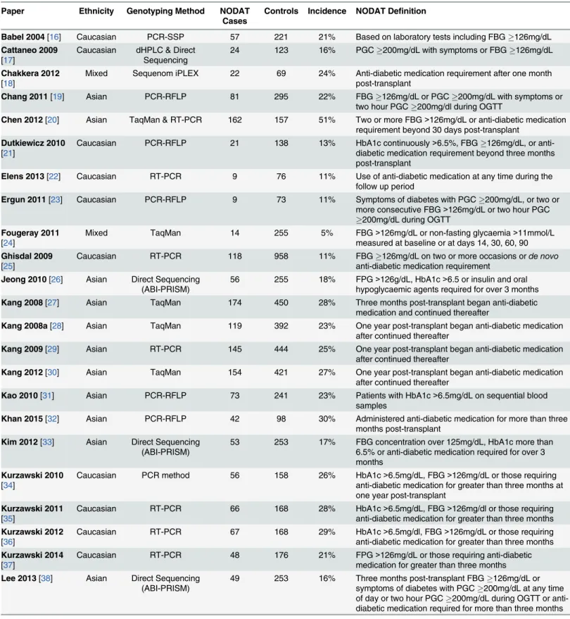
Of the 36 studies deemed eligible for inclusion, 16 studies were carried out in Asian
popula-tions, 16 in Caucasian populations and 4 in populations of mixed ethnicity.
Table 1
outlines
the characteristics of each of the eligible studies.
The literature review revealed 18 genetic variants considered for association with NODAT
that were reported in a minimum of three studies across 12 genes (
Table 2
and
Fig 2
).
Of these analysed variants, three were significantly associated with NODAT based on
meta-analysis;
CDKAL1
rs10946398 p = 0.006 OR = 1.43, 95% CI = 1.11–1.85 (n = 696 individuals),
KCNQ1
rs2237892 p = 0.007 OR = 1.43, 95% CI = 1.10–1.86 (n = 1,270 individuals), and
TCF7L2
rs7903146 p = 0.01 OR = 1.41, 95% CI = 1.07–1.85 (n = 2,967 individuals) (
Fig 3
).
Power calculations (
S1 Table
) suggest this study is adequately powered to identify a risk
var-iant; for example, considering 2360 cases and 607 controls there was
>
80% power to identify a
risk variant with odds ratio 1.5 and minor allele frequency of 5%.
Discussion
Main Findings
significantly associated with NODAT–
TCF7L2
rs7903146,
CDKAL1
rs10946398 and
KCNQ1
rs2237892 at the significance level p
<
0.05.
Many of the studies used in this investigation focused on genes previously associated with
type 2 diabetes (T2D)[
29
,
36
]. NODAT and T2D have a number of important similarities. Both
are characterised by insulin resistance and insulin hypo-secretion and share similar risk factors
including increased age, family history of diabetes and non-white ethnicity [
17
].
TCF7L2
(transcription factor 7-like-2) has been previously linked to T2D, and has been
cited as one of the most important signals associated with T2D [
51
]. The T allele was identified
as a diabetes risk factor in the pre-GWAS era and was later replicated across a number of
groups with different ethnic ancestry [
52
,
53
]. It is not yet completely understood how
TCF7L2
influences risk of T2D but a number of theories have been put forward. It may affect blood
glu-cose homeostasis by altering levels of glucagon-like peptide 1 in the gut, or it may decrease
insulin secretion
via
the pancreatic beta, adipose or liver cells [
54
]. rs7903146 is located in
an intron; a non-protein coding region of the gene [
55
]. There is no obvious mechanism by
which a mutation at this locus could affect NODAT or T2D development, however the variant
rs7903146 may either be in linkage disequilibrium with a causal allele or may itself influence
gene expression through regulatory mechanisms.
CDKAL1
(cyclin dependent kinase 5 regulatory subunit associated protein 1 like 1) has
been associated with impaired insulin secretion and the development of T2D in both
Euro-pean and Han Chinese populations by GWAS [
56
] and the variant rs10946398 has been
found to be significantly associated with T2D by meta-analysis [
57
].
CDKAL1
encodes a
methylthiotransferase which is thought to regulate the CDK5 protein which stimulates
pro-duction of insulin as well as other processes in the pancreatic beta cells [
58
]. In this manner,
by impairing insulin production via over-expression of CDK5,
CDKAL1
may increase risk of
T2D [
57
] and NODAT. The rs10946398 variant is found in exon 5 of the
CDKAL1
gene. An
alternative splicing product of
CDKAL1
(CDKAL1v1) is increased in individuals homozygous
for the minor C allele at this locus. It has therefore been suggested that this particular variant
influences splicing of the gene [
59
].
KCNQ1
is also an established T2D risk factor and has been associated with gestational
dia-betes [
60
–
62
]. Variants of
KCNQ1
cause a variety of disorders including hereditary long QT
Fig 1. Flowchart describing the process of selection of eligible articles and variants for inclusion in the meta-analysis.Table 1. Summary of eligible studies describing the ethnicity, genotyping method and total numbers of cases (NODAT patients) and controls (non-NODAT kidney transplant recipients).
Paper Ethnicity Genotyping Method NODAT
Cases Controls Incidence NODAT Definition
Babel 2004[16] Caucasian PCR-SSP 57 221 21% Based on laboratory tests including FBG126mg/dL
Cattaneo 2009 [17]
Caucasian dHPLC & Direct Sequencing
24 123 16% PGC200mg/dL with symptoms or FBG126mg/dL
Chakkera 2012 [18]
Mixed Sequenom iPLEX 22 69 24% Anti-diabetic medication requirement after one month post-transplant
Chang 2011[19] Asian PCR-RFLP 81 295 22% FBG126mg/dL or PGC200mg/dL with symptoms or
two hour PGC200mg/dl during OGTT
Chen 2012[20] Asian TaqMan & RT-PCR 162 157 51% Two or more FBG>126mg/dL or anti-diabetic medication
requirement beyond 30 days post-transplant Dutkiewicz 2010
[21]
Caucasian PCR-RFLP 21 138 13% HbA1c continuously>6.5%, FBG126mg/dL, or
anti-diabetic medication requirement beyond three months post-transplant
Elens 2013[22] Caucasian RT-PCR 9 76 11% Use of anti-diabetic medication at any time during the
follow up period
Ergun 2011[23] Caucasian PCR-RFLP 9 73 11% Symptoms of diabetes with PGC200mg/dL, or two or
more consecutive FBG>126mg/dL or two hour PGC
200mg/dL during OGTT
Fougeray 2011
[24] Mixed TaqMan 14 255 5% FBG
>126mg/dL or non-fasting glycaemia>11mmol/L measured at baseline or at days 14, 30, 60, 90 Ghisdal 2009
[25] Caucasian RT-PCR 118 958 11% FBG
126mg/dL on two or more occasions orde novo anti-diabetic medication requirement
Jeong 2010[26] Asian Direct Sequencing
(ABI-PRISM) 56 255 18% FPG
>126g/dL, HbA1c>6.5 or insulin and oral hypoglycaemic agents required for over 3 months
Kang 2008[27] Asian TaqMan 174 450 28% Three months post-transplant began anti-diabetic
medication and continued thereafter
Kang 2008a[28] Asian TaqMan 119 392 23% One year post-transplant began anti-diabetic medication
after continued thereafter
Kang 2009[29] Asian RT-PCR 145 444 25% One year post-transplant began anti-diabetic medication
after continued thereafter
Kang 2012[30] Asian TaqMan 154 421 27% One year post-transplant began anti-diabetic medication
after continued thereafter
Kao 2010[31] Asian PCR-RFLP 73 241 23% Patients with HbA1c>6.5mg/dL on sequential blood
samples
Khan 2015[32] Asian PCR-RFLP 42 98 30% Administered anti-diabetic medication for more than three
months post-transplant
Kim 2012[33] Asian Direct Sequencing
(ABI-PRISM) 53 253 17% FBG concentration over 125mg/dL, HbA1c more than6.5% or anti-diabetic medication required for over 3 months
Kurzawski 2010
[34] Caucasian PCR method 56 158 26% HbA1c
>6.5mg/dL, FBG>126mg/dL or those requiring anti-diabetic medication for greater than three months at one year post-transplant
Kurzawski 2011 [35]
Caucasian RT-PCR 66 168 28% HbA1c>6.5mg/dL, FBG>126mg/dl or those requiring anti-diabetic medication for greater than three months Kurzawski 2012
[36]
Caucasian RT-PCR 67 168 29% HbA1c>6.5mg/dl, FBG>126mg/dL or those requiring anti-diabetic medication for greater than three months Kurzawski 2014
[37]
Caucasian RT-PCR 48 176 21% FPG>126mg/dL or those requiring anti-diabetic medication for greater than three months
Lee 2013[38] Asian Direct Sequencing
(ABI-PRISM)
49 253 16% Three months post-transplant FBG126mg/dL or
symptoms of diabetes with PGC200mg/dL at any time
of day or two hour PGC200mg/dL during OGTT or
syndrome (Romano-Ward syndrome)[
63
]. It is expressed in the pancreatic islet cells as well as
the heart and encodes a protein which combines with KCNE proteins to form voltage charged
potassium channels found in the membranes. The KCNQ1 proteins form the structure of the
channel while the KCNE proteins regulate the activity of the channel [
64
]. Pancreatic beta cell
survival rate is thought to be affected by these potassium channels. It is thought that
dysfunc-tion of these potassium channels could alter cell membrane potential and contribute to
devel-opment of T2D or NODAT. A specific KCNQ1 blocker 293B has been shown to increase
insulin production [
65
]. The variant rs2237892 C risk allele has been shown to be associated
with fasting plasma glucose concentration, suggesting that C homozygous individuals have
impaired baseline insulin secretion. The gene is also under the control of tissue specific
imprinting [
66
].
These genetic variants are all established T2D risk factors and several variants have been
implicated in potential mechanisms contributing to diabetes. Therefore, it is not surprising
that these variants are linked to NODAT, another form of diabetes, since the mechanisms
con-trolling insulin production and maintenance of stable glucose levels will both be similar in T2D
and NODAT. The meta-analyses conducted on the other variants identified in the literature
did not reach statistical significance. This may have been for several reasons, including the
Table 1. (Continued)Paper Ethnicity Genotyping Method NODAT
Cases Controls Incidence NODAT Definition
McCaughan
2014[1] Caucasian & Sequenom iPLEXIllumina 660K Array 26 230 10% New requirement for anti-diabetic medication aftertransplant Nicoletto 2013
[39] Caucasian Sequenom iPLEXRT-PCR 83 187 31% Second recorded FBG of 126mg/dL or more Özdemir 2011
[40] Caucasian PCR Method 23 27 46% Symptoms of diabetes with PGC
200mg/dL, or record of
two or more consecutive FBG>126mg/dL or two hour PGC200mg/dL during OGTT and anti-diabetic
medication requirement Szuszkiewicz
2011[41] Mixed PCR-RFLP 36 79 31% Anti-diabetic medication requirement, FBG
126mg/dL
and two hour PGC200mg/dL when available from
patient history
Tavira 2011[42] Caucasian PCR-RFLP 115 205 36% FBG>126g/dL after three consecutive measurements
Tavira 2012[43] Caucasian PCR-RFLP 115 205 36% FBG>126g/dL after three consecutive measurements
Tsai 2011[44] Asian PCR-RFLP 85 198 30% FBG126mg/dL or symptoms of diabetes and PGC
200mg/dL at any time of day or two hour PGC200mg/
dL during OGTT
Vattam 2013[45] Asian PCR-RFLP 42 98 30% PGC200mg/dL with diabetes symptoms or FBG
126mg/dL or two hour PGC200mg/dL during OGTT
Wang 2011[46] Mixed Direct Sequencing 51 72 41% FBG126mg/dL three months post-transplant
Weng 2012[47] Asian PCR-RFLP 27 251 10% PGC200mg/dL with diabetes symptoms or FBG
126mg/dL or two hour PGC200mg/dL during OGTT
Yang 2011[48] Caucasian RT-PCR 133 170 44% Two or more occasions of FPG level>126mg/dL one
month or more after transplant
Yao 2013[49] Asian PCR-RFLP 16 89 15% PGC200mg/dL with diabetes symptoms or FBG
126mg/dL or two hour PGC200mg/dL during OGTT
Yu 2011[50] Asian PCR-RFLP 97 301 24% FBG126mg/dL on at least two occasions or to require
anti-diabetic medication
PCR-SSP, Polymerase chain reaction, single specific primer; dHPLC, Denaturing high performance liquid chromatography; RFLP, PCR Restriction Fragment Length Polymorphism; RT-PCR, Real Time PCR; FBG, Fasting Blood Glucose; PGC, Plasma Glucose Concentration; OGTT, Oral Glucose Tolerance Test; HbA1c, Haemoglobin A1c
small number of studies, small numbers of study participants, or varying phenotypic
defini-tions. Of note, our meta-analysis incorporates data from both candidate gene and
genome-wide association studies.
A number of variants which were associated with NODAT in previous studies were not
found to be associated with NODAT following meta-analysis. Notable variants which did not
reach significance after meta-analysis include
KCNJ11
rs5219 and
ADIPOQ
rs1501299.
KCNJ11
rs5219 is an established T2D risk factor in a gene encoding a voltage gated potassium
channel.
ADIPOQ
rs1501299 has been previously associated with NODAT as well as breast
cancer, prostate cancer and T2D complications such as heart disease. Adiponectin encoded by
ADIPOQ
is involved in lipid metabolism and insulin sensitivity and making this an attractive
candidate for association with NODAT. Neither of these particular variants reached the
p
<
0.05 significance threshold following meta-analysis which could mean they are not
associ-ated with NODAT or that the association is only present in certain populations.
Limitations of the Review
This study does have a number of limitations. The definition of NODAT differs from centre to
centre as highlighted by the varying prevalence of NODAT reported in
Table 1
which ranges
from 5–51%. A potential explanation for this large variation in reported prevalence is the
dif-ferences in how NODAT is diagnosed i.e. heterogeneity of the clinical phenotype. Some
authors employed diagnostic criteria for diabetes as defined by the World Health Organisation
and American Diabetic Association, although the final interpretation of these standards did
vary in published studies of NODAT. Others authors used pragmatic clinical criteria for
NODAT diagnosis defining the affected patients as requiring the
de novo
prolonged use of
insulin and/or oral hypoglycaemic medication following transplant. A more rigorously defined
Table 2. Variants replicated in a minimum of three publications with associated odds ratios, 95% confidence intervals and p values following meta-analysis.Gene Variant Odds Ratio 95% CI p Value Minor Allele Minor Allele Frequency (Control Group)
CDKAL1 rs10946398 1.43 1.11–1.85 0.006 C 42.65%
KCNQ1 rs2237892 1.43 1.10–1.86 0.007 T 43.39%
TCF7L2 rs7903146 1.41 1.07–1.85 0.01 T 18.41%
KCNJ11 rs5219 1.28 0.92–1.76 0.14 T 35.50%
PPARG rs4253728 1.55 0.78–3.11 0.21 A 23.71%
TNFA rs1800629 0.81 0.56–1.17 0.25 A 14.07%
HHEX rs1111875 1.14 0.89–1.44 0.30 C 49.92%
HHEX rs5015480 1.24 0.77–1.97 0.38 C 33.70%
IGF2BP2 rs1470579 1.15 0.84–1.59 0.39 C 33.26%
KCNJ11 rs5215 1.09 0.88–1.34 0.42 C 36.07%
CDKN2A/B rs10811661 1.1 0.79–1.54 0.57 C 25.87%
IGF2BP2 rs4402960 0.96 0.80–1.14 0.61 T 31.85%
SLC30A8 rs13266634 0.87 0.48–1.55 0.63 T 35.99%
PPARG rs1801282 1.05 0.80–1.37 0.73 G 10.71%
TCF7L2 rs12255372 1.06 0.77–1.47 0.73 T 24.11%
ADIPOQ rs1501299 1.06 0.71–1.56 0.79 T 8.33%
CDKAL1 rs7754840 1.04 0.79–1.37 0.80 C 31.50%
FTO rs8050136 1.01 0.82–1.24 0.95 A 32.28%
Variants highlighted in bold are those which reached significance at the 5% level.
Fig 2. Genetic variants explored for association with NODAT in at least three publications.
NODAT phenotype may facilitate more reproducible results between studies. There were a
small number of studies available for many of the variants; larger, carefully phenotyped studies
would provide better power to identify alleles robustly associated with NODAT. There was a
varying degree of heterogeneity noted between studies, some of which was likely due to
differ-ent ethnicities considered. In addition, the variations in prescribed immunosuppressive
regi-mens, and their differential effects on NODAT incidence, were not accounted for in many of
the studies. It is of note that the Belfast derived data was from the single transplant centre for
Northern Ireland.
TCF7L2
rs7903146 variant was only nominally associated with NODAT
(p = 0.01), but this association was replicated with the same direction of effect across five
inde-pendent collections. Possible interactions among the genetic variants identified have not been
investigated and this is a further limitation of the study.
Conclusions
This is a thorough overview of all reported genetic factors influencing the development of
NODAT in the current literature. Analysis revealed a significant association between NODAT
and three established T2D risk factor variants. Functional studies will be required to further
investigate these variants and associated pathways to gain a complete perspective on their
effects. In order to obtain more consistency between studies and identify risk alleles with
smaller effect sizes, larger participant numbers through multi-centre collaboration and
har-monised phenotypic definition of NODAT is required. An adequately powered, dense
genome-wide association study will provide more information using a carefully defined
NODAT phenotype.
Hypothesis free approaches such as the GWAS carried out by McCaughan and colleagues
for NODAT are advantageous to identify new biological targets and therapeutic pathways and
should also be carried out in other populations and ethnicities to better understand the genetic
architecture underlying this disease [
1
].
Supporting Information
S1 Table. Power Calculations for Genetic Variants.
This table describes the power which this
study had to identify significant genetic variants. The power was based on 607 cases, 2360
Fig 3. Forest Plots illustrating odds ratios and 95% confidence intervals for the three variants significantly associated with NODAT in random effects meta-analysis; black diamonds represent overall odds ratios for each of the variants. ACDKAL1rs10946398 BKCNQ1rs2237892 CTCF7L2 rs7903146.controls and dependent on the Minor Allele Frequency, significance sought, and effect size
(reported for each variant in
Table 2
).
(XLSX)
S2 Table. PRISMA Checklist.
Describes how this manuscript conforms to the PRISMA 2009
guidelines.
(DOCX)
Acknowledgments
KAB is supported by a Queen’s University Belfast, School of Medicine, Dentistry and
Biomedi-cal Science International PhD Fellowship as well as receiving support from the Northern
Ire-land Kidney Research Fund. The NIKRF had no role in the study design.
Author Contributions
Conceived and designed the experiments: KAB APM AJM. Performed the experiments: KAB.
Analyzed the data: KAB APM AJM. Contributed reagents/materials/analysis tools: KAB APM
AJM. Wrote the paper: KAB APM AJM.
References
1. McCaughan J, McKnight AJ, Maxwell AP. Genetics of new-onset diabetes after transplantation. J Am Soc Nephrol. 2014; 25(5):1037–49. doi:10.1681/ASN.2013040383PMID:24309190
2. Mora PF. New-onset diabetes after renal transplantation. J Investig Med. 2010; 58(6):755–63. PMID: 20517164
3. Kesiraju S, Paritala P, Rao Ch UM, Sahariah S. New onset of diabetes after transplantation—An over-view of epidemiology, mechanism of development and diagnosis. Transpl Immunol. 2014; 30(1):52–8. PMID:24184293
4. Hecking M, Werzowa J, Haidinger M, Hörl WH, Pascual J, Budde K, et al. Novel views on new-onset diabetes after transplantation: Development, prevention and treatment. Nephrol Dial Transplant. 2013; 28(3):550–66. doi:10.1093/ndt/gfs583PMID:23328712
5. Davidson J., Wilkinson A. New-onset diabetes after transplantation 2003 international consensus guidelines: An endocrinologist’s view. Diabetes Care. 2004; 27(3):805–12. PMID:14988309 6. Shabir S, Jham S, Harper L, Ball S, Borrows R, Sharif A. Validity of glycated haemoglobin to diagnose
new onset diabetes after transplantation. Transpl Int. 2013; 26(3):315–21 doi:10.1111/tri.12042PMID:
23279163
7. Sharif A, Hecking M, De Vries AP, Porrini E, Hornum M, Rasoul-Rockenschaub S, et al. Proceedings from an international consensus meeting on post transplantation diabetes mellitus: Recommendations and future directions. Am J Transplant. 2015; 14(9):1992–2000.
8. Luan FL, Steffick DE, Ojo AO. New-onset diabetes mellitus in kidney transplant recipients discharged on steroid-free immunosuppression. Transplantation. 2011: 91(3):334–41
9. Gaynor JJ, Ciancio G, Guerra G, Sageshima J, Hanson L, Roth D, et al. Multivariable risk of developing new onset diabetes after transplant-results from a single-center study of 481 adult, primary kidney transplant recipients. Clin Transplant. 2015; 29(4):301–10. doi:10.1111/ctr.12510PMID:25581205
10. Palepu S. New-onset diabetes mellitus after kidney transplantation: Current status and future direc-tions. World J Diabetes. 2015; 6(3):445. doi:10.4239/wjd.v6.i3.445PMID:25897355
11. Menzaghi C, Ercolino T, Salvemini L, Coco A, Kim SH, Fini G, et al. Multigenic control of serum adipo-nectin levels: Evidence for a role of the APM1 gene and a locus on 14q13. Physiol Genomics. 2004; 19 (2):170–4. PMID:15252189
12. Wiecek A. Genotypes of renin-angiotensin system and plasma adiponectin concentration in kidney transplant patients. Ann Transplant. 2013; 18:593–603. doi:10.12659/AOT.884022PMID:24185422
14. Hara K, Fujita H, Johnson T, Yamauchi T, Yasuda K, Horikoshi M, et al. Genome-wide association study identifies three novel loci for type 2 diabetes. Hum Mol Genet. 2014; 23(1):239–46. doi:10.1093/ hmg/ddt399PMID:23945395
15. The Cochrane Collaboration. Review Manager (RevMan) [Computer program]. Version 5.3. 2014. 16. Babel N, Cherepnev G, Kowalenko A, Horstrup J, Volk H-D, Reinke P. Nonimmunologic complications
and gene polymorphisms of immunoregulatory cytokines in long-term renal transplants. Kidney Int. 2004; 66:428–32. PMID:15200452
17. Cattaneo D, Ruggenenti P, Baldelli S, Motterlini N, Gotti E, Sandrini S, et al. ABCB1 genotypes predict cyclosporine-related adverse events and kidney allograft outcome. J Am Soc Nephrol. 2009; 20 (6):1404–15. doi:10.1681/ASN.2008080819PMID:19470683
18. Chakkera HA, Hanson RL, Raza SM, Distefano JK, Millis MP, Heilman RL, et al. Pilot Study: Associa-tion of tradiAssocia-tional and genetic risk factors and new-onset diabetes mellitus following kidney transplanta-tion. Transplant Proc. 2012; 41(10):4172–7.
19. Chang HR, Yang SF, Tsai JP, Hsieh MC, Wu SW, Tsai HC, et al. Plasminogen activator inhibitor-1 5G/ 5G genotype is a protecting factor preventing post transplant diabetes mellitus. Clin Chim Acta. 2011; 412(3–4):322–6. doi:10.1016/j.cca.2010.10.029PMID:21070757
20. Chen Y, Sampaio M, Yang J, Min D, Hutchinson I. Genetic polymorphisms of the transcription factor NFATc4 and development of new-onset diabetes after transplantation in Hispanic kidney transplant recipients. Transplantation. 2012; 93(3):325–30. PMID:22234350
21. Dutkiewicz G, Domanski L, Pawlik A, Binczak-Kuleta A, Safranow K, Ciechanowicz A, et al. Polymor-phisms of superoxide dismutase, glutathione peroxidase and catalase genes in patients with post-transplant diabetes mellitus. Arch Med Res. 2010; 41(5):350–5. doi:10.1016/j.arcmed.2010.06.006
PMID:20851292
22. Elens L, Sombogaard F, Hesselink D, van Schaik RHN, van Gelder T. Single-nucleotide polymor-phisms in P450 oxidoreductase and peroxisome proliferator-activated receptor-αare associated with the development of new-onset diabetes after transplantation in kidney transplant recipients treated with tacrolimus. Pharmacogenet Genomics. 2013; 23(12):649–57. PMID:24113216
23. Ergün I, Keven K, Sengül S, Karabulut HG, Kurultak I, Soypacaci Z, et al. Endothelial nitric oxide synthase gene intron 4 polymorphism predicts new onset diabetes mellitus after transplantation in kid-ney allograft recipients treated with cyclosporin A. Int Urol Nephrol. 2011; 43(2):543–8. doi:10.1007/ s11255-010-9786-8PMID:20559724
24. Fougeray S, Loriot M, Nicaud V, Legendre C, Thervet E, Pallet N. Increased body mass index after kid-ney transplantation in activating transcription factor 6 single polymorphism gene carriers. Transplant Proc. 2011; 43(9):3418–22. doi:10.1016/j.transproceed.2011.09.022PMID:22099811
25. Ghisdal L, Baron C, Le Meur Y, Lionet A, Halimi JM, Rerolle JP, et al. TCF7L2 polymorphism associ-ates with new-onset diabetes after transplantation. J Am Soc Nephrol. 2009; 20(11):2459–67. doi:10. 1681/ASN.2008121314PMID:19713311
26. Jeong KH, Moon JY, Chung JH, Kim YH, Lee TW. Significant Associations between CCL5 polymor-phisms and post transplantation diabetes mellitus in Korean renal allograft recipients. Am J Nephrol. 2010; 32:356–61. doi:10.1159/000319704PMID:20805685
27. Kang ES, Nam CM, Kim MS, Ahn CW, Kim YS, Cha BS, et al. A variant of the transcription factor 7-Like 2 (TCF7L2) gene and the risk of post transplantation diabetes mellitus in renal allograft recipients. Diabetes Care. 2008; 31(1):63–8. PMID:17934151
28. Kang ES, Kim SM, Kim YS, Kim CH, Han SJ, Chun SW, et al. A polymorphism in the zinc transporter gene SLC30A8 confers resistance against post transplantation diabetes mellitus in renal allograft recip-ients. Diabetes. 2008; 57(4):1043–7. PMID:18162509
29. Kang ES, Kim SM, Kim CH, Chung MN, Han SJ, Hur KY, et al. Association of common type 2 diabetes risk gene variants and post transplantation diabetes mellitus in renal allograft recipients in Korea. Transplantation. 2009; 88(5):693–8. PMID:19741467
30. Kang ES, Magkos F, Kim BS, Zhai R, Su L, Kim YS, et al. Variants of the adiponectin and adiponectin receptor-1 genes and post transplantation diabetes mellitus in renal allograft recipients. J Clin Endocri-nol Metab. 2012; 97(1):E129–35. doi:10.1210/jc.2011-1796PMID:22049178
31. Kao CC, Lian JD, Chou MC, Chang HR, Yang SF. Tumor necrosis factor alpha promoter polymorphism in post transplantation diabetes mellitus of renal transplant recipients. Transplant Proc. 2010; 42 (9):3559–61. doi:10.1016/j.transproceed.2010.06.032PMID:21094815
33. Kim YG, Ihm CG, Lee TW, Lee SH, Jeong KH, Moon JY, et al. Association of genetic polymorphisms of interleukins with new-onset diabetes after transplantation in renal transplantation. Transplantation. 2012; 93(9):900–7. PMID:22377791
34. Kurzawski M, Dziewanowski K, Kedzierska K, Gornik W, Banas A, Drozdzik M. Association of calpain-10 gene polymorphism and post transplant diabetes mellitus in kidney transplant patients medicated with tacrolimus. Pharmacogenomics J. 2010; 10(2):120–5. doi:10.1038/tpj.2009.44PMID:19752882
35. Kurzawski M, Dziewanowski K, Kêdzierska K, Wajda A, Lapczuk J, Droÿdzik M. Association of tran-scription factor 7-like 2 (TCF7L2) gene polymorphism with post transplant diabetes mellitus in kidney transplant patients medicated with tacrolimus. Pharmacol Reports. 2011; 63:826–33.
36. Kurzawski M, Dziewanowski K,Łapczuk J, Wajda A, Droździk M. Analysis of common type 2 diabetes mellitus genetic risk factors in new-onset diabetes after transplantation in kidney transplant patients medicated with tacrolimus. Eur J Clin Pharmacol. 2012; 68(12):1587–94. doi: 10.1007/s00228-012-1292-8PMID:22569928
37. Kurzawski M, Malinowski D, Dziewanowski K, Droździk M. Impact of PPARA and POR polymorphisms on tacrolimus pharmacokinetics and new-onset diabetes in kidney transplant recipients. Pharmaco-genet Genomics. 2014; 24:397–400. PMID:24921414
38. Lee SR, Moon JY, Lee SH, Ihm CG, Lee TW, Kim SK, et al. Angiotensinogen polymorphisms and post-transplantation diabetes mellitus in Korean renal transplant subjects. Kidney Blood Press Res. 2013; 37(2–3):95–102. doi:10.1159/000343404PMID:23594830
39. Nicoletto BB, Souza GC, Fonseca NKO, Centenaro A, Manfro RC, Canani LHS, et al. Association between 276G/T adiponectin gene polymorphism and new-onset diabetes after kidney transplantation. Transplantation. 2013; 96(12):1059–64. PMID:23985723
40. Özdemir BH, Özdemir FN, Ataç FB, Özdemir AA, Haberal M. Angiotensinogen t235 and angiotensin-converting enzyme insertion/deletion polymorphisms associated with the development of post trans-plantation diabetes mellitus in renal allograft recipients. Transplant Proc. 2011; 43(2):572–4. doi:10. 1016/j.transproceed.2011.01.046PMID:21440764
41. Szuszkiewicz M, Bell J, Vazquez M, Adams-Huet B, Grundy SM, Chandalia M, et al. ENPP1/PC-1 K121Q and other predictors of post transplant diabetes. Metab Syndr Relat Disord. 2011; 9(1):25–9. doi:10.1089/met.2010.0041PMID:20958205
42. Tavira B, Diaz-Corte C, Ortega F, Arias M, Torres A, Diaz J, et al. KCNQ1 gene variants and risk of new-onset diabetes in tacrolimus-treated renal-transplanted patients. Clin Transplant. 2011; 25:E284– 91. doi:10.1111/j.1399-0012.2011.01417.xPMID:21355884
43. Tavira B, Coto E, Torres A, Díaz-Corte C, Díaz-Molina B, Ortega F, et al. Association between a com-mon KCNJ11 polymorphism (rs5219) and new-onset post transplant diabetes in patients treated with Tacrolimus. Mol Genet Metab. 2012; 105(3):525–7. doi:10.1016/j.ymgme.2011.12.020PMID:
22264780
44. Tsai JP, Yang SF, Wu SW, Hung TW, Tsai HC, Lian JD, et al. Glutathione S-transferase gene polymor-phisms are not major risks for susceptibility to post transplantation diabetes mellitus in Taiwan renal transplant recipients. J Clin Lab Anal. 2011; 25(6):432–5. doi:10.1002/jcla.20498PMID:22086798 45. Vattam KK, Khan IA, Movva S, Mukkavali KK, Poornima S, Rao P, et al. IGF2 ApaI A/G polymorphism
evaluated in ESRD individuals as a biomarker to identify patients with new onset diabetes mellitus after renal transplant in Asian Indians. Open J Nephrol. 2013; 3, 104
46. Wang P, Hudspeth E. Increased body mass index but not common vitamin D receptor, peroxisome pro-liferator-activated receptorγ, or cytokine polymorphisms confers predisposition to post transplant dia-betes. Arch Pathol Lab Med. 2011; 135(12):1581–4. doi:10.5858/arpa.2011-0160-OAPMID: 22129188
47. Weng SC, Shu KH, Tarng DC, Wu MJ, Chen CH, Yu TM, et al. Gene polymorphisms are associated with post transplantation diabetes mellitus among Taiwanese renal transplant recipients. Transplant Proc. 2012; 44(3):667–71. doi:10.1016/j.transproceed.2011.11.011PMID:22483464
48. Yang J, Hutchinson II, Shah T, Min DI. Genetic and clinical risk factors of new-onset diabetes after transplantation in Hispanic kidney transplant recipients. Transplantation. 2011; 91:1114–9. PMID: 21544032
49. Yao B, Chen X, Shen FX, Xu W, Dong TT, Chen LZ, et al. The incidence of post transplantation diabe-tes mellitus during follow-up in kidney transplant recipients and relationship to Fok1 vitamin D receptor polymorphism. Transplant Proc. 2013; 45(1):194–6. doi:10.1016/j.transproceed.2012.08.019PMID:
23375298
51. Hattersley AT. Prime suspect : the TCF7L2 gene and type 2 diabetes risk. Nephrology. 2007; 117(8):6– 8.
52. Grant SF, Thorleifsson G, Reynisdottir I, Benediktsson R, Manolescu A, Sainz J, et al. Variant of tran-scription factor 7-like 2 (TCF7L2) gene confers risk of type 2 diabetes. Nat Genet. 2006; 38(3):320–3. PMID:16415884
53. Tong Y, Lin Y, Zhang Y, Yang J, Zhang Y, Liu H, et al. Association between TCF7L2 gene polymor-phisms and susceptibility to type 2 diabetes mellitus: a large Human Genome Epidemiology (HuGE) review and meta-analysis. BMC Med Genet. 2009; 10:15. doi:10.1186/1471-2350-10-15PMID:
19228405
54. Dormans JP, Gunawardena AT, Hakonarson H, Hecht JT. The type 2 diabetes associated rs7903146 T allele within TCF7L2 is significantly under-represented in hereditary multiple exostoses: insights into pathogenesis. Bone. 2015;( 72):123–7. doi:10.1016/j.bone.2014.11.024PMID:25498973
55. Nicod N, Pradas-Juni M, Gomis R. Role of the single nucleotide polymorphism rs7903146 of TCF7L2 in inducing nonsense-mediated decay. Springerplus. 2014: Jan 22; 3:41 doi:10.1186/2193-1801-3-41
PMID:24498581
56. Steinthorsdottir V, Thorleifsson G, Reynisdottir I, Benediktsson R, Jonsdottir T, Walters GB, et al. A var-iant in CDKAL1 influences insulin response and risk of type 2 diabetes. Nat Genet. 2007; 39(6):770–5. PMID:17460697
57. Dehwah MS, Wang M, Huang QY. CDKAL1 and type 2 diabetes: A global meta-analysis. Genet Mol Res. 2010; 9(2):1109–20. doi:10.4238/vol9-2gmr802PMID:20568056
58. Brambillasca S, Altkrueger A, Colombo SF, Friederich A, Eickelmann P, Mark M, et al. CDK5 regulatory subunit-associated protein 1-like 1 (CDKAL1) is a tail-anchored protein in the endoplasmic reticulum (ER) of insulinoma cells. J Biol Chem. 2012; 287(50):41808–19 doi:10.1074/jbc.M112.376558PMID: 23048041
59. Zhou B, Wei FY, Kanai N, Fujimura A, Kaitsuka T, Tomizawa K. Identification of a splicing variant that regulates type 2 diabetes risk factor CDKAL1 level by a coding-independent mechanism in human. Hum Mol Genet. 2014; 23(17):4639–50 doi:10.1093/hmg/ddu184PMID:24760768
60. Bazzi MD, Nasr FA, Alanazi MS, Alamri A, Turjoman AA. Association between FTO, MCR, SLC30A8, and KCNQ1 gene variants and type 2 diabetes in Saudi population. Genet Mol Res. 2014; 1(4):10194– 203.
61. Ao D, Wang H, Wang L, Song J, Yang H, Wang Y. The rs2237892 Polymorphism in KCNQ1 influences gestational diabetes mellitus and glucose levels: A case-control study and meta-analysis. PLoS One. 2015; 10(6):e0128901. doi:10.1371/journal.pone.0128901PMID:26039078
62. Huerta-Chagoya A, Vázquez-Cárdenas P, Moreno-Macías H. Genetic determinants for gestational dia-betes mellitus and related metabolic traits in Mexican women. PLoS One. 2015; 10(5):1–17.
63. Tester DJ, Ackerman MJ. Genetics of long Qt syndrome. Methodist Debakey Cardiovasc J. 2014; 10 (1):29–33. PMID:24932360
64. Abbot GW. KCNE1 and KCNE3: The yin and yang of voltage-gated K+ channel regulation. Gene;2015: S0378-119(15)01168-3
65. Lui L, Wang F, Lu H, Ren X, Zou J. Chromanol 293B, an inhibitor of KCNQ1 channels, enhances glu-cose-stimulated insulin secretion and increases glucagon-like peptide-1 level in mice. Islets. 2014; 6(4) e962386 doi:10.4161/19382014.2014.962386PMID:25437377
66. Schultz B, Gallicio G, Cesaroni M, Lupey L, Engel N. Enhancers compete with a long non-coding RNA for regulation of the Kcnq1 domain. Nucl. Acids Res. 2015; 43 (2): 745–759 doi:10.1093/nar/gku1324