ABSTRACT
Physicochemical properties of endodontic sealers
of different bases
Gabriela Alexandra MARÍN-BAUZA1, Yara Teresinha Correa SILVA-SOUSA2 , Suely Aparecida da CUNHA1, Fuad Jacob
Abi RACHED-JUNIOR3, Idomeo BONETTI-FILHO4, Manoel Damião SOUSA-NETO5, Carlos Eduardo Saraiva MIRANDA6
1- DDS, Postgraduate student, School of Dentistry, University of Ribeirão Preto, Ribeirão Preto, SP, Brazil. 2- DDS, MSc, PhD, Titular Professor, School of Dentistry, University of Ribeirão Preto, Ribeirão Preto, SP, Brazil. 3- DDS, MSc, PhD, Assistant Professor, School of Dentistry, University of Ribeirão Preto, Ribeirão Preto, SP, Brazil.
4- DDS, MSc, PhD, Adjunct Professor, Araraquara School of Dentistry, Univ. Estadual Paulista - UNESP, Araraquara, SP, Brazil. 5- DDS, MSc, PhD, Associate Professor, Ribeirão Preto School of Dentistry, University of São Paulo, Ribeirão Preto, SP, Brazil. 6- MSc, PhD, Adjucnt Professor, University of Ribeirão Preto, School of Pharmaceutics Science, Ribeirão Preto, SP, Brazil.
Corresponding address: Prof. Yara Teresinha Correa Silva-Sousa - Rua Célia de Oliveira Meirelles, 350 - 14024-070 - Ribeirão Preto - São Paulo - Brasil - Phone/fax: +55 16 3603 6763 - e-mail: [email protected]
O
!"#"$and dimensional change following setting (DC) of different sealers (AH Plus®%"&"
Apexit Plus®, Sealapex®, Endométhasone®'&""®) according to American National
*#+- !" - -.*+-!- & /01 2" and methods: Five samples of each material were used for each test. For ST, cast rings &"""4""1"" on a glass plate. After 180 s, another plate with 20 g and a load of 100 g were applied on the material, and the diameters of the discs formed were measured. In RD, circular molds &""""9#!;1$ #""&"""""" and another glass plate was positioned on the set, pressed and stored at 37°C. Samples were weighed, placed in water, dried and reweighed. The water used for SB was analyzed 1!>#""&""" covered by glass plates and stored at 37°C. Samples were measured and stored in water for 30 days. After this period, they were dryed and measured again. Results: Regarding ST, AH Plus®, Apexit®'&"® sealers are in accordance with ANSI/ADA standards.
'@G#;#J%"&"K"" and Sealapex® did not set. Considering RD, SB and DC, all sealers were in accordance
-.*+-!-1"&#;L+ and
QU+ ions was released from Apexit Plus®'&""®, respectively. Conclusion: Except
for DC, all other physicochemical properties of the tested sealers conformed to ANSI/ADA requirements.
Key words:'1"#1"&"""1
INTRODUCTION
Since the first studies on the prognosis of endodontic therapy, the quality of root canal &"""; success22. The presence of voids in the root canal
&"" ;" " "; perpetuation of periapical lesions20.
Among the several factors that can interfere
" &"" " stand out1,3,14,16,17. These materials should present
biocompatibility15,21 and suitable antimicrobial28
and physiochemical properties5,19. In this context,
" " "& to the chemical composition in zinc oxide-eugenol-based, calcium hydroxide-containing, glass-ionomer-based, epoxy-resin-based and methacrylate-resin-based sealers19,24.
The zinc oxide-eugenol-based sealers were introduced in endodontics by Grossman, in 1936, to be used in conjunction with the gutta-percha or silver cones in root canal fillings. Endofill (Dentsply-Mallefer, Dentsply Indústria e Comércio Ltda., Petrópolis, RJ, Brazil) and Endométhasone®
(Spécialities Septodont, Saint-Maurdes-Fossés, Cedex, France) are commonly used zinc oxide-eugenol-based sealers, both available in a powder-liquid presentation.
Sealers that contain calcium hydroxide were idealized with the purpose of improving the biological properties and ensuring a good seal of root canal system. Among these products, Apexit Plus® (Ivoclar, Vivadent, Fürstentum, Schaan,
Liechtenstein) and Sealapex® (Sybron-Endo,
Glendora, CA, USA) are available in the market in a paste-paste presentation26,27.
& { " Schröeder, in 1954, and contained epoxy resin and bisphenol. Since them, studies have contributed to the improvement of sealers’ quality, leading to the development of an epoxy resin-based sealer with good physicochemical properties, AH Plus®
(Dentsply, DeTrey GmbH, Konstanz, Germany)14.
Much research has been done to develop products derived from biomaterials, in order to increase the compatibility to biological tissues11-13,18.
*}~GK""""%"&" was developed at the Araraquara School of Dentistry (SP, Brazil). This sealer is based on a polyurethane vegetable resin, Ricinus communis extract, and has
18;&"
capacity13,18 and some antimicrobial activity11,12.
In 1983, the American National Standards Institute/American Dental Association (ANSI/ ADA) released a series of norms and tests (named & /0 "# " properties of endodontic sealers, aiming at 9"& #" 1 & revised in 2000 and it includes the following tests: film thickness, setting time, flow, radiopacity, solubility and dimensional change following setting2.
Due to the diversity of composition of the available sealers and considering the ANSI/ADA standards, the objective of this study was to evaluate in vitro the physiochemical properties
of sealers of different bases: Endofill® and
Endométhasone® (zinc oxide and eugenol), Apexit
Plus® and Sealapex® "# K %"&"
(Ricinus communis polymer) and AH Plus® (epoxy
resin), in order to contribute to the selection of the
appropriate material for clinical practice.
MATERIAL AND METHODS
"#" dimensional change after setting for AH Plus®,
%"&" -K %"#®, Sealapex®, Endométhasone®
'&""® sealers were measured according
to ANSI/ADA standards for dental root canal sealing materials by one examiner blinded to the &;"1!";" are displayed in Figure 1.
All tested materials were prepared in accordance to the manufacturers’ instructions. AH Plus®, III and
IV are presented as two pastes and, for each tested sample, 15 mm of the sealer (1:1) was dispensed onto a mixing pad and mixed until obtaining a # K#1 * # ** %"&" 1} mL of the liquid was mixed to 0.44 mL of the paste, measure through a graduate syringe and the mixture was mixed on a glass plate.
For Endométhasone® '&""®, 3 drops
of the liquid and a portion of the powder were released in glass plate and mixed in a gradual way until getting the ideal consistence, characterized by a cement thread of approximately 2 cm, without falling during 15 s.
For the physicochemical tests, the arithmetic ;&";" and considered as the result of the test.
Setting time
Five plaster of cast rings having an internal diameter of 10 mm and a thickness of 2 mm were prepared for each group. The external borders of #"&KK"" 0/KU/K} 1 #" &"" material and transferred to a chamber with 95% relative humidity and a temperature of 37°C. After 150±10 s from the onset of mixing, a Gilmore-type ";}1/ of 2.0±0.1 mm in diameter was carefully lowered vertically onto the horizontal surface of the testing sample. The needle tip was cleaned and the probing was repeated until indentations ceased to be visible. The time used from the start of mixing to this point was recorded. If the results differed by more than ±5%, the test was repeated.
Flow test
measured using a digital calliper with a resolution of 0.01 mm (Mitutoyo MTI Corporation, Tokyo, Japan). If both measurements were consistent to within 1 mm, the results were recorded. If the major and minor diameter discs were not uniformly circular or did not match within 1 mm, the test was repeated.
Radiopacity test
Five acrylic plates (2.2 cm x 4.5 cm x 1 mm), containing four wells measuring 1 mm in depth and 5 mm in diameter, were prepared and placed over a glass plate covered by cellophane sheet. In group I, the freshly mixed sealer was introduced into the wells using a syringe, to avoid the formation of bubbles. In groups II, III the respective material " # &"" ""1 -glass plate covered with cellophane was placed on top until complete setting and any excess sealer removed. Each plate was kept in an incubator (37°C, 95% relative humidity) for a period corresponding to three times the setting time. Each well was &"";";""# according to the setting time of the material, from the longest to the shortest, so that the samples would be ready for radiographic evaluation after &";"""1
Each one of the acrylic plates containing the &"" " of the radiographic exposure, alongside to another
acrylic plate (1.3 cm x 4.5 cm x 1 mm), containing an aluminum stepwedge, made of 1100 alloy, with the thickness varying from 1 to 10 mm, in uniform steps of 1 mm each. This set of acrylic plates was placed in front of this phosphor plate and a digital radiograph was taken (Digora system, Soredex Orion Corporation, Helsinki, Finland). Care was taken to place the samples next to the aluminum step wedge and in the middle of the phosphor plate. Radiographic images were obtained using the Spectro 70x X-ray machine (Dabi Atlante, Ribeirão Preto, SP, Brazil), at 70 kVp and 8 mA. The object-to-focus distance was 30 cm and the exposure time was 0.2 s. Exposed imaging plates of the test samples were immediately scanned after exposure (Digora™ Scanner) and analyzed using Digora™ for Windows 5.1 software.
Dimensional change following setting
#";# of 3.58-mm high cylindrical test bodies measuring 3 mm in diameter, were placed on a glass plate &""1#" &"" " K ; ;" K sealers and a microscope slide, also wrapped in cellophane, was pressed onto the upper surface of #"1"#&" joined with a C-shaped clamp and transferred to an incubator (37°C, 95% relative humidity) left to
Sealers Composition Manufacturer
AH Plus Paste A: Bisphenol-A epoxy resin, Bisphenol-F epoxy resin, calcium tungstate, zirconium oxide, silica and iron oxide pigments. Paste B: Dibenzydiamine, aminoadamante, trycyclodecane- diamine, calcium tungstate, zirconium oxide, silica and silicone oil.
Dentsply, DeTrey, Konstanz, Germany
Paste based on trifunctional polyol, synthetic from
Ricinus communis (castor oil) combined to zinc
oxide and calcium carbonate. Liquid: ampoule with synthesized pre-polymer from diphenylmethane
Araraquara Polímeros Químicos Ltda,
Araraquara, Brazil
Apexit Plus Calcium hydroxide, calcium oxide, hydrated collophonium, disalicylate, bismuth hydroxide, bismuth
carbonate, silicon dioxide, phosphoric acid alkyl ester.
Ivoclar/Vivadent,Fürstentum, Liechtenstein
Sealapex Calcium hydroxide, barium sulfate, zinc oxide, titanium dioxide and zinc stearate.
SybronEndo, Glendora, CA, USA
Endométhasone Zinc oxide, dexamethasone, hydrocortisone acetate, diode thymol, paraformaldehyde, lead oxide, barium
sufate, magnesium stearate, bismuth subnitrate. Eugenol.
Spécialities Septodont, Saint-Maurdes-Fósses, France
Zinc oxide, hydrogenated resin, bismuth subcarbonate, barium sulfate, sodium borate. Eugenol
and oil of sweet almonds
Dentsply, Petrópolis Ind. e Com. Ltda, Rio de Janeiro, Brazil
stand for a period corresponding to three times 1-;; the moulds, containing the samples, were grinded with 600 grit wet sandpaper. The samples were removed from the mould, measured with a digital caliper, stored in a 50-mL vessel containing 2.24 mL of distilled and deionized water, and kept in an incubator (37°C, 95% relative humidity) for 30 days. The sample was then removed from the container, blotted dry on absorbent paper, and measured again for length. The percentage of the dimensional alterations was calculated using the equation:
where L30 is the length of the sample after 30 days of storage and L is the initial length of the sample.
Solubility
A 1 . 5 - m m - t h i c k c y l i n d r i c a l Te f l o n %"#"!#%-$*-L Sweden) mould measuring 7.75 mm in inner &"";"K"1 mould was supported by a larger glass plate and covered with a cellophane sheet. An impermeable nylon thread was placed inside the material and another glass plate, also covered with cellophane &" #" manually in such a way that the plates touched the entire mould in a uniform manner. The assembly was placed in an incubator (37°C, 95% relative humidity) and left to stand for a period corresponding to three times the setting time. As soon as the samples were removed from the mould, they were weighed three times each in an analytical balance (HM-200; A&D Engineering Inc., Bradford, MA, USA), and the mean reading recorded.
The samples were suspended by nylon thread and placed two-by-two inside a plastic vessel with a wide opening containing 7.5 mL of distilled and deionized water, taking care to avoid any contact between them and the inner surface of the container. The containers were sealed and left for 7 days in an incubator (37°C, 95% relative humidity). After this period, the samples were removed from the containers, rinsed with distilled and deionized water, blotted dry with absorbent paper, and placed #&;U1-; weighed again. The weight loss of each sample, expressed as percentage of the original mass (m%=mi-mf), was taken as the solubility of the sealer.
The solution in which the specimens were immersed was analyzed by atomic absorption spectrometry (Perkin Elmer Instruments GmbH, Überlingen, Germany) to quantify the levels of Na2+, K+, Ni2+Q2+ ions released in the solution.
Merck solutions with 1000 mg/L concentration
(Merck, Darmstadt, Germany) were used to prepare the standard solution of the different metals. Metals’ analytical curves were obtained from appropriate dilutions of each respective stock solution. This spectrophotometer is supplied with hollow cathode lamps with different light spectrums exclusively for measuring metallic ions. The obtained solutions were sprayed into the atomic absorption spectrophotometer for ions #&1"" for each specimen. The arithmetic mean was done and considered as the results of the concentration of Na2+, K+Q2+ and Ni2+K+1
Statistical analysis
Five specimens from each group were tested and the means were statistically compared. The Kolmogorov-Smirnov showed that the results were consistent with a normal distribution curve. The parametric statistical analysis was performed by one-way ANOVA and Tukey-Kramer post-hoc test)
&""/4% InStat, GraphPad Software Inc., San Diego, CA, USA).
RESULTS
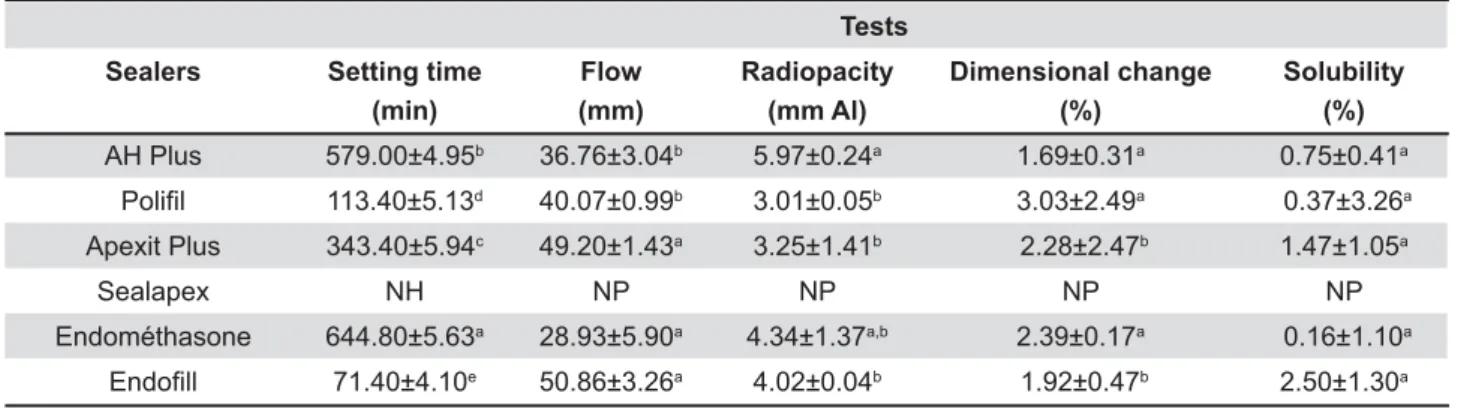
Table 1 shows the mean values and standard deviations for the physicochemical properties " following setting and solubility) of the tested sealers.
Setting time
-.*+-!-&2 requires that the
setting time of a sealer shall be within 10% of that stated by the manufacturers. In this study, AH Plus®
and Apexit Plus® are in agreement with ANSI/ADA
standards. The manufacturers of Endométhasone®
'&""® do not mention the setting time of
" %"&" "" K" phase. Statistical analysis demonstrated that all sealers displayed mean values statistically different "1/'@®
'&""® presented, respectively, the longest
and the shortest setting time. The Sealapex® did
not set after 168 h and, for this reason, it was not subjected to the other tests.
Flow test
-.*+-!- &2 requires that a
sealer shall have a diameter of no less than 20 mm and all groups of this study conformed to the standards. Statistical analysis showed that '&""® and Apexit Plus® presented the highest
"#1/""" between themselves. AH Plus®%"&"
between themselves (p>0.05). Endométhasone®
exhibited the lowest mean values.
Radiopacity test
All materials showed radiopacity above the 3 mm "##-.*+-!-& 57 (2000). AH Plus® presented higher values
statistically similar to Endométhasone® (p>0.05)
&" ;; ; # 1/1'@® showed intermediate
values, statistically similar to the other sealers (p>0.05).
Dimensional change after setting
ANSI/ADA states that the maximum limit is 1% for linear shrinkage and 0.1% for expansion. The dimensional change of all sealers was greater than the values considered acceptable by ANSI/ ADA. AH Plus®%"&"'@® were
statistically similar among themselves (p>0.05) and ;;;"1/1 Apexit Plus® '&""® presented the lowest
mean values and were statistically similar among themselves (p>0.05).
Solubility
A root canal sealer should not exceed 3% by mass when the solubility of the set material is tested1. All sealers are in agreement with ANSI/
ADA standards. Statistical analysis showed that sealers were statistically similar among themselves (p>0.05).
The distilled and deionized water used for the solubility test was analyzed by atomic absorption spectrometry. The amount of ions released in the immersion solution, in each group, is displayed in Table 2. The concentration of Na+ ions was higher for
Apexit Plus®, followed by AH Plus®, Endométhasone®
'&""®. The concentration of Na+ ions was not
; %"&" " # ; successive dilutions, the concentration of this ion in the immersion solution was much higher than the concentration of the most concentrated pattern of the analytical curve. For K+ ions, the release was
higher in Endométhasone, followed by Apexit Plus®,
AH Plus®%"&"'&""®1Q2+, the
"&;'&"";"" '@%"&"1.2+ ions, all groups
presented concentration inferior to 0.6 mg/L.
DISCUSSION
The properties of root canal sealers can be divided into the following categories: physicochemical, antimicrobial, and biological. When studying the ";&"""" establish research parameters for the development of new products and to evaluate those already
Tests Sealers Setting time
(min)
Flow (mm)
Radiopacity (mm Al)
Dimensional change (%)
Solubility (%)
AH Plus 579.00±4.95b 36.76±3.04b 5.97±0.24a 1.69±0.31a 0.75±0.41a
113.40±5.13d 40.07±0.99b 3.01±0.05b 3.03±2.49a 0.37±3.26a
Apexit Plus 343.40±5.94c 49.20±1.43a 3.25±1.41b 2.28±2.47b 1.47±1.05a
Sealapex NH NP NP NP NP
Endométhasone 644.80±5.63a 28.93±5.90a 4.34±1.37a,b 2.39±0.17a 0.16±1.10a
71.40±4.10e 50.86±3.26a 4.02±0.04b 1.92±0.47b 2.50±1.30a
Same letters indicate statistical similarity (p>0.05) NH: not set; NP: not performed
Table 1- Mean values and standard deviation of the physiochemical properties for each sealer
Sealers
Metallic ions AH Plus !"" Apexit Plus Endométhasone #""
K+ 0.58±0.36 0.32±0.12 1.46±0.92 14.20±1.72 0.21±0.04
Na+ 3.07±2.10 ND 4.63±2.33 2.88±1.54 1.28±0.72
Ni2+ !" !" !" !" !"
Zn2+ ! 2.15±0.10 ! 4.55±0.83 5.72±2.13
ND: not determined
existing on the market24, thus achieving better
clinical results in clinical practice.
The physiochemical tests conducted in this study &/0;-.*+-!-2,
;""&>"{ Júnior, et al.5 (2007), who suggested a reduction
of 80% in volume of the test samples dimensions, aiming to contribute to the rational use of endodontic materials without affecting the results. - & ; images of the sealers. The images were obtained using the Digora digital system (Digora software for Windows), which requires a shorter exposure time and the operating system captures, processes, stores and measures the images. It also presents a scale that achieves 256 gray levels4.
According to the ANSI/ADA, the setting time of a sealer should vary only 10% in relation to the established by the manufacturer. In the present study, AH Plus® and Apexit Plus® are in agreement
with ANSI/ADA standards. The manufacturers of '@ '&"" ; " %"&" "" experimental phase, thus, no setting time has been &1
The setting time is primarily a control test on the stable behavior of a product and is dependent on the sealer components, particle size, room temperature, and relative humidity8,17. The chemical agents used
to promote the radiopacity of Endométhasone®
(lead oxide and bismuth subnitrate) and AH Plus®
(zirconium oxide and calcium tungstate) can be responsible for the longer setting time, since these radiopacifying agents have low solubility in water.
Apexit Plus® sealer has calcium oxide in its
composition, which is converted into calcium hydroxide when in contact with water, which delay the setting process due to the inclusion of another chemical reaction. Sealapex did not set in the experimental phase of this study (1 week-period), which is in agreement with previous reports1,10.
'&"" probably due to the absence of calcium hydroxide and the presence of a single radiopacifying agent on its composition24.
ANSI/ADA establishes as 20 mm the acceptable minimum value for the diameter of the disc formed by the sealer. In this study, all sealers conformed -.*+-!-1#& these results19,281*"#'&""G
"# ; hydrogenated resin on its formula. For Apexit Plus®,
it is important to highlight the presence of epoxy resin29, probably responsible for the increase of
sealer viscosity6,23,241%"&"#"
that the physiochemical characteristics of the " " ; 1 lower values achieved by Endométhasone® could
be attributed to the absence of hydrogenated resin or epoxy resin on its composition.
" " #" " #;& radiopacity to allow for a clear distinction between the materials and surrounding anatomic structures and to facilitate the evaluation of the quality of the &""#"# radiographic examination4. ANSI/ADA establishes
that the radiopacity should be larger or equal of 3 mm of aluminum. All materials of the present study were in accordance with the ANSI/ADA &1%###-%"#®4,19,28,
Sealapex®9,26 '&""®4&#"1
difference in the results can be attributed to the radiopacifying agents present in the sealers7. AH
Plus® has calcium tungstate and zirconium oxide on
its composition. Endométhasone® has lead oxide
and bismuth subnitrate4.
Dimensional change demonstrates, in percentage, the shrinkage or expansion of the material following setting. ANSI/ADA states that
the maximum limit is 1% for linear shrinkage and 0.1% for expansion5. The dimensional change
of all tested sealers was greater than the values considered acceptable by ANSI/ADA.
AH Plus®, Endométhasone®%"&"
expansion, while Apexit Plus® and Endofill®
presented contraction. In other literature reports, K " & ; - %"#®19,28 and
'&""61%"&""K"
due to water sorption by its components (zinc oxide and calcium carbonate) after polymerization. However, this is an experimental sealer and, consequently, the mechanism related to water sorption and diffusion in the matrix is still not completely elucidated. The contraction presented by Apexit Plus® can be attributed to its high
solubility, which affected the dimensional stability ;"1'&""® also presented contraction
and it lost an equivalent amount of zinc as that of Endométhasone. The zinc found in the immersion solution can be related to the continuous loss of eugenol from the matrix1,14,24.
ANSI/ADA2 establishes that solubility of sealers
should not exceed 3% by mass. Solubility results of all groups were within ANSI/ADA recommendations. AH Plus®, Apexit Plus® and
'&""® "#"9 "1 %"&"
and Endométhasone® presented expansion due to
water sorption. Previous studies of solubility only found material loss by solubilization of the sealer structure5,14,19,28. In this study, the sealers were
statistically similar regarding solubility. However, AH Plus® presented the lowest mean values, probably
due to the presence of HEMA in its composition. "#" '&""®
The analysis of the immersion solutions revealed that overall the sealers presented low levels of metals concentration. The amount Ni2+ ions found
" "" # ; #& #"1 Q2+ levels were higher
for Endométhasone® '&""®, which is an
expected result since they are zinc oxide-eugenol-based sealers.
Further studies should aim for better understanding of physical, mechanical and chemical properties of the endodontic sealers, supporting researchers and clinicians to determine their ideal ""&""#1
CONCLUSIONS
Based on the results of this laboratory research, it may be concluded that:
AH Plus®, Apexit Plus® '&""® sealers
are in accordance with ANSI/ADA standards. Endométhasone’s manufacturer did not mention %"&" K"" and Sealapex® did not set;
All tested sealers were in accordance with ANSI/ -!-;"#"J
The dimensional change of all sealers did not ;#"&""-.*+-!-#1
REFERENCES
1- Allan NA, Walton RC, Schaeffer MA. Setting times for endodontic sealers under clinical usage and in vitro condition. J Endod. 2001;27:421-3.
U{-!"-1-.*+-!-&/0{ Endodontic Sealing Material. Chicago: ADA; 2000.
3- Bodrumlu E, Sumer AP, Gungor K. Radiopacity of a new root canal sealer, Epiphany. Oral Surg Oral Med Oral Pathol Oral Radiol Endod. 2007;104:58-61.
4- Carvalho-Junior JR, Correr-Sobrinho L, Correr AB, Sinhoreti MA, >#{.2!1;&""" using digital radiography. Int Endod J. 2007;40:514-20. 5- Carvalho-Junior JR, Correr-Sobrinho L, Correr AB, Sinhoreti MA, Consani S, Sousa-Neto MD. Solubility and dimensional change after setting of root canal sealers: a proposal for smaller dimensions of test samples. J Endod. 2007;33:1110-6.
6- Carvalho-Junior JR, Guimarães LF, Correr-Sobrinho L, Pécora JD, Sousa-Neto MD. Evaluation of solubility, desintegration, and dimensional alterations of a glass ionomer root canal sealer. Braz Dent J. 2003;14:114-8.
7- Coomaraswamy KS, Lumley PJ, Hofmann MP. Effect of bismuth K & " ; endodontic Portland cement-based (MTA-like) system. J Endod. 2007;33:295-8.
8- Gambarini G, Romeo U, Tucci E, Gerosa R, Nocca G, Lupi A, et al. Cytotoxicity of Epiphany SE endodontic sealer: a comparative
in vitro study. Med Sci Monit. 2009;15:PI15-8.
9- Guerreiro-Tanomaru JM, Duarte MA, Gonçalves M, Tanomaru-Filho M. Radiopacity evaluation of root canal sealers containing calcium hydroxide and MTA. Braz Oral Res. 2009;23:119-23. 10- Ingle J, Newton C, West JG. Obturation of the radicular space. In: Ingle J, Bakland L, editors. Endodontics. Hamilton: BC Decker Inc.; 2002. p.571-668.
11- Leite FR, Ramalho LT. Bone regeneration after demineralized bone matrix and castor oil (Ricinus communis) polyurethane implantation. J Appl Oral Sci. 2008;16:122-6.
12- Leonardo MR, Silva LA, Tanomaru-Filho M, Bonifácio KC, Ito LY. In vitro evaluation of the antimicrobial activity of a castor oil-based irrigant. J Endod. 2001;27:717-9.
13- Martins GR, Carvalho CA, Valera MC, Oliveira L, Buso L, Carvalho AS. Sealing ability of castor oil polymer as a root end &"""1-""1U~J}0 UU{1
14- Nunes VH, Silva RG, Alfredo E, Sousa-Neto MD, Silva-Sousa YTC. Adhesion of Epiphany and AH Plus sealers to human root dentin treated with different solutions. Braz Dent J. 2008;19:46-50.
15- Onay EO, Ungor M, Ozdemir BH. In vivo evaluation of the biocompatibility of a new resin-based obturation system. Oral Surg Oral Med Oral Pathol Oral Radiol Endod. 2007;104:60-6. 16- Ørstavik D. Materials used for root canal obturation: technical, biological and clinical testing. Endod Topics. 2005;12(6):25-38. 17- Ørstavik D, Nordahl I, Tibballs JE. Dimensional change following setting of root canal sealer materials. Dent Mat. 2001;17:512-9.
18- Pinheiro CR, Guinesi AS, Camargo EJ, Pizzolitto AC, Bonetti-" *1 $Bonetti-" Bonetti-" Bonetti-"# ; Bonetti-" &Bonetti-"Bonetti-" different endodontic sealers. Oral Surg Oral Med Oral Pathol Oral Radiol Endod. 2009;108:56-60.
19- Resende LM, Rached-Junior FJ, Versiani MA, Souza-Gabriel AE, Miranda CE, Silva-Sousa YT, et al. A comparative study of physicochemical properties of AH Plus, Epiphany, and Epiphany SE root canal sealer. Int Endod J. 2009;42:785-93.
20- Ricucci D, Lin LM, Spångberg LS. Wound healing of apical tissues after root canal therapy: a long-term clinical, radiographic, and histopathologic observation study. Oral Surg Oral Med Oral Pathol Oral Radiol Endod. 2009;108:609-21.
21- Scarparo RK, Grecca FS, Fachim EV. Analysis of tissue reactions to methacrylate resin-based, epoxy resin-based and oxide-eugenol endodontic sealers. J Endod. 2009;35:229-32.
22- Sjögren ULF, Hagglund B, Sundquist G, Wing K. Factors affecting the long term results of endodontic treatment. J Endod. 1990;16:498-504.
23- Sousa-Neto MD, Guimarães LF, Gariba Silva R, Saquy %> %@ !1 # ; ;; ; hydrogenated resins on the powder-liquid ratio of Grossman cements. Braz Dent J. 1998;9:11-8.
24- Sousa-Neto MD, Guimarães LF, Saquy PC, Pécora JD. Effect of different grades of gum rosins and hydrogenated resins on the solubility, disintegration, and dimensional alterations of Grossman cement. J Endod. 1999;25:477-80.
25- Sousa-Neto MD, Passarinho-Neto JG, Carvalho-Júnior JR, Cruz-Filho AM, Pécora JD, Saquy PC. Evaluation of effect of EDTA, EGTA and CDTA on dentin adhesiveness and microleakage with different root canal sealers. Braz Dent J. 2002;13:123-8.
26- Tanomaru-Filho M, Jorge EG, Guerreiro-Tanomaru JM, 4" 21 "# ; " &"" materials by digitalization of images. J Endod. 2007;33:249-51. 27- Tanomaru-Filho M, Jorge EG, Tanomaru JMG, Gonçalves M. Evaluation of the radiopacity of calcium hydroxide- and glass-ionomer-based root canal sealers. Int Endod J. 2008;41:50-3. 28- Versiani MA, Carvalho-Júnior JR, Padilha MI, Lacey S, Pascon EA, Sousa-Neto MD. A comparative study of physicochemical properties of AH Plus and Epiphany root canal sealants. Int Endod J. 2006;39:464-71.