Zika Virus Infection in Pregnant Women and
Microcephaly
Infecção do vírus Zika em gestantes e microcefalia
Geraldo Duarte
1Antonio Fernandes Moron
2Artur Timerman
3César Eduardo Fernandes
4Corintio Mariani Neto
5Gutemberg Leão de Almeida Filho
6Heron Werner Junior
7Hilka Flavia Barra do Espírito Santo
8João Alfredo Piffero Steibel
9João Bortoletti Filho
2Juvenal Barreto Borriello de Andrade
10Marcelo Burlá
11Marcos Felipe Silva de Sá
1Newton Eduardo Busso
12Paulo César Giraldo
13Renato Augusto Moreira de Sá
14Renato Passini Junior
13Rosiane Mattar
2Rossana Pulcineli Vieira Francisco
151Universidade de São Paulo, Ribeirão Preto, SP, Brazil 2Universidade Federal de São Paulo, São Paulo, SP, Brazil 3Hospital Professor Edmundo Vasconcelos, São Paulo, SP, Brazil 4Faculdade de Medicina do ABC, Santo André, SP, Brazil 5Hospital Maternidade Leonor Mendes de Barros, São Paulo, SP,
Brazil
6Universidade Federal do Rio de Janeiro, Rio de Janeiro, RJ, Brazil 7Alta Excelência Diagnóstica, Rio de Janeiro, RJ, Brazil
8Universidade Federal do Amazonas, Manaus, AM, Brazil 9Pontifícia Universidade Católica, Porto Alegre, RS, Brazil, 10Consultório Médico Juvenal Barreto Borriello de Andrade, São
Paulo, SP, Brazil
Rev Bras Ginecol Obstet 2017;39:235–248.
Address for correspondence Geraldo Duarte, Avenida Bandeirantes, 3900, 14049-900, Ribeirão Preto, SP, Brazil
(e-mail: [email protected]).
Keywords
►
pregnancy
complications
►
Zika virus
►
arbovirus infections
►
microcephaly/
ultrasonography
►
real-time polymerase
chain reaction
►
deafness/ etiology
►
blindness/ etiology
Abstract
From the discovery of the Zika virus (ZIKV) in 1947 in Uganda (Africa), until its arrival in South
America, it was not known that it would affect human reproductive life so severely. Today,
damage tothe central nervous system is known to be multiple, and microcephaly is considered
the tip of the iceberg. Microcephaly actually represents the epilogue of this infection
’
s
devastating process on the central nervous system of embryos and fetuses. As a result of
central nervous system aggression by the ZIKV, this infection brings the possibility of
arthrogryposis, dysphagia, deafness and visual impairment. All of these changes of varying
severity directly or indirectly compromise the future life of these children, and are already
considered a congenital syndrome linked to the ZIKV. Diagnosis is one of the main dif
fi
culties
in the approach of this infection. Considering the clinical part, it has manifestations common
to infections by the dengue virus and the chikungunya fever, varying only in subjective
intensities. The most frequent clinical variables are rash, febrile state, non-purulent
11Clínica Marcelo Burlá, Rio de Janeiro, RJ Brazil
12Faculdade de Ciências Médicas da Santa Casa de São Paulo, São
Paulo, SP, Brazil
13Universidade Estadual de Campinas, Campinas, SP, Brazil 14Universidade Federal Fluminense, Niterói, RJ, Brazil 15Universidade de São Paulo, São Paulo, SP, Brazil
This review is part of the Series, Guidelines and Recommenda-tions of the Federação das Associações de Ginecologia e Obstetrícia - FEBRASGO, and it was prepared by the FEBRASGO National Provisional Specialized Commission for the Study of Zika Virus, Pregnancy and Microcephaly.
received February 21, 2017 accepted March 17, 2017
DOIhttps://doi.org/ 10.1055/s-0037-1603450. ISSN 0100-7203.
Copyright © 2017 by Thieme Revinter Publicações Ltda, Rio de Janeiro, Brazil
conjunctivitis and arthralgia, among others. In terms of laboratory resources, there are also
limitations to the subsidiary diagnosis. Molecular biology tests are based on polymerase chain
reaction (PCR) with reverse transcriptase (RT) action, since the ZIKV is a ribonucleic acid (RNA)
virus. The RT-PCR shows serum or plasma positivity for a short period of time, no more than
fi
ve days after the onset of the signs and symptoms. The ZIKV urine test is positive for a longer
period, up to 14 days. There are still no reliable techniques for the serological diagnosis of this
infection. If there are no complications (meningoencephalitis or Guillain-Barré syndrome),
further examination is unnecessary to assess systemic impairment. However, evidence is
needed to rule out other infections that also cause rashes, such as dengue, chikungunya,
syphilis, toxoplasmosis, cytomegalovirus, rubella, and herpes. There is no speci
fi
c antiviral
therapy against ZIKV, and the therapeutic approach to infected pregnant women is limited to
the use of antipyretics and analgesics. Anti-in
fl
ammatory drugs should be avoided until the
diagnosis of dengue is discarded. There is no need to modify the schedule of prenatal visits for
pregnant women infected by ZIKV, but it is necessary to guarantee three ultrasound
examinations during pregnancy for low-risk pregnancies, and monthly for pregnant women
with con
fi
rmed ZIKV infection. Vaginal delivery and natural breastfeeding are advised.
Resumo
Desde a descoberta do vírus Zika (VZIK) em 1947 em Uganda, na África, até sua chegada na
América do Sul, não se tinha notícia de que ele seria capaz de comprometer a vida reprodutiva
em humanos de forma tão severa. Hoje, sabe-se que os danos sobre o sistema nervoso central
são múltiplos, e a microcefalia é considerada a ponta do iceberg, visto que na realidade ela
representa o epílogo de um processo devastador desta infecção sobre o sistema nervoso
central do embrião e do feto. Em decorrência da agressão do sistema nervoso central pelo
VZIK, esta infecção pode provocar artrogripose, disfagia, surdez e comprometimento visual.
Todas estas alterações, de gravidade variável, direta ou indiretamente comprometem a vida
futura dessas crianças, já sendo considerada uma síndrome congênita ligada ao VZIK. Uma das
principais di
fi
culdades na abordagem dessa infecção é relativa ao diagnóstico. Considerando a
parte clínica, observa-se que ela apresenta manifestações comuns às infecções pelos vírus da
dengue e da febre chikungunya, variando apenas em suas intensidades subjetivas. As variáveis
clínicas mais frequentes são o exantema, febrícula, conjuntivite não purulenta e artralgia. No
tocante aos recursos laboratoriais, também existem limitações ao diagnóstico subsidiário. As
provas de biologia molecular se fundamentam na reação em cadeia da polimerase (RCP) com
ação da transcriptase reversa (TT), visto que o VZIK é um vírus ácido ribonucleico (ARN). A
TR-RCP apresenta positividade sérica ou plasmática por um período curto de tempo, não
ultrapassando cinco dias após início dos sinais e sintomas. Esta pesquisa do VZIK na urina
fi
ca
positiva por período mais prolongado, chegando a 14 dias. Ainda não existem técnicas seguras
para diagnóstico sorológico dessa infecção. Não havendo complicações (meningoencefalite
ou síndrome de Guillain-Barré), di
fi
cilmente são necessários mais exames complementares
para avaliar o comprometimento sistêmico. No entanto, são necessárias provas para descartar
as outras infecções que causam exantema, como dengue, chikungunya, sí
fi
lis, toxoplasmose,
citomegalovírus, rubéola e herpes. Sabe-se que não existe terapia antiviral especí
fi
ca contra o
VZIK, e a abordagem terapêutica de gestantes portadoras da infecção limita-se ao uso de
antitérmicos e analgésicos. Orienta-se evitar anti-in
fl
amatórios até que o diagnóstico de
dengue seja descartado. Sobre a condução do pré-natal, não há necessidade de modi
fi
car o
cronograma de consultas pré-natais para gestantes que foram infectadas pelo VZIK, mas é
necessária a garantia de três exames ecográ
fi
cos durante a gravidez para gestantes de baixo
risco, e mensais para a gestante com infecção con
fi
rmada pelo VZIK. A via de parto é vaginal, e
está liberado o aleitamento natural.
Palavras-chave
►
complicações na
gravidez
►
vírus da Zika
►
infecções por arbovírus
►
microcefalia/
ultrassonogra
fi
a
►
reação em cadeia da
polimerase em tempo
real
Introduction
The dengue virus (DENV) has severely affected the Brazilian population for several decades, but there was no adequate social, political or sanitary response to deal with the vector control of this infection. The situation was not tackled with effective strategies, just like what occurred at the beginning of the last century for yellow fever control. This relative sanitary inertia, along with the constant adaptations ofAedes mosquitoes have made this insect a feared enemy able to dribble all timid interventions for its control, allowing the repetition of increasingly expanded epidemic peaks of arbo-viruses. In addition to these variables, globalization brought diseases with which the Brazilian population had not had prior contact, such as those caused by the Zika virus (ZIKV) and the chikungunya virus (CHIKV), which can be transmit-ted by humans or animals. However, attention with arbovi-ruses was only raised in face of evidences of the association between the ZIKV and the occurrence of microcephaly.1–5
This fact has created a legitimate demand for the union of forces of professionals from all areas of health to care for people affected by this infection, whether mothers, their children or their families.6,7
Nowadays, there is no more questioning if ZIKV infection is responsible for embryonic, fetal and neonatal harm, but only if this diagnostic duality derives from a pure, accidental or incidental causal association.8Nervous system diseases (embryonic, fetal and postnatal) resulting from ZIKV infec-tion induced a considerable number of studies, pedagogical publications on the subject, and the creation of several care and surveillance protocols.9–16
Microcephaly resulting from ZIKV infection is the epi-logue of a vast process of diffuse involvement of the embry-onic/fetal nervous system that manifests clinically with the reduction of the cephalic perimeter. Over time, other abnor-malities have been identified, such as deafness, visual defi -ciencies of varying degrees, dysphagia and arthrogryposis, resulting in a complex of alterations currently called ‘ con-genital Zika virus syndrome’. As some of these changes occur in the postnatal period, the adequate care to mothers even-tually affected by this infection should include guidance and the correct referral of‘all children exposed to this infection’, not only those with organic birth defects.10
Nothing is definitive beyond the objective disposition to inform about the adequate care of mothers possibly affected by this infection and to provide the correct referral of the children eventually affected by this infection. This is a type of document that needs to be continuously re-evaluated and updated.
Etiological Agent
Considering the phylogenetic aspects, ZIKV is an arbovirus of theFlavivirusgenus of theFlaviviridaefamily, transmitted by mosquitoes of theAedesgenus, among whichAedes aegyptiis the most common in Brazil. Like otherFlavivirus, its genome is composed of single-stranded ribonucleic acid (RNA) con-taining 1,0794 Kb. It includes an open reading frame that encodes a polyprotein and two non-codingflanking regions
in its replication process. Then, the polyprotein is cleaved in capsid (C) proteins, membrane precursor (Mpr), envelope (E), and in seven non-structural (NS) proteins called NS1, NS2a, NS2b, NS3, NS4a, NS4b and NS5.17,18This high number of common cellular structures to all arboviruses explains the difficulty to elaborate diagnostic serological tests without cross-reactivity between one another.
Particularities of ZIKV
The initial description of the ZIKV occurred in 1947 in Uganda in a forest called Zika, and its isolation was found in sentinel rhesus monkeys of a yellow fever study.19Thefirst human case occurred in Nigeria in 1954, and its dispersion throughout the African continent was slow. Until 2007, the literature docu-ments that the number of people affected by this virus did not exceed 50. After reports of sporadic cases in Asian countries, thefirst epidemic of this virus was observed in 2007 in Yap Island, located in the Pacific Ocean.20In 2013, other epidemic peaks occurred in French Polynesia and Easter Island, and the virusfinally arrived in Brazil between 2013 and 2014. The virus has undergone genomic recombination in this trajectory, and nowadays there are two lineages, one African and the other Asian, the latter responsible by the epidemic in Brazil.21,22 There is already information about other lineages,23 but officially, there are only two recognized lineages so far. These mutations seem to be responsible for the pathogenic profile that directly or indirectly associates the ZIKV with the occur-rence of damage to the central nervous system of human fetuses. In this context, the significant increase of cases of Guillain-Barré syndrome and encephalitis in people affected by this infection is noteworthy.24 Several other alterations occurred, promoting an even greater adaptation of the ZIKV to Aedesgenus mosquitoes.20Reflecting on this vector premise, the spectrum of difficulties in this area seems to have potential to increase, and within the guidelines of attention to pregnant women, vector control strategies within the reach of patients, their families and the general community cannot be forgotten. According to official information, the entry of ZIKV in Brazil seems to have occurred from states in the Northeast, and its dispersion coincides with an increased incidence of micro-cephaly cases in communities infested byAedes aegypti.25
ZIKV Transmission
There is no doubt that the most important form of ZIKV transmission is through the bite of theAedesgenus mosquito, the same transmitter of DENV and CHIKV, among others.26 Therefore, the combat of the mosquitoes and their prolifera-tion is considered the only truly effective intervenprolifera-tion to control these diseases so far.
dysphagia, severe auditory and ocular alterations, among others).27–29 The growing fear after confirmation of ZIKV
sexual transmission forced the world’s leading health agencies to develop care protocols that are constantly modified by additional knowledge on the subject.30,31This fact has brought objective difficulties for couples who planned their pregnan-cies for current times, either naturally or with assisted repro-duction resources. The sexual transmission of the ZIKV has been determined to occur from men (symptomatic and asymptomatic) to women, from symptomatic women to men, and in penis-anus relationships between men.32
The presence of the ZIKV has also been confirmed in urine, breast milk, tears and saliva, but it is not yet possible to qualify thesefluids as‘effective vehicles’in the transmission of the infection.33Despite the presence of the virus in breast milk, the general orientation is not to discontinue breast-feeding in cases of puerperal women infected with the ZIKV.6,7,11Although rare, transmission by blood transfusion and organ transplantation has also been reported on some occasions.34–36
Despite knowledge advances on ZIKV infection, to date there is no reliable information on whether the immunity conferred by the natural virus infection is permanent and can prevent new infections. However, the greater concern re-gards the previous circulation of this virus only in some regions of Brazil, making most of the population still suscep-tible to this infection and without any type of immunity against it. Even after thefirst epidemic peak in the summer of 2015/2016, a large number of pregnant women are still
expected to become infected in the summer of 2016/2017 in Brazil, because the exhaustion of those susceptible to this infection has not yet been observed.
Diagnosis of ZIKV Infection
Clinical diagnosis is initially considered to diagnose ZIKV infection, remembering the limitations resulting from the asymptomatic form of the infection.37,38The epidemiologi-cal diagnosis complements the cliniepidemiologi-cal diagnosis, with the aim of clarifying the pregnant woman’s trajectory in terms of trips and contact with sick people, or who have visited high endemic areas.
Clinical Diagnosis of ZIKV Infection
In most cases, ZIKV infection is a clinically self-limiting disease, but still considered an acute febrile illness. Since the beginning of the spread of the virus in Brazil, experts have observed the disease pattern of low fever (lower than 38.5°C) or no fever, lasting 1 to 2 days, accompanied by a pruritic and coarse rash of variable duration (2 to 7 days), mild muscle pain, joint pain and joint edema of mild to moderate intensity. Non-purulent conjunctivitis is observed in most cases.39
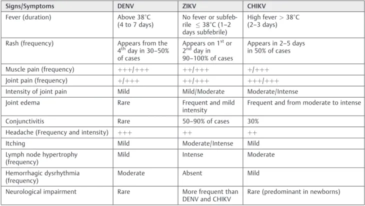
In Brazil, the main differential diagnoses of ZIKV infec-tion are the other arboviruses caused by DENV and CHIKV.►Table 1shows a summary of the clinical
manifes-tations of these three diseases based on their diagnosis. In
Table 1 Frequency and comparative intensity of the most common signs and symptoms in ZIKV, DENV and CHIKV infections
Signs/Symptoms DENV ZIKV CHIKV
Fever (duration) Above 38°C
(4 to 7 days) No fever or subfeb-rile 38°C (1–2 days subfebrile)
High fever>38°C (2–3 days)
Rash (frequency) Appears from the 4thday in 30
–50%
of cases
Appears on 1stor 2ndday in 90–100% of cases
Appears in 2–5 days in 50% of cases
Muscle pain (frequency) þþþ/þþþ þþ/þþþ þ/þþþ
Joint pain (frequency) þ/þþþ þþ/þþþ þþþ/þþþ
Intensity of joint pain Mild Mild/Moderate Moderate/Intense
Joint edema Rare Frequent and mild
intensity Frequent and from moderate to intense
Conjunctivitis Rare 50–90% of cases 30%
Headache (Frequency and intensity) þþþ þþ þþ
Itching Mild Moderate/Intense Mild
Lymph node hypertrophy
(frequency) Mild Intense Moderate
Hemorrhagic dysrhythmia
(frequency) Moderate Absent Mild
Neurological impairment Rare More frequent than
DENV and CHIKV Rare (predominant in newborns)
Abbreviations: CHIKV, chikungunya virus; DENV, dengue virus; ZIKV, zika virus.
ZIKV infection, the exanthematic picture is very marked, with conjunctival hyperemia, but without significant alter-ation in joint involvement. In general, the symptoms disap-pear in three to seven days after their onset.38,39During the anamnesis, it is important to obtain information about the general causes of the rash, such as exogenous intoxications, allergies to medicines or allergenic substances. These re-sponses are only possible if objectively surveyed, and must be in the differential diagnosis of arboviral infections. Even considering the rarity, eventual clinical manifestations of toxoplasmosis, syphilis, herpes, rubella and cytomegalovirus are also part of this information.10,12
Epidemiological Diagnosis
The clinical diagnosis can be enriched with epidemiological information of fundamental importance in the differential diagnosis of rheumatic causes. The following information is particularly important: about the contact with people diag-nosed as having DENV, CHIKV and/or ZIKV infections; con-tact with people with other exanthematous diseases; use of medication/alcohol/illicit drugs during pregnancy; and resi-dence or travel to areas of ZIKV infestation during pregnancy.6,38
The high frequency of rashes in symptomatic cases of ZIKV infection was the main factor that made it a clinical marker to include pregnant women in the diagnosticflows of this infection. In the presence of this signal, the pregnant woman should notified the Municipal Epidemiological
Sur-veillance (ES) that informs the reference Epidemiological Surveillance Group (ESG), after completing the ES specific form.7,12With these measures, it is expected that at least the symptomatic cases will be notified (►Fig. 1). This
important step should include the help from ES professio-nals of the basic network, hospitals and municipalities for the completion of these forms.
Laboratory Diagnosis of ZIKV Infection
To confirm the clinical and epidemiological suspicion of ZIKV infection, three blood samples should be requested. Thefirst sample is used to identify NS-1 (differential diagnosis with DENV); the second sample evaluates the differential diagno-ses with syphilis, toxoplasmosis, rubella, type 2 herpes and cytomegalovirus (to request these exams, one should con-sider if the pregnant woman already has these serologies indicating previous contact with the etiological agents, if she has proof of vaccine against rubella, and the availability of these tests in the particular community); and the third sample is used to confirm the presence of ZIKV RNA. If there is no absolute certainty that the maternal rash is clearly derived from exogenous intoxications, the use of medica-tions or other non-infectious causes, performing these tests is recommended. In practice, all pregnant women with a rash should undergo these exams.
It is just a matter of time before the performance of all of the previously mentioned exams becomes mandatory. When a specific and safe serological test for immunoglobulin M
(IgM) dosing against ZIKV is possible, it may not be necessary to request the serologies that make differential diagnosis between the diseases causing syphilis, toxoplasmosis, rubel-la, cytomegalovirus and herpes (STORCH) syndrome, cur-rently called Z-STORCH.10 In spite of all promises of the Brazilian Official Health Offices about the existence of accu-rate diagnostic tests for ZIKV infection (rapid tests), in practice, they are not available for the Brazilian Unified Healthcare System (SUS, in the Portuguese acronym).
To date, for confirmation of ZIKV infection in pregnant women with a rash, the preferred screening test is the reverse transcriptase polymerase chain reaction (RT-PCR) of ZIKV RNA. Given the short duration of the viremic period, viral RNA detection is possible only for a seven-day period after the onset of the symptoms. However, aiming for greater effectiveness of the technique, it is recommended to examine the material by thefifth day after the onset of the clinical manifestations. For urine RT-PCR, the period is a little longer, up to the 14th day after the onset of the symptoms with commercially available exams.40
At the request of the Centers for Disease Control and Prevention (CDC), in September 2016, the Food and Drug Administration (FDA) of the United States released the Trioplex Real-time RT-PCR Assay for use in emergencies such as ZIKV infection during pregnancy. This RT-PCR eval-uates the presence of DENV, CHIKV and ZIKV in a single sample, in a single test. However, this test has not yet been released for commercialization in Brazil.41
The serological tests commercially available in Brazil have low specificity for detection of ZIKV-specific antibodies. Cross-reactivity with infections caused by other arboviruses (DENV, CHIKV and yellow fever virus, among others) absurd-ly increases costs, and results in diagnosis of ZIKV infection by exclusion, making the diagnostic processfinancially un-affordable. At present, the method for large-scale antibody titration recommended by the Brazilian Ministry of Health is based on in-house Enzyme-Linked Immunosorbent Assay (ELISA) techniques, a protocol established by the CDC (CDC, 2016).42This test is already standardized in some state-run laboratories in Brazil, but with limited availability to the public. It would primarily serve pregnant women with a history of exanthematous disease, although outside the ideal collection period for the RT-PCR test for ZIKV, or those with present diagnosis of fetal microcephaly during pregnancy and without previous diagnosis of ZIKV infection. However, laboratories cannot even meet the demand for these specific groups. In summary, in practice, we do not yet have a serological test available for our SUS population.
From what is known to date, there is no need to change the laboratory routine of each prenatal service (each municipal-ity has its prenatal laboratory priorities). The need to include any other test in the list of routine laboratory exams for prenatal care is still unknown.10
Diagnosis of Systemic Involvement
Although characterized as atypical and rare, severe forms of ZIKV infection in pregnant women have been described, such
as extreme dehydration, severe joint involvement, encepha-litis and Guillain-Barré syndrome.43These forms can be so severe they can cause death in both adults and fetuses.6,24,27 In these cases, the help of professionals connected to neurol-ogy and advanced life support is imperative.
If there is a clinical indication to evaluate the systemic compromise in cases of pregnant women with suspected ZIKV infection, the most commonly used tests are those measuring the body’s response to the infection. However, there is no laboratory specificity for this evaluation resulting from ZIKV infection; hence, the definition of which tests to request will depend on the organic system affected and the health professional’s decision, including the decision of the need for tests. In general, the hemogram may indicate moderate leukopenia and thrombocytopenia, but it may also assist in differentiating viral infection from bacterial infection. A more pronounced reduction in platelet count would indicate bleeding risk and a greater likelihood of DENV infection. In turn, the assessment of the hepatic function in ZIKV infection will identify a slight or no elevation of enzymes, bilirubin, and markers of inflammatory activity. Depending on the degree of systemic involvement, renal function may indicate a slight elevation of urea, creatinine, and sodium and potassium changes.6,11,39One should note that these changes are rare in ZIKV infection.
Prophylaxis of the
Aedes Aegypti
Bite
Vector control is surely the most important prophylactic measure to avoid ZIKV infection. Preventing theAedes ae-gyptimosquitoes’ reproduction would be the most logical measure to be adopted; however, considering Brazil as a whole, the attempts to do so have been unsuccessful so far. As the flight range of the Aedes mosquito is 50 m, it is important to try to control outbreaks and breeding places in this radius of distance,44 since this is possible in some communities and could serve as an example to others.
When control of the vector breeding sites fails, people should be careful and try to avoid contact with the mosquito. The strategies to prevent infection include avoiding the same environment of people with suspected ZIKV infection, and avoiding trips to areas of high incidence of the disease. In this group of interventions, preventing mosquitoes from enter-ing homes by usenter-ing window screens in senter-ingle-story houses or in buildings near high areas that harborAedesoutbreaks may aid in prophylaxis.
without basic sanitation, and with inefficient garbage collection.
Prophylaxis measures againstAedes aegyptibites are com-pleted with the use of repellent in any exposed area of the skin. When wearing wide woven fabric clothing, the repellent should be used underneath the fabric, as there are doubts about its actual effectiveness when used over the fabric. The most suitable repellents for use in pregnant women are those containing icaridin, N,N-diethyl-3-methylbenzamide (DEET) and ethyl 3-[acetyl(butyl)amino]propanoate (IR3535).16 In communities with higher temperatures, these repellents should be reapplied in shorter periods than recommended. Care should be taken to avoid contact of these compounds with the eyes, mouth and nose. The intake of thiamine and B1-complex vitamins is not proven to be effective as a repellent, and its indication of use is not approved by the Brazilian National Sanitary Surveillance Agency (Anvisa, in the Portu-guese acronym).
Microcephaly and ZIKV Infection
According to information from the Brazilian Center for Emergency Operations in Public Health on Microcephaly, up to epidemiological week 52 (25 to 31/12/2016), of 10,867 reported cases of microcephaly, 2,366 had already been confirmed as having infectious etiologies, with a high prob-ability that the ZIKV was the cause of those etiologies. The remaining cases have been discarded or are still under investigation.45
Concept and Prognosis of Microcephaly
From the conceptual point of view, microcephaly represents a disruption of the neurological development causing the cephalic perimeter (CP) measurement of the fetus or new-born to be two standard deviations (SDs) below the normali-ty limit for gestational age and sex.46,47Some authors and services also consider percentiles, observing that two SDs would correspond to percentile three.48,49
Regarding prognosis, mild microcephaly is when the CP measurement is between two to three SDs below the mean for age and sex (fetal or neonatal), and severe microcephaly is when the CP measurement is below three SDs.11,50 In general, the more severe and stronger the microcephaly, the more compromised is the prognosis.51In severe micro-cephaly, in rare cases the neuropsychomotor development does not confirm this association. However, it is difficult to predict the prognosis in mild microcephaly.52
The central nervous system begins its differentiation early, but there is risk of some impairment even after the end of pregnancy. Disruptive alterations due to ZIKV infec-tion occur less frequently in thefinal trimesters of pregnan-cy, but little is known about functional changes. As is known, myelination processes continue after birth. Studies have demonstrated the potential of the ZIKV to invade and destroy nerve cells in experimental models, which may explain the role of this virus in the genesis of microcephaly.53According to Honein et al,54the rate of any organic change in fetuses of
ZIKV-infected mothers during pregnancy is of 6%. However, if the infection occurred during thefirst trimester, this rate can reach 11%, because it occurs in a phase of high potential of cellular differentiation.55 In the city of Ribeirão Preto, Brazil, the rate of severe fetal involvement in mothers with symptomatic ZIKV infection is of 6.2%.
According to Oliveira Melo et al,56microcephaly associat-ed with ZIKV would be just the tip of the iceberg. Only the evolution of the knowledge on the subject enabled the confirmation of additional harm to the perinatal health of these children resulting from this infection, such as severe ocular involvement, deafness, diffuse damage in the central nervous system, arthrogryposis and severe dysphagia.9,28,29 For Suy et al,57the evidence indicates that cases of vertical transmission and severe fetal impairment are linked to a longer period of maternal ZIKV viremia.
Causes of Microcephaly
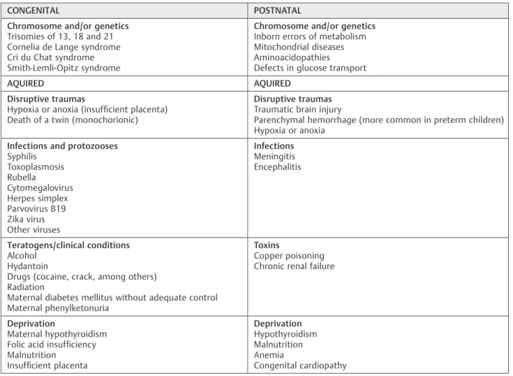
Some authors consider the etiological factor to qualify microcephaly as a malformation or disruptive injury. Mal-formation is considered when the cause is genetic (intrinsic), and disruptive injury, when the cause is extrinsic, like infections.37It is important to remember that the presence of an etiological factor of microcephaly, like maternal infec-tion by the ZIKV, does not make the risk of this harm to the fetus or newborn mandatory.►Table 2is a summary of the
most frequent causes among the more than 500 known causes of microcephaly.52,58,59
Reference Curves for the Diagnosis of Fetal
Microcephaly
The entire diagnostic expectation of fetal microcephaly rests on the ultrasound examination, both for the obstetrician and the parents. Given this high anxiety situation of the family related to the severity of the diagnosis, sonographers seek the most reliable parameters, so that the exam result is the closest to biological reality.
The measurements of CP normality depend on gestational age and sex, and there is no way to correctly diagnose microcephaly (deviation from normality) without the exact dating of the pregnancy, which can be obtained with the date of the last menstrual period and thefirst trimester echocar-diogram. In this period, in experienced hands, the margin of error of the ultrasonography evaluating the embryonic crown-rump length is minimum, and does not exceed three days.51In more advanced pregnancies and when the patient does not know the last menstrual period date, the ultraso-nography is still a good resource to approximate gestational age, but with reduced effectiveness. In this situation, without the last menstrual period date, a good alternative is to combine the measurements of the CP with the fetal length, which can improve the ultrasound results.60
(directly or indirectly) the growth of the CP throughout pregnancy,62–64several representative colegiates of health in
the country11and worldwide15choose to use the curves of Intergrowth 21.47,48All existing curves have pros and cons that limit or stimulate their adoption in a systematic way and as a protocol. For the situation in question, those with more positive factors were the curves of Intergrowth 21.49This is a reference curve for cranial circumference growth. It is easy to use and fairly representative, since it was elaborated with samples from several countries, including Brazilian children, and contemplates the evaluation of preterm fetuses.
Reference measurements of fetal cranial circumference of extreme preterm, preterm, and full-term fetuses, consider-ing fetal gender, can be found at: https://intergrowth21.tghn. org/articles/intergrowth-21st-fetal-growth-standards/
Thefiles in this website are in Excel (Microsoft, Redmond, WA, US) spreadsheets, and can be easily accessed, including the calculator that allows the remote calculation of micro-cephaly by introducing the fetal anatomical variables (weight, femoral length, cranial circumference). Check http://intergrowth21.ndog.ox.ac.uk/pt/ManualEntry
The results obtained can be in percentiles or SD (Z score). In fact, they inform in which SD is the cranial
circumference in relation to the expected standard for that specific gestational age. Results in percentiles can be found at: https://intergrowth21.tghn.org/site_media/ media/articles/INTERGROWTH21st_Fetal_charts_Head_ Circumfrance_11062015.pdf
Following the intrauterine pattern of fetal circumference, the same principles guided the choice for reference standards to diagnose microcephaly after birth. The curves of Inter-growth 21,65elaborated to evaluate preterm and full-term infants in the postnatal period, were then used once more.
Ultrasound Screening of Fetal Microcephaly
For microcephaly screening during low-risk prenatal care, the minimum ultrasound protocol during pregnancy should consider at least three exams. Thefirst examination should be performed around the 12th week of pregnancy (10–14 weeks); the second (morphological) should be performed around the 22nd week (18–24 weeks); and the third should be performed around the 32nd week (28–34 weeks). Obviously, other ultrasound examinations may be requested, but there is no added value without a precise indication to justify it.
Table 2 Most frequent causes of microcephaly (congenital and postnatal)
CONGENITAL POSTNATAL
Chromosome and/or genetics Trisomies of 13, 18 and 21 Cornelia de Lange syndrome Cri du Chat syndrome Smith-Lemli-Opitz syndrome
Chromosome and/or genetics Inborn errors of metabolism Mitochondrial diseases Aminoacidopathies
Defects in glucose transport
AQUIRED AQUIRED
Disruptive traumas
Hypoxia or anoxia (insufficient placenta) Death of a twin (monochorionic)
Disruptive traumas Traumatic brain injury
Parenchymal hemorrhage (more common in preterm children) Hypoxia or anoxia
Infections and protozooses Syphilis
Toxoplasmosis Rubella
Cytomegalovirus Herpes simplex Parvovirus B19 Zika virus Other viruses
Infections Meningitis Encephalitis
Teratogens/clinical conditions Alcohol
Hydantoin
Drugs (cocaine, crack, among others) Radiation
Maternal diabetes mellitus without adequate control Maternal phenylketonuria
Toxins
Copper poisoning Chronic renal failure
Deprivation
Maternal hypothyroidism Folic acid insufficiency
Malnutrition Insufficient placenta
Deprivation Hypothyroidism Malnutrition Anemia
Congenital cardiopathy
In trying to screen the damage to the fetus’ central nervous system, the objective of all ultrasound examinations is to identify as early as possible the suggestive markers of injury associated with ZIKV infection.57These include the reduction of the cephalic circumference, dilation of the cerebral ventricles, parenchymal, periventricular or basal nuclei calcifications, porencephaly or destructive injuries (mainly in the posterior fossa), enlargement of the subarach-noid space, and arthrogryposis. Among the non-specific signs that may occur during ZIKV infection in pregnant women, the following stand out: restriction of fetal growth (mainly after 32 weeks), oligohydramnios, placental calcifi -cations, and fetal death. If the ultrasound examination detects any of these abnormalities in pregnant women with no clinical suspicion of ZIKV infection (which may be due to an asymptomatic infection), it is necessary to assess the fetal damage and seek the differential diagnosis of the cause.
When detecting alterations in the fetus’central nervous system, if possible, the pregnant woman should be referred to services with greater potential of care resources. The image examination should be complemented by magnetic resonance imaging (MRI)61in an attempt to diagnose alter-ations difficult to evaluate by ultrasound, such as encepha-lomalacia, real ventriculomegaly dimension, cortical atrophy, cerebellar hemisphere hypoplasia and brain stem atrophy, among others.
For pregnant women diagnosed with ZIKV infection, the ultrasound exam is performed monthly. Similarly, if fetal abnormality is detected, the pregnant woman should be referred to services with better resources and have her image examination complemented by an MRI.
Pre-gestational Conduct
Confirmation of the sexual transmission of the ZIKV has brought additional concerns to gynecologists/obstetricians. This fact has brought objective difficulties to couples who have planned pregnancies for the present time, both natu-rally or using assisted reproduction resources. It has also challenged the main health agencies in the world to develop care protocols that are constantly updated as a result of additional knowledge on the subject.11,30,31
As ZIKV can be transmitted through sexual activity, it is essential to anticipate reproductive care for the preconcep-tional period. Therefore, women should be oriented about the risks of becoming pregnant without planning, empha-sizing the use of an effective contraceptive method in times of ZIKV infection by avoiding pregnancy without planning for proper gestational care.66According to Atkinson et al,6753% of men with symptomatic ZIKV infection have seminal virus elimination.
From a practical point of view, McCarthy68 suggests it seems logic that prospective mothers at risk of ZIKV infection should postpone the pregnancy project, wait for the end of the turmoil caused by the ZIKV infection epidemic, and for the discovery of ways to effectively prevent the vertical transmission of the virus.69However, there is a great
misconception about the meaning and effectiveness of guidelines radically against the couple’s decision to get pregnant. In general, those able to plan the pregnancy have a clear idea of the risk of ZIKV infection. Depending on their life moment, such as the limit of reproductive life, they will accept the ability to avoid the risk of infection regardless of contraindications to pregnancy at this time. In these cases, when taking the risk, it is crucial that the couple is very well advised on how to avoid the infection.
In addition to the general guidelines for ZIKV vector control (creating physical barriers), and to avoid the bite of the mosquitoes (with the use of repellents and clothes that protect the skin to the maximum extent possible), men who had the infection or have been in an area of high endemicity are advised to use a condom for six months from the infectious event, or to return from the area of greater risk, because there has been documentation of viral elimination in semen for 92 days,70motivating the current orientation of 180 days of protected sex after the partner’s infection. However, this orientation may change because Barzon et al71 have found seminal elimination of the virus until 181 days after the infection. Similar findings have been described by Atkinson et al,68who state this phenomenon may last up to six months after a symptomatic ZIKV infection. In case the partner of a man who had ZIKV infection chooses pregnancy, the use of condoms becomes imperative in the gestational period. If the woman has been infected by the ZIKV, she is requested not to become pregnant for 60 days, until new evidence can establish this period with greater accuracy.
International reports indicate that up to 80% of ZIKV infections may be asymptomatic,38which would logically place all our women at potential risk of becoming sexually infected in our day and age. In this case, the great challenge is not to allow pregnant women to be exposed to the risk of becoming infected by the ZIKV eventually conveyed by their partners’semen. In addition, the couple should be objectively oriented and informed (in writing, obligatorily) of the limits of laboratory techniques in the diagnosis of asymptomatic infections caused by this virus. However, based on Anvisa guidelines,72the couple should only be consideredfit for an assisted fertilization proce-dure when serum tests indicate no acute infection, rep-resented by the presence of IgM, using the tests available in Brazil.
Conduct for Pregnant Women
Prenatal Care for Pregnant Women with No
Previous History of ZIKV Infection and No
Clinical Manifestations
According to the current knowledge about ZIKV infection in pregnant women, the strategy for this group prioritizes prophylaxis, providing guidance on self-protection against the mosquito. To this end, there should be orientation on barrier measures (door and window screens, mosquito nets with thin fabrics on beds), clothing that protects as much body surface as possible, and the use of repellents.16 The most indicated are those containing icaridin, DEET or IR3535. Care should be taken to avoid contact with the eyes, mouth and nose. We must recall that ingestion of sweat-excreted B complex compounds (vitamin B1) has no proven efficacy as a repellent.
After confirming the sexual transmission of the ZIKV and knowing that a large part of people who acquire the infection are asymptomatic, there should be guidance on sexual absti-nence or imperative use of condoms if the partner has symp-toms suggestive of ZIKV infection or a confirmed disease.73In these cases, authors as Oster et al,30and Citil-Dogan et al74are radical, and recommend the use of condoms until the end of pregnancy, because the duration of ZIKV elimination through the semen is not known exactly. Having traveled to regions of higher prevalence of infection in Brazil loses importance a little, but should still be considered, since there are regions where ZIKV infection has not been detected.
If serological tests were available to diagnose possible asymptomatic infections by the ZIKV during prenatal care, they could be used with the strict objective of providing differentiated prenatal care. It would not be appropriate to screen pregnant women simply to know if they have had the infection previously, also because it is not known if a prior infection represents immunization for the rest of life. An-other risk of universal and non-objective serological screen-ing is the potential lack of attention and neglect to avoid infection by other arboviruses in case of confirmation of previous infection by the ZIKV. Although so far they do not have any association with fetal malformations, both CHIKV and DENV infections have compromised gestational and perinatal prognoses. It is important to remember that the vertical transmission rate of CHIKV varies from 27.5 to 48.3%, with a neonatal mortality rate of 5.1%.74 In turn, maternal infection by DENV causes a miscarriage rate 3.3 times higher than that observed among pregnant women in control groups.75
To date, there is no justification to change the set of routine laboratory exams for prenatal care used in the prenatal care community. The prenatal consultation interval (return consultations) and vaccination schedule are also not modified. However, at least three obstetrical ultrasound examinations are guaranteed for pregnant women. Thefirst is around the 12th week of pregnancy (between 10 and 14 weeks, very important for gestational dating); the second is morphological, and performed around the 22nd week (between 18 and 24 weeks); and the third is performed around the 32nd week (between 28 to 34 weeks). Obviously,
other ultrasound exams may be required in case of other specific indications.
For the group of asymptomatic pregnant women at the moment, but who mention previous clinical manifestations compatible with prior ZIKV infection during pregnancy, there must be orientation regarding care to avoid mosquito bites, as it is not certain if the infection was really by the ZIKV. These pregnant women should maintain the same care as asymptomatic pregnant women, that is,‘absolute priority to prophylaxis, providing guidance on how to protect them-selves against the mosquito’.‘The imperative use of condoms if the partner has symptoms suggestive of ZIKA/confirmed disease, or sexual abstinence’is also advised. For the labora-tory diagnosis of these pregnant women, there is the limita-tion imposed by the RT-PCR diagnosis (until the 5th day in the blood and up to 14th day in the urine). This laboratory resource will have little chance of assisting in the diagnosis. The use of currently available serological tests is also limited because there is no determination of their positive or nega-tive predicnega-tive values. In summary, the care for these women would be based solely on ultrasound examinations and regular prenatal care, without changes in the calendar of appointments (return consultations), complementary exams and vaccination schedule. If fetal changes are detected, the patient moves to high-risk prenatal care. For delivery, the obstetric orientation is maintained for these pregnant wom-en, without indication of cesarean delivery.10 In the puer-peral period there are no changes either, including natural breastfeeding.
Care to Pregnant Women with Clinical
Manifestation of ZIKV Infection
Considering the current slow speed in the large-scale diag-nosis of ZIKV infection,10and the possibility that up to 80% of infections may be asymptomatic, a clinical marker of the infection for the entry of pregnant women into diagnostic and care flows was adopted. Rash is the most prevalent among these markers, hence the reason for its choice. To normalize the careflow, the initial approach of all pregnant women should be in the health units within their communi-ties, where exams will be collected and sent to the previously established reference laboratories.
Regardless of where and how will the prenatal care of pregnant women with ZIKV infection be provided, provision of care and psychological support to these women and their families is essential.10
Clinical and Laboratory Diagnosis of ZIKV
Infection
The resources of anamnesis, physical examination and epi-demiological information are used to diagnose ZIKV infec-tion. In the clinical diagnosis, low fever, non-purulent conjunctivitis, arthralgia and malaise stand out. For details, check the section dedicated to diagnosis of ZIKV infection.
probability that plasma or serum RT-PCR can detect viral RNA.73As already mentioned, this test has high sensitivity and specificity rates up to thefifth day after the appearance of the rash.
For pregnant women with ZIKV infection, it is necessary to search for other diseases that may cause rashes and micro-cephaly, which requires the organization of a specificflow to reduce their anxiety, avoid wasting time and optimize costs. In view of the limited access to molecular biology techniques for all these pregnant women, it is advisable to simultaneously investigate some of the diseases that may cause rashes by means of common serological tests, rapid tests or by looking for specific markers (DENV, CHIKV, syphilis, rubella, toxoplas-mosis, cytomegalovirus, and herpes).
Treatment of Pregnant Women with ZIKV
Infection
To date, there is no description of any specific medication to combat or control ZIKV infection. The use of antipyretics and common analgesics (dipyrone and acetaminophen) is rec-ommended to control the febrile and painful symptoms. Until dengue infection is discarded, the use of acetylsalicylic acid or other non-steroidal anti-inflammatory drugs (NSAIDs) should be avoided.11,74Water intake is encouraged to avoid dehydration. In general, ZIKV infection does not require hospitalization, and its complication rate is low. However, when complications occur, they are serious, such as meningoencephalitis and Guillain-Barré syndrome.25,40
Pregnant women with severe pruritus require a symp-tomatic treatment. Hydroxyzine may be used, with the drawback of drowsiness. If a non-sedative treatment is necessary, loratadine or cetirizine may be used.76,77Cases demanding the use of topical or even systemic corticoste-roids are rare.
Aiming to avoid the spread of infection by ZIKV, the care should continue to prevent patients from being bitten again, potentially infecting new vectors. This measure helps to control the spread of ZIKV infection and prevents pregnant women from being victims of other arboviruses. If for some reason hospitali-zation of the pregnant woman with ZIKV infection is necessary up to the seventh day of the onset of the symptoms, all measures should be taken to avoid the spread of the virus in the hospital environment. Among such measures, the hospitalization unit should have screen protection in the windows, the patients’skin should be protected, and repellents should be applied on the unprotected areas of the skin.
Given the lack of knowledge about the dimensions of the risk of sexual transmission of the ZIKV from women to their partners, for safety reasons, there should be sexual absti-nence or consistent use of condoms for at least 60 days. This topic must be reviewed in the future.30–32
Prenatal Care of Pregnant Women with ZIKV
Infection
It is noteworthy that the fetal damage credited to ZIKV infection so far depends on which gestational age occurred
the infection. It seems that the earlier the contact with the virus, the more severe is the fetal damage.54For this reason, information about the last menstrual period is key. In lack of it, ultrasound documentation of the gestational age is im-perative. In the prenatal follow-up of pregnant women diagnosed with ZIKV infection, there is no biochemical laboratory specificity.
When defining the place of follow-up for the pregnant women with ZIKV infection, we have learned the lesson that the place of care provision depends on the possibilities of the hierarchical flow of health in the particular communities where they live. If there is the possibility of making referrals to greater-complexity care, this should be done preferably to the tertiary level. If there is no such possibility, the follow-up can be performed in the unit of care, but with the guarantee of monthly ultrasound examinations until birth to identify or eliminate any type of fetal damage. Diagnosing any fetal abnormality requires referrals to greater-complexity ser-vices according to the usualflow in a particular community for cases of fetal changes in the central nervous system, notably microcephaly.10 Prenatal return consultations will follow the usual routine for risk pregnancies if no change is diagnosed.
To date, there is no information contraindicating vaginal delivery, even if the mother is in the acute phase of the ZIKV infection. In the puerperal period, there is no different measure, and natural breastfeeding is allowed. In case of acute infection, care should be taken to prevent the patient from being bitten in the hospital setting, putting other patients in the same environment at risk. It is important to reinforce that the hospitalization unit of these patients should be equipped with screen protection in windows, the patients’ skin should be protected with appropriate clothing, and repellents must be applied on the unprotected areas of the skin.
Prenatal Care of Pregnant Women whose
Fetus are Diagnosed with Microcephaly
In another moment, the diagnosis of fetal changes in ultra-sound examinations will also be used to include pregnant women in the care line for pregnant women diagnosed with ZIKV infection (confirmed or not). This is independent from the presence or information of a recent or past rash. Thefirst step is the confirmation of the diagnosis of microcephaly. After confirmation, care should be provided in a high-com-plexity environment, because microcephaly has perinatal prognoses that qualify the gestation as a gestation of high perinatal risk. If there are conditions to access genetic services, the evaluation of the case by a professional in this area is of great value, as this will expedite theflow for the differential diagnosis of microcephaly of chromosomal or genetic causes.10
having microcephaly. For neonates above 37 weeks, the WHO curve (CP measurement of 2 SDs below the mean) will be used, also considering sex and gestational age.
In the prenatal care of pregnant women whose fetuses were diagnosed with microcephaly, the existingflows and referrals for services that serve pregnant women demanding greater complexity of resources, aiming at the birth and follow-up of the newborn, will be used. During the prenatal care of mothers with fetuses with microcephaly caused by the ZIKV, the protocols of each service, with no differences or specificities, will be followed. In general, the concern in these cases is with fetal vitality. However, specific institutional efforts should be directed to provide psychotherapeutic support for these mothers, and extend it to the families directly involved with this health problem. Upon confirming social vulnerability, social workers should also be involved.10
Childbirth of Pregnant Women Exposed to
ZIKV Infection
To date, there is no information on any particularity regard-ing the delivery of pregnant women with ZIKV infection. Until the available knowledge indicates a different interven-tion, vaginal delivery will be advised. In cases of fetal microcephaly, there is no particularity in the delivery, re-gardless of the etiology. However, in the presence of micro-cephaly and depending on the severity of the process, it is necessary to use strategies for deliveries considered as high-risk, which require experienced health personnel and elec-tronic resources for fetal vitality. Likewise, neonatal care should be performed by a professional trained in the care of high-risk neonates.10
The diagnosis of microcephaly during prenatal care allows the referral of pregnant women to reference services. How-ever, the delivery of a child with undiagnosed microcephaly will possibly be performed in a typical maternity setting, eventually causing some discomfort and difficulties with the early stimulation of the newborn.78
For the diagnosis of microcephaly after birth, the choice among reference standards was for the Intergrowth 21 postnatal curves designed to evaluate preterm and full-term infants in the postnatal period.49The WHO curve can also be used for full term children.46
Puerperal Period of Pregnant Women
Exposed to ZIKV
There is no particularity about the puerperal period of women who have been or are in the period of infection by the ZIKV. Although the ZIKV is eliminated through the breast milk, there is no contraindication to breastfeeding. Natural breastfeeding is objectively advised, especially in cases of microcephaly, as these neonates will surely need greater nutritional care. Psychotherapeutic support is also strongly advised, because it plays an important role in this phase.10 Another important detail is the reassurance for puerperal women regarding the availability of basic care services, specialized rehabilitation services, tests, diagnostic services,
hospital services, as well as orthoses and prostheses in case their child needs it. This is undoubtedly the most vulnerable point in all attempts to organize a care program for the mother/child/family affected by the ZIKV. This is the biggest challenge against the vertical transmission of the ZIKV for health institutions in Brazil, since adequate care for the young children suffering from this epidemic requires much more than our health system is prepared to offer. These children have organic, cognitive and psychomotor disorders that demand special care programs,78 making scarcer the resources that were already insufficient to meet the previous health demands in the country.55
Acknowledgements
This review is part of the Guidelines and Recommenda-tions of the Brazilian Federation of Gynecology and Ob-stetrics Associations–FEBRASGO, and was prepared by the National Provisional Specialized Commission for the Study of the Zika Virus, Pregnancy and Microcephaly.
References
1 Brasil. Ministério da Saúde do Brasil. Ministério da Saúde
con-firma relação entre vírus Zika e microcefalia [Internet]. Brasília (DF): Ministério da Saúde; 2015; [cited in Dec 22, 2015]. Available at: http://portalsaude.saude.gov.br/index.php/cidadao/principal/ agencia-saude/21014-ministerio-da-saude-confi rma-relacao-entre-virus-zika-e-microcefalia
2 European Centre for Disease Prevention and Control (ECDC). Rapid risk assessment: Microcephaly in Brazil potentially linked to the Zika virus epidemic, 2015. Stockholm: ECDC; 2015 3 Mlakar J, Korva M, Tul N, et al. Zika virus associated with
microcephaly. N Engl J Med 2016;374(10):951–958
4 Centro de Operações de Emergências em Saúde Pública Sobre Microcefalias (COESP). Informe Epidemiológico N° 23. Semana Epidemiológica 16/2016 (17/04 a 23/04/2016). Monitoramento dos casos de microcefalia no Brasil [Internet]. [citado Apr 24, 2016]. Available at: http://portalsaude.saude.gov.br/images/pdf/ 2016/abril/27/COES-Microcefalias—Informe-Epidemiol– gico-23–SE-16-2016–25abril2016-20h07.pdf
5 Rasmussen SA, Jamieson DJ, Honein MA, Petersen LR. Zika Virus and Birth defects–Reviewing the Evidence for Causality. N Engl J Med 2016;374(20):1981–1987
6 Ministério da Saúde (MS) Secretaria de Vigilância em Saúde, Departamento de Vigilância das Doenças Transmissíveis. Proto-colo de Vigilância e Resposta à Ocorrência de Microcefalia Re-lacionada à Infecção Pelo Vírus Zika. Ministério da Saúde, Secretaria de Vigilância em Saúde, Departamento de Vigilância das Doenças Transmissíveis. Brasília (DF); Ministério da Saúde: 1–71, 2015b
7 São Paulo State Government. Secretaria de Estado da Saúde, Coordenadoria de Controle de Doenças Centro de Vigilância Epidemiológica, Instituto Adolfo Lutz. Vigilância das microcefa-lias relacionadas à infecção pelo vírus Zika. São Paulo; SESP-SP: 1–13, 2015. [Informe Técnico N° 1 de 14/12/2015]
8 Tetro JA. Zika and microcephaly: causation, correlation, or coin-cidence? Microbes Infect 2016;18(03):167–168
10 Duarte G. Infecção pelo vírus zika durante a gravidez. Femina 2016;44:26–48
11 Brasil. Ministério da Saúde. Secretaria de Vigilância em Saúde, Departamento de Vigilância das Doenças Transmissíveis. Proto-colo de vigilância e resposta à ocorrência de microcefalia e/ou alterações do sistema nervoso central (SNC). Versão 2. Brasília (DF): Ministério da Saúde; 2016. p.1–55
12 São Paulo State Government. Secretaria de Estado da Saúde. Centro de Vigilância Epidemiológica da SESP-SP. Protocolo de vigilância para gestantes com exantema. São Paulo: Secretaria de Estado da Saúde; 2016
13 Olson CK, Iwamoto M, Perkins KM, et al. Preventing transmission of zika virus in labor and delivery settings through implementa-tion of standard precauimplementa-tions - United States, 2016. MMWR Morb Mortal Wkly Rep 2016;65(11):290–292
14 Centers for Disease Control and Prevention (CDC). Zika virus transmission [Internet]. [cited 2015 Dec 18]. Available from: http://www.cdc.gov/zika/transmission/index.html
15 World Health Organization (WHO). Pregnancy management in the context of Zika virus. Interim guidance uptodate. Geneva: WHO; 2016 WHO/ZIKV/MOC/16.2; page 1–7
16 Duarte G. Cartilha sobre a infecção pelo vírus Zika para orientação das gestantes. Texto elaborado pela Comissão Nacional Especia-lizada Provisória Zika Vírus, Gravidez e Microcefalia. Cartilha sobre a infecção pelo vírus Zika para orientação das gestantes [Internet]. [cited 2017 Fev 18]. Available at: http://www.febrasgo. org.br/site/wp-content/uploads/2016/02/Cartilha-sobre-a-infec-cao-pelo-virus-Zika-para-orientacao-das-gestantes.pdf 17 Kuno G, Chang GJ, Tsuchiya KR, Karabatsos N, Cropp CB. Phylogeny
of the genus Flavivirus. J Virol 1998;72(01):73–83
18 Klema VJ, Padmanabhan R, Choi KH. Flaviviral Replication Com-plex: Coordination between RNA Synthesis and 5’-RNA Capping. Viruses 2015;7(08):4640–4656
19 Dick GW, Kitchen SF, Haddow AJ. Zika virus. I. Isolations and serological specificity. Trans R Soc Trop Med Hyg 1952;46(05): 509–520
20 Imperato PJ. The convergence of a virus, mosquitoes, and human travel in globalizing the zika epidemic. J Community Health 2016; 41(03):674–679
21 Enfissi A, Codrington J, Roosblad J, Kazanji M, Rousset D. Zika virus genome from the Americas. Lancet 2016;387(10015):227–228 22 Faria NR, Azevedo RdoS, Kraemer MU, et al. Zika virus in the
Americas: Early epidemiological and genetic findings. Science 2016;352(6283):345–349
23 Naccache SN, Thézé J, Sardi SI, et al. Distinct zika virus lineage in Salvador, Bahia, Brazil. Emerg Infect Dis 2016;22(10):1788–1792 24 Pan American Health Organization (PAHO). Alerta Epidemioló-gica-Síndrome neurológico, anomalías congénitas e infección por virus Zika. Implicaciones para la salud pública en las Américas [Internet]. Washington: Pan American Health Organization; 2015. [cited 2016 Nov 26]. Available at: http://www.paho.org/ hq/index.php?option=com_docman&task=doc_view&Ite-mid=270&gid=32404&lang=es
25 Kleber de Oliveira W, Cortez-Escalante J, De Oliveira WT, et al. Increase in Reported Prevalence of Microcephaly in Infants Born to Women Living in Areas with Confirmed Zika Virus Transmis-sion During the First Trimester of Pregnancy–Brazil, 2015. MMWR Morb Mortal Wkly Rep 2016;65(09):242–247
26 Musso D, Cao-Lormeau VM, Gubler DJ. Zika virus: following the path of dengue and chikungunya? Lancet 2015;386(9990):243–244 27 França GV, Schuler-Faccini L, Oliveira WK, et al. Congenital Zika
virus syndrome in Brazil: a case series of thefirst 1501 livebirths with complete investigation. Lancet 2016;388(10047):891–897 28 Miranda-Filho DdeB, Martelli CM, Ximenes RA, et al. Initial
Description of the Presumed Congenital Zika Syndrome. Am J Public Health 2016;106(04):598–600
29 de Paula Freitas B, de Oliveira Dias JR, Prazeres J, et al. Ocular Findings in Infants With Microcephaly Associated With Presumed
Zika Virus Congenital Infection in Salvador, Brazil. JAMA Ophthal-mol 2016;134(05):529–535
30 Oster AM, Russell K, Stryker JE, et al. Update: Interim Guidance for Prevention of Sexual Transmission of Zika Virus–United States, 2016. MMWR Morb Mortal Wkly Rep 2016;65(12): 323–325
31 Petersen EE, Meaney-Delman D, Neblett-Fanfair R, et al. Update: Interim guidance for preconception counseling and prevention of sexual transmission of zika virus for persons with possible zika virus exposure–United States, September 2016. MMWR Morb Mortal Wkly Rep 2016;65(39):1077–1081
32 World Health Organization (WHO). Prevention of sexual trans-mission of Zika virus. Interim guidance update. 2016; WHO/ZIKV/ MOC/16.1 Rev.3, page: 1–5. [cited 2017 Jan 8]. Available from: http://apps.who.int/iris/bitstream/10665/204421/1/WHO_-ZIKV_MOC_16.1_eng.pdf?ua=1
33 Grischott F, Puhan M, Hatz C, Schlagenhauf P. Non-vector-borne transmission of Zika virus: A systematic review. Travel Med Infect Dis 2016;14(04):313–330
34 Nogueira ML, Estofolete CF, Terzian AC, et al. Zika virus infection and solid organ transplantation: A new challenge. Am J Transplant 2017;17(03):791–795
35 Musso D, Nhan T, Robin E, et al. Potential for Zika virus transmis-sion through blood transfutransmis-sion demonstrated during an outbreak in French Polynesia, November 2013 to February 2014. Euro Surveill 2014;19(14):20761
36 Cunha MS, Esposito DL, Rocco IM, et al. First complete genome sequence of Zika virus (Flaviviridae, Flavivirus) from an auto-chthonous transmission in Brazil. Genome Announc 2016;4(02): 16–32
37 Cardoso CW, Paploski IA, Kikuti M, et al. Outbreak of exanthe-matous illness associated with Zika, Chikungunya, and Dengue viruses, Salvador, Brazil. Emerg Infect Dis 2015;21(12): 2274–2276
38 Duffy MR, Chen TH, Hancock WT, et al. Zika virus outbreak on Yap Island, Federated States of Micronesia. N Engl J Med 2009; 360(24):2536–2543
39 Piersen TC, Diamond MS. Flaviviruses. In: Knipe DM, Howley PM, eds. Fields Virology. Philadelphia, PA: Wolters Kluver/Lippincott/ Williams & Wilkins; 2013. p. 747–95
40 Centers for Disease Control and Prevention (CDC). Interim gui-dance for zika virus testing of urine - United States, 2016. MMWR Morb Mortal Wkly Rep 2016;65(18):474
41 Centers for Disease Control and Prevention (CDC). Trioplex Real-time RT-PCR Assay. For use under an emergency use authoriza-tion only [Internet]. [cited 2016 Nov 22]. Available from: http:// www.fda.gov/downloads/MedicalDevices/Safety/EmergencySi-tuations/UCM491592.pdf
42 Centers for Disease Control and Prevention (CDC). Diagnostic testing [Internet]. [cited 2016 Nov 22]. Available from http:// www.cdc.gov/zika/hc-providers/diagnostic.html
43 Oehler E, Watrin L, Larre P, et al. Zika virus infection complicated by Guillain-Barre syndrome–case report, French Polynesia, De-cember 2013. Euro Surveill 2014;19(09):20720
44 Zara AL, Santos SM, Fernandes-Oliveira ES, Carvalho RG, Coelho GE. [Aedes aegypti control strategies: a review]. Epidemiol Serv Saude 2016;25(02):391–404
45 Centro de Operações de Emergências em Saúde Pública sobre Microcefalias (COES–Microcefalias, 2016) [Internet]. [cited 2017 Jan 19]. Available from: http://combateaedes.saude.gov. br/images/pdf/Informe-Epidemiologico-n57-SE-52_2016-09jan2017.pdf
47 World Health Organization (WHO), Centers for Disease Control and Prevention (CDC), International Clearinghouse for Birth Defects Surveillance and Research (ICBDSR). Birth Defects Sur-veillance: A Manual for Programme Managers. Geneva: World Health Organization; 2014. p.1–116
48 Papageorghiou AT, Ohuma EO, Altman DG, et al; International Fetal and Newborn Growth Consortium for the 21st Century (INTERGROWTH-21st). International standards for fetal growth based on serial ultrasound measurements: the Fetal Growth Longitudinal Study of the INTERGROWTH-21st Project. Lancet 2014;384(9946):869–879
49 Intergrowth-21st. Intergrowth-21st Fetal Growth Standards. Accessed in Apr 14, 2016. Available at http://intergrowth21. ndog.ox.ac.uk/
50 World Health Organization (WHO). Assessment of infants with microcephaly in the context of Zika virus. Interim guidance. Geneva: WHO; 2016. WHO/ZIKV/MOC/16.3; Rev. 1, page 1–2 51 Papageorghiou AT, Thilaganathan B, Bilardo CM, et al. ISUOG
Interim Guidance on ultrasound for Zika virus infection in preg-nancy: information for healthcare professionals. Ultrasound Ob-stet Gynecol 2016;47(04):530–532
52 Ashwal S, Michelson D, Plawner L, Dobyns WB; Quality Standards Subcommittee of the American Academy of Neurology and the Practice Committee of the Child Neurology Society. Practice para-meter: Evaluation of the child with microcephaly (an evidence-based review): report of the Quality Standards Subcommittee of the American Academy of Neurology and the Practice Committee of the Child Neurology Society. Neurology 2009;73(11):887–897 53 Qian X, Nguyen HN, Song MM, et al. Brain-Region-Specific
Orga-noids Using Mini-bioreactors for Modeling ZIKV Exposure. Cell 2016;165(05):1238–1254
54 Honein MA, Dawson AL, Petersen EE, et al; US Zika Pregnancy Registry Collaboration. Birth defects among fetuses and infants of US women with evidence of possible zika virus infection during pregnancy. JAMA 2017;317(01):59–68
55 Duarte G. Challenges of Zika Virus Infection in Pregnant Women. Rev Bras Ginecol Obstet 2016;38(06):263–265
56 Oliveira Melo AS, Malinger G, Ximenes R, Szejnfeld PO, Alves Sampaio S, Bispo de Filippis AM. Zika virus intrauterine infection causes fetal brain abnormality and microcephaly: tip of the iceberg? Ultrasound Obstet Gynecol 2016;47(01):6–7
57 Suy A, Sulleiro E, Rodó C, et al. Prolonged zika virus viremia during pregnancy. N Engl J Med 2016;375(26):2611–2613
58 Abuelo D. Microcephaly syndromes. Semin Pediatr Neurol 2007; 14(03):118–127
59 Bailão LA, Osborne NG, Rizzi MC, Bonilla-Musoles F, Duarte G, Bailão TC. Ultrasound markers of fetal infection part 1: viral infections. Ultrasound Q 2005;21(04):295–308
60 Papageorghiou AT, Kemp B, Stones W, et al; International Fetal and Newborn Growth Consortium for the 21st Century (INTER-GROWTH-21st). Ultrasound-based gestational-age estimation in late pregnancy. Ultrasound Obstet Gynecol 2016;48(06): 719–726
61 Leibovitz Z, Daniel-Spiegel E, Malinger G, et al. Prediction of microcephaly at birth using three reference ranges for fetal head circumference: can we improve prenatal diagnosis? Ultra-sound Obstet Gynecol 2016;47(05):586–592
62 Fenton TR, Kim JH. A systematic review and meta-analysis to revise the Fenton growth chart for preterm infants. BMC Pediatr 2013;13:59
63 Chervenak FA, Jeanty P, Cantraine F, et al. The diagnosis of fetal microcephaly. Am J Obstet Gynecol 1984;149(05):512–517 64 Chervenak FA, Rosenberg J, Brightman RC, Chitkara U, Jeanty P. A
prospective study of the accuracy of ultrasound in predicting fetal microcephaly. Obstet Gynecol 1987;69(06):908–910
65 Villar J, Giuliani F, Bhutta ZA, et al; International Fetal and New-born Growth Consortium for the 21(st) Century (INTERGROWTH-21(st)). Postnatal growth standards for preterm infants: the Preterm Postnatal Follow-up Study of the INTERGROWTH-21 (st) Project. Lancet Glob Health 2015;3(11):e681–e691 66 Petersen EE, Polen KN, Meaney-Delman D, et al. Update: Interim
Guidance for Health Care Providers Caring for Women of Re-productive Age with Possible Zika Virus Exposure – United States, 2016. MMWR Morb Mortal Wkly Rep 2016;65(12): 315–322
67 Atkinson B, Thorburn F, Petridou C, et al. Presence and persistence of zika virus RNA in semen, United Kingdom, 2016. Emerg Infect Dis 2017;23(04):611–615; [ Epub ahead of print] DOI: 10.3201/ eid2304.161692
68 McCarthy M. Couples at risk from exposure to Zika virus should consider delaying pregnancy, says CDC. BMJ 2016;352:i1813 69 Fuwa K, Hayakawa S. Mechanisms and possible controls of the
in utero Zika virus infection: Where is the Holy Grail? Am J Reprod Immunol 2017;77(02) Epub2016Dec14. Doi: 10.1111/ aji.12605
70 Mansuy JM, Suberbielle E, Chapuy-Regaud S, et al. Zika virus in semen and spermatozoa. Lancet Infect Dis 2016;16(10): 1106–1107
71 Barzon L, Pacenti M, Franchin E, et al. Infection dynamics in a traveller with persistent shedding of Zika virus RNA in semen for six months after returning from Haiti to Italy, January 2016. Euro Surveill 2016;21(32):30316
72 Agência Nacional de Vigilância Sanitária (Anvisa), Ministério da Saúde. Funcionamento dos Bancos de Células e Tecidos Germi-nativos. Resolução DC/Anvisa, n° 72 de 30/03/2016, Brasília: Anvisa, 2016 [Internet]. Publicada no DOU, n° 62, de 01/04/16, Seção 1: p.103 [cited 2017 Fev 19]. Disponível no ftp://ftp.saude. sp.gov.br/ftpsessp/bibliote/informe_eletronico/2016/iels.abr.16/ Iels60/U_RS-MS-ANVISA-RDC-72_300316.pdf
73 Oduyebo T, Petersen EE, Rasmussen SA, et al. Update: Interim Guidelines for Health Care Providers Caring for Pregnant Women and Women of Reproductive Age with Possible Zika Virus Ex-posure–United States, 2016. MMWR Morb Mortal Wkly Rep 2016;65(05):122–127
74 Citil Dogan A, Wayne S, Bauer S, et al. The Zika virus and pregnancy: evidence, management, and prevention. J Matern Fetal Neonatal Med 2017;30(04):386–396
75 Torres JR, Falleiros-Arlant LH, Dueñas L, Pleitez-Navarrete J, Salgado DM, Castillo JB. Congenital and perinatal complications of chikungunya fever: a Latin American experience. Int J Infect Dis 2016;51:85–88