Microbiology
Fernanda Campos Machado(a)
Dionéia Evangelista Cesar(b)
Amanda Vervloet Dutra Agostinho Assis(c)
Cláudio Galuppo Diniz(d)
Rosangela Almeida Ribeiro(c)
(a)Postgraduate Program in Health, School of Medicine, Univ Federal de Juiz de Fora - UFJF, Juiz de Fora, MG, Brazil.
(b)Department of Ecology and Molecular Biology of Microorganisms, Biological Sciences Institute, Univ Federal de Juiz de Fora - UFJF, Juiz de Fora, MG, Brazil. (c)Department of Social and Child Dentistry,
School of Dentistry, Univ Federal de Juiz de Fora - UFJF, Juiz de Fora, MG, Brazil. (d)Department of Parasitology, Microbiology
and Immunology, Biological Sciences Institute, Univ Federal de Juiz de Fora - UFJF, Juiz de Fora, MG, Brazil.
Corresponding Author: Fernanda Campos Machado E-mail: [email protected]
Detection and enumeration of
periodontopathogenic bacteria in
subgingival biofilm of pregnant women
Abstract: The aim of this study was to use the luorescence in situ hy-bridization (FISH) technique to test the hypothesis of qualitative and quantitative differences of 8 periodontopathogens between pregnant and non-pregnant women. This cross-sectional study included 20 preg-nant women in their second trimester of pregnancy and 20 non-pregpreg-nant women. Probing depth, bleeding on probing, clinical attachment level, and presence of calculus were recorded. Subgingival plaque samples were collected and the FISH technique identiied the presence and numbers of
Aggregatibacter actinomycetemcomitans, Tannerella forsythia, Campy-lobacter rectus, Porphyromonas gingivalis, Treponema denticola, Fuso-bacterium nucleatum, Prevotella intermedia and Prevotella nigrescens. The Mann-Whitney U-test was applied to compare the data between the two groups. The mean age, ethnicity, marital status, education, and eco-nomic level in both groups were similar. The clinical parameters showed no signiicant differences between pregnant and non-pregnant women. The numbers of subgingival periodontopathogens were not found to be signiicantly different between groups, despite the higher mean counts of
P. intermedia in pregnant women. Colonization patterns of the different bacteria most commonly associated with periodontal disease were not different in the subgingival plaque of pregnant and non-pregnant women.
Descriptors: Pregnancy; Microbiology; Bioilms; In situ Hybridization, Fluorescence.
Introduction
The hormonal changes in pregnant women appear to be correlated with the enhanced growth of certain Gram-negative anaerobic bacteria in the oral cavity, stimulating the growth of Prevotella intermedia in particular.1-3 Higher levels of Campylobacter rectus and Prevotella
ni-grescens have also been found among these women.4,5 However, this
is-sue remains controversial.
In addition, during pregnancy, other periodontal pathogens, such as
Tannerella forsythia, Porphyromonas gingivalis, Aggregatibacter acti-nomycetemcomitans, and Treponema denticola, should also be taken into account because of the relationship between certain subgingival spe-cies and preterm birth.6-8 Further studies are needed to identify the
etio-logic factors that might explain this relationship.9
In previous studies with pregnant women, periodontopathogens
Declaration of Interests: The authors certify that they have no commercial or associative interest that represents a conflict of interest in connection with the manuscript.
Submitted: Mar 15, 2012
have been identiied by traditional anaerobic cul-ture methods, PCR-based methods, or DNA-DNA hybridization.1-5,10,11 About 50% of oral bacterial
strains cannot be cultured by conventional methods. For some periodontopathogens, molecular tech-niques, such as PCR, real-time PCR, or DNA-DNA hybridization, have been used successfully to de-tect microorganisms that are dificult to culture.4,5,9
Nonetheless, these methods are only qualitative or semi-quantitative, or are used to quantify the num-bers of ampliied copies of genes and/or gene expres-sion, and thereby do not provide information about the morphology, speciic numbers, and spatial dis-tribution of microorganisms. In this context, one technique that could be used is luorescence in situ
hybridization (FISH) with synthetic ribosomal RNA (rRNA)-target oligonucleotide probes. In this tech-nique, the cell remains intact and each individual cell can be counted.12,13
The FISH technique could provide accurate in-formation on periodontal microbiota in normal pregnancy, to improve our knowledge of the rela-tionship between pregnancy and periodontal disease and to help identify the microbiota that might be linked to risks for birth complications.
We conducted the present study using the FISH technique to test the hypothesis of qualitative and quantitative differences of A. actinomycetemcomi-tans, T. forsythia, C. rectus, P. gingivalis, T. denti-cola, Fusobacterium nucleatum, P. intermedia and
P. nigrescens between pregnant and non-pregnant women.
Methodology
We adopted a cross-sectional research design and identiied a convenience sample. The study was approved by the Ethics Committee in Human Research of the Federal University of Juiz de Fora (UFJF), MG, Brazil (protocol number 130/2010). All participants signed an informed consent form. The present data represent cases enrolled between June and December 2010. Expectant mothers were recruited during routine prenatal health visits, and the non-pregnant women were recruited from the Reception Department of the School of Dentistry of UFJF.
Information about age, marital status, education and economic levels, and medical history was as-sessed by interview.
Study population
The pregnant group in the study population (Pr group) was comprised of 20 consecutive women from 24 to 32 years old at gestational stage between 14 and 24 weeks. Gestational age was conirmed by ultrasound. Twenty non-pregnant women within the same age range were recruited as controls (N-Pr group).
All participants were in good general health and had a minimum of 20 permanent teeth. Women with chronic disease, smokers, alcohol dependency, reported use of systemic antibiotics and/or psycho-tropic or anticonvulsant medication in the preceding 3 months, as well as those who required prophylac-tic antibioprophylac-tics for periodontal examination or who had received professional tooth-cleaning in the pre-ceding 6 months, or who were orthodontic patients, were excluded.
Periodontal examination
Full-mouth data were recorded according to the following clinical parameters:
• probing depth (PD),
• bleeding on probing (BOP),14
• clinical attachment level (CAL) and • presence of calculus (PC).
All teeth were examined, with the exception of third molars, by means of a North Carolina peri-odontal probe (PCP-UNC 15, Hu-Friedy Manufac-turing Inc., Chicago, USA). Six sites per tooth were examined, and the highest scores of PD, CAL, BOP, and PC were registered for each tooth. Periodontal examination was conducted by a single trained and calibrated dentist (FCM). Intra-examiner agreement showed Cohen’s Kappa coeficients of 0.86 for PD and 0.90 for CAL.
Microbiological analysis
The plaque sampling followed the protocol estab-lished by Madianos et al.8 Supragingival bioilm was
removed with sterile gauze, and subgingival bioilm samples were collected from the mesial surfaces of 2 opposite and contralateral irst molars (or, if miss-ing, the next most posterior tooth) by means of ster-ile Gracey curettes (Hu-Friedy Manufacturing Inc.). The bioilm samples were transferred into a 1.5-mL screw-cap microtube and immediately transported on dry ice to the laboratory.
The microtubes were weighed before and imme-diately after the sampling, to standardize the collec-tion and facilitate the quantitative analysis. Samples were ixed and stored in paraformaldehyde solution, 2% inal concentration. The material was kept in a freezer at -20°C until it was used for microbiologi-cal analysis.
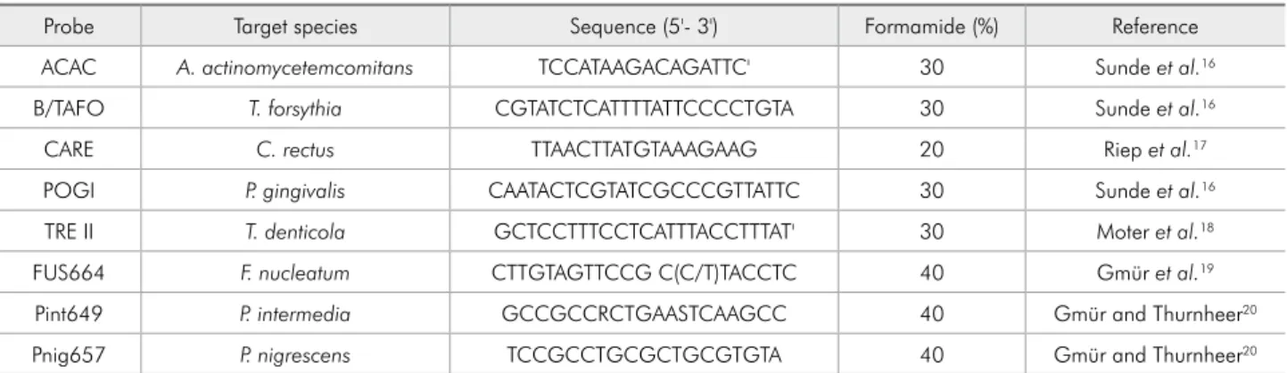
The FISH technique was carried out to identify and enumerate the periodontal pathogens in the subgingival bioilm of the women. Eight species-speciic 16S rRNA oligonucleotide probes (Operon Technologies Inc., Alameda, USA), labeled with Cy3 luorescent dye, were used (Table 1).16-20
A negative control probe (5'-CCTAGTGAC-GCCGTCGAC-3'), with a sequence that should not bind to any prokaryote rRNA, was used.21
The samples were prepared by sonication and centrifugation,22 after which a 5-mL sample was
il-trated into 0.2-µm membrane ilters.
FISH was performed according to a method de-scribed by Cottrel and Kirchman.21 The ilters were
divided into 9 equal pieces. Each piece was placed
on a slide, overlaid with a hybridization solution at a inal concentration of 2.5 ng/µL of Cy3-labeled oli-gonucleotide probe, and incubated in a sealed con-tainer overnight at 42°C. The hybridization solution contained 0.9 M NaCl, 20 mM Tris-HCl (pH 7.4), 0.01% sodium dodecyl sulfate, and the concentra-tion of formamide determined to achieve speciicity for each targeted group of bacteria (Table 1). After hybridization, the sample was transferred to a wash-ing solution and stained with 2 µg of DAPI (4',6'-di-amidino-2-phenylindole) per mL, so that the bacte-rial cells could be counted.
Total bacterial cells and cells from each species were counted by means of an Olympus BX60 micro-scope itted with a DAPI ilter 31000 and Cy3 ilter 41007a (Chroma, Bellows Falls, USA), respectively. The numbers of cells observed in 10 randomized mi-croscopic ocular grid ields per sample were counted by a single trained observer (FCM). The inal num-ber of bacteria was calculated from the multipli-cation of the dilutions performed throughout the treatment of the sample, based on the initial weight. Results are presented in cells/g.
For documentation, photomicrographs were tak-en with an Evolution VF Color Cooled camera (Me-dia Cybernetics Inc., Bethesda, USA) attached to the microscope.
Statistical analysis
The statistical analysis was performed with SPSS 14.0 (SPSS Inc., Chicago, USA). Nominal data were described by relative and/or absolute frequencies. Numerical data were described by mean, standard
Table 1 - Sequences of 16S rRNA oligonucleotide probes and formamide concentrations for fluorescence in situ hybridization.
Probe Target species Sequence (5'- 3') Formamide (%) Reference
ACAC A. actinomycetemcomitans TCCATAAGACAGATTC' 30 Sunde et al.16
B/TAFO T. forsythia CGTATCTCATTTTATTCCCCTGTA 30 Sunde et al.16
CARE C. rectus TTAACTTATGTAAAGAAG 20 Riep et al.17
POGI P. gingivalis CAATACTCGTATCGCCCGTTATTC 30 Sunde et al.16
TRE II T. denticola GCTCCTTTCCTCATTTACCTTTAT' 30 Moter et al.18
FUS664 F. nucleatum CTTGTAGTTCCG C(C/T)TACCTC 40 Gmür et al.19
Pint649 P. intermedia GCCGCCRCTGAASTCAAGCC 40 Gmür and Thurnheer20
deviation, and minimum and maximum values. The
non-parametric Mann-Whitney U-test was used for
data comparison between the Pr and N-Pr groups. Data were considered as statistically signiicant at p -values < 0.05.
Results
The mean age of the pregnant women was 26.4 years (SD ±2.5), while that of the non-pregnant women was 27.4 years (SD ±2.2). In the Pr group, the mean gestational age was 18.9 weeks (SD ±3.4).
No signiicant differences were observed between the Pr and N-Pr groups in terms of age (p = 0.584), ethnicity (p = 0.393), marital status (p = 0.251), ed-ucation (p = 0.478), and economic level (p = 0.315).
Periodontal status
Both groups had similar periodontal conditions. There were no signiicant differences between Pr
and N-Pr groups in numbers of teeth and their clini-cal parameters (Table 2). Among the Pr and N-Pr groups, 14 and 16 women, respectively, had no peri-odontal disease (p = 0.256).
Microbiological results
The mean total bacterial count was
492 × 107 ± 233 × 107 cells/g and 624 × 107 ±
301 × 107 cells/g for the Pr and N-Pr groups,
respec-tively. Statistical analysis demonstrated no signii-cant difference between groups (p = 0.13).
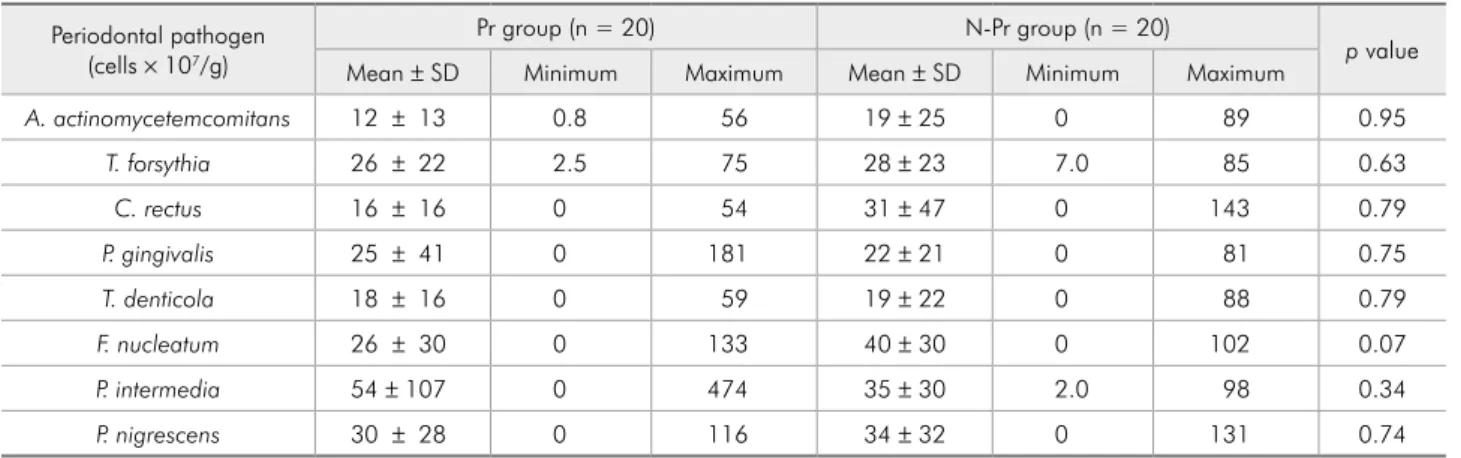
Analysis by the Mann-Whitney U-test failed to demonstrate signiicant differences between groups in the numbers of all bacterial species evaluated (Ta-ble 3).
Figure 1 shows a ield from a representative plaque sample stained for total bacterial cells (with DAPI) and for P. intermedia (with probe Pint649). The images demonstrate the presence of these
bacte-Variable Pr group (n = 20) N-Pr group (n = 20) p value
Mean ± SD Mean ± SD
Mean number of teeth 26.7 ± 1.3 26.1 ± 2.2 0.53
Teeth with BOP (%) 48.1 ± 30.9 49.3 ± 29.2 0.79
Teeth with PC (%) 12.3 ± 11.9 13.4 ± 20.2 0.61
Teeth with PD = 4–5 mm (%) 6.3 ± 15.4 1.7 ± 4.1 0.55
Teeth with PD ≥ 6 mm (%) 0.9 ± 3.3 0.2 ± 0.9 0.53
Teeth with CAL = 3–5 mm (%) 1.2 ± 3.3 0 0.15
*BOP = bleeding on probing; PC = presence of calculus; PD = probing depth; CAL = clinical attachment level; SD = standard deviation; n = number.
Table 2 - Clinical periodontal parameters of pregnant and
non-pregnant women
Table 3 - Mean, standard deviation (SD), and minimum and maximum values of the counts of periodontal pathogens from pregnant (Pr group) and non-pregnant (N-Pr group) women.
Periodontal pathogen (cells × 107/g)
Pr group (n = 20) N-Pr group (n = 20)
p value
Mean ± SD Minimum Maximum Mean ± SD Minimum Maximum
A. actinomycetemcomitans 12 ± 13 0.8 56 19 ± 25 0 89 0.95
T. forsythia 26 ± 22 2.5 75 28 ± 23 7.0 85 0.63
C. rectus 16 ± 16 0 54 31 ± 47 0 143 0.79
P. gingivalis 25 ± 41 0 181 22 ± 21 0 81 0.75
T. denticola 18 ± 16 0 59 19 ± 22 0 88 0.79
F. nucleatum 26 ± 30 0 133 40 ± 30 0 102 0.07
P. intermedia 54 ± 107 0 474 35 ± 30 2.0 98 0.34
rial cells in pregnant and non-pregnant women.
Discussion
To our knowledge, this is the irst study that has used the FISH technique to assess periodontopatho-gens during pregnancy.
All women included in the present study were from 24 to 32 years old, and most had no periodon-tal disease. Both groups were similar in terms of clinical periodontal parameters and were matched by age, ethnicity, marital status, education, and eco-nomic level. In older people, periodontal disease is likely to be more pronounced.23 Also, different
spe-ciic stages in life, such as puberty and menopause, can modulate periodontal tissue response and there-fore contribute to periodontal disease.24
Addition-ally, oral health status could also be related to the level of education, ethnicity, marital status, and economic level.23 The young mean age of this study
population, together with the similarities in the so-cio-demographic variables and clinical periodontal parameters, might have resulted in the absence of differences in terms of the composition of the sub-gingival microbiota.
Although exacerbated gingival inlammation in pregnant women has been clinically and histo-logically well-documented, its aetiology has not yet been clearly established, and it is not known why only some pregnant women develop frank signs of gingival inlammation.11,25 Changes in the sub- or
supragingival bioilm1-3 have been proposed as a
po-tential mechanism; however, there are only limited
b
d a
c
Figure 1 - DAPI staining for total bacterial cell counts and P. intermedia of subgingival plaque samples from pregnant (a, b)
data on the composition of the subgingival micro-biota during pregnancy.
An increase in P. intermedia in the subgingival bioilm of pregnant women during the second tri-mester was irst reported in 1980.1-3 In our study,
el-evated counts of P. intermedia were observed in the Pr-group; however, no signiicant differences were seen. Other investigators also showed no signiicant microbiological differences between pregnant and non-pregnant women.5,10
Differences in methods used to assess bacteria may partly explain the results reported. In most cas-es, the previously mentioned studies1-3,10,11 used
clas-sic microbiological analyses, such as culture meth-ods, which are hampered by the complexity of the periodontal microbiota and the fastidious nature of these microorganisms.11,13 These methods can also
be laborious, time-consuming and prone to statis-tical and methodological errors.13 More recently,
techniques that do not require previous culturing, like PCR-based assays and DNA-DNA hybridiza-tion, have been used to detect periodontal patho-gens in pregnant women,4,5,9 but so far they have the
disadvantage of yielding only qualitative or semi-quantitative data.13,20 FISH combines the precision
of molecular genetics with the visual information from microscopy, allowing direct visualization and identiication of individual cells within their natural microhabitat or diseased tissue.13 Additionally, this
technique showed a high standard deviation and a high variability of bacterial loads in this population by the count of absolute values, not only by the pres-ence or abspres-ence of microorganisms, as with other molecular techniques.
Increasing evidence is accumulating that oral bacteria may translocate directly into the pregnant uterus, causing localized inlammation and adverse pregnancy outcomes, notwithstanding the presence of clinical periodontitis.26F. nucleatum,P.
gingiva-lis, A. actinomycetencomitans, T. denticola, C. rec-tus, and T. forsythia are examples of such species.6-8
The high levels of these pathogens observed in preg-nant women in the present study, independent of the presence of periodontitis or the lack of differences in comparison with non-pregnant women, may be suggested as an increased risk of adverse pregnancy
outcomes. Early diagnosis and extra care in control of these microorganisms might be necessary.
One can imagine that levels of periodontal bac-teria would vary according to gestational phase. Nevertheless, previous studies have shown that the highest proportions of periodontal pathogens and the major changes in clinical parameters occur in the second trimester of pregnancy,2,5,9 which was an
inclusion criterion for this study. Continued longitu-dinal studies with the FISH technique would enable investigators to verify the actual subgingival micro-biological patterns during pregnancy and post par-tum. In addition, the periodontal pathogens in oth-er samples, e.g., saliva or gingival crevicular luid, should be investigated by FISH.
Furthermore, we do not ignore the fact that peri-odontal microbiological patterns could be associ-ated with plaque index, which was not recorded in our study. Despite that limitation, the results of this study of pregnant and non-pregnant women provide additional insights into their periodontal microbio-ta, identiied and quantiied by the FISH technique. The fact that the subgingival microbiota in healthy pregnant women did not differ from that in the non-pregnant women may suggest that the hormonal changes in pregnancy exert little, if any, inluence on colonization/growth patterns of the 8 different bacteria most commonly associated with periodontal disease, at least in women with little evidence of periodontitis.
Conclusions
The study did not conirm the hypothesis of qualitative and quantitative differences, between pregnant and non-pregnant women, in terms of the 8 periodontopathogens evaluated by the FISH tech-nique.
Acknowledgements
References
1. Jensen J, Liljemark W, Bloomquist C. The effect of female sex hormones on subgingival plaque. J Periodontol. 1981 Oct;52(10):599-602.
2. Kornman KS, Loesche WJ. The subgingival microbial flora during pregnancy. J Periodontal Res. 1980 Mar;15(2):111-22. 3. Muramatsu Y, Takaesu Y. Oral health status related to sub-gingival bacterial flora and sex hormones in saliva during pregnancy. Bull Tokyo Dent Coll. 1994 Aug;35(3):139-51. 4. Yokoyama M, Hinode D, Yoshioka M, Fukui M, Tanabe S,
Grenier D, et al. Relationship between Campylobacter rectus and periodontal status during pregnancy. Oral Microbiol Im-munol. 2008 Feb;23(1):55-9.
5. Gürsoy M, Haraldsson G, Hyvönen M, Sorsa T, Pajukanta R, Könönen E. Does the frequency of Prevotella intermedia increase during pregnancy?. Oral Microbiol Immunol. 2009 Aug;24(4):299-303.
6. Hasegawa K, Furuichi Y, Shimotsu A, Nakamura M, Yoshi-naga M, Kamitomo M, et al. Associations between systemic status, periodontal status, serum cytokine levels, and delivery outcomes in pregnant women with a diagnosis of threatened premature labor. J Periodontol. 2003 Dec;74(12):1764-70. 7. Offenbacher S, Jared HL, O’Reilly PG, Wells SR, Salvi GE,
Lawrence HP, et al. Potential pathogenic mechanisms of peri-odontitis associated pregnancy complications. Ann Periodon-tol. 1998 Jul;3(1):233-50.
8. Madianos PN, Lieff S, Murtha AP, Boggess KA, Auten RL, Beck JD, et al. Maternal periodontitis and prematurity. Part II: Maternal infection and fetal exposure. Ann Periodontol. 2001 Dec;6(1):175-82.
9. Adriaens LM, Alessandri R, Sporri S, Lang NP, Persson GR. Does pregnancy have an impact on the subgingival micro-biota?. J Periodontol. 2009 Jan;80(1):72-81.
10. Jonsson R, Howland BE, Bowden GH. Relationships between periodontal health, salivary steroids, and Bacteroides inter-medius in males, pregnant and non-pregnant women. J Dent Res. 1988 Aug;67(8):1062-9.
11. Carrillo-de-Albornoz A, Figuero E, Herrera D, Bascones-Martinez A. Gingival changes during pregnancy: II. Influence of hormonal variations on the subgingival biofilm. J Clin Periodontol. 2010 Mar;37(3):230-40.
12. Amann RI, Ludwig W, Schleifer KH. Phylogenetic identifica-tion and in situ detecidentifica-tion of individual microbial cells without cultivation. Microbiol Rev. 1995 Mar;59(1):143-69. 13. Moter A, Gobel UB. Fluorescence in situ hybridization (FISH)
for direct visualization of microorganisms. J Microbiol Meth-ods. 2000 Jul;41(2):85-112.
14. Ainamo J, Bay I. Problems and proposals for recording gin-givitis and plaque. Int Dent J. 1975 Dec;25(4):229-35. 15. López NJ, Smith PC, Gutierrez J. Higher risk of preterm birth
and low birth weight in women with periodontal disease. J Dent Res. 2002 Jan;81(1):58-63.
16. Sunde PT, Olsen I, Gobel UB, Theegarten D, Winter S, De-belian GJ, et al. Fluorescence in situ hybridization (FISH) for direct visualization of bacteria in periapical lesions of asymp-tomatic root-filled teeth. Microbiology. 2003 May;149(Pt 5):1095-102.
17. Riep B, Edesi-Neuss L, Claessen F, Skarabis H, Ehmke B, Flemmig TF, et al. Are putative periodontal pathogens reliable diagnostic markers?. J Clin Microbiol. 2009 Jun;47(6):1705-11.
18. Moter A, Hoenig C, Choi BK, Riep B, Göbel UB. Molecular epidemiology of oral treponemes associated with periodontal disease. J Clin Microbiol. 1998 May;36(5):1399-403. 19. Gmür R, Wyss C, Xue Y, Thurnheer T, Guggenheim B.
Gin-gival crevice microbiota from Chinese patients with gingivi-tis or necrotizing ulcerative gingivigingivi-tis. Eur J Oral Sci. 2004 Feb;112(1):33-41.
20. Gmür R, Thurnheer T. Direct quantitative differentiation between Prevotella intermedia and Prevotella nigrescens in clinical specimens. Microbiology. 2002 May;148(Pt 5):1379-87.
21. Cottrell MT, Kirchman DL. Community composition of ma-rine bacterioplankton determined by 16S rRNA gene clone libraries and fluorescence in situ hybridization. Appl Environ Microbiol. 2000 Dec;66(12):5116-22.
22. Epstein SS, Rossel J. Enumeration of sandy sediment bacteria: search for optimal protocol. Mar Ecol Prog Ser. 1995;117:289-98.
23. Taani DQ, Habashneh R, Hammad MM, Batieha A. The periodontal status of pregnant women and its relationship with socio-demographic and clinical variables. J Oral Rehabil. 2003 Apr;30(4):440-5.
24. Markou E, Eleana B, Lazaros T, Antonios K. The influence of sex steroid hormones on gingiva of women. Open Dent J. 2009 Jun 5;3:114-9.
25. Mealey BL, Moritz AJ. Hormonal influences: effects of diabe-tes mellitus and endogenous female sex steroid hormones on the periodontium. Periodontol 2000. 2003 Jun;32(1):59-81. 26. Han YW. Oral health and adverse pregnancy outcomes -