Weekly monitoring of the effects of conventional
external beam radiation therapy on patients with head
and neck, chest, and pelvis cancer by means of blood
cells count*
Avaliação semanal dos efeitos da radioterapia externa convencional pela contagem dos leucócitos e plaquetas de pacientes portadores de câncer nas áreas de cabeça e pescoço, tórax e pelve
Maria da Salete Fonseca dos Santos Lundgren1, Maria do Socorro de Mendonça Cavalcanti2, Divaldo de Almeida Sampaio3
OBJECTIVE: To evaluate the necessity of weekly monitoring by means of leukocyte and platelet counts of patients with head and neck, chest, and pelvis cancer submitted to conventional radiotherapy. MATERIALS AND METHODS: A hundred and one adult patients with cancer of head and neck (n = 11), chest (n = 35) and pelvis (n = 55), submitted to radiotherapy were assessed by means of leukocyte and platelet counts on a weekly basis, with a comparison between the results before and during the treatment and in correlation with the area treated, patient’s sex and age group. RESULTS: The most significant decrease in leukocytes was observed in the fourth week, when lymphocytes, total leukocytes, neutrophils, monocytes and plate-lets presented a decrease of 53.5%, 26.8%, 19.4%, 22.2% and 14.6%, respectively, in comparison with the values found before the beginning of the therapy. Geometric means for pelvis during the treatment were lower than those for chest, and head and neck. Lymphocytes demonstrated to be more sensitive to radiation therapy. No alteration was found in leukocyte or platelet counts in correlation with patients’ sex or age. CONCLUSION: Based on the results of the present study, weekly leukocyte and platelet counts do not seem to be useful in the assessment patients submitted to conventional radiotherapy for localized cancer.
Keywords: Leukocyte count; Complete blood count; External beam radiotherapy; Radiotherapy toxicity. OBJETIVO: Avaliar a necessidade de monitoração semanal, pela contagem de leucócitos e plaquetas, dos pacientes portadores de câncer das áreas de cabeça e pescoço, tórax e pelve submetidos a radioterapia externa convencional. MATERIAIS E MÉTODOS: Cento e um adultos, portadores de câncer das áreas de cabeça e pescoço (11 pacientes), tórax (35 pacientes) e pelve (55 pacientes), submetidos a radioterapia, avaliados semanalmente com leucograma e contagem de plaquetas, comparando-se as contagens das célu-las antes do início do tratamento com as obtidas nas semanas ao longo do tratamento, área tratada, sexo e faixa etária. RESULTADOS: A maior queda dos leucócitos e plaquetas ocorreu na quarta semana, quando linfócitos, leucócitos totais, neutrófilos, monócitos e plaquetas apresentaram diminuição de 53,5%, 26,8%, 19,4%, 22,2% e 14,6%, respectivamente, ao serem comparados aos valores do início do tratamento. Du-rante o tratamento, as médias geométricas da pelve foram estatisticamente menores do que as de tórax e cabeça e pescoço. Os linfócitos foram os mais sensíveis à irradiação. Não houve alteração da contagem de leucócitos e plaquetas relacionadas ao sexo ou à faixa etária. CONCLUSÃO: A partir dos resultados obtidos não parece ser necessária a contagem semanal de leucócitos e plaquetas para pacientes submetidos a radio-terapia externa convencional em campos localizados.
Unitermos: Leucograma; Hemograma completo; Radioterapia externa; Toxicidade radioterápica.
Abstract
Resumo
* Study developed at Instituto de Radioterapia Waldemir Miranda, Recife, PE, Brazil.
1. Master in Medical Sciences, Head for the Radiotherapy Unit at Hospital Universitário Oswaldo Cruz, Recife, PE, Brazil.
2. PhD in Biological Sciences, Coordinator for Post-Gradua-tion at Universidade de Pernambuco, Recife, PE, Brazil.
3. PhD in Hematology, Hematologist at Centro de Hematologia de Pernambuco (Hemope), Recife, PE, Brazil.
Mailing address: Dra. Salete Lundgren. Instituto de
Radiote-ventional external beam radiation therapy covering localized areas of the bone mar-row is similar to the alterations occurring during the whole bone marrow irradiation. However these alterations do not occur immediately and may arise weeks or even months following the radiotherapy(1,2). In
the case of localized radiotherapy, the rapia Waldemir Miranda. Rua Pacífico dos Santos, 60, Derby.
Recife, PE, Brazil, 52010-030. E-mail: [email protected]
Received February 27, 2006. Accepted after revision May 7, 2007.
Lundgren MSFS, Cavalcanti MSM, Sampaio DA. Avaliação semanal dos efeitos da radioterapia externa convencional pela conta-gem dos leucócitos e plaquetas de pacientes portadores de câncer nas áreas de cabeça e pescoço, tórax e pelve. Radiol Bras. 2008; 41(1):29–33.
INTRODUCTION
con-analysis of a medullary sample taken from the irradiated area of a patient does not re-flect the conditions of his/her whole bone marrow, and the apparent injury to a part of the medullary function does not seem to be significant for the whole hematological condition. Some studies have demonstrated that peripheral blood count results were within the regular parameters at the mo-ment of the localized medullary hypopla-sia(3–5). Therefore, after radiotherapy, the
medullary regeneration depends not only on the absorbed dose, but also on the amount of irradiated hematopoietic tissue. Few studies in the literature report the he-matological effects when small irradiation fields are utilized, usually for treatment of
localized cancer(6). Weekly peripheral
blood counts have become a routine for patients undergoing radiation therapy, as a result of the hematopoietic system sensitiv-ity and based on studies with whole-body irradiation.
MATERIALS AND METHODS
The present study was developed in the period between March and October/2004, with 101 male and female patients > 21 years of age, affected by cancer of head and neck, chest and pelvis, with an area treated > 100 cm², in five weekly fractions at a daily radiation dose ranging between 180 cGy and 200 cGy (total dose = 4500 to
5000 cGy)(7–9), during five weeks.
High-energy X-radiation was delivered by a 6 MV Clinac 600C Varian linear accel-erator, and the patients had their blood counts performed before and weekly dur-ing the treatment. Exclusion criteria were: patients previously or concomitantly sub-mitted to chemotherapy; patients subsub-mitted to hypo- or hyper-fractioned radiotherapy, or multiple-site radiotherapy; patients with a known disease of the hematopoietic sys-tem; patients with an initial leukocytes count < 4000 cells/mm³; patients with an initial platelet counts < 150000 cells/mm³. Data analyses and comparisons were made on an Excel worksheet, and geomet-ric mean was adopted as the measurement of the typical value for each type of blood cell, considering the remarkable asymme-try between data(10). Regression models for
longitudinal data were adjusted to analyze
the effects of variables such as patients’ sex, mean age and area treated on the blood counts results, utilizing generalized estima-tion equaestima-tions (GEE). All of the patients included in the sample of the present study signed a term of free and informed consent. The study was approved by the Committee for Ethics in Research of the Institution.
RESULTS
Leukocyte and platelet counts (606 blood counts) of 101 adult patients (69 women and 32 men), with ages ranging between 29 and 85 years (mean age = 59.3 years, standard deviation = 12.6 years) were evaluated. All of the patients with tumors of head and neck (n = 11; 10.9%),
chest (n = 35; 34.6%) and pelvis (n = 55;
54.5%), were submitted to conventional external beam radiotherapy. Relative and absolute values corresponding to the geo-metric means of total leukocytes, neutro-phils, eosinoneutro-phils, lymphocytes, mono-cytes and platelets before the radiotherapy (pre-treatment) and during the treatment (first, second, third, fourth and fifth weeks) of the patients with cancer of head and neck, chest and pelvis submitted to conven-tional radiotherapy are shown on Table 1. Basophils were not described, considering hat 70% of the values corresponded to zero at all moments of measurement, resulting in geometric means and medians equal to zero, so the arithmetic mean was not sig-nificant. The major decrease in the
geomet-ric means of blood counts occurred in the fourth week of treatment, when lympho-cytes, total leukolympho-cytes, neutrophils, mono-cytes and platelets presented, respectively, 53.5%, 26.8%, 19.4%, 22.2%, and 14.6% decrease as compared with the pre-treat-ment relative values. Eosinophils presented the major decrease (9.2%) in values in the first week of treatment.
The geometric means of the number of total leukocytes presented no significant variation in relation to patients’ sex and age range, but the variation in relation to the area treated was statistically significant. During the treatment, the geometric means of area treated in the pelvis were signifi-cantly lower than those for area treated in head and neck (Figure 1).
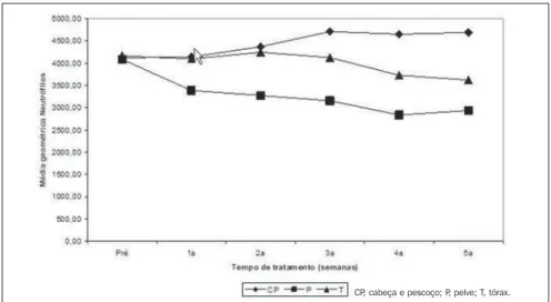
The geometric means of the number of neutrophils presented no significant varia-tion with patients’ sex and age range, but the variation in relation to the area treated was statistically significant. During the treatment, the geometric means of area treated in the pelvis were significantly lower than those for area treated in head and neck (Figure 2).
The geometric means of the number of lymphocytes presented no significant variation with patients’ sex and age range, but the variation in relation to the area treated was statistically significant. During the treatment, the geometric means of area treated in the pelvis were significantly lower than those for area treated in head and neck (Figure 3).
Table 1 Absolute and relative variations of geometric means of total leukocytes, neutrophils, eosino-phils, lymphocytes, monocytes and platelets, before and during the treatment with conventional external beam radiotherapy. Blood cells Total leucocytes Relative variation* Neutrophils Relative variation Eosinophils Relative variation Lymphocytes Relative variation Monocytes Relative variation Platelets Relative variation Pre 6864.6 100.0% 4118.1 100.0% 190.1 100.0% 1908.5 100.0% 347.6 100.0% 252.4 100.0% Weeks 1st 5847.4 85.2% 3696.4 89.8% 176.5 92.8% 1284.9 67.3% 294.9 84.8% 241.1 95.5% 2nd 5671.5 82.6% 3696.2 89.8% 196.9 103.6% 1091.2 57.2% 301.9 86.8% 221.3 87.7% 3rd 5436.8 79.2% 3621.6 87.9% 214.0 112.6% 941.0 49.3% 314.2 90.3% 216.4 85.7% 4th 5027.9 73.2% 3321.0 80.6% 189.4 99.6% 888.2 46.5% 270.3 77.8% 215.6 85.4% 5th 5182.7 75.5% 3401.1 82.6% 191.7 100.8% 957.3 50.2% 282.7 81.3% 230.4 91.3%
The geometric means of the number of monocytes presented a significantly de-creasing trend along the treatment period, but there was no significant variation in relation the patients’ sex, age range and area treated (Figure 4).
The geometric means of the number of platelets presented a significantly decreas-ing trend along the treatment period. How-ever, this trend discontinued in the third week of treatment (Figure 5). There was no significant variation of geometric means of the number of platelets in relation the pa-tients’ sex, age range and area treated.
The geometric means of the number of eosinophils present no statistically signifi-cant variation along the treatment period in relation to the patients´ sex, age range and area treated.
DISCUSSION
In the present study the total leukocytes, eosinophils, neutrophils, lymphocytes, monocytes and platelets counts were ana-lyzed, considering that these are the cells described as radiation-sensitive, and there-fore a decrease in their values is expected as a result of radiotherapy. Erithrocytes were not evaluated, considering that no decrease is expected in their values because of the compensatory capacity of these cells, with increase in their proliferation, releas-ing mature cells into the peripheral blood and maintaining their levels within the normal parameters.
Also, a statistically significant decrease was observed in the total leukocytes, neu-trophils, lymphocytes, monocytes and platelets counts as from the first week of treatment, but eosinophils presented no statistically significant difference along the treatment period. Similar results have been found by other authors studying patients with cancer of head and neck, chest and pelvis submitted to conventional radio-therapy(6,11–14). An analysis of the
geomet-ric means of blood cells counts along the treatment period, demonstrated that lym-phocytes presented the major decrease (53.5%) as compared with the pre-treat-ment value, in coincidence with the results obtained by Yang et al.(6) and Zachariah et
al.(14), with a decrease ranging between
51% and 67%. Leukocytes presented a
Figure 3. Variation of geometric means of the lymphocytes number according the area treated, in rela-tion to the treatment period (weeks).
CP, cabeça e pescoço; P, pelve; T, tórax.
Figure 2. Variation of geometric means of the neutrophils number according the area treated, in relation to the treatment period (weeks).
CP, cabeça e pescoço; P, pelve; T, tórax.
Figure 1. Variation of geometric means of the total leukocytes number according the area treated, in relation to the treatment period.
Figure 5. Variation of geometric means of the platelets number according the area treated, in relation to the treatment period (weeks).
Figure 4. Variation of geometric means of the monocytes number according the area treated, in relation to the treatment period (weeks).
26.8% decrease, similar to the decrease observed in those studies (between 14% and 30%). Neutrophils decreased 19.4% in the present study, and those authors re-ported a decrease ranging between 14% and 28%. Monocytes decreased 22%, but those authors did not report their values. Platelets decreased 14.6%, in coincidence with the value (12%) reported by Stutz & Slawson(11), Ampil et al.(12), Yang et al.(6), Blank et al.(13) and Zachariah et al.(14). The data observed in the present study, as well as those in the literature, demonstrate that lymphocytes are the most radiation-sensi-tive peripheral blood cells.
The highest decrease in the number of peripheral blood cells occurred in the fourth week of treatment, as compared with the pre-treatment. This finding also has been found by Zachariah et al.(14), in a study
counts and patient’s sex also was analyzed, and no statistically significant variation was found in the geometric means of the number of these cells. In the analysis of the peripheral blood cells count in relation to the patients´ age range, also no statistically significant variation was found in the geo-metric means of the number of peripheral blood cells, corroborating the findings re-ported by Yang et al.(6). Analyzing the
varia-tions in the peripheral blood cells counts in relation the area treated, the major decrease was found in the geometric means of total leukocytes, neutrophils and lymphocytes when the area treated was the pelvis, fol-lowed by chest and head and neck, possi-bly because of the amount of bone marrow present in the respective areas(17).
Yang et al.(6) have recommended a blood
cells count prior the initiation of the radio-therapy and in the first week of treatment, and Zachariah et al.(14) recommend the blood
cells count prior the initiation of the treat-ment, and in the first and third weeks after radiotherapy is initiated. The data found in the present study suggest that a leukocyte and platelet count should be requested prior the initiation of radiotherapy, and an addi-tional test could be requested in the fourth week after initiation of the treatment.
CONCLUSION
The major decrease in leukocytes and platelets count occurred as from the third and particularly fourth weeks following the initiation of the radiotherapy. Weekly leu-kocytes and platelets counts do not seem to be useful in the assessment of patients sub-mitted to conventional external radio-therapy for localized cancer. However, a baseline leukocyte and platelet count is recommended prior the initiation of the radiotherapy and in the first week during the treatment.
REFERENCES
1. Heier HE. The influence of therapeutic irradiation of blood and peripheral lymph lymphocytes. Lymphology. 1978;11:238–42.
2. Plowman PN. The effects of conventionally frac-tionated, extended portal radiotherapy on the human peripheral blood count. Int J Radiat Oncol Biol Phys. 1983;9:829–39.
3. Sykes MP, Chu FCH, Wilkerson WG. Local bone-marrow changes secondary to therapeutic irradia-tion. Radiology. 1960;75:919–24.
with 299 patients with cancer of head and neck, chest and pelvis submitted to conven-tional external beam radiation therapy, demonstrating the major decrease in the pe-ripheral blood cells count in the third week after the beginning of the treatment. Differ-ently, Yang et al.(6), in a study with 117 patients with cancer of head and neck, chest and pelvis submitted to conventional external beam radiation therapy, has ob-served that the major decrease in the pe-ripheral blood cells count occurred in the first week of treatment. Studies in the lit-erature have reported that the decrease in the peripheral blood cells counts starts in the first weeks of conventional external beam radiation therapy(11,15,16).
4. Sykes MP, Savel H, Chu FCH, et al. Long-term effects of therapeutic irradiation upon bone mar-row. Cancer. 1964;17:1444–8.
5. Cowall DE, MacVittie TJ, Parker GA, et al. Ef-fects of low-dose total-body irradiation on canine bone marrow function and canine lymphoma. Exp Hematol. 1981;9:581–7.
6. Yang FE, Vaida F, Ignacio L, et al. Analysis of weekly complete blood counts in patients receiv-ing standard fractionated partial body radiation therapy. Int J Radiat Oncol Biol Phys. 1995;33: 607–17.
7. Cardoso MFA, Novikoff S, Tresso A, et al. Preven-ção e controle das seqüelas bucais em pacientes irradiados por tumores de cabeça e pescoço. Ra-diol Bras. 2005;38:107–15.
8. Esteves SCB, Oliveira ACZ, Cardoso H, et al. Braquiterapia de alta taxa de dose no tratamento do carcinoma da próstata: análise da toxicidade
aguda e do comportamento bioquímico. Radiol Bras. 2006;39:127–30.
9. Chen MJ, Nishimoto IN, Novaes PERS, et al. Radioterapia adjuvante no tratamento do câncer de endométrio: experiência com a associação de radioterapia externa e braquiterapia de alta taxa de dose. Radiol Bras. 2005;38:403–8.
10. Davis CS. Statistical methods for the analysis of repeated measurements. New York: Springer; 2002.
11. Stutz FH, Slawson RG. Local radiotherapy and the peripheral white blood cell count: review of 203 treatment record. Mil Med. 1976;141:390–1.
12. Ampil FL, Burton GV, Li BDL. “Routine” weekly blood counts during breast irradiation for early stage cancer: are they really necessary? Breast J. 2001;7:450–2.
13. Blank KR, Cascardi MA, Kao GD. The utility of serial complete blood county monitoring in
pa-tients receiving radiation therapy for localized prostate cancer. Int J Radiat Oncol Biol Phys. 1999;44:317–21.
14. Zachariah B, Jacob SS, Gwede C, et al. Effect of fractionated regional external beam radiotherapy on peripheral blood cell count. Int J Radiat Oncol Biol Phys. 2001;50:465–72.
15. Saunders AM. White blood cells: what to do be-yond measurement. Blood Cells 1980;6:357–64.
16. Tell R, Heiden T, Grarath F, et al. Comparison be-tween radiation-induced cell cycle delay in lym-phocytes and radiotherapy response in head and neck cancer. Br J Cancer. 1998;77:643–9.