Keywords
Heart failure/therapy; autonomic nervous system; baroreflex/drugs effects.
Behavior of Central and Peripheral Chemoreflexes in Heart Failure
Guilherme Veiga Guimarães
1,2, Juliana Fernanda Canhadas Belli
1,2, Fernando Bacal
1, Edimar Alcides Bocchi
1Instituto do Coração do Hospital das Clínicas da Faculdade de Medicina da Universidade de São Paulo1; Laboratório de Atividade Física e
Saúde do Centro de Práticas Esportivas da Universidade de São Paulo2, São Paulo, SP - Brazil
Mailing address: Guilherme Veiga Guimarães •
Rua Dr. Baeta Neves, 98 - Pinheiros - 05444-050 - São Paulo, SP - Brazil E-mail: [email protected]
Manuscript received June 23, 2009; revised manuscript received August 20, 2009; accepted July 10, 2009.
alterations result in systolic and/or diastolic dysfunction and, consequently, in the loss of functional capacity, decrease in quality of life and increased morbidity and mortality3,4.
The HF syndrome is associated with hemodynamic disorders followed by systemic alterations, endothelial dysfunction, neurohormonal activation with increased catecholamine release, higher brain natriuretic peptide (BNP) levels5 and release of pro-inflammatory factors that contribute to heart dilation and poorer performance at physical exercises5,6. Patients with HF present higher levels of ventilation for a certain workload when compared to normal individuals7. This fact results in low ventilatory efficiency and is related with higher ventilation related to CO2 production, which is an important predictor of poor prognosis, in addition to being a limiting factor for the practice of exercises8,9.
Hyperventilation can occur due to several causes and among them, a hyperactive chemoreceptor reflex, one of the several abnormalities in the cardiovascular reflex control related to the increased sympathetic tonus in HF7,10. The signal hyperactivation originated from the receptors located in the skeletal muscles (mechanoreceptors - metaboreceptors) is a recently proposed hypothesis to explain the origin of symptoms of intolerance to physical exertion8.
The sympathoinhibitory cardiovascular reflexes, such as the arterial baroreceptor reflex, are significantly suppressed in HF. In turn, the sympathoexcitatory reflexes, including the arterial chemoreceptor reflex and the sympatho-afferent cardiac reflexes, are increased in HF. Although the functional alterations of the reflexes have been used independently to illustrate the sympathetic excitation observed in HF, the interaction between these reflexes in normal and pathological conditions, especially their contribution to the sympathoexcitatory state found in HF, has not been broadly studied11. Therefore, the objective of this study is to carry out a review of the action mechanism of peripheral and central chemoreceptors.
Cardiopulmonary regulation
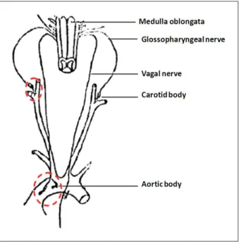
The cardiopulmonary recptores are located, basically, in association with the large arterial vessels in the chest and neck and transmit stimuli to the respiratory center to help regulate the respiratory activity. Most of them are found in the aortic and carotid bodies, along their afferent nervous connections with the respiratory cento. The carotid bodies are located bilaterally in the bifurcations of the main carotid arteries and their afferent fibers pass through Hering’s nerves to the glossopharyngeal nerves and then to the medulla oblongata. The aortic bodies are located along the aortic arch and their nervous fibers pass into the medulla oblongata through
Abstract
The heart failure (HF) syndrome can be defined as the final pathway of any type of heart disease. The sympatho-inhibitory cardiovascular reflexes, such as the arterial baroreceptor reflex, are significantly decreased in HF. Patients with HF present higher ventilation for a certain workload when compared with normal individuals. This fact generates low ventilatory efficiency and is related to higher ventilation associated with the carbon dioxide production, which is a predictor of bad prognosis, in addition to being a limiting factor for the practice of exercises. There is evidence that the autonomic imbalance contributes to the pathogenesis and the progression of heart failure. The chemoreflexes are the main mechanisms of control and regulation of the ventilatory responses to the changes in concentrations of arterial oxygen and carbon dioxide. The chemoreflex activation causes an increase in the sympathetic activity, heart rate, arterial pressure and minute volume. However, the increase in the minute volume and the arterial pressure, due to negative feedback, cause inhibition of the sympathetic response at the chemoreflex activation. In spite of the functional alterations of the reflexes, their behavior in normal and pathological conditions, especially their contribution to the sympathoexcitatory state observed in HF has not been broadly studied.
Therefore, this review aims at integrating the knowledge on central and peripheral chemoreflexes in HF syndrome, as well as clarifying the influence of the heart failure drug therapy on the chemoreflexes.
Introduction
the vagus nerve and then to the spinal cord through the sympathetic nerves12 (Figure 1). Three sets of cardiopulmonary receptors can be identified: 1) non-myelinated vagal cardiac afferents, which are small nervous terminals scattered in the cardiac chambers, sensitive to mechanical distension due to alterations in the atrial pressure or diastolic-end pressure and, as they cause similar responses to the arterial baroreceptors, it is supposed that both use the same neuronal pathways; 2) myelinated vagal cardiac afferents, located especially at the junction of large vessels and atria, spontaneously active during systole and diastole and that supply information to the central nervous system on the degree of atrial filling and heart rate. In situations of increased volemia, they cause a decrease in the renal sympathetic nervous activity and, consequently, a decrease in the release of vasopressin by the neurohypophysis. In this condition there is also an increase in the levels of brain natriuretic peptide (BNP) levels, which induces increases in diuresis and natriuresis, in addition to the inhibition of renin and aldosterone release and 3) spinal afferents of which trajectory coincides with that of sympathetic ones and are distributed along the coronary arteries, the cardiac chambers and great vessels of the chest.
Chemoreflex control
The autonomous nervous system allows the body to adjust its circulation and ventilation to maintain the oxygen supply to the tissues. The autonomic balance is maintained by the complex interaction of the arterial baroreflex, central and peripheral chemoreflex, ergoreflex and lung stretch reflex (Figure 2). There is evidence that the autonomic imbalance contributes to the pathogenesis and progression of heart failure15.
The chemoreflexes are the main mechanisms of control and regulation of the ventilatory responses to the changes in arterial oxygen and CO2 concentrations
16. The peripheral
Figure 1 -Chemoreceptors located outside the central nervous system, responsive to the alterations in concentrations of oxygen, carbon dioxide and hydrogen ions. They are located in association with the great arteries of the chest and neck, here represented by the carotid and aortic bodies, along their afferent nervous connections, via the vagal and glossopharyngeal nerves, respectively, with the respiratory center in the medulla oblongata. Adapted from Guyton AC and Hall JE15.
Figure 2 -Mechanisms of autonomic control in heart failure. The sensitivity of the arterial baroreceptors and cardiopulmonary receptors is decreased, whereas the sensitivity of the chemoreceptors is increased. The response to this altered balance includes the generalized increase in the sympathetic activity, resulting in increased arterial pressure, ventilation, renal vascular resistance and peripheral vascular resistance, whereas there is a decrease in the parasympathetic activity, resulting in the increased heart rate.
chemoreceptors located in the carotid and aortic bodies, with afferents to the respiratory center in the medulla oblongata and the nucleus of the solitary tract, respond primarily to hypoxia16,17. The central chemoreceptors, located on the ventral surface of the spinal cord, respond primarily to hypercapnia18. Both responses to alterations in the concentrations of O2 and CO2, respectively, increase pulmonary ventilation. Almost simultaneously, another set of neurons that reach the nucleus of the solitary tract induces the increase in the sympathetic nervous activity. Both chemoreceptor mechanisms have a strong influence on neural control of circulation, especially in situations involving significant changes in the arterial concentrations of oxygen and/or CO2. The chemoreflex activation causes an increase in the sympathetic activity, heart rate, arterial pressure and minute volume. However, the increase in the minute volume and the arterial pressure, by negative feedback, causes inhibition of the sympathetic response to the chemoreflex activation. Therefore, chemoreflexes trigger several cardiovascular and respiratory responses, with complex interactions between their responses. To define any abnormality in the chemoreflex function, it is essential to consider the individual contribution of each component that integrates this response. In patients with HF, the stimulation of the central and peripheral chemoreceptors causes a marked increase in pulmonary ventilation and the sympathetic nervous activity, characterized by a selective potentiation of these responses14,19.
Contribution of the chemoreceptors under
normoxia
The chemoreflexes (central and peripheral) have a crucial role in the control of alveolar ventilation to guarantee that the gas exchanges in the lungs continuously supply the metabolic demand through the oxygen uptake and CO2 removal.
Frequently overestimated, however, is the fact that these reflexes also have a significant effect on the cardiac and vascular control to regulate the blood flow and then the levels of tissue gas exchange. An important component for the activation of the chemoreflex is an increase in the sympathetic activity on the vascular beds. This response helps the maintenance of the arterial pressure in the presence of the direct vasodilating effects of hypoxemia or hypercapnia and then helps to maintain the pressure adequate for the blood flow and tissue gas exchange.
The idea that the chemoreflexes contribute to the sympathetic activity at rest in pathological states has been the hypothesis that the chemoreflexes would not be activated at rest (normoxia and isocapnia) and, therefore, they would have little influence on the sympathetic tonus under normal conditions. However, this hypothesis is quite controversial. In normal individuals at rest, the hyperoxia, which inhibits the activity of peripheral echemoreceptors, decreases the sympathetic nervous activity20. However, individuals with sleep apnea present an increased chemoreflex activity under normoxia. Thus, it can be suggested that the chemoreflexes contribute to the increased sympathetic tonus in heart failure, even in the absence of hypoxia21.
Chemoreflexes and ventilatory
abnormalities
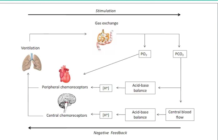
The respiration is stimulated by the chemoreflexes that include the central and peripheral receptors, the central nervous system and the respiratory muscles. The chemoreflexes are part of the negative feedback of a feedback cycle. The cycle is completed by the progression in which the alveolar ventilation controls the O2 extraction and the elimination of CO2. The cycle progression associates the dependence of PCO2 and PO2 in ventilation22. The simulation of chemoreceptors results in an increased alveolar ventilation that leads to an increased elimination of CO2 and decrease in PCO2, stimulating the chemoreceptors por meiothrough the increased concentration of H+ ions; therefore, concluding the negative feedback designation of the system23 (Figure 3). The increased sensitivity of the central and peripheral chemoreflexes can have an important role in the ventilatory control alteration observed in heart failure and a significant correlation can be found between the ventilatory response to exercise and the hypoxic chemosensitivity at rest24. The inhibition of the peripheral chemoreflex has been shown to increase the performance at exercise and decrease the ventilatory response in patients with HF. These observations suggest that the marked hyperpnea at exercise can be influenced by the increased peripheral chemoreflex activity in these patients25,26.
A significant positive correlation was observed between the central hypercapnic ventilatory response and the sleep time percentage in patients with HF and Cheyne-Stokes respiration. The fact that supplemental O2 ane CO2 during sleep increase the Cheyne-Stokes respiration suggests a contribution of the increased chemosensitivity in this disorder. This increase can also explains the tendency of lower-than-normal PaCO2 obseraesduring sleep and wakefulness in individuals with Cheyne-Stokes respiration26.
In heart failure of Chagasic etiology, the ventilatory and autonomic responses increased with the stimulation of the peripheral chemoreflex. On the other hand, the ventilatory and autonomic responses decreased during hypercapnia, associated with the activation of central chemoreceptors in this group of patients27,28.
Additionally, it is worth mentioning that, together with Chagas’ disease, diabetes mellitus is a disease that can course with autonomic neuropathy, leading to dysautonomy and very often, patients with HF present diabetes as a comorbidity.
There have been few studies associating diabetes and the chemoreflex function. Studies with experimental models have shown that the chemoreflex is decreased29. In humans, it has been observed a decrease in the peripheral chemoreflex in individuals with type 2 diabetes, especially regarding the response to hypoxia30.
Influence of drug therapy on the central and
peripheral chemoreflexes
Figure 3 -Flow chart illustrating the inluence of chemoreceptors in ventilation control. The pulmonary ventilation controls PCO2 and PO2; part of the cycle stimulation. The arterial and central concentration of the hydrogen ion ([H+]) is determined by their respective PCO2, ionic difference and concentrations of albumin ([Alb]) and phosphate ([PO4-]). The concentration of ions H+ and PO
2 control the ventilation through the ventilatory stimulation of the chemorelexes, apart from the negative feedback of the cycle.
angiotensin II receptor blockers (ARB). The treatment with beta-blockers significantly altered the morbidity and mortality of patients with HF, as shown in the COPERNICUS31, CIBIS-II32 and MERIT-HF studies33. The beta-blockers decrease the progression of left ventricular dysfunction, decrease the sympathetic activity and thus improve the prognosis of patients HF34. Their beneficial effects are evident, regardless of age or the low, intermediate or high risk that the patients present for HF35. However, that is not translated into exercise capacity improvement, at either submaximal or maximal exertion.
The beta-blockers can act on the beta-1 and/or beta-2 adrenergic receptors. The respiratory system is characterized by a prevalence of beta-2 receptors, which are characterized by regulating the bronchial tonus, whereas the alveolar beta receptors regulate the reabsorption of fluids that affect gas diffusion; however, more than 90% of the beta receptors are located in the alveoli, in which the beta-2 type predominate (70%)36,37. Although the treatment with beta-blockers is an effective therapy for HF, to date, only a few studies have evaluated the effect of beta-blockers on exercise-induced hyperventilation38. Additionally, there are no data regarding the effect of beta-blockers during exercise under hypoxia, when the arterial hypoxemia increases the activity of the chemoreceptors39.
The angiotensin-converting enzyme inhibitors (ACEI) inhibit the conversion of angiotensin I (AI) into angiotensin II (AII), decreasing the levels of the latter in the blood. The formation
of AII involves the sequential cleavage of angiotensinogen. The angiotensinogen is transformed into angiotensin I through the action of renin, synthesized by the kidney, and then it is hydrolyzed into angiotensin II, through the action of the angiotensin-converting enzyme (ACE). The renin-angiotensin-aldosterone system can be blocked at different places and by several mechanism. The angiotensin-converting enzyme activity is predominantly found in the endothelium of lung vessels. The angiotensin II promotes aldosterone release by the adrenal glands. The agents that inhibit ACE interfere with the conversion of AI into AII, the latter being considered one of the most important systemic hormones, among which are noradrenaline, vasopressin and endothelin.
The angiotensin II receptor blockers (ARB) are drugs that act on AT1 receptors, responsible for all known actions of AII, including vasoconstriction, aldosterone release and effects on the myocardium and vasculature (Figure 4). There are hypotheses to explain their influence on the dynamics of central and peripheral chemoreflexes. Although the cell and molecular mechanisms involved in the increased sensitivity of the carotid chemoreceptors during HF are yet to be elucidated, there is evidence that the increased levels of angiotensin II in the central nervous system accentuates the peripheral chemoreflex sensitivity and increases the peripheral sympathetic nervous activity40,41.
hypersensitivity of chemoreceptors in HF to the increased levels of AII and the decreased levels of nitric oxide42,43.
Recent studies demonstrated that the endogenous levels of AII and the expression of AT1 receptors are increased in the carotid bodies in animal models of HF and that mediates the increased sensitivity of chemoreceptors to hypoxia. Therefore, the drug treatment of with ARB can affect the chemoreflexes in HF, but most studies are experimental animal models, which limits the extrapolation of these conclusions regarding these mechanisms to humans41,44.
Perspectives
In spite of the divergent results, currently the evidence points out to an increase in the activity of central and peripheral chemoreflexes in HF, which is correlated with disease severity. Although the mechanisms responsible for the altered central chemoreflex sensitivity are still unclear, the resulting alterations can be responsible, in part, for the increased ventilatory response at exercise, dyspnea, Cheyne-Stokes respiration and sympathetic hyperactivation observed in heart failure. As for the drugs used in the clinical management of HF, although
they decrease the sympathetic hyperstimulation and the sensitivity of the central and peripheral chemoreflexes, they do not improve exercise performance, even if they attenuate symptoms such as dyspnea.
Therefore, the genesis of the signs and symptoms of the heart failure syndrome requires further clarification, so that we can intervene and treat the syndrome in order to improve prognosis, quality of life and the performance at activities of daily living.
Potential Conflict of Interest
No potential conflict of interest relevant to this article was reported.
Sources of Funding
There were no external funding sources for this study.
Study Association
This study is not associated with any post-graduation program.
Figure 4 -The beta-blockers (β-block) and the angiotensin II receptor blockers (ARB) can act on the cardiac, renal, vascular or central adrenergic receptors (AR).
References
1. Hunt SA, Abraham WT, Chin MH, Feldman AM, Francis GS, Ganiats TG, et al. ACC/AHA 2005 Guideline update for the diagnosis and management of chronic heart failure in the adult. American College of Cardiology / American Heart Association. Task Force on Practice Guidelines Writing Committee to Update the 2001 Guidelines for the Evaluation and Management of Heart Failure: developed in collaboration with the American College of Chest Physicians and the International Society for Heart and Lung Transplantation: endorsed by the Heart Rhythm Society. Circulation. 2005; 112 (12): e154-235. 2. Felker GM, Adams KF Jr, Konstam MA, O’Connor CM, Gheorghiade M. The
problem of decompensated heart failure: nomenclature, classification, and risk stratification. Am Heart J. 2003;145 (Suppl 2):18-25.
3. Jessup M, Brozena S. Heart failure. N Engl J Med. 2003; 348 (20): 2007-18. 4. Ramos RB, Fabri J Jr, Mansur AP. A insuficiência cardíaca no Brasil e no mundo e avaliação de sua influência socioeconômica. In: Nobre F, Serrano Jr CV. Tratado de cardiologia SOCESP. São Paulo: Manole; 2005.
5. Guimarães JI, Mesquita ET, Bocchi EA, Vilas-Boas F, Montera MW, Moreira MCV, et al. Revisão das II Diretrizes da Sociedade Brasileira de Cardiologia para o diagnóstico e tratamento da insuficiência cardíaca. Arq Bras Cardiol. 2002; 79 (supl. 4): 1-30.
6. Piepoli MF, Dimopoulos K, Concu A Crisafulli A. Cardiovascular and ventilatory control during exercise in chronic heart failure: role of muscle reflexes. Int J Cardiol. 2008; 130 (1): 3-10.
7. Agostoni P, Contini M, Magini A, Apostolo A, Cattadori G, Bussotti M, et al. Carvedilol reduces exercise-induced hyperventilation: a benefit in normoxia and a problem with hypoxia. Eur J Heart Fail. 2006; 8 (7): 729-35. 8. Kleber FX, Vietzke G, Wernecke KD, Bauer U, Opitz C, Wensel R, et al.
Impairment of ventilatory efficiency in heart failure: prognostic impact. Circulation. 2000; 101 (24): 2803-9.
9. Agostoni P, Pellegrino R, Conca C, Rodarte JR, Brusasco V. Exercise hyperpnea in chronic heart failure: relationships to lung stiffness and expiratory flow limitation. J Appl Physiol. 2002; 92 (4): 1409-16.
10. Ponikowski P, Chua TP, Francis DP, Capucci A, Coats AJS, Piepoli MF. Muscle ergoreceptor overactivity reflects deterioration in clinical status and cardiorespiratory reflex control in chronic heart failure. Circulation. 2001; 104 (19): 2324-30.
11. Wang WZ, Gao L, Wang HJ, Zucker IH, Wang W. Interaction between cardiac sympathetic afferent reflex and chemoreflex is mediated by the NTS AT1 receptors in heart failure. Am J Physiol Heart Circ Physiol. 2008; 295 (3): H1216–H1226.
12. Guyton AC, Hall JE. Tratado de fisiologia médica. 11ª. ed. São Paulo: Elsevier; 2006.
13. Dibner-Dunlap ME, Smith ML, Kinugawa T, Thames MD. Enalaprilat augments arterial and cardiopulmonary baroreflex control of sympathetic nerve activity in patients with heart failure. J Am Coll Cardiol. 1996; 27 (2): 358-64. 14. Corrêa LMA, Santos TSNP, Moura THP, Negrão CE. Alterações autonômicas
na insuficiência cardíaca: benefícios do exercício físico. Rev SOCERJ. 2008; 21 (2): 106-11.
15. Abboud FM, Thames MD. Interaction of cardiovascular reflexes in circulatory control. In: Sheperd JT, Abboud FM, Geiger SR. (editors). Handbook of physiology section 2: the cardiovascular system. Bethesda: American Physiological Society; 1983. p. 557-622.
16. Wade JG, Larson CP Jr, Hickey RF, Ehrenfeld WK, Severinghaus JW. Effect of carotid endarterectomy on carotid chemoreceptor and baroreceptor function in man. N Engl J Med. 1970; 282 (15): 823-9.
17. Lugliani RB, Whipp BJ, Seard C, Wasserman K. Effect of bilateral carotid-body resection on ventilatory control at rest and during exercise in man. N Engl J Med. 1971; 285 (20): 1105-11.
18. Gelfand R, Lambertsen CJ. Dynamic respiratory response to abrupt change of inspired CO2 at normal and high PO2. J Appl Physiol. 1973; 35 (6): 903-13. 19. Narkiewicz K, Pesek CA, van de Borne PJH, Kato M, Somers VK. Enhanced
sympathetic and ventilatory responses to central chemoreflex activation in heart failure. Circulation. 1999; 100 (3): 262-7.
20. Seals DR, Johnson DG, Fregosi RF. Hyperoxia lowers sympathetic nerve activity at rest but not during exercise in humans. Am J Physiol. 1991; 260 (5 Pt 2): R873-8.
21. Schultz HD, Sun SY. Chemoreflex function in heart failure. Heart Fail Rev. 2000; 5 (1): 45-56.
22. Duffin J. Role of acid-base balance in the chemoreflex control of breathing. J Appl Physiol. 2005; 99 (6): 2255-65.
23. Ainslie PN, Duffin J. Integration of cerebrovascular CO2 reactivity and chemoreflex control of breathing: mechanisms of regulation, measurement and interpretation. Am J Physiol Regul Integr Comp Physiol. 2009; 296 (5): R1473-95.
24. Barrett KM, Ackerman RH, Gahn G, Romero JM, Candia M. Basilar and middle cerebral artery reserve: a comparative study using transcranial Doppler and breathholding techniques. Stroke. 2001; 32 (12): 2793-6. 25. Atkinson JL, Anderson RE, Sundt TM Jr. The effect of carbon dioxide on the
diameter of brain capillaries. Brain Res. 1990; 517 (12): 333-40. 26. Hanly P, Zuberi N, Gray R. Pathogenesis of Cheyne-Stokes respiration in
patients with congestive heart failure: relationship to arterial PCO2. Chest. 1993; 104 (4): 1079-84.
27. Barreto-Filho JAS, Consolim-Colombo FM, Lopes HF, Martins Sobrinho CR, Guerra-Riccio GM, Krieger EM. Dysregulation of peripheral and central chemoreflex responses in chagas’ heart disease patients without heart failure. Circulation. 2001; 104 (15): 1792-8.
28. Barreto Filho JA. Quimiorreflexo na cardiopatia da doença de Chagas. [Tese]. São Paulo: Faculdade de Medicina da Universidade de São Paulo; 1998. 29. De Angelis K, Schaan BA, Rodrigues B, Malfitano C, Irigoyen MC. Disfunção
autonômica cardiovascular no diabetes mellitus experimental. Arq Bras Endocrinol Metab. 2007; 51: 2.
30. Weisbrod CJ, Eastwood PR, O’Driscoll G, Green DJ. Abnormal ventilatory responses to hypoxia in Type 2 diabetes. Diab Med. 2005; 22 (5): 563-8. 31. Packer M, Fowler MB, Roecker EB, Coats AJ, Katus HA, Krum H, et al.
Effect of carvedilol on the morbidity of patients with severe chronic heart failure: results of the carvedilol prospective randomized cumulative survival (COPERNICUS) Study. Circulation. 2002; 106 (17); 2194-9.
32. The Cardiac Insufficiency Bisoprolol Study II (CIBIS-II) a randomized trial. Lancet. 1999; 353 (9146): 9-13.
33. Effect of metoprolol CR/XL in chronic heart failure: Metoprolol CR/XL Randomised Intervention Trial in Congestive Heart Failure (MERIT-HF). Lancet. 1999; 353 (9169): 2001-7.
34. Greenberg B. Nonselective versus selective beta-blockers in the management of chronic heart failure: clinical implications of the carvedilol or metoprolol European Trial. Rev Cardiovasc Med. 2004; 5 (Suppl 1): S10-7.
35. Remme WJ. Beta-blockade as first-line therapy in the elderly heart failure patient - the proper approach or asking for trouble? Cardiovasc Drugs Ther. 2008; 22 (5): 347-50.
36. Agostoni P, Contini M, Cattadori G, Apostolo A, Sciomer S, Bussotti M, et al. Lung function with carvedilol and bisoprolol in chronic heart failure: is beta selectivity relevant? Eur J Heart Fail. 2007; 9 (8): 827-33.
37. Puri S, Dutka DP, Baker BL, Hughes JMB, Cleland JGF. Acute saline infusion reduces alveolar-capillary membrane conductance and increases airflow obstruction in patients with left ventricular dysfunction. Circulation. 1999; 99 (9): 1190-6.
38. Witte KKA, Thackray SRD, Nikitin NP, Cleland JGF, Clark AL. The effects of alpha and beta blockade on ventilatory responses to exercise in chronic heart failure. Heart. 2003; 89 (10): 1169-73.
39. Prabhakar NR, Peng YJ. Peripheral chemoreceptors in health disease. J Appl Physiol. 2004; 96 (1): 359-66.
41. Li YL, Xia XH, Zheng H, Gao L, Li YF, Liu D, et al. Angiotensin II enhances carotid body chemoreflex control of sympathetic outflow in chronic heart failure rabbits. Cardiovasc Res. 2006; 71 (1): 129-38.
42. Liu JL, Irvine S, Reid IA, Patel KP, Zucker IH. Chronic exercise reduces sympathetic nerve activity in rabbits with pacing-induced heart failure: a role for angiotensin II. Circulation. 2000; 102 (15): 1854-62.
43. van deWal RM, Plokker HW, Lok DJ, Boomsma F, van der Horst FA, van Veldhuisen DJ, et al. Determinants of increased angiotensin II levels in severe chronic heart failure patients despite ACE inhibition. Int J Cardiol. 2006; 106 (3): 367-72.