Clinicoradiological Session
Case 1/2011 - 15-Year-Old Male Adolescent with Congenital
Tricuspid Insufficiency Simulating Ebstein’s Anomaly
Edmar Atik
Clínica Privada do Dr. Edmar Atik
Mailing address: Edmar Atik •
Rua Dona Adma Jafet, 74 conj. 73 - Bela Vista - 01308-050 - São Paulo, SP - Brazil
E-mail: [email protected], [email protected]
Manuscript received July 29, 2010; revised manuscript received December 14, 2010; accepted December 14, 2010.
Keywords
Heart defects, congenital; Ebstein anomaly; tricuspid valve insufficiency; ventricular dysfunction, right.
septum, with discrete mobility, thus leading to the diagnosis of Ebstein’s anomaly. Right ventricular cavity corresponded to 54 mm, left ventricle to 36 mm and right ventricular function by Simpson’s method was 44.0%. Over time, there was a progressive right ventricular dilation of 25 mm in diameter at month 14, and 54 mm at age 15. During these periods, the left ventricle maintained the same diameter of 37 mm (Figure 2).
Magnetic resonance imaging - confirmed the diagnosis with a sharp increase of right cavities and right ventricular dysfunction.
Diagnosis
Ebstein’s anomaly with severe tricuspid regurgitation and right ventricular dysfunction in patient with few symptoms.
Clinical reasoning
The diagnosis of tricuspid regurgitation was easily established and Ebstein’s anomaly suggested from the outset by echocardiography. However, this image may be confused and more rigorous and precise criteria should be devised.
Differential diagnosis
Confused with Ebstein’s anomaly by echocardiography, any other abnormality of the tricuspid valve, either congenital or acquired, could be clinically exteriorized in the same manner and with the same implications regarding the conduct established.
Conduct
The surgical correction of the defect was considered in view of abnormalities resulting from progression of chronic tricuspid regurgitation since birth, and right ventricular dysfunction.
Upon the surgery, the tricuspid valve was trivalvular but with severe dysplasia. The valves were tapered valve and the septal valve was not coupled to the ventricular septum, but it was rectified due to marked dilation of the tricuspid ring and dilated right ventricle. Upon ruling out the diagnosis of Ebstein’s anomaly, the anterior and posterior valves were removed and replaced with bioprosthesis number 31, on cardiopulmonary bypass at 28o C.
The immediate outcome was complicated by fibrillo-flutter on the first and second postoperative days, twice controlled with amiodarone and electrical reversion. Heart murmur disappeared and further examination still showed the same aspects found before the surgery.
Clinical aspects
Since birth, the presence of tricuspid insufficiency due to an alleged Ebstein’s anomaly was known. This was exteriorized by a heart murmur in the first hours of life. Over time, the patient remained with mild fatigue upon exertion and no specific medication and has developed normal weight and height. Six months ago, due to progressive increase in right heart chambers and the finding of right ventricular dysfunction by nuclear magnetic resonance, surgical correction of the defect was considered.
On physical examination, the patient presented good general condition, eupneic, blushed, normal pulses, weighing 72 kg and height of 1.73 m. BP was at 110/80 mmHg and HR at 82 bpm. The aorta could not be felt at suprasternal notch. Precordium: Mild systolic impulsions at the left sternal border and diffuse apical impulse in the fourth and fifth intercostal spaces. The heart sounds were normal and systolic and diastolic murmurs could be heard, +/++ intensity, harsch, in the third and fourth intercostal spaces at the sternal border irradiating to the mitral area and right sternal border. Liver could not be felt.
Complementary tests
Electrocardiogram - since birth, showed signs of right bundle branch block with QRS duration of 0.14’’.
Chest radiography - shows marked increase in the cardiac area due to right chambers, excavated medial arch and decreased pulmonary vascular markings (Figure 1).
Echocardiography - showed severe tricuspid regurgitation and increased right cavities. There was a marked dilatation of the tricuspid ring and septal valve coupled to the ventricular
Clinico Radiological Session
Edmar Atik
Arq Bras Cardiol 2011; 96(1): e1-e2
Figure 1 -Chest radiography in the immediate postoperative period shows sharp increase in the right cavities, excavated medial arch and decreased pulmonary vascular markings, such as those found before the surgery, suggesting Ebstein’s anomaly.
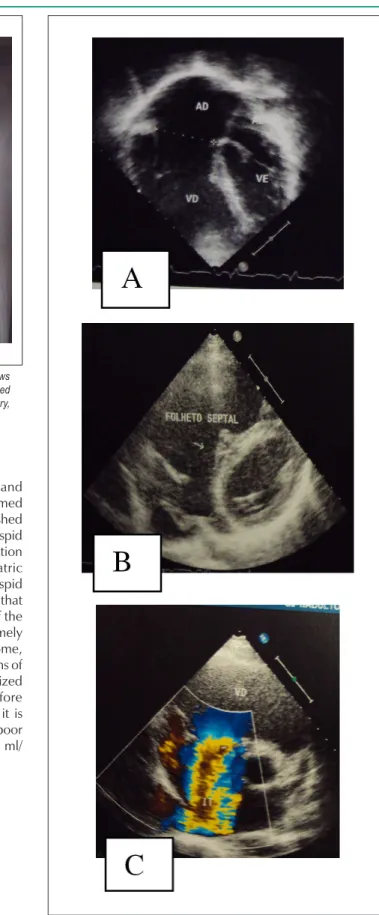
Figure 2 -Echocardiogram shows a sharp increase of the right cavities in 4-chamber apical section, in A, the septal tricuspid valve was not very mobile and “attached” to the ventricular septum without coaptation with the other valves in parasternal short-axis in B, and marked tricuspid regurgitation in C.
Comments
The septal tricuspid valve with reduced mobility and rectified distorted the diagnostic impression that assumed Ebstein’s anomaly. The true anatomical diagnosis, established upon the surgery, dismissed this anomaly as a cause of tricuspid regurgitation, present since birth. The tricuspid malformation found upon the surgery is rarely found in the pediatric specialty, but it behaves like any cause of congenital tricuspid regurgitation with well developmental tolerance, given that the patient had few symptoms, despite the magnitude of the defect. This anomaly requires the establishment of a timely surgical intervention in order to prevent an adverse outcome, which happens when the right ventricle shows marked signs of severe deterioration of functions. This is why it is recognized that the surgical procedure should be performed before the appearance of any ventricular dysfunction. Finally, it is important to remember that those prohibitive rates for poor prognosis refer to values greater than 180 ml/m2 and 80 ml/
m2 of right ventricular diastolic and systolic volumes.