ARTICLE
Respiratory changes in Parkinson’s disease
may be unrelated to dopaminergic dysfunction
Alterações respiratórias na doença de Parkinson podem não ter relação com a
disfunção dopaminérgica
Luciana Ulhôa Guedes1, Juliana Melo Rodrigues2, Aline Andrioni Fernandes3, Francisco E. Cardoso4,
Verônica Franco Parreira5
James Parkinson already described the presence of respi-ratory abnormalities in Parkinson’s disease (PD)1. It has been
shown that patients with PD may have an array of respira-tory abnormalities and it is well recognized that aspiration pneumonia and pulmonary embolism are the main causes of death in PD2,3. However, it has also been suggested that
re-spiratory symptoms may not be reported or noticed early
because individuals with PD may reduce their levels of activ-ity, lead a very sedentary life, thus never being able to experi-ence any respiratory deicit4-6.
Not many studies describe details concerning respiratory changes in patients with PD. he indings reported in litera-ture include tachypnea, dyspnea and irregularities in the re-spiratory rhythm, restrictive pulmonary function, dysfunction
1Rehabilitation Science Graduation Program, School of Physical Education, Physical Therapy and Occupational Therapy; Universidade Federal de Minas
Gerais (UFMG), Belo Horizonte MG, Brazil;
2Sarah Kubitschek Hospital, Belo Horizonte MG, Brazil;
3Student, Department of Physical Therapy, School of Physical Education, Physical Therapy and Occupational Therapy; UFMG, Belo Horizonte MG, Brazil; 4Full Professor, Movement Disorders Unit; Neurology Service; Department of Internal Medicine; Medical School; UFMG, Belo Horizonte MG, Brazil;
5Full Professor, Department of Physical Therapy, School of Physical Education, Physical Therapy and Occupational Therapy; UFMG, Belo Horizonte MG, Brazil.
Correspondence: Verônica Franco Parreira; Laboratório de Avaliação e Pesquisa em Desempenho Cardiorrespiratório, Departamento de Fisioterapia, UFMG; Avenida Antônio Carlos 6.627; 31270-901 Belo Horizonte MG - Brazil; E-mail: [email protected] / [email protected] Conflict of interest: There is no conflict of interest to declare.
Received 09 June 2012; Received in final form 04 July 2012; Accepted 11 July 2012
ABSTRACT
Objective: To investigate the maximal inspiratory pressure (MIP) and maximal expiratory pressure (MEP) in patients with Parkinson’s disease (PD) during the on and off periods of levodopa and to compare with healthy controls. Methods: Twenty-six patients were analyzed with Hoehn and Yahr scores (2–3) and 26 age and gender matched-controls. Statistical analysis was performed with Student’s t-test for paired and in-dependent samples. Results: MIP and MEP values in patients were significantly lower than the values obtained in controls both for off and on stages — excepted for MIP in women (p=0.28). For patients with PD, the studied parameters did not differ between stages on and off, with the exception of MEP in women (p=0.00). Conclusions: Patients with PD have respiratory pressure lower than controls, even in early stages of the disease, and dopamine replacement has little impact over these respiratory pressures. These findings suggest that respiratory changes in PD may be unrelated to dopaminergic dysfunction.
Key words: Parkinson’s disease, levodopa, breathing, muscle strength, assessment.
RESUMO
Objetivo: Investigar as pressões inspiratórias máximas (PImáx) e as pressões expiratórias máximas (PEmáx) em pacientes com doença de Parkinson (DP) durante períodos on e off e comparar com controles Métodos: Foram estudados 26 pacientes com scores de Hoehn e Yahr (2–3) e 26 indivíduos saudáveis pareados sexo e idade. A análise estatística foi realizada com o teste t de Student para amostras pareadas e para amostras independentes. Resultados: Os valores de PImáx e PEmáx nos pacientes foram significativamente menores que os valores observados nos controles, tanto no período off como no período on — exceto PImáx nas mulheres (p=0,28). Nos pacientes com DP, os parâmetros estudados não diferiram entre os estágios off e on (exceto PEmáx nas mulheres-p=0,00). Conclusões: Pacientes com DP têm pressões respiratórias inferiores a controles mesmo em estágios iniciais da doença, e a reposição de dopamina tem pouco impacto sobre pressões respiratórias. Esses achados sugerem que as alterações respiratórias na DP podem não estar relacionadas às disfunções dopaminérgicas.
in upper airways, as well as decreased respiratory pres-sure1,3,4. Measuring maximal inspiratory pressure (MIP) and
maximal expiratory pressure (MEP) is useful to quantify the degree of respiratory impairment in individuals with PD6,7,
and it is also a good method to assess the functional eicien-cy of respiratory muscles. Observations of respiratory muscle weakness are consistent with the reduced ability to gener-ate normal forced expiratory maneuvers8. Muscle weakness
in patients with PD restricts the ability to overcome rigidity and potentially contributes to reduced lung volume and re-spiratory pressure, which will impact on swallow, cough and speech functions7.
Black and Hyatt9 developed a noninvasive, fast and
reli-able method to measure MIP and MEP. hese varireli-ables re-lect the static pressure generated in the mouth during maxi-mal inspiratory and expiratory eforts10. he changes of these
parameters have been well characterized in a myriad of neu-romuscular disorders11.
Several investigators have studied MIP and MEP in PD. In some of these studies, authors have failed to ind diferences between these parameters in comparison with controls and/or predicted values. For instance, MIP values of nine patients with PD according to Hoehn and Yahr (HY) scores ranged from 1 to 3, and they did not difer from values of control subjects in one study8. In another study with 16 patients (HY 1 to 3), MIP and
MEP were within the range predicted for their age and gender12.
Similarly, 40 patients with PD (HY 1 to 3) had MIP and MEP comparable to those found in controls13.
Silverman et al.7 evaluated MIP and MEP of 28 patients
with PD (mean age of 64 years and HY 2 to 3), comparing them with reference values established by Enright et al.14.
hey found that in the group of patients with PD 69.2% of MIP values were within or above normal ranges. On the oth-er hand, only 28.6% showed MEP within normal ranges. No participants had MEP values above normal. he authors con-cluded that MIP was less afected at baseline than MEP, since most participants were within normal ranges.
On the other hand, other studies have found diferences between patients with PD and controls. Vincken et al.1, for
in-stance, determined respiratory pressure of patients with PD as HY 1 to 5. Regardless of the presence of upper airway evi-dence, they found an average reduction of 61% of both MIP and MEP in comparison to values predicted for matched-controls. Similar indings were reported by Sabaté et al.5, who
studied 58 patients with PD (HY 1 to 5, mean age of 68 years, and average duration of illness of 5.8 years). MIP and MEP values in PD were 25±17 H2Ocm and 38±13 H2Ocm,
respec-tively, whereas in controls these pressures were 52±24 H2Ocm
and 63±23 H2Ocm. he diferences were statistically
signii-cant. hese results were reproduced by another investigation with 23 patients with PD, which found signiicant reduction of MEP both in comparison to controls and predicted values. Weiner et al.15 studied 20 patients with PD, HY 2 to 3, mean
age of 66 years and compared them to 20 healthy age and
gender matched-controls. MIP and MEP values were signii-cantly lower in patients with PD. Similarly, the maximum re-spiratory pressure of 66 patients with PD (HY 1 to 4, mean age of 63 years and average duration of illness of 6 years) was sig-niicantly lower than the values obtained in the age matched control group as well as when compared to predicted values6.
he mechanism responsible for respiratory pressure changes is yet to be determined and is described in at least part of the patients with PD. Some suggest that these ind-ings are a result of deformities in the spine and chest walls, commonly seen in PD5. here is also evidence that support
the role of central nervous system dysfunction in the patho-genesis of these indings. In one investigation performed with 10 patients (HY 2 to 4), the authors described a statistically signiicant increase of MIP and MEP after administration of apomorphine. hese results suggest that nigrostriatal dys-function is at least partly responsible for the decrease of re-spiratory pressure in patients with PD4. here are few
publi-cations in literature concerning the efect of levodopa on MIP and MEP15,16.
he aim of this study was to determine MIP and MEP in patients with PD, comparing these values with those found in matched-controls and to evaluate the efect of levodopa administration over these variables.
METHODS
Participants
We enrolled 26 patients who met the UK Brain Bank Criteria for PD17, HY score 2 to 3, aged between 50 and 75
years, stable antiparkinsonian regimen for at least 30 days. hey must not have undergone surgery for PD, with mini-mental status examination superior to 24, body mass index lower than 30 kg/m2, absence of cardio-respiratory disorders;
they also could not be smokers. We also studied 26 age and gender matched healthy controls. he study was approved by the Ethics Committee of our Institution.
Measurements
Maximum respiratory pressure was measured by a mano-vacuometer with operational break of -300 to +300 H2Ocm
(GeRar®São Paulo, Brazil), using the protocol described by Neder et al.18. here were two initial training sessions
fol-lowed by ive sessions with a one-minute interval between them. With this procedure, respiratory pressure ranged less than 10% in at least three of the sessions. he highest values of MIP and MEP were used for statistical analysis. Patients with PD had their pressure measured during the practical-ly deined of state (12 hours of antiparkinsonian drug with-drawal), and during the on state (60 minutes after the intake of medications). Prior to the irst measurement, a complete Uniied Parkinson’s Disease Rating Scale (UPDRS)19 was
Control subjects had respiratory pressure measurements per-formed once, according to the same guidelines. Regression equations proposed by Neder et al.18 to calculate the
predict-ed values of the maximum respiratory pressure in relation to age and gender for the Brazilian population were used.
Statistical analysis
Statistical analysis was performed with SPSS (Version 10.0, Illinois, USA). Shapiro-Wilk test was used to check the normal distribution of variables whereas Student’s t-test was employed for paired samples in order to compare states on
and of in patients with PD and control group. Because of the gender’s inluence on respiratory pressure, we studied females and males separately. Signiicance level was estab-lished at 5%.
RESULTS
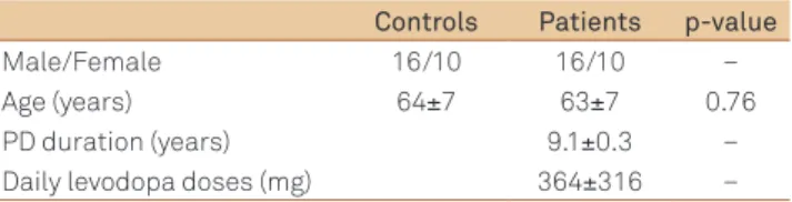
Table 1 presents the demographic features of the subjects enrolled in the study: 26 patients with PD and 26 age and gen-der matched-controls.
Table 2 shows UPDRS scores of off and on states. It was observed that scores were significantly higher during the
off state in comparison to the on state, for both females and males.
Table 3 shows MIP and MEP values for patients and con-trols. Both women and men with PD presented with lower values during the of state in relation to controls. Concerning the on stage, there are no signiicant diferences of MIP be-tween females with PD and controls. Comparison of on and
of state variables between patients with PD conirmed the little impact of the antiparkinsonian treatment on respira-tory pressure because only the MEP of women signiicantly increased after using the medications.
Table 4 demonstrates that overall the respiratory pres-sures of both patients and controls are below those expected for the Brazilian population. he exceptions are MEP values of male and female controls.
DISCUSSION
Our indings conirm the observation of others authors, according to which respiratory pressure in PD is lower than that found in healthy age and gender matched-controls, as well as in relation to values predicted for the population1,3,5,6.
Since we have studied subjects with HY 2 to 3, our results in-dicate that respiratory dysfunction is already present in ear-ly stages of PD. We have also replicated the literature ind-ing showind-ing that gender inluences respiratory pressures9,20.
Harik-Khan et al.20, for instance, demonstrated that MIP
val-ues in healthy men are 30% higher than those found in age-matched women.
It is interesting that some investigations about respiratory pressure present in literature failed to ind diferences between patients with PD and values predicted for the general popu-lation or controls8,12,13. he discrepancy in results of diferent
studies most likely relects methodological issues: 1) Some of the studies7,12 compared the values of variables with those
pre-dicted for the population, not including controls. As we have shown (Table 4), there is a diference between the values pre-dicted for the Brazilian population and the controls included in our study; 2) Other investigations used non-matched con-trols; 3) Because of the inluence of the gender over the respi-ratory pressure, demonstrated by our study and others7,9,15,20,
it is mandatory not only to use age, but also gender matched-controls, which did not happen in many of the studies on PD6;
4) In some of the studies, it is not clear whether or not the pressures were measured when the patients were on the efect of the antiparkinsonian medications; 5) Diagnostic criteria of PD have not been described in some of the investigations. his raises the issue of the possibility to include patients with par-kinsonian disorders other than PD; 6) Finally, there has been a wide severity variation concerning the parkinsonian syn-drome among the patients included in some of these studies, with HY score ranging from 1 to 5.
Few studies investigate the efect of levodopa on respi-ratory pressure. One report, however, described that sub-cutaneous injections of apomorphine, a powerful direct do-pamine receptor agonist, induced a statistically signiicant increase of MIP and MEP in 10 patients with PD (HY 2 to 4)4. here is also a single case report which described the
reversibility of upper airway obstruction in a patient with PD after using levodopa16. On the contrary, Weiner et al.15
observed MIP and MEP comparing of and on periods in 20 patients with PD. hese variables tended to increase during
on periods, however, it did not reach statistical signiicance. In this study, we have found that levodopa caused MIP
Controls Patients p-value
Male/Female 16/10 16/10 –
Age (years) 64±7 63±7 0.76
PD duration (years) 9.1±0.3 –
Daily levodopa doses (mg) 364±316 –
Table 1. Demographic and clinical features of studied subjects.
Data presented as mean±standard deviation; PD: Parkinson’s disease.
off on p-value
Total females 51.2 ± 19.3 42.7 ± 17.3 0.001 Total males 60. 3 ± 14,1 44.4± 12.8 0.001 Part III females 26.5 ± 13.1 18.0 ± 11.6 0.001 Part III males 34.3 ± 8.3 18.3 ± 6.4 0.001
Table 2. Unified Parkinson’s disease rating scale scores of patients.
values in women to signiicantly increase. One may spec-ulate that this lack of diference between on and of states relects insuicient dopaminergic stimulation. However, the statistically signiicant diferences between on and of
UPDRS scores indicate that the dose of levodopa and other antiparkinsonian drugs was capable of inducing a clinically relevant change in the functional status of the nigrostriatal system. his inding in our study raises the issue that re-spiratory dysfunction in patients with PD may be unrelat-ed to dopaminergic dysfunction. Such situation would be similar to the occurrence of dysphagia, gait disorder includ-ing freezinclud-ing (motor block) and other features of PD, which are little or not changed by dopamine replacement therapy. In one study, the authors showed that despite unequivocal improvement of motor indings with levodopa, swallowing function presented little improvement21.
Similarly, step length and freezing of gait are only part-ly improved by levodopa replacement22. It is worth
men-tioning that in contrast to dysphagia and gait disorder, which occur after long duration of PD, our findings sug-gest that respiratory pressure changes are an early symp-tom of PD. Finally, data from other studies suggest that MIP and MEP changes may worsen with the progression of the illness6,14.
he pathogenesis of respiratory pressure changes in PD is yet to be determined. Some authors have shown respi-ratory function improvement in PD with dopamine treat-ment1,4, whereas others did not ind any respiratory
im-provement with the action of levodopa15,23. owever, these
indings were not explained by the improvement in pulmo-nary function or respiratory muscle. he authors suggested that it was possibly caused due to a central efect. he pres-ence of dopamine receptors in central and peripheral struc-tures24 may suggest that dopamine dysfunction leading to a
direct negative efect on the respiratory structures, as well as to rigidity and bradykinesia of the respiratory muscles, is the causative mechanism of MIP and MEP abnormali-ties4. his hypothesis is weakened, however, by the
disso-ciation between the action of levodopa on UPDRS scores and the respiratory values found in our study. he sugges-tion that deformities in the thoracic cage lead to respirato-ry abnormalities5 also seems unlikely, since none of our
pa-tients presented with such indings. Considering the little improvement of respiratory changes in PD after the use of levodopa, one may speculate that dopaminergic and non-dopaminergic central changes play a role in the pathogen-esis of these abnormalities.
Finally, none of our patients presented respiratory com-plaints (data not shown) despite the low values of respira-tory pressure. In fact, just moderate to severe reductions in respiratory pressure, 40% below normal values, are regard-ed as a respiratory disorder25. Nevertheless, the decrease of
these variables is a risk factor for pulmonary dysfunction, since it increases the risk of atelectasis and others24.
Finally, our results demonstrated that the pressure of patients with Parkinson’s disease is lower than of that in control subjects, even in early stages of the disease. Dopamine replacement has little impact on these respi-ratory variables, suggesting that early intervention to im-prove respiratory function in PD may be necessary.
Controls Patients-Off Patients-on p-value
MIP women 70.0±14.1 50.0±22.9 55.6±18.8
0.04* 0.28 0.33
MEP women 105.6 ±19.4 55.6±18.8 64.4±21.9
0.00* 0.00** 0.00***
MIP men 86.9 ± 6.2 61.9±17.2 65.0±17.9
0.00* 0.00** 0.42
MEP men 120.0 ±29.4 74.4±15.9 81.9±14.7
0.00* 0.00** 0.07 Table 3. Values of maximal inspiratory pressure and maximal expiratory pressure in Parkinson’s disease patients and controls.
Data presented as mean±standard deviation (cmH2O); off refers to “without levodopa”; on refers to “with levodopa”; *p-value for patients-off versus controls;
**p-value for patients-on versus controls; ***p-value for patients-off versus patients-on.
MIP: maximal inspiratory pressure; MEP: maximal expiratory pressure.
Controls (%) off (%) on (%)
MIP women 94.7 66.7 62.3
MEP women 133.6 71.7 80.6
MIP men 80.2 62.3 62.1
MEP men 133.6 71.7 80.6
Table 4. Percentage of maximal inspiratory pressure and maximal expiratory pressure in Parkinson’s disease patients and controls.
% refers to values predicted for the Brazilian population; off refers to “without levodopa”; on refers to “with levodopa”.
References
1. Vincken WG, Gauthier SG, Dollfuss RE, Hanson RE, Darauay CM, Cosio MG. Involvement of upper-airway muscles in extrapyramidal disorders. A cause of airflow limitation. N Engl J Med 1984;311:438-442. 2. Hovestadt A, Bogaard JM, Meerwaldt JD, van der Meché FG, Stigt
J. Pulmonary function in Parkinson’s disease. J Neurol Neurosurg Psychiatry 1989;52:329-333.
3. Fontana GA, Pantaleo T, Lavorini F, Benvenuti F, Gangemi S. Defective motor control of coughing in Parkinson’s disease. Am J Respir Crit Care Med 1998;158:458-464.
4. de Bruin PF, de Bruin VM, Lees AJ, Pride NB. Effects of treatment on airway dynamics and respiratory muscle strength in Parkinson’s disease. Am Rev Respir Dis 1993;148:1576-1580.
5. Sabaté M, Rodríguez M, Méndez E, Enríquez E, González I. Obstructive and restrictive pulmonary dysfunction increases disability in Parkinson disease. Arch Phys Med Rehabil 1996;77:29-34.
6. Haas BM, Trew M, Castle PC. Effects of respiratory muscle weakness on daily living function, quality of life, activity levels, and exercise capacity in mild to moderate Parkinson’s disease. Am J Phys Med Rehabil 2004;83:601-607.
7. Silverman EP, Sapienza CM, Saleem A, et al. Tutorial on maximum inspiratory and expiratory mouth pressures in individuals with idiopathic Parkinson disease (IPD) and the preliminary results of an expiratory muscle strength training program. NeuroRehabilitation 2006;21:71-79.
8. Tzelepis GE, McCool FD, Friedman JH, Hoppin FG Jr. Respiratory muscle dysfunction in Parkinson’s disease. Am Rev Respir Dis 1988;138:266-271.
9. Black LF, Hyatt RE. Maximal respiratory pressures: normal values and relationship to age and sex. Am Rev Respir Dis 1969;99:696-702. 10. McConnell AK, Copestake AJ. Maximum static respiratory pressures
in healthy elderly men and women: issues of reproducibility and interpretation. Respiration 1999;66:251-258.
11. Berry JK, Vitalo CA, Larson JL, Patel M, Kim MJ. Respiratory muscle strength in older adults. Nurs Res 1996;45:154-159.
12. Canning CG, Alison JA, Allen NE, Groeller H. Parkinson’s disease: an investigation of exercise capacity, respiratory function, and gait. Arch Phys Med Rehabil 1997;78:199-207.
13. Cardoso SR, Pereira JS. [Analysis of breathing function in Parkinson’s disease]. Arq Neuropsiquiatr 2002;60:91-95.
14. Enright PL, Kronmal RA, Manolio TA, Schenker MB, Hyatt RE.
Respiratory muscle strength in the elderly. Correlates and reference values. Cardiovascular Health Study Research Group. Am J Respir Crit Care Med 1994;149:430-438.
15. Weiner P, Inzelberg R, Davidovich A, et al. Respiratory muscle performance and the Perception of dyspnea in Parkinson’s disease. Can J Neurol Sci 2002;29:68-72.
16. Vincken WG, Darauay CM, Cosio MG. Reversibility of upper airway obstruction after levodopa therapy in Parkinson’s disease. Chest 1989;96:210-212.
17. Hughes AJ, Daniel SE, Kilford L, Lees AJ. Accuracy of clinical diagnosis of idiopathic Parkinson’s disease: a clinico-pathological study of 100 cases. J Neurol Neurosurg Psychiatry 1992;55: 181-184.
18. Neder JA, Andreoni S, Lerario MC, Nery LE. Reference values for lung function tests. II. Maximal respiratory pressures and voluntary ventilation. Braz J Med Biol Res 1999;32:719-727.
19. Fahn S, Elton R, and members of the UPDRS Development Committee. Unified Parkinson’s Disease Rating Scale. In: Fahn S, Marsden C, Calne D, Goldstein M (eds). Recent development in Parkinson`s disease. Florham Park, NJ: Macmillan Health Care Information; 1987: 153-164.
20. Harik-Khan RI, Wise RA, Fozard JL. Determinants of maximal inspiratory pressure. The Baltimore Longitudinal Study of Aging. Am J Respir Crit Care Med 1998;158:1459-1464.
21. Hunter PC, Crameri J, Austin S, Woodward MC, Hughes AJ. Response of parkinsonian swallowing dysfunction to dopaminergic stimulation. J Neurol Neurosurg Psychiatry 1997; 63:579-583.
22. Morris M, Iansek R, McGinley J, Matyas T, Huxham F. Three-dimensional gait biomechanics in Parkinson’s disease: evidence for a centrally mediated amplitude regulation disorder. Mov Disord 2005;20:40-50.
23. Obenour WH, Stevens PM, Cohen AA, McCutchen JJ. The causes of abnormal pulmonary function in Parkinson’s disease. Am Rev Respir Dis 1972;105:382-387.
24. Rice JE, Antic R, Thompson PD. Disordered respiration as a levodopa-induced dyskinesia in Parkinson’s disease. Mov Disord 2002;17: 524-527.